Diarrhea outbreaks in suckling piglets due to rotavirus group
C single and mixed (rotavirus groups A and B) infections
1Elis Lorenzetti2, Danilo T. Stipp2,3, Flávia Possatti2, Joice E.T. Campanha2, Alice F. Alfieri2 and Amauri A. Alfieri2*
ABSTRACT.- Lorenzetti E., Stipp D.T., Possatti F., Campanha J.E.T., Alfieri A.F. & Alfieri A.A. 2014. Diarrhea outbreaks in suckling piglets due to rotavirus group C single and mixed (rotavirus groups A and B) infections. Pesquisa Veterinária Brasileira 34(5):391-397. Laboratory of Animal Virology, Department of Veterinary Preventive Medicine, Uni-versidade Estadual de Londrina, Rodovia Celso Garcia Cid, Campus Universitário, Cx. Postal 10011, Londrina, PR 86057-970, Brazil. E-mail: alfieri@uel.br
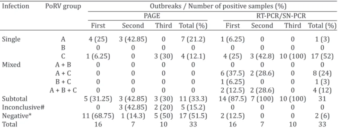
Porcine group A rotavirus (PoRVA) is a major cause of neonatal diarrhea in suckling and recently weaned piglets worldwide. The involvement of non-group A rotavirus in cases of neonatal diarrhea in piglets are sporadic. In Brazil there are no reports of the porcine rotavirus group C (PoRVC) as etiologic agent of the diarrhea outbreaks in piglets. The aim of this study was to describe the identification of rotavirus group C in single and in mixed infection with rotavirus groups A and B in three neonatal diarrhea outbreaks in suckling (≤21-day-old) piglets, with 70% to 80% and 20% to 25% of morbidity and lethality rates, respectively, in three pig herds located in the state of Santa Catarina, Brazil. The diagnosis of PoRV in the diarrheic fecal samples was performed using polyacrylamide gel electropho-resis (PAGE) to identify the presence of porcine rotavirus groups A, B (PoRVB), and C, and by RT-PCR (PoRVA and PoRVC) and semi-nested (SN)-PCR (PoRVB) to partially amplify the VP4 (VP8*)-VP7, NSP2, and VP6 genes of PoRVA, PoRVB, and PoRVC, respectively. One RT-PCR (PoRVA and PoRVC) and SN-RT-PCR (PoRVB) product of each group of rotavirus of each diarrhea outbreak was submitted to nucleotide (nt) sequence analysis. Based on the PAGE technique, 4 (25%) and 1 (6.25%) of the 16 diarrheic fecal samples evaluated in the first outbreak presented PoRVA and PoRVC electropherotype, respectively, and 11 (68.75%) were negative. In the second outbreak, 3 (42.85%) of the 7 fecal samples evaluated pre-sented PoRVA electropherotype, and in 3 (42.85%) and in 1 (14.3%) fecal samples were de-tected inconclusive and negative results, respectively. Three (30%) of the 10 fecal samples of the third outbreak presented PoRVC electropherotype; 5 (50%) and 2 (20%) samples showed negative and inconclusive results, respectively. Based on the RT-PCR and SN-PCR assays in the first neonatal diarrhea outbreak, PoRVC was detected in 13 (81.2%) of the 16 diarrheic fecal samples evaluated. PoRVC single infection was identified in 4 (25%) of these samples and mixed infections with PoRVA and PoRVB in 9 (56.2%) fecal samples. All of the seven diarrheic fecal samples evaluated from the second neonatal diarrhea outbreak were positive for PoRVC, whereas its mixed infection with other PoRV groups was detected in 4 (57.2%) samples. In the third outbreak, PoRVC in single infection was detected in all of the 10 diarrheic fecal samples analyzed. In the nt sequence analysis, the PoRVA strains of the first and second outbreaks demonstrated higher nt identity with G4P[6] and G9P[23] genotypes, respectively. The PoRVB strains (first and second outbreaks) and the PoRVC
1 Received on October 2, 2013.
Accepted for publication on January 27, 2014.
2 Laboratório de Virologia Animal, Departamento de Medicina Vete-rinária Preventiva, Universidade Estadual de Londrina (UEL), Rodovia Celso Garcia Cid, PR-445, Km 380, Campus Universitário, Caixa Pos-
tal 10011, Londrina, PR 86057-970, Brazil. *Corresponding author: alfieri@uel.br
RESUMO.- [Surtos de diarreia em leitões lactentes por rotavírus grupo C em infecções singulares e mistas (ro-tavirus grupos A e B).] O rotavírus suíno grupo A (PoRVA) é uma das principais causas de diarreia neonatal em leitões lactentes e recém-desmamados em todo o mundo. As des-crições do envolvimento de rotavírus não-grupo A em qua-dros de diarreia neonatal em leitões são esporádicas. No Brasil não há relatos do envolvimento do rotavírus suíno grupo C (PoRVC) na etiologia dos surtos de diarreia em lei-tões. O objetivo deste estudo foi descrever a identificação de rotavírus grupo C em infecções singulares e mistas com os rotavírus grupos A e B em três surtos de diarreia neona-tal em leitões lactentes (≤21 dias de idade), com taxas de morbidade de 70% a 80% e de letalidade de 20% a 25%, em três rebanhos suínos localizados no estado de Santa Ca-tarina, Brasil. O diagnóstico de PoRV nas amostras de fezes diarreicas foi realizado por eletroforese em gel de poliacri-lamida (PAGE) para identificar a presença dos grupos A, B (PoRVB), e C de rotavírus suíno e por RT-PCR (PoRVA e PoR-VC) e semi-nested (SN)-PCR (PoRVB) com a amplificação parcial dos genes VP4 (VP8*)-VP7, NSP2 e VP6 de PoRVA, PoRVB e PoRVC, respectivamente. Um produto de RT-PCR (PoRVA e PoRVC) e SN-PCR (PoRVB) de cada grupo de rota-vírus de cada um dos três surtos de diarreia foi submetido à análise da sequência de nucleotídeos (nt). Com base na técnica de PAGE, 4 (25%) e 1 (6,25%) das 16 amostras de fezes analisadas no primeiro surto apresentaram eletrofe-rotipo característico de PoRVA e PoRVC, respectivamente, e 11 (68,75%) amostras fecais foram negativas. No segundo surto, 3 (42,85%) das 7 amostras de fezes analisadas apre-sentaram perfil eletroforético de PoRVA; em 3 (42,85%) e em 1 (14,3%) amostras de fezes foram detectados resulta-dos inconclusivos e negativos, respectivamente. Três (30%) das 10 amostras de fezes do terceiro surto apresentaram eletroferotipo característico de PoRVC; 5 (50%) e 2 (20%) amostras apresentaram resultados negativos e inconclusi-vos, respectivamente. Com base nos resultados da RT-PCR e SN-PCR, no primeiro surto de diarreia neonatal o PoRVC foi detectado em 13 (81,2%) das 16 amostras de fezes diarrei-cas analisadas, sendo que em 4 (25%) amostras foi identi-ficada infecção singular e em 9 (56,2%) amostras infecção mista com PoRVA e PoRVB. Todas as sete amostras de fezes diarreicas provenientes do segundo surto de diarreia ne-onatal foram positivas para o PoRVC, enquanto infecções mistas com outros grupos de PoRV foram detectadas em 4 (57,2%) amostras. No terceiro surto o PoRVC foi detec-tado em infecção singular em todas as dez amostras de fezes diarreicas analisadas. Na análise da sequência de nt as cepas de PoRVA do primeiro e segundo surtos
demons-traram maior identidade de nt com os genotipos G4P[6] e G9P[23], respectivamente. As cepas de PoRVB (primeiro e segundo surtos) e as cepas de PoRVC (primeiro, segundo e terceiro surtos) mostraram maior identidade de nt com cepas de PoRVB e PoRVC que pertencem aos genotipos N4 e I1, respectivamente. Esta é a primeira descrição realizada no Brasil do envolvimento de PoRVC na etiologia de surtos de diarreia em leitões lactentes. Os resultados deste estudo demonstram que o PoRVC, tanto em infecções singulares quanto em infecções mistas, é um importante enteropató-geno envolvido em surtos de diarreia neonatal em leitões e que o uso de técnicas de diagnóstico mais sensíveis permite caracterizar que infecções mistas, com dois ou até mesmo com três grupos de PoRV, podem ser mais comuns do que anteriormente relatado.
TERMOS DE INDEXAÇÃO: Diarreia, rotavirose, RT-PCR, infecção mista, suínos.
INTRODUCTION
Neonatal diarrhea is the most important health problem in suckling and recently weaned piglets throughout the world (Tubbs et al. 1993). Diarrhea is the result of the combina-tion of several factors, including infectious agents, host im-munity, and management procedures (Wittum et al. 1995). Neonatal diarrhea increases the morbidity and mortali-ty rates in the maternimortali-ty units and nurseries of pig farms worldwide, causing direct and indirect economic losses to the pig industry (Dewey et al. 1995, Wittum et al. 1995).
Newborn piglets are susceptible to infection by several enteric microorganisms, including bacteria (enterotoxigen-ic Escherichia coli and Clostridium perfringens type C), pro-tozoans (Cryptosporidium spp and Isospora suis), and virus-es (rotavirus, coronavirus, and calicivirus) (Barry, Alfieri & Alfieri 2008, Zlotowski et al. 2008, Jeong et al. 2009, Lin -ares et al. 2009, Médici et al. 2011, Marthaler et al. 2012).
Rotaviruses are the main viral etiologic agents of diar-rhea in children and the young animals of a wide variety of mammalian and avian species throughout the world (Estes & Kapikian 2007). Rotaviruses belong to the Reoviri-dae family and are characterized by double-stranded RNA (dsRNA) containing 11 genomic segments and by capsids composed of three concentric protein layers. Based on the antigenic properties of the capsid protein in the middle layer (VP6), rotaviruses are classified into 5 distinct groups (A to E), and two tentative species (F and G) (ICTV 2013). Recently, a new group (H) was described by Matthijnssens et al. (2012). The A, B, C, and H rotavirus (RV) groups have been found in humans and animals, whereas groups D-G strains (first, second, and third outbreaks) showed higher nt identity and clustered in the
phylogenetic tree with PoRVB and PoRVC strains that belong to the N4 and I1 genotypes, respectively. This is the first description in Brazil of the involvement of PoRVC in the etio-logy of diarrhea outbreaks in suckling piglets. The results of this study demonstrated that PoRVC, in both single and mixed infections, is an important enteropathogen involved in neonatal diarrhea outbreaks in piglets and that the use of more sensitive diagnostic tech-niques allows the identification of mixed infections involving two or even three groups of PoRV, which may be more common than previously reported.
have been identified only in animals (Estes & Kapikian 2007). However, rotavirus group A (RVA) is the main group of rotaviruses that cause diarrhea in human (infants) and young animal hosts (Estes & Kapikian 2007).
Porcine group A rotavirus (PoRVA) infections are most frequently identified in episodes of diarrhea in piglets worldwide, including in Brazil (Linares et al. 2009, Halaihel et al. 2010, Médici et al. 2011, Lorenzetti et al. 2011); how-ever, the involvement of porcine rotavirus groups B and C (PoRVB and PoRVC) in weaning and post-weaning piglets with diarrhea has also been reported mainly in transversal epidemiological studies (Magar et al. 1991, Zlotowski et al. 2008, Médici et al. 2010a, 2010b, Médici et al. 2011, Mar-thaler et al. 2012).
RVC has been associated with enteritis in humans, pigs, calves, dogs, and ferrets, and this group of RV is considered an important emerging agent of viral diarrhea in these spe-cies (Torres-Medina 1987, Teixeira et al. 1998, Otto et al. 1999, Médici et al. 2011, Park et al. 2011). In Brazil, RVC has been identified as a cause of diarrhea in human and animal hosts (Teixeira et al. 1998, Médici et al. 2010b, 2011).
Most studies have reported the involvement of a single PoRV group with the occurrence of diarrhea in piglets, al-though some studies showed infection with a mixed of PoRV groups in piglets with clinical signs of diarrhea (Kim et al. 1999, Barreiros et al. 2003, Martella et al. 2007, Linares et al. 2009, Médici et al. 2011). A combination of different groups of PoRVs can intensify the severity of the piglets’ diarrhea, although mixed infections have been detected in piglets without these clinical signs (Marthaler et al. 2012).
According to the two outer capsid proteins, VP7 and VP4, the RVA is classified into G (glycoprotein) and P (pro -tease-sensitive) genotypes, respectively (Estes & Kapikian 2007). To date, 27 G and 37 P genotypes of RVA have been described in human and animal hosts (Matthijnssens et al. 2011, Trojnar et al. 2013). Based on the VP6 gene, the RVC strains have been classified into three genotypes, designat -ed I1, I2, and I3. The genotypes I1, I2, and I3 are compos-ed of porcine, human, and bovine strains, respectively (Ya-mamoto et al. 2011). The NSP2 gene of RVB was recently classified into four genotypes, designated N1, N2, N3, and N4. The genotype N1 is composed by human and murine strains, the genotype N2 by porcine and bovine strains and the genotypes N3 and N4 are formed only by porcine strains (Suzuki et al. 2012).
The aim of this study was to describe the identification of rotavirus group C in single and in mixed infection with rotavirus groups A and B in three neonatal diarrhea out-breaks in suckling piglets in Brazilian pig herds.
MATERIALS AND METHODS
The neonatal diarrhea outbreaks occurred in June 2007 (first), November 2011 (second), and August 2013 (third) in three di-fferent pig farms located in the state of Santa Catarina, south of Brazil, and exhibited 70% to 80% and 20% to 25% of morbidity and lethality rates, respectively. These complete-cycle pig farms of medium size (± 400 sows) had a confinement system (all-in/ all-out) and good nutritional and health management practices including sows vaccination against etiological agents of
neona-tal diarrhea such as Escherichia coli (K88, K99, 987P, and F41), PoRVA (genotypes G4 and G5), and Clostridium perfringens type C. Sampling consisted of 33 diarrheic fecal samples with a watery consistency from suckling (≤3-week-old) piglets, with 16 fecal samples coming from the first diarrhea outbreak, 7 samples from the second, and 10 samples from the third. All diarrheic fecal sam-ples analyzed in this study were collected from live piglets.
Viral dsRNA was obtained from the fecal samples using a com-bination of phenol/chloroform/isoamyl alcohol (25:24:1) and si -lica/guanidinium isothiocyanate nucleic acid extraction methods, modified as described by Alfieri et al. (2006). The OSU strain am -plified in MA104 cells was used as the positive control for PoRVA and two porcine fecal samples with electropherotype characteris-tics of group B or C that were confirmed by nucleotide sequence analysis were used as positive controls for PoRVB and PoRVC, res-pectively (accession numbers: EF577257 and EU002783) (Médici et al. 2010a,b). Aliquots of Tris-Ca2+ buffer were used as a negative
control. Positive and negative controls were used for all of the viral RNA extraction, RT-PCR, and semi-nested (SN)-PCR procedures.
All diarrheic fecal samples were tested by 7.5% polyacrylami-de gel electrophoresis (PAGE) followed by silver staining as polyacrylami- des-cribed by Herring et al. (1982) and Pereira et al. (1983), respecti-vely, to verify the presence of porcine rotavirus groups A, B, and C. The diagnoses of PoRV in the diarrheic fecal samples were also were conducted using RT-PCR for RVA, with primers that am-plified 876-bp and 1,062-bp products from the VP4 (VP8*) and VP7 genes, respectively (Gouvea et al. 1990, Gentsch et al. 1992). SN-PCR was performed for RVB using primers that amplified a 434-bp product of the NSP2 gene (Gouvea et al. 1991), and RT--PCR was performed for RVC using primers that amplified a 270-bp product of the VP6 gene (Alfieri et al. 1999).
The RT-PCR (PoRVA and PoRVC) and SN-PCR (PoRVB) pro-ducts were analyzed using 2% agarose gel electrophoresis, stai-ning with ethidium bromide, and visualization under UV light.
One RT-PCR (PoRVA and PoRVC) and SN-PCR (PoRVB) product of each group of rotavirus of each diarrhea outbreak was purified using the GFX PCR DNA and Gel Band Purifica tion Kit (GE Health -care, Little Chalfont, Buckinghamshire, UK) and quantified using a Qubit® Fluorometer (Invitrogen Life Technologies, Eugene, OR,
USA). The DNA sequences were obtained using a BigDye®
Termi-nator v3.1 Cycle Sequencing Kit (Applied Biosystems, Foster City, CA, USA) and an ABI3500 Genetic Analyzer sequencer (Applied Biosystems, Foster City, CA, USA). Sequence quality analyses were conducted using Phred and CAP3 software (http://asparagin.ce -nargen.embrapa.br/phph/). Sequence similarity searches were performed using the BLAST program (http://blast.ncbi.nlm.nih. gov/), and identity matrix analyses were performed using BioE-dit version 7.1.3.0. Phylogenetic trees were constructed using the neighbor-joining method and the Kimura two-parameter model in MEGA software version 5.05. The bootstrap probabilities of each node were calculated using 1,000 replicates.
The sequences of PoRVA, PoRVB, and PoRVC described in the present study have been deposited in the GenBank database un-der the accession numbers: KF991082 to KF991090.
RESULTS
exhibited PoRVC electropherotype, and 2 (20%) fecal sam-ples presented inconclusive results (Table 1).
Based on the RT-PCR and SN-PCR assays, 14 (87.5%) of the 16 diarrheic fecal samples evaluated in the first ne -onatal diarrhea outbreak were PoRV-positive. PoRVC was the most prevalent group, being identified in 13 (81.2%) of these fecal samples. Single infections of PoRVC were iden-tified in 4 (25%) of the fecal samples and mixed infections with other PoRV groups were identified in 9 (56.2%) of the samples (Table 1). In the second outbreak, PoRV was identified in all (n=7) of the fecal samples analyzed. PoRVC was identified in all of fecal samples from this diarrhea ou -tbreak, as a single infection in 3 (42.8%) samples and as a mixed infection with other PoRV groups in 4 (57.2%) of the fecal samples (Table 1). All of the 10 diarrheic fecal samples evaluated from the third outbreak were positive for PoRVC in single infection (Table 1).
Sequence analysis of the VP7 gene of RVA and compa-rison with the 27 known G genotypes revealed that the BRA900/2007-Po (first outbreak) PoRVA strain displayed 83% and 84.2% nucleotide (nt) identity with the human (ST3 strain) and porcine (Gottfried strain) prototypes of the G4 genotype, respectively, and the BRA82/2011-Po (se -cond outbreak) PoRVA strain displayed 88.4% and 91% nt identity with the human (WI61 strain) and porcine (JP29-6 strain) prototypes of the G9 genotype, respectively (Fig.1).
Comparison of the VP4 gene (VP8* subunit) of RVA with the 37 known P genotypes revealed that the BRA900/2007 --Po (first outbreak) PoRVA strain exhibited 88.4% and 79.6% nt identity with the human (ST3 strain) and porcine (Gottfried strain) prototypes of the P[6] genotype, respec -tively, and the BRA82/2011-Po (second outbreak) PoRVA strain exhibited 91.5% nt identity with the porcine (A34 strain) prototype of the P[23] genotype (Fig.2).
The NSP2 sequence of the two Brazilian wild-type PoR-VB strains of the first and second outbreaks (BRA900/2007 --Po and BRA82/2011--Po) displayed 79.3 to 90.5 nt identi -ty with the PoRVB strains that belong to the N4 geno-type and showed 100% nt identity each other. These sequences exhibited high nt identity with the Brazilian PoRVB strains (89% to 90.5%) (Fig.3).
Sequence analysis of the VP6 gene of the three Bra-zilian wild-type PoRVC strains of the three outbreaks (BRA905/2007-Po, BRA82/2011-Po, and BRA1437/2013 --Po) displayed 83.8% to 96.9% nt identity with the PoRVC strains that belong to the I1 genotype. The VP6 sequence of the three PoRVC strains described in this study displayed Fig.1. Phylogenetic tree based on the partial nucleotide sequences (nt 103-1020) of the VP7 gene from PoRVA strains described in this study and those of representative strains of each 27 ge-notypes recognized thus far. The tree was constructed using neighbor-joining and Kimura two-parameter as a nucleotide substitution model. The bootstrap values are shown at the branch nodes (values < 50% not shown). The Brazilian PoRVA strains from the first and second diarrhea outbreaks are ma -rked with a filled square and a filled circle, respectively.
Table 1. Porcine rotavirus (PoRV) groups identified by PAGE technique, RT-PCR and SN-PCR assays in three diarrhea outbreaks in suckling pigs
Infection PoRV group Outbreaks / Number of positive samples (%)
PAGE RT-PCR/SN-PCR
First Second Third Total (%) First Second Third Total (%)
Single A 4 (25) 3 (42.85) 0 7 (21.2) 1 (6.25) 0 0 1 (3)
B 0 0 0 0 0 0 0 0
C 1 (6.25) 0 3 (30) 4 (12.1) 4 (25) 3 (42.8) 10 (100) 17 (52)
Mixed A + B 0 0 0 0 0 0 0 0
A + C 0 0 0 0 6 (37.5) 2 (28.6) 0 8 (24)
B + C 0 0 0 0 1 (6.25) 0 0 1 (3)
A + B + C 0 0 0 0 2 (12.5) 2 (28.6) 0 4 (12) Subtotal 5 (31.25) 3 (42.85) 3 (30) 11 (33.3) 14 (87.5) 7 (100) 10 (100) 31 Inconclusive# 0 3 (42.85) 2 (20) 5 (15.2) 0 0 0 0 Negative* 11 (68.75) 1 (14.3) 5 (50) 17 (51.5) 2 (12.5) 0 0 2 (6)
Total 16 7 10 33 16 7 10 33
high (85.3% to 96.9%) nt identity with the Brazilian PoRVC strains (Fig.4).
DISCUSSION
Identification of enteric pathogens as causative agents of diarrhea in piglets has been conducted worldwide. The majority of the surveys evaluated only one pathogen, re-sulting in insufficient information about the frequency and severity of diarrhea caused by concomitant infections with multiple enteropathogens (Barreiros et al. 2003, Linares et al. 2009); however, more recently some studies have iden-tified several different enteric agents in pig fecal samples (Martella et al. 2007, Jeong et al. 2009, Halaihel et al. 2010, Médici et al. 2011, Marthaler et al. 2012).
In Brazil, the association of different PoRV groups in piglet enteritis has been reported in transversal epidemio-logical studies carried out in distinct pig farms, not all of which were involved in a diarrhea outbreak (Médici et al. 2011). Considering the variety of enteric pathogens that Fig.2. Phylogenetic tree based on the partial nucleotide sequences
(nt 60-778) of the VP4 (VP8*) gene from PoRVA strains des-cribed in this study and those of representative strains of each 37 genotypes recognized thus far. The tree was constructed using neighbor-joining and Kimura two-parameter as a nu-cleotide substitution model. The bootstrap values are shown at the branch nodes (values < 50% not shown). The Brazilian PoRVA strains from the first and second diarrhea outbreaks are marked with a filled square and a filled circle, respectively.
Fig.3. Phylogenetic tree based on the partial nucleotide sequences (nt 58-405) of the NSP2 gene from PoRVB strains described in this study and representative strains of the 4 genotypes recog-nized thus far. The tree was constructed using neighbor-joi-ning and Kimura two-parameter as a nucleotide substitution model. The bootstrap values are shown at the branch nodes (values < 50% not shown). The Brazilian PoRVB strains from the first and second diarrhea outbreaks are marked with a fil -led square and a fil-led circle, respectively.
can be isolated from the fecal samples of suckling piglets, the lack of recent multiple etiological studies is compro-mising the success of preventive measures.
PoRVC has been described as a cause of diarrhea out-breaks in weaning and post-weaning pigs, which in some cases leads to substantial morbidity rates (Morin et al. 1990, Kim et al. 1999, Martella et al. 2007). Single infec-tions of PoRVC have been detected in piglets, but this PoRV group was more frequently detected in piglets in associa-tion with other PoRV groups or with other enteric viruses (Médici et al. 2011, Marthaler et al. 2012). Martella et al. (2007) reported that in piglets, PoRVC was more frequently detected in association with other viruses, such as PoRVA and calicivirus, than as a single infectious agent.
The high morbidity (70% to 80%) and lethality (20% to 25%) rates observed in the three neonatal diarrhea out-breaks reported in this study might have involved a patho-genic PoRVC strain in the third outbreak and by the simul-taneous infections with different groups of PoRV in the other two outbreaks. Dual infections involving bovine RVA and RVC have been reported in neonatal diarrhea episodes in gnotobiotic calves, generally with intestinal lesions, such as villous atrophy and lymphoid hyperplasia, which were more severe than the single-infection lesions (Chang et al. 1999).
The lower frequency of reports of piglet diarrhea caused by PoRVB and PoRVC cannot be a consequence of a low in-fection rate but rather of the use of less sensitive diagnostic techniques. Polyacrylamide gel electrophoresis is the most practical and low-cost method used for RV diagnosis in ani-mal production; however, this diagnostic method exhibits low sensitivity and thus PoRVB and PoRVC infections are frequently not identified (Xu et al. 1990, Médici et al. 2011). The several enzyme-linked immunosorbent assay (ELISA) kits available (commercial or in-house) are specifically de -signed to identify only RVA. Some studies have confirmed that the frequency of diagnosis of RVB and RVC as single or mixed infections increased with the use of more sensitive diagnostic techniques, such as RT-PCR assay (Alfieri et al. 1999, Médici et al. 2011).
The G4P[6] and G9P[23] genotypes of RVA detected in the first and second diarrhea outbreaks, respectively, have been detected in Brazilian pig herds (Lorenzetti et el. 2011, Tonietti et al. 2013). Similarly, the genotypes N4 of RVB (NSP2 gene) and I1 of RVC (VP6 gene) have been described in fecal samples of piglets in Brazilian pig herds (Médici et al. 2010a,b).
The immune pressure induced by mass vaccination could promote reassortment, rearrangement, and zoonotic transmission of RVA strains between human and animal hosts (Matthijnssens et al. 2009). Lorenzetti et al. (2011) suggested that the immune pressure induced by commer-cial vaccine on neonatal diarrhea control might have al-lowed the selection and emergence of PoRVA strains. In this study, the sows vaccinated with an attenuated commercial vaccine for neonatal diarrhea control that included PoRVA protected most of the piglets for group A rotavirus infec-tion and might have influenced in the selecinfec-tion or emer -gence of PoRVC strains.
The present study indicated that PoRVC, both in sin-gle and mixed infections with PoRVA and PoRVB was the main PoRV group detected in the three neonatal diarrhea
outbreaks and that the use of more sensitive diagnostic te-chniques allowed the identification of mixed infections of PoRV groups and the elucidation of the etiology of diarrhea. PoRVC can also be considered an important viral etiologic agent in the diarrhea outbreaks in suckling piglets, and therefore more studies of infections involving the atypical PoRV groups are needed. To the best of our knowledge, this is the first study about the involvement of PoRVC in diar-rhea outbreaks in suckling piglets in Brazil.
Acknowledgements.- This study was supported by the following Brazi-lian institutes: the National Counsel of Scientific and Technological Deve -lopment (CNPq), the Brazilian Federal Agency for Support and Evaluation of Graduate Education (CAPES), Financing of Studies and Projects (FI-NEP), and the Araucaria Foundation (FAP/PR). A.A. Alfieri and A.F. Alfieri are recipients of CNPq fellowships.
REFERENCES
Alfieri A.A., Leite J.P.G., Alfieri A.F., Jiang B., Glass R.I. & Gentsch J.R. 1999. Detection of field isolates of human and animal group C rotavirus by re -verse transcription-polymerase chain reaction and digoxigenin-labeled oligonucleotide probes. J. Virol. Methods 83:35-43.
Alfieri A.A., Parazzi M.E., Takiuchi E., Médici K.C. & Alfieri A.F. 2006. Fre -quency of group A rotavirus in diarrhoeic calves in Brazilian cattle her-ds, 1998-2002. Trop. Anim. Health Prod. 38:521-526.
Barreiros M.A., Alfieri A.A., Alfieri A.F., Médici K.C. & Leite J.P.G. 2003. An outbreak of diarrhoea in one-week-old piglets caused by group A ro-tavirus genotypes P[7],G3 and P[7],G5. Vet. Res. Commun. 27:505-512. Barry A.F., Alfieri A.F. & Alfieri A.A. 2008. Calicivirus animal. Semina: Ciênc.
Agrárias 29:933-946.
Chang K.O., Nielsen P.R., Ward L.A. & Saif L.J. 1999. Dual infection of gno -tobiotic calves with bovine strains of group A and porcine-like group C rotaviruses influences pathogenesis of the group C rotavirus. J. Virol. 73:9284-9293.
Dewey C.E., Wittum T.E., Hurd H.S., Dargatz D.A. & Hill G.W. 1995. Herd- and litter-level factors associated with the incidence of diarrhea mor-bidity and mortality in piglets 4-14 days of age. J. Swine Health Prod. 3:105-112.
Estes M.K. & Kapikian A.Z. 2007. Rotaviruses, p.1917-1974. In: Knipe D.M., Howley P.M., Griffin D.E., Lamb R.A., Martin M.A., Roizman B. & Straus S.E. (Eds), Fields Virology. Vol.2. 5th ed. Lippincott, Williams and & Wi -lkins/ Wolters Kluwer, Philadelphia, PA.
Gentsch J.R., Glass R.I., Woods P., Gouvea V., Gorziglia M., Flores J., Das B.K. & Bhan M.K. 1992. Identification of group A rotavirus gene 4 types by polymerase chain reaction. J. Clin. Microbiol. 30:1365-1373.
Gouvea V., Glass R.I., Woods P., Taniguchi K., Clark H., Forrester B. & Fang Z.Y. 1990. Polymerase chain reaction amplification and typing of rota -virus nucleic acid from stool specimens. J. Clin. Microbiol. 28:276-282. Gouvea V., Allen J.R., Glass R.I., Fang Z.Y., Bremont M., Cohen J., McCrae M.A.,
Saif L.J., Sinarachatanant P. & Caul O. 1991. Detection of group B and C rotaviruses by polymerase chain reaction. J. Clin. Microbiol. 29:519-523. Halaihel N., Masía R.M., Fernández-Jiménez M., Ribes J.M., Montava R., De Blas I., Gironés O., Alonso J.L. & Buesa J. 2010. Enteric calicivirus and rotavirus infections in domestic pigs. Epidemiol. Infect. 138:542-548. Herring A.J., Inglis N.F., Ojeh C.K., Snodgrass D.R. & Menzies J. 1982. Rapid
diagnosis of rotavirus infection by direct detection of viral nucleic acid in silver-stained polyacrylamide gels. J Clin. Microbiol. 16:473-477. ICTV - International Committee on Taxonomy of Viruses. <http://www.
ncbi.nlm.nih.gov/ICTVdb/> Accessed 10 Dec. 2013.
Jeong Y., Park S., Hosmillo M., Shin D., Chun Y., Kim H., Kwon H., Kang S., Woo S., Park S., Kim G., Kang M. & Cho K. 2009. Detection and molecular characterization of porcine group C rotaviruses in South Korea. Vet. Mi-crobiol. 138:217-224.
rotaviruses associated with diarrhea outbreaks in feeder pigs. J. Clin. Microbiol. 37:1484-1488.
Linares R.C., Barry A.F., Alfieri A.F., Médici K.C., Ferronato C., Grieder W. & Alfieri A.A. 2009. Frequency of group A rotavirus in piglet stool sam -ples from non-vaccinated Brazilian pig herds. Braz.Arch. Biol. Technol. 52:63-68.
Lorenzetti E., Medeiros T.N.S., Alfieri A.F. & Alfieri A.A. 2011. Genetic he-terogeneity of wild-type G4P[6] porcine rotavirus strains detected in a diarrhea outbreak in a regularly vaccinated pig herd. Vet. Microbiol. 154:191-196.
Magar R., Robinson Y. & Morin M. 1991. Identification of atypical rotavi -ruses in outbreaks of preweaning and postweaning diarrhea in Quebec swine herds. Can. J. Vet. Res.55:260-263.
Martella V., Bányai K., Lorusso E., Bellacicco A.L., Decaro N., Camero M., Bozzo G., Moschidou P., Arista S., Pezzotti G., Lavazza A. & Buonavoglia C. 2007. Prevalence of group C rotaviruses in weaning and post-weaning pigs with enteritis. Vet. Microbiol. 123:26-33.
Marthaler D., Rossow K., Gramer M., Collins J., Goyal S., Tsunemitsu H., Kuga H., Suzuki T., Ciarlet M. & Matthijnssens J. 2012. Detection of subs-tantial porcine group B rotavirus genetic diversity in the United States, resulting in a modified classification proposal for G genotypes. Virology 433:85-96.
Matthijnssens J., Bilcke J., Ciarlet M., Martella V., Bányai K., Rahman M., Zeller M., Beutels P., Damme P.V. & Ranst M.V. 2009. Rotavirus disease and vaccination: Impact on genotype diversity. Fut. Microbiol. 4:1303-1316.
Matthijnssens J., Ciarlet M., McDonald S.M., Attoui H., Bányai K., Brister J.R., Buesa J., Esona M.D., Estes M.K., Gentsch J.R., Iturriza-Gómara M., Johne R., Kirkwood C.D., Martella V., Mertens P.P., Nakagomi O., Parreño V., Rahman M., Ruggeri F.M., Saif L.J., Santos N., Steyer A., Taniguchi K., Patton J.T., Desselberger U. & Ranst M.V. 2011. Uniformity of rotavirus strain nomenclature proposed by the rotavirus classification working group (RCWG). Arch. Virol. 156:1397-1413.
Matthijnssens J., Otto P., Ciarlet M., Desselberger U., Ranst M.V. & Johne R. 2012. VP6 sequence-based cut-off values as a criterion for rotavirus species demarcation. Arch. Virol. 157:1177-1182.
Médici K.C., Barry A.F., Alfieri A.F. & Alfieri A.A. 2010a. Genetic analysis of the porcine group B rotavirus NSP2 gene from wild-type Brazilian strains. Braz. J. Med. Biol. Res. 43:13-16.
Médici K.C., Barry A.F., Alfieri A.F. & Alfieri A.A. 2010b. VP6 gene diversity in Brazilian strains of porcine group C rotavirus. Genet. Mol. Res. 9:506-513.
Médici K.C., Barry A.F., Alfieri A.F. & Alfieri A.A. 2011. Porcine rotavirus groups A, B, and C identified by polymerase chain reaction in a fecal sample collection with inconclusive results by polyacrylamide gel elec-trophoresis.J. Swine Health Prod. 19:146-150.
Morin M., Magar R. & Robinson Y. 1990. Porcine group C rotavirus as a
cause of neonatal diarrhea in a Quebec swine herd. Can. J. Vet. Res. 54: 385-389.
Otto P., Schulze P. & Herbst W. 1999. Demonstration of group C rotaviruses in fecal samples of diarrheic dogs in Germany. Arch. Virol. 144:2467-2473.
Park S-I., Jeong Y-J., Kim H-J., Park J-G., Kang S-Y., Woo S-K., Kim C-H., Jung C-H., Kang M-I. & Cho K-O. 2011. Genetically diverse group C rotaviruses cause sporadic infection in Korean calves. J. Vet. Med. Sci. 73:479-482. Pereira H.G., Azeredo R.S., Leite J.P.G., Candeias J.A.N., Rácz M.L., Linhares
A.C., Gabbay Y.B. & Trabulsi J.R. 1983. Electrophoretic study of the ge -nome of human rotaviruses from Rio de Janeiro, São Paulo and Pará, Brazil. J. Hyg. 90:117-125.
Suzuki T., Soma J., Kuga K., Miyazaki A. & Tsunemitsu H. 2012. Sequence and phylogenetic analyses of nonstructural protein 2 genes of species B porcine rotaviruses detected in Japan during 2001-2009. Virus Res. 165:46-51.
Teixeira J.M.S., Camara G.N.N.L., Pimentel P.F.V., Ferreira M.N.R., Ferreira M.S.R., Alfieri A.A., Gentsch J.R. & Leite J.P.G. 1998. Human group C ro-tavirus in children with diarrhea in the Federal District, Brazil. Braz. J. Med. Biol. Res. 31:1397-1403.
Tonietti P.O., Hora A.S., Silva F.D.F., Ruiz V.L.A. & Gregori F. 2013. Phyloge-netic analyses of the VP4 and VP7 genes of Porcine Group A Rotaviruses in São Paulo state, Brazil: first identification of G5P[23] in Piglets. J. Clin. Microbiol. 51:2750-2753.
Torres-Medina A. 1987. Isolation of an atypical rotavirus causing diarrhea in neonatal ferrets. Lab. Animal Sci. 37:167-171.
Trojnar E., Sachsenröder J., Twardziok S., Reetz J., Otto P.H. & Johne R. 2013. Identification of an avian group A rotavirus containing a novel VP4 gene with a close relationship to those of mammalian rotaviruses. J. Gen. Virol. 94:136-142.
Tubbs R.C., Hurd H.S., Dargatz D.A. & Hill GW. 1993. Preweaning morbidi-ty and mortalimorbidi-ty in the United States swine herds. Swine Health Prod. 1:21-28.
Xu L., Harbour D. & McCrae M.A. 1990. The application of polymerase chain reaction to the detection of rotaviruses in faeces. J. Virol. Methods 27:29-37.
Wittum T.E., Dewey C.E., Hurd H.S., Dargatz D.A. & Hill G.W. 1995. Herd and litter-level factors associated with the incidence of diarrhea morbidity and mortality in piglets 1-3 days of age. J. Swine Health Prod. 3:99-104. Yamamoto D., Ghosh S., Kuzuya M., Wang Y-H., Zhou X., Chawla-Sarkar M.,
Paul S.K., Ishino M. & Kobayashi N. 2011. Whole-genome characteriza -tion of human group C rotaviruses: identifica-tion of two lineages in the VP3 gene. J. Gen. Virol.92:361-369.