(Annals of the Brazilian Academy of Sciences)
Printed version ISSN 0001-3765 / Online version ISSN 1678-2690 www.scielo.br/aabc
Common Deletion (CD) in mitochondrial DNA of irradiated rat heart
RAQUEL G. SIQUEIRA1,2, DAYSE A. DA SILVA2, LUIZ D.B. DE MELO3, ELIZEU F. DE CARVALHO2, SAMARA C. FERREIRA-MACHADO1 and CARLOS E.V. DE ALMEIDA1
1Laboratório de Ciências Radiológicas, Departamento de Biofísica, Instituto de Biologia Roberto Alcântara Gomes,
Universidade do Estado do Rio de Janeiro, Rua São Francisco Xavier, 524, Pavilhão Haroldo Lisboa da Cunha, Térreo, Maracanã, 20550-900 Rio de Janeiro, RJ, Brasil
2
Laboratório de Diagnósticos por DNA, Departamento de Biologia Geral, Instituto de Biologia Roberto Alcântara Gomes, Universidade do Estado do Rio de Janeiro, Rua São Francisco Xavier, 524, Pavilhão Haroldo Lisboa da Cunha,
Térreo, Maracanã, 20550-010 Rio de Janeiro, RJ, Brasil
3Laboratório de Parasitologia Molecular, Departamento de Biofísica, Instituto de Biofísica Carlos Chagas Filho,
Universidade Federal do Estado do Rio de Janeiro, Cidade Universitária, 21949-900 Rio de Janeiro, RJ, Brasil
Manuscript received on August 17, 2012; accepted for publication on April 25, 2013
ABSTRACT
The purpose of this study was to map the common deletion (CD) area in mtDNA and investigate the levels of this deletion in irradiated heart. The assays were developed in male Wistar rats that were irradiated with three different single doses (5, 10 or 15 Gy) delivered directly to the heart and the analyses were performed at various times post-irradiation (3, 15 or 120 days). The CDs area were sequenced and the CD quantified by real-time PCR. Our study demonstrated that the CD levels progressively decreased from the 3rd
until the 15th
day after irradiation, and then increased thereafter. Additionally, it was observed that the levels of CD are modulated differently according to the different categories of doses (moderate and high). This study demonstrated an immediate response to ionizing radiation, measured by the presence of mutations in the CD area and a decrease in the CD levels.
Key words: cardiovascular complications, common mitochondrial DNA deletion, mediastinal tumours, radiotherapy.
Correspondence to: Raquel Gomes Siqueira E-mail: [email protected]
INTRODUCTION
During radiotherapy (RT) of mediastinal tumours (lymphomas, breast cancer, and lung cancer), frequently a part of the heart is included in
the treatment field and may receive significant
doses of ionizing radiation (Hilbers et al. 2012). Clinical reports indicate that a considerable number of patients who receive this therapy develop cardiovascular complications. Radiation damage may affect the pericardium, myocardium
or coronary vasculature characterised by fibrotic
changes or small vessel damage (Rijswijk et al. 2008, Doyen et al. 2010).
Recent studies showed that the dose radiation typically used in RT, promotes a larger number of ionizations within the cytoplasm (and in the organelles residing therein), as compared to the nucleus (Nikjoo and Lindborg 2013). Therefore, looking more closely to an important organelle for energy metabolism of cardiac tissue, such as the mitochondria, it will provide important insight in how cardiac tissue might respond to RT.
The mitochondria contain their own DNA (mtDNA) which encodes components of the respiratory chain, being of extreme importance for the synthesis of energy. Studies have shown that this molecule may be damaged by exogenous harmful agents such as ionizing radiation, since mtDNA lacks the protection of histones, contains no introns, is exposed continuously to endogenous reactive oxygen species (ROS) and
has a less efficient repair system than nuclear
DNA (Prithivirajsingh et al. 2004).
Although the mitochondrial genome encom-passes a small fraction of the total genetic material in a cell, any damage or alteration to it can still have serious implications for a cell’s viability and/ or survival (Murphy et al. 2005, Evdokimovsky et al. 2011) and can lead to the development of degenerative diseases with slowly progressing conditions often associated with impaired oxidative phosphorylation, especially in tissues with high energy expenditures such as the heart.
Various mutations have been induced in mtDNA after RT (Wardell et al. 2003). Among the various mutations, a large-scale deletion of 4977 bp in humans and 4834 bp in rodents (8103-8118 bp, 12937-12952 bp), the so-called common deletion (CD), has been reported after low-level ionizing radiation exposure (Ikushima et al. 2002, Murphy et al. 2005). However, the dynamic of CD levels after moderate and high-level ionizing radiation exposure, such as that of RT, has not been reported. Therefore, the purpose of this study was to map the CD region in mtDNA and investigate the level of this deletion in mitochondrial DNA of cardiac tissue of a radiobiological model in Wistar rats employing doses similar to those used in RT.
MATERIALS AND METHODS
ANIMALS AND IRRADIATION
Male Wistar rats were obtained at three months of age and weighed approximately 250g. Rats were
maintained on a 12h light/dark cycle with food and water provided ad libitum. Animals were distributed into four groups (n = 15 per group), and each group received a single dose irradiation (0, 5, 10 or 15 Gy). The heart position was marked after Computed Tomography (HiSpeed CT/Dual, GE Healthcare,
USA). Cardiac-specific irradiation was performed
using a Varian Clinac 2100 C linear accelerator (Variant Medical Systems, CA, USA) with a 6MV X-ray, and a dose rat of 240 centi-Gy/min. Individual rats were
irradiated in supine position using an anterior field size
of 3 x 3 cm2 with bolus depth of 0.5 cm. This study was approved by the local ethical council CEUA/010/2012 (Universidade do Estado do Rio de Janeiro).
Before irradiation, animals were anesthetised with 10% ketamina / 2% xilazina (0.1 mg/kg), intraperitoneally. Single doses of 10-15 Gy were calculated to be approximately equivalent to the total fractionated doses of 30-50 Gy (2 Gy per fraction), according to the Linear Quadratic model and an a/b ratio of 2:3 as described by Schultz-Hector et al. (1992).
DNAISOLATION
Euthanasia was performed 3, 15 or 120 days post-irradiation. The animals were anesthetised with 10% ketamina / 2% xilazina (0.1 mg/kg), intraperitoneally and approximately 100 mg of the left ventricle were collected. Then, the samples of cardiac tissue were washed with ice-cold saline, minced and homogenised at 0-4 °C with a glass homogeniser by hand. After this procedure, total DNA was extracted by a standard extraction protocol using proteinase K and sodium dodecyl sulphate (SDS) according to methods previously described (Miller 1998). The concentration of DNA was measured by Biophotometer (Eppendorf) using 260 nm optical density.
PRESENCE OF THE COMMON DELETION BY SEQUENCING
The presence of mtDNA in each sample was proven
control region present in 100% of all normal mtDNA (D-loop region) by conventional PCR. The primers to the D-loop and the CD from the rat mitochondrial genome (GenBank Accession No. X14848) were designed using the Primer 3 software (http:// frodo. wi.mit.edu/). In addition, we used BLAST to
confirm that the primers chosen for both regions had
exclusive homology with their target in rat mtDNA. The primers are shown in Table I.
visualised with ethidium bromide. Each product
obtained with primers for the CD was gel-purified
and directly sequenced using an ABI PRISM 310 (Applied Biosystems) with ABI PRISM310 Genetic Analyzer software.
ABSOLUTE QUANTIFICATION OF THE COMMON DELETION BY THE STANDARD CURVE METHOD
The quantity of the CD in heart was measured by
quantitative co-amplification of the mitochondrial
D-loop and the mitochondrial deletion using a real-time PCR absolute DNA quantification method. To do so, it was necessary to construct a standard curve using serial dilutions of plasmids containing D-loop and CD fragments as previously
described by Ye et al. (2008) with modifications.
The products created from a conventional PCR positive control sample were excised from the
agarose gel and purified using the DNALL-IN-ONE DNA Purification Kit (Biotools, Brazil) according to the manufacturer’s instructions. The purified
PCR products were cloned into the pGEM®-T 3000 bp plasmid (Promega) in accordance with the manufacturer’s instructions. Blue-white screening was performed before plasmids were isolated. The
cloned products were amplified by conventional
PCR using D-loop and CD primers for sequencing.
The confirmed clones were renamed pD-loop for
the total mtDNA and pCD for the common deletion. The recombinant plasmid DNA was isolated and
purified using QIAprep Spin and QIAGEN type
100 (Qiagen). The concentration of the DNA was measured by Biophotometer (Eppendorf) using 260 nm optical density, and the copy number was calculated according to the known molecular weight of the plasmid. Serial dilutions of each standard were made in the range of 10-107 copies per 1 µl according to Applied Biosystems instructions (Creating standards curves with genomic DNA or plasmids DNA templates for use in quantitative PCR. Tutorial Review, http://www6.appliedbiosystems. com/support/tutorials/pdf/quant_pcr.pdf).
Primer Sequence 5’→3’ Binding
site Amplicon D-loop F conv GGT TCT TAC TTC
AGG GCC ATC
15772 - 15792
519 bp D-loop R conv GTG GAA TTT TCT
GAG GGT AGG C
16269 - 16290 Del F conv GTT CCC ATC AAT
TCT ATT CCC AT
7978 - 8000
307 bp Del R conv GAA GCC TGC
TAG GAT GCT TC
13099 - 13118
TABLE I
Primers used for conventional PCR amplification of
total mtDNA (D-loop) and deleted mtDNA (CD).
The conventional PCR amplification of
the D-loop region was carried out in a 25 µl reaction volume consisting of 1 U Platinum Taq
DNA polymerase (Invitrogen), 5 M each D-loop primer (Applied Biosystems), 4x reaction buffer, containing dNTPs (Invitrogen) and 25 ng of total DNA. The thermocycling conditions included an initial denaturation phase at 94°C for 2 min, then 35 cycles of denaturation at 94°C for 15 s, annealing at 60°C for 30 s, and extension at 72°C for 1 min,
followed by a final extension at 72°C for 5 min. The primers used for identification of the
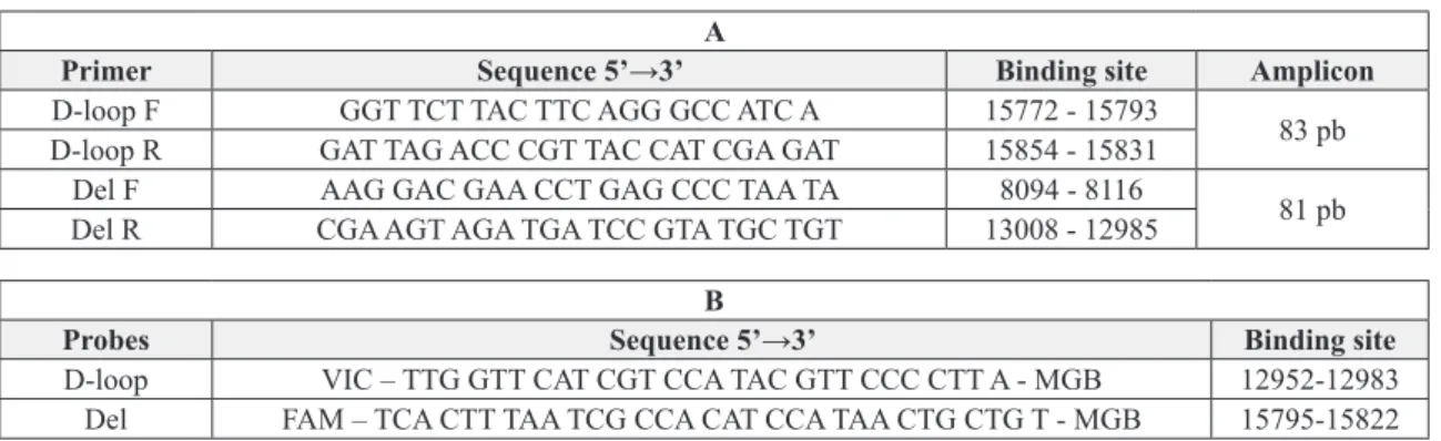
Primers and probes for the D-loop region and the CD region were previously described by Branda et al. (2002) (Table II). The proportion of the CD was
quantified with a deletion 6’-FAM-labelled fluorogenic
probe (position 12952-12983) and the total mtDNA
was detected with a VIC-labelled fluorogenic probe
(position 15795-15822). The PCR products from total mtDNA and the CD were 83 bp and 81 bp amplicons,
respectively. Standard PCR amplification was carried
out in a 15 µl reaction volume consisting of 1 µl of template DNA (10-107copies of plasmid or 50 ng of total DNA), TaqMan Universal Master mix (7 µl),
5 µM each CD primer, 5 µM each D-loop primer, and 10 µM each CD and D-loop probe. All samples were tested in triplicate and all experiments were done under multiplex conditions, that is, primers and probes for both total mtDNA and CD were present in each reaction. The thermocycling conditions included an initial denaturation phase and hot start at 95°C for 10 min; then 45 cycles of denaturation at 95°C for 15 s and annealing and extension at 60°C for 30 s. The
fluorescence spectra were monitored by Sequence
Detection System Real Time ABI PRISM 7500 with 7500 System SDS Software v1.4 (Applied Biosystems).
TABLE II
A. Primers used in real time PCR for amplification of a fragment of the D-loop mtDNA and a fragment indicative of the presence of the 4834 mtDNA deletion (Branda et al. 2002). B. Probes used in real time PCR for hybridization D-loop region
and the region of occurrence of the 4834 mtDNA deletion (Branda et al. 2002).
A
Primer Sequence 5’→3’ Binding site Amplicon
D-loop F GGT TCT TAC TTC AGG GCC ATC A 15772 - 15793
83 pb D-loop R GAT TAG ACC CGT TAC CAT CGA GAT 15854 - 15831
Del F AAG GAC GAA CCT GAG CCC TAA TA 8094 - 8116
81 pb
Del R CGA AGT AGA TGA TCC GTA TGC TGT 13008 - 12985
B
Probes Sequence 5’→3’ Binding site
D-loop VIC – TTG GTT CAT CGT CCA TAC GTT CCC CTT A - MGB 12952-12983 Del FAM – TCA CTT TAA TCG CCA CAT CCA TAA CTG CTG T - MGB 15795-15822
STATISTICAL ANALYSIS
The CD percentage was calculated as: the copy number of the CD molecules divided by total
mtDNA molecules x 100. The significance of the
differences between the values was tested with analysis of variance (ANOVA) and the Tukey-Kramer multiple comparison test using statistical software GraphPad Prism 5.0 (California, USA).
RESULTS
THE COMMON DELETION IN THE HEART OF IRRADIATED
ANIMALS
Pronounced PCR amplification of the CD was
observed in all heart samples including the control
ones. Sequencing of these amplification products
confirmed the CD in all samples, and additional
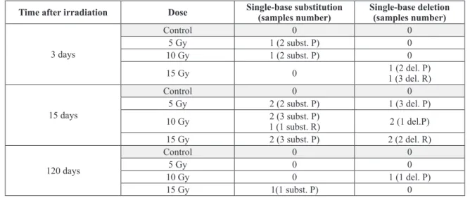
mutations were also present. The additional mutations were single-base substitutions and single-base deletions in the 16 bp sequence that
flank the CD as well as in nearer regions. These
additional mutations were present only in irradiated samples, but there was no correlation to dose or to time after irradiation (Table III).
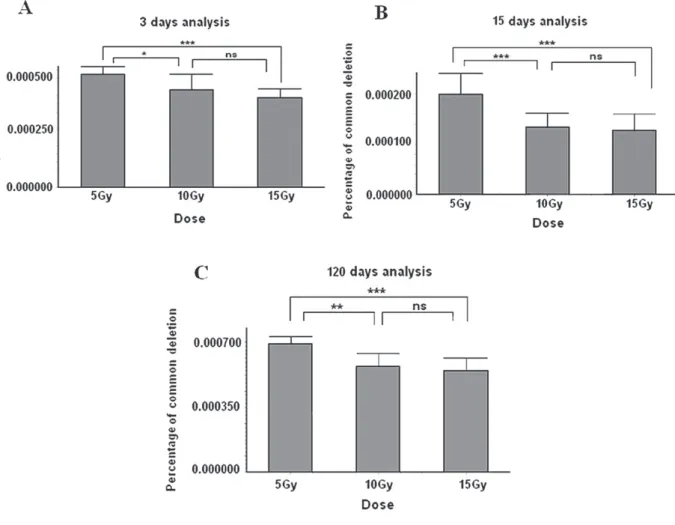
The real-time PCR quantitative analysis re-vealed that the percentage of CDs in total rat heart mtDNA varied from 1.06x10⁻⁴% to 7.99x10⁻⁴% in control and irradiated samples.
increase in the CD levels at 3, 15 and 120 days. On the other hand, the amount of the CD in the
irradiated groups decreased significantly over time,
when compared to the controls (Figure 1).
The results at three days after the 5 Gy heart irradiation show a small decrease in the amount of deleted mtDNA. However, this difference was not
significant in relation to the corresponding control
Time after irradiation Dose Single-base substitution
(samples number)
Single-base deletion (samples number)
3 days
Control 0 0
5 Gy 1 (2 subst. P) 0
10 Gy 1 (2 subst. P) 0
15 Gy 0 1 (2 del. P)
1 (3 del. R)
15 days
Control 0 0
5 Gy 2 (2 subst. P) 1 (3 del. P)
10 Gy 2 (3 subst. P)
1 (1 subst. R) 2 (1 del.P)
15 Gy 2 (3 subst. P) 2 (2 del. R)
120 days
Control 0 0
5 Gy 0 0
10 Gy 0 1 (1 del. P)
15 Gy 1(1 subst. P) 0
TABLE III
Single-base substitutions and single-base deletions were observed in the 16 bp sequence that characterize the CD (R) and in adjacent regions (P). Five samples were analyzed in each group.
Figure 1 - Quantification by Real Time PCR of CD levels in cardiac tissue of rats irradiated with
group (P > 0.05). In addition, the doses of 10 Gy and 15 Gy induced large reductions in the CD level
P < 0.05 for 10 Gy and P < 0.01 for 15 Gy) at this point of time (Figure 1).
At 15 days after the irradiation a strong reduction in CD levels was observed in all the irradiated heart samples. All the doses yielded
significant differences relative to the corresponding
control ones (P < 0.001) (Figure 1).
The analysis at 120 days after the irradiation showed that after the initial sharp drop in CD levels, the cardiac tissue partially recovered
the number of deleted molecules. All the doses
showed significant differences relative to the
corresponding control ones (P < 0.001) (Figure 1). This multiple comparison analysis between the irradiated groups showed that the dose of 5 Gy induced the lowest reduction of CD levels in heart compared to the highest doses at all time points. We also observed that the effects of the highest
doses (10 and 15 Gy) were not significantly
different from each other (P > 0.05), indicating that these doses have similar biological effects (Figure 2).
DISCUSSION
In our study initially the CD was identified in
cardiac tissue of both irradiated and control groups. The quantitative analysis showed an increase in CD frequency in the control groups by the time passing, which is in agreement with previous studies that related the accumulation of this deletion in tissues with low rates of cellular division, such as the cardiac tissue to the natural process of ageing (Zhong et al. 2011). On the other hand, only the irradiated cardiac tissue presented mutations in
the 16 bp sequences that flank the CD as well as
mutations in nearer regions, indicating radiation-induced damage in mtDNA.
In the irradiated cardiac tissue a reduction of CD levels (relative to control rats) was observed with all doses used and at all points of time after irradiation, indicating the existence of
radiation-induced modifications in the cardiac tissue, reflected
in the physiological alteration of the CD levels. Few studies have been conducted involving ionizing radiation and the CD, however they have used cell cultures and low to moderate doses (0.05-5 Gy) (Murphy et al. 2005, Pogozelski et al. 2006, Wang et al. 2007). In these studies the CD accumulations were observed only in irradiated cells with low doses; to moderate dose of 5Gy accumulation of CD was also observed, but their levels declined 24 hours after irradiation, in this case, Wang et al. (2007) associated the decreases in the levels of this deletion to cell death.
It is known that the accumulation of CDs is associated with oxidative stress generated by ionizing radiation (Murphy et al. 2005, Ikushima et al. 2002). However, these CD generated by oxidative stress were only seen in cells treated with low doses of ionizing radiation, on the contrary a reduced in CD levels are observed in cells treated with high doses of ionizing radiation (Wang et al. 2007). In our study, likewise, moderate and high doses induced reductions in these CD levels.
Additionally, the results of sequencing, which revealed the presence of additional mutations in the irradiated samples, may help in understanding the reductions in the levels of deletion observed until the 15th day after irradiation. This reduction may have occurred by action of a mechanism of selection, where there were preferential gradual degradation of mtDNA with excessive accumulation of mutations (common deletion pre-existing + radiation induced mutations). Shokolenko et al. 2009 suggest a mechanism for the protection of mtDNA against oxidative insults, whereby a higher incidence of lesions to this molecule induces degradation of damaged mtDNA and prevents the accumulation of mutagenic base lesions. Furthermore, Kim et al. 2007 related another mechanism of degradation of no functional mitochondria (carrying molecules containing excess mutations), this mechanism has been described to elimination of aged and dysfunctional mitochondria; it is also called mitophagy, where autophagic and delivery to lysosomes for hydrolytic degradation occurs.
More significant reductions in cardiac tissue
CD levels occurred when doses of 10 and 15 Gy were used. Their effects were similar at all points of time. It is probable that these doses induced intense damage in the mtDNA. This observation demonstrates that levels of CD are modulated differently according to the different categories of doses (low, moderate and high).
the deleted mtDNA is more easily replicated than wild-type mtDNA (Elson et al. 2001). It is possible that the increase in CD levels observed at 120 days after irradiation of cardiac tissue occurred due to increases in the number of mitochondria in conjunction with the replicative advantage of deleted mtDNA.
Although it can be considered a compensatory mechanism, a previous study developed by our group showed that the increase in the number of mitochondria (and mtDNA) does not necessarily imply in the recovery of the damaged cardiac tissue, since a deficit in myocardial contraction was observed in Wistar rats 120 days after irradiation with a dose of 15 Gy (Ferreira-Machado et al. 2010).
It is important to mention that this was a preliminary study about the effects of RT on the mtDNA in cardiac tissue, where radiation-induced damage in this molecule were observed. Considering the important role that the mitochondria have in cell energy metabolism, especially when it comes to muscle cell, further studies are needed to investigate the extent of injuries to this organelle, to its genetic material and its consequences cardiac tissue. Furthermore, the hypotheses proposed in our study must be supported by these future studies.
ACKNOWLEDGMENTS
We acknowledge Dr. Rodolpho Albano (Instituto de Biologia Roberto Alcântara Gomes, Universidade do Estado do Rio de Janeiro, Brasil) who helped with cloning experiments. This project has been supported by Coordenção de Aperfeiçoamento de Pessoal de Nível Superior (CAPES/Brazil).
RESUMO
O propósito deste estudo foi mapear a região de “deleção comum” no mtDNA e investigar os níveis desta deleção no coração irradiado. Os ensaios foram desenvolvidos em ratos Wistar machos que foram irradiados com três diferentes doses únicas (5, 10 ou 15 Gy), direcionadas ao coração,
e as análises foram realizadas em diferentes tempos após irradiação (3, 15 ou 120 dias). A região de “deleção comum” foi sequenciada e esta deleção foi quantificada através de PCR em tempo real. Nosso estudo mostrou que os níveis da “deleção comum” foram reduzidos progressivamente do 3º até o 15º dia após irradiação, e então aumentaram após este período. Adicionalmente, foi observado que os níveis da “deleção comum” são diferentemente modulados de acordo com as diferentes categorias de doses (moderada ou alta). Este estudo mostrou que há uma resposta imediata a radiação ionizante, mensurada através da presença de mutações na área de “deleção comum” e do decréscimo nos níveis desta deleção.
Palavras-chave: complicações cardiovasculares, deleção comum no DNA mitochondrial, tumores na região do mediastino, radioterapia.
REFERENCES
BRANDA RF, BROOKS EM, CHEN Z, NAUD SJ AND NICKLAS JA.
2002. Dietary modulation of mitochondrial DNA deletions and copy number after chemotherapy in rats. Mutat Res 501(1-2): 29-36.
DOYEN J, GIRAUD P AND BELKACEMI Y. 2010. Normal tissue tolerance to external beam radiation therapy: cardiac structures. Cancer Radiother 14(4-5): 319-326.
ELSON JL, SAMUELS DC, TURNBULL DM AND CHINNERY
PF. 2001. Random intracellular drift explains the clonal expansion of mitochondrial DNA mutations with age. Am J Hum Gen 68(3): 802-806.
EVDOKIMOVSKY EV, USHAKOVA TE, KUDRIAVTCEV AA AND
GAZIEV AI. 2011. Alteration of mtDNA copy number, mitochondrial gene expression and extracellular DNA content in mice after irradiation at lethal dose. Radiat Environ Biophys 50(1): 181-188.
FERREIRA-MACHADO SC ET AL. 2010. Up-regulation of angiotensin-converting enzyme and angiotensin II type 1 receptor in irradiated rats. Int J Radiat Biol 86(10): 880-887. HILBERS FS, BOEKEL NB, VAN DEN BROEK AJ, VAN HIEN
R, CORNELISSEN S, ALEMAN BM, VAN 'T VEER LJ,
VAN LEEUWEN FE AND SCHMIDT MK. 2012. Genetic variants in TGFβ-1 and PAI-1 as possible risk factors for cardiovascular disease after radiotherapy for breast cancer. Radiother Oncol 102(1): 115-21.
IKUSHIMA T, ANDOH T, KAIKAWA T AND HASHIGUCHI K.
2002. Induction of a large deletion in mitochondrial genome of mouse cells induced by X-ray irradiation. Int Congr 1236: 331-334.
KIM I, RODRIGUEZ-ENRIQUEZ S AND LEMASTERS JJ. 2007.
MILLER SA. 1998. Simples Salting out Procedure for extraction DNA from human nucleated cells. Nucleic Acids Res 16: 1215.
MURPHY JEJ, NUGENT S, SEYMOURA C AND MOTHERSILL C.
2005. Mitochondrial DNA point mutations and a novel dele-tion induced by direct low-LET radiadele-tion and by medium from irradiated cells. Mutat Res 585(1-2): 127-136.
NIKJOO H AND LINDBORG L. 2013. RBE of low energy electrons and photons. Phys Med Biol 55(10): 65-109
POGOZELSKI W, ARPAIA N AND O’DONNELL R. 2006.
Induction of the common deletion in mitochondrial DNA
by gama radiation and absolute quantification by real-time
PCR. Abstracts / Mitochondrion 6: 281.
PRITHIVIRAJSINGH S, STORY MD, BERGH SA, GEARA FB,
ANG KK, ISMAIL SM, STEVENS CW, BUCHHOLZ TA
AND BROCK WA. 2004. Accumulation of the common mitochondrial DNA deletion induced by ionizing radiation. FEBS Lett 571(1-3): 227-232.
RIJSWIJK S, HUIJBREGTS MAJM, LUST E AND STRACK
VAN SCHIJNDEL RJM. 2008. Mini-review on cardiac complications after mediastinal irradiation for Hodgkin lymphoma. Neth J Med 66(6): 234-237.
RUSSELL LK, FINCK BN AND KELLY DP. 2005. Mouse models of mitochondrial dysfunction and heart failure. J Mol Cell Cardiol38(1): 81-91.
SCHULTZ-HECTOR S. 1992. Radiation-induced heart disease: review of experimental data on dose response and pathogenesis. Int J Radiat Biol 61(2): 149-160.
SHOKOLENKO I, VENEDIKTOVA N, BOCHKAREVA A, WILSON
GL AND ALEXEYEV MF. 2009. Oxidative stress induces degradation of mitochondrial DNA. Nucleic Acids Res 37(8): 2539-2548.
WANG L, KUWAHARA Y, LI L, BABA T, SHIN R, OHKUBO Y,
ONO K AND FUKUMOTO M. 2007. Analysis of Common Deletion (CD) and a novel deletion of mitochondrial DNA induced by ionizing radiation. Int J Radiat Biol 83(7): 433-442.
WARDELL TM, FERGUSON E, CHINNERY PF, BORTHWICK GM,
TAYLOR RW, JACKSON G, CRAFT A, LIGHTOWLERS RN,
HOWELL N AND TURNBULL DM. 2003. Changes in the human mitochondrial genome after treatment of malignant disease. Mutat Res 525(1-2): 19-27.
YE C, SHU X, WEN W, PIERCE L, COURTNEY R, GAO Y, ZHENG
W AND CAI Q. 2008. Quantitative analysis of mitochondrial DNA 4977-bp deletion in sporadic breast cancer and benign breast diseases. Breast Cancer Res Treat 108(3): 427-434.
ZHONG Y, HU YJ, YANG Y, PENG W, SUN Y, CHEN B, HUANG