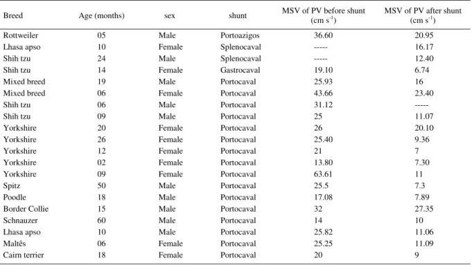
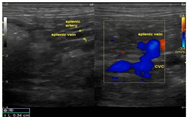
Dopplervelocimetric evaluation of portal vein as a diagnostic tool for portosystemic shunt diagnosis in dogs
Texto
Imagem


Documentos relacionados
OBJECTIVE: To compare superior ophthalmic vein blood flow parameters measured with color Doppler imaging in patients with congestive Graves’ orbitopathy before and after treatment
Abdominal ultrasonography and computed tomography (CT) showed communication between the portal vein and the middle hepatic vein, indicating an intrahepatic portosystemic venous
Detection of palate defects directly in sagittal images or flow in sagittal CDUS showed comparable, or better, accuracy than axial scans for CL with CP (detecting alveolar defects
A venography was performed (Figure 1), which showed agenesis of the inferior vena cava and dilated hemiazygos vein, with venous drainage into the common trunk, the superior
tive day following laparoscopic splenectomy, showing thrombosis of the splenic vein, main portal vein, right and left portal veins, and portions of the smaller intrahepatic
absence of the inferior vena cava in its habitual topography, right com- mon iliac vein with reduced diameter, venous drainage by a vessel on the left side of the aorta.. Figure 2 –
Objective : To determine the prevalence of deep vein thrombosis in paraplegic patients whose paraplegia was caused by traumas, using color Doppler ultrasonography for
Objectives: To assess the association between segmental GSV aplasia and the presence of varicose veins and/or GSV insuiciency in lower limbs using color Doppler ultrasonography,