Migration of endotacker into the bladder 7 years after
laparoscopic retroperitoneal burch application
_______________________________________________
Ahmet Salvarci
1, Yunus Agrali
21 Department of Urology, Konya Hospital, Semsi Tebrizi Mh. Serafettin Caddesi, Konya, Turkey and 2
Department of General Surgery, Konya Hospital, Semsi Tebrizi Mh. Serafettin Caddesi, Konya, Turkey
ABSTRACT
ARTICLE
INFO
______________________________________________________________ ______________________
Laparoscopy began to be used widely since the second half of 1990s as an alternative to laparotomy or vaginal approaches in incontinence and pelvic diseases in women, based on its claimed better success rates. Injuries were reported in the bladder, gas-trointestinal system and the entry of the Verress cannula in early and late laparoscopic applications. De-novo urging, voiding dysfunctions, marked recurrences and surgical inefficiencies were observed in 5-year follow-ups after laparoscopic incontinence sur-gery. Although tension-free midurethral sling operations replaced open laparoscopic colposuspensions nowadays, laparoscopic colposuspension is still preferred in cases where simultaneous laparoscopic paravaginal repair or sacrocolpopexy is considered or where synthetic graft implantation is contraindicated.
Moreover, meshes and endotackers are still frequently used in many laparoscopic ap-plications in various clinics. The migration of the tacker used in mesh fixation in a patient where retroperitoneal laparoscopic Burch was performed 7 years ago due to stress urinary incontinence and the extraction of the ossified tacker from the bladder will be presented.
Key words:
laparoscopy, Burch, tacker, bladder, migration
Int Braz J Urol. 2015; 41: 382-7
_____________________
Submitted for publication: June 06, 2014
_____________________
Accepted after revision: July 17, 2014
INTRODUCTION
Although more than 100 operations have been defined as success for preventing inconti-nence in women, laparoscopic Burch operation was launched as the first-line option due to its high success rate and low complication rate (1). At first, it was defined as the laparoscopic Burch colposuspension procedure in 1991 (2). The fact that cystocele could also be corrected in the la-paroscopic Burch operation was described as an advantage. The extraperitoneal approach was re-commended more especially for patients who un-derwent abdominal and retro-pubic surgery and for whom a simultaneous pelvic surgery was not planned (3). It was presented as an advantageous approach due to easy entry into the Retzius
CASE
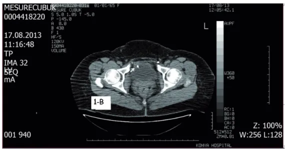
A 48-year old female patient with com-plaints of blood clots in the urine, burning during urination, frequent urination and incontinence was examined in the out-patient department. It was learned that retroperitoneal laparoscopic Bur-ch was performed on the patient by obstetricians 7 years ago due to incontinence and bladder prolap-se. The patient reported that she constantly felt ur-gency after the surgery. She also indicated that she had urgency type incontinence after the surgery. The patient reported that in addition to the use of gentamicin, ciprofloxacin for recurring urinary tract infections after the laparoscopic Burch pro-cedure, and even ceftriaxone due to pyelonephri-tis, she received continuous anticholinergic thera-py for irritative urinary complaints and ongoing incontinence. Grade II cystocele was observed at physical examination. Marshall-Marchetti-Krantz and Bonney test resulted (+). Urethral hypermo-bility was observed at the Q type test. The blood urea was 18 mg/dl, creatinine was 1.1 mg/dl and the leucocyte level was 3200/µL. A high level of leucocytes, erythrocytes and oxalate crystals were detected in the urinalysis. E. coli 100.000 cfu/ml proliferation was observed in the urine culture. No pathology was observed in the upper urinary sys-tem at ultrasonography, while it was reported that there was a stone in the bladder (Figure-1a). Prior to the assessment of the pelvic tomography, the radiologist recorded that tackers were observed around the bladder and urethra upon being in-formed about laparoscopic Burch and endotackers and indicated that probably one of these was insi-de the bladinsi-der while another one was behind the bladder (Figures 1b, c and d). A stone adhered on the right lateral wall of the bladder was observed during cystoscopy performed on the patient with spinal anesthesia (Figure-2). When the stone was removed from the bladder wall with forceps, the metallic helical structure of its adhered part was revealed and it was understood that this was an ossified tacker utilized in laparoscopy which had migrated in the bladder (Figure-2). The tacker was extracted via forceps accompanied by cystoscopy. The integrity of the bladder wall was monitored in the control cystoscopy conducted 45 days after
the cystoscopic removal of the ossified endotacker which had migrated into the bladder. Grade III cystocele was detected in the vaginal examination of the patient. The patient’s mixed incontinence was verified urodynamically and vaginal anterior colpography as well as transobturator tape were performed on the patient in the same session. In the physical examination of the patient, who had no incontinence complaints after surgery, it was observed that the Marshall-Marchetti-Krantz and Bonney tests were normal and that her urethral hypermobility to the Q type test recovered. No proliferation was observed in the urine culture in the post-surgical follow-ups.
A consent was received from the patient, indicating that her data would be archived and that her post-surgery data would be used for a scientific presentation.
DISCUSSION
Laparoscopic retroperitoneal Burch colpo-suspension has entered into our practice for the purpose of achieving urinary continence, simulta-neous repair of the cystocele and lowering morbi-dity. The duration of the operation which was lon-ger in the initial cases, shortened with technical support and with experience. Bladder perforation, hypotension, pneumomediastinum and pneumo-thorax were reported during the procedure in the initial cases (7-9). Urgent laparotomy was
formed in complications such as bowel damage, large vessel injury and venous thromboembolism on 280 patients who underwent laparoscopic Bur-ch (22.2%) within the group of 1.265 patients on whom laparoscopy was performed in the multi--centric prospective case loading analysis (10). In the comparison of sutures and tackers in colpo-suspensions comprising 254 women, it was also claimed that tacker was more risky and that the recurrence of incontinence was higher after tacker (11). Retroperitoneal hematoma as well as
blad-der injury were observed in the same patient af-ter laparoscopic Burch performed with tacker (12). Bladder damage, hemorrhage, urinary infection, fever, sepsis, de novo urgency, subcutaneous em-physema were reported in a 800-case series where laparoscopic Burch procedure was performed (13). Tackers are used widely used in general surgery for mesh fixation at laparoscopic inguinal hernia repairs and single-incision laparoscopic surgeries are performed with the assistance of an arthrosco-pic cannula and tacker (14, 15). Iatrogenic bladder
Figure 1b - Tomography (CT) image of endotacker inside the bladder.
Figure 1d - CT image of endotacker migrated behind the bladder.
Figure 2 - Migrated tacker adhered on the bladder wall at cystoscopy and post-surgery images.
stone formation associated with non-absorbable sutures passing through the bladder after the Bur-ch colposuspension applied again in the treatment of urinary incontinence was reported previously and dispareunia lasting 2 years as well as irritati-ve symptoms were obserirritati-ved in these patients (16). It was proposed to perform cystoscopy
post-sur-gically in these patients, where necessary, and to apply the sutures more laterally from the bladder.
our case. It was indicated only in one report that bladder stones on the surgical tape were removed via cystoscopy 6 years after laparoscopic colpo-suspension for stress incontinence occurring as a long term complication and this was stated to be the first such case (17). Although we initially thought that the tacker was placed upon direc-tly perforating the bladder in our patient, the fact that no pathology was detected in the bladder in numerous ultrasonographies and cystoscopies performed in different centers throughout 7 ye-ars made us shy away from this thought. We were more convinced that the tacker partially perfora-ted the bladder during the surgical procedure and entered into the bladder upon eroding with time. We believe that our thought is more substantia-ted with the ossified suspended tacker image on the right lateral wall of the bladder (Figure-2). Al-though this is a tacker whose tip is in partial con-tact with the bladder, the fact that it is inside the bladder supports our claim of migration. Further-more, the fact that one of the tackers placed on the right side was outside its normal position towards the back of the bladder, gave the impression of a tacker which was either not suitably positioned or missed or again of a perivesically migrated tacker (Figure-1d). It was observed in this case that the endotacker may have migrated inside the bladder after years and it was considered that it could have been one of the late complications of laparoscopic Burch procedure.
Nowadays, tension-free midurethral slings (such as transobturator tapes) have replaced lapa-roscopic and open colposuspensions. Due to their easy application, minimum invasiveness, low re-currence rate in 10-year follow-ups and contri-bution to the correction of sphincter impairment unlike other operations, tension-free midurethral slings have become the first-line choice. It was also recorded that tension-free mid sling may be applied in the same session with anterior colpo-raphy in the presence of cystocele and that the success rate was further increased. However, in the consideration of laparoscopic vaginal repair or laparoscopic sacrocolpopexy or in vaginal ap-plications where synthetic graft implantation is contraindicated in obstetrics and urology in the laparoscopic applications of various clinics,
lapa-roscopic colposuspension Burch procedures and endotacker comprising kits are still used simul-taneously nowadays. In the light of our clinical experience, we prefer mostly transobturator tape (TOT) and transvaginal tape (TVT) procedures in urinary incontinence surgery especially for sup-porting external urinary sphincter, ensuring ure-thral hypermobility stabilization and repairing in the same session the cystocele which determines success in such surgical procedures. It was obser-ved in the patient that the endotacker migrated behind and inside the bladder similarly to the migration of teflon which is a synthetic material used in the treatment of vesicoureteral reflux in the past. We recommend TOT and TVT even in laparoscopic gynecological approaches if urinary incontinence is to be treated in the same session.
In conclusion, we believe that it should not be forgotten that endotackers may migrate as a late complication in radiological images at diffe-rent anatomic sites including stones in the bladder of patients with a history of laparoscopic inconti-nence where endotackers are used.
CONFLICT OF INTEREST
None declared.
REFERENCES
1. Guner H, Yildiz A, Erdem A, Erdem M, Tiftik Z, Yildirim M. Surgical treatment of urinary stress incontinence by a suburethral sling procedure using a Mersilene mesh graft. Gynecol Obstet Invest. 1994;37:52-5.
2. Vancaillie TG, Schuessler W. Laparoscopic bladderneck suspension. J Laparoendosc Surg. 1991;1:169-73.
3. Frick AC, Paraiso MF. Laparoscopic management of incontinence and pelvic organ prolapse. Clin Obstet Gynecol. 2009;52:390-400.
4. Bulent Tiras M, Sendag F, Dilek U, Guner H. Laparoscopic burch colposuspension: comparison of effectiveness of extraperitoneal and transperitoneal techniques. Eur J Obstet Gynecol Reprod Biol. 2004;116:79-84.
6. Zullo F, Palomba S, Piccione F, Morelli M, Arduino B, Mastrantonio P. Laparoscopic Burch colposuspension: a randomized controlled trial comparing two transperitoneal surgical techniques. Obstet Gynecol. 2001;98:783-8. 7. Radomski SB, Herschorn S. Laparoscopic Burch bladder
neck suspension: early results. J Urol. 1996;155:515-8. 8. Wolf JS Jr, Carrier S, Stoller ML. Intraperitoneal versus
extraperitoneal insufflation of carbon dioxide as for laparoscopy. J Endourol. 1995;9:63-6.
9. Kiilholma P, Mäkinen J, Chancellor MB, Pitkänen Y, Hirvonen T. Modified Burch colposuspension for stress urinary incontinence in females. Surg Gynecol Obstet. 1993;176:111-5.
10. Johnston K, Rosen D, Cario G, Chou D, Carlton M, Cooper M, Reid G. Major complications arising from 1265 operative laparoscopic cases: a prospective review from a single center. J Minim Invasive Gynecol. 2007;14:339-44.
11. Moehrer B, Ellis G, Carey M, Wilson PD. Laparoscopic colposuspension for urinary incontinence in women. Cochrane Database Syst Rev. 2002;(1):CD002239. Update in: Cochrane Database Syst Rev. 2006;(3):CD002239. 12. Lee CL, Yen CF, Wang CJ, Lee PS, Chiu HC. Trocar-assisted
sling suspension for stress urinary incontinence: three-year follow-up. J Am Assoc Gynecol Laparosc. 2004;11:525-9. 13. Debodinance P, Delporte P, Engrand JB, Boulogne M.
[Complications of urinary incontinence surgery: 800 procedures]. J Gynecol Obstet Biol Reprod (Paris). 2002;31:649-62.
14. Ertem M, Ozben V, Yilmaz S, Ozveri E. The use of tacker and arthroscopy cannules in SILS cholecystectomy. J Laparoendosc Adv Surg Tech A. 2010;20:551-4.
15. Sajid MS, Ladwa N, Kalra L, McFall M, Baig MK, Sains P. A meta-analysis examining the use of tacker mesh fixation versus glue mesh fixation in laparoscopic inguinal hernia repair. Am J Surg. 2013;206:103-11.
16. Moyano Calvo JL, Romero Díaz A, Ortiz Gamis A, Martínez Moran A, Castiñeiras Fernández J. Iatrogenic bladder lithiasis in the Burch technique. An infrequent complication. Arch Esp Urol. 2000;53:468-9.
17. Yesilli C, Seckiner I, Mungan NA, Akduman B. Stone formation on surgical staple in the bladder: a long-term complication of laparoscopic colposuspension. Surg Laparosc Endosc Percutan Tech. 2007;17:568-9.
_______________________
Correspondence address:
Ahmet Salvarci, MD Department of Konya Hospital Semsi Tebrizi Mh. Serafettin Caddesi, 95 Karatay, Konya, 42103, Turkey Fax: + 90 505 544-2333