ABSTRACT
http://dx.doi.org/10.1590/1678-775720160154
Effect of silver nanoparticles on the physicochemical
and antimicrobial properties of an orthodontic
adhesive
Felipe Weidenbach DEGRAZIA19LFHQWH&DVWHOR%UDQFR/(,781(1, Isadora Martini GARCIA15RGULJR$OH[$57+852,
6XVDQD0DULD:HUQHU6$08(/1, Fabrício Mezzomo COLLARES1
1- Universidade Federal do Rio Grande do Sul, Faculdade de Odontologia, Laboratório de Materiais Odontológicos, Porto Alegre, RS, Brasil. 2- Universidade Federal do Rio Grande do Sul, Faculdade de Odontologia, Laboratório de Bioquímica e Microbiologia Oral, Porto Alegre, RS, Brasil.
Corresponding address: Fabrício Mezzomo Collares - Laboratório de Materiais Odontológicos - Faculdade de Odontologia
Universidade Federal do Rio Grande do Sul - Rua Ramiro Barcelos, 2492 - Rio Branco - 90035-003 - Porto Alegre - RS - Brazil - Phone: 55-51-33085198 - e-mail: [email protected]
Submitted: March 31, 2016 - Accepted: May 30, 2016
O
spot lesions. Objective: This study aimed to incorporate silver nanoparticle solutions (AgNP) in an orthodontic adhesive and evaluate its physicochemical and antimicrobial properties. Material and Methods: Silver nanoparticle solutions were added to a commercial adhesive in different concentrations (w/w): 0%, 0.11%, 0.18%, and 0.33%. Shear bond strength (SBS) test was performed after bonding metal brackets to enamel. Raman spectroscopy was used to analyze in sit u the degree of conversion (DC) of the adhesive layer. The surface free energy (SFE) was evaluated after the measurement of contact angles. Growth inhibition of St rept ococcus m ut ans in liquid and solid media was determined by colony-forming unit count and inhibition halo, respectively. One-way ANOVA was performed for SBS, DC, SFE, and growth inhibition. Results: The incorporation of AgNP solution decreased the SBS (p<0.001) and DC in situ (p<0.001) values. SFE decreased after addition of 0.18% and 0.33% AgNP. Growth inhibition of S. mutans in liquid media was obtained after silver addition (p<0.05). Conclusions: The addition of AgNP solutions to Transbond™ XT adhesive primer inhibited S. m ut ans growth. SBS, DC, and SFE values decreased after incorporation up to 0.33% AgNP solution without compromising the chemical and physical properties of the adhesive.Ke yw or ds: Metal nanoparticles. Anti-bacterial agents. Shear strength. Dental cements.
I N TROD UCTI ON
The formation of white spot lesions (WSLs) on dental enamel during orthodontic treatment is considered one of the worst problems faced in dental clinical care. This process mainly occurs due to low oral pH and lactic acid produced by
St r ep t ococcu s m u t an s metabolism22. Previous
studies reported an increase of bacterial growth at the interface between adhesive resins used to bond orthodontic attachments to enamel23,29.
Furthermore, even with patient compliance, the mechanical removal of plaque around orthodontic
cariogenic challenge.
The increased appearance of bacteria resistant to commercially antimicrobial agents has led to a crescent necessity for natural and nontoxic sources16
showed moderate evidence of prevention in
9.
Fluoride-releasing materials showed fast decrease of antibacterial activity24. In this regard, novel
materials aiming to reduce the adhesion of cariogenic streptococci to orthodontic adhesives for longer periods are essential to prevent enamel demineralization.
or zinc17. This allows for important clinical effects
with reduced toxicity. Furthermore, according to Zhang, et al.30 (2013), the incorporation of AgNP to
an adhesive did not affect the cytotoxicity regarding human gingival fibroblast viability. As shown elsewhere20, the antibacterial activity of silver
nanoparticle incorporated into adhesive cement may be prolonged up to 4 months. Nevertheless,
lead to formation of voids and thus weaken the polymeric matrix. For this reason, a simple method to promote AgNP dispersion with an aqueous solution was recently reported10.
Although the addition of water into the non-polymerized resin may harm cross-linking formation, it facilitates silver nanoparticles dispersion and, consequently, improves the antibacterial effect. Previous studies showed growth inhibition effects against S. m u t an s by silver ions release20 and
bacterial adhesion assay6. However, ion release
may lead to polymer degradation, whereas the bacterial adhesion assay on adhesive surface does not consider the bacterial colonization that occurs at the interface between enamel and adhesive. Furthermore, studies1,10 evaluating mechanical
properties of orthodontic adhesives showed no antibacterial assay after the incorporation of low concentrations of silver nanoparticles.
Therefore, the aim of this study was to evaluate the antibacterial effect on liquid and solid media, and physical-chemical properties of an orthodontic adhesive system after incorporation of different concentrations of silver nanoparticle solutions. The null hypothesis was that AgNP incorporation would promote antibacterial activity without compromising the physical-chemical properties of the orthodontic bonding system.
M ATERI AL AN D M ETH OD S
Pr e pa r a t ion of e x pe r im e n t a l a dh e sive s
To produce the experimental adhesive base, Transbond™ XT primer and adhesive were purchased from 3M Unitek Corp. (Monrovia, CA, USA – LOT N546755 and N556918). Silver nanopowder (particle size <150 nm – Figure 1) was purchased from Sigma-Aldrich Corp. (St. Louis, MO, USA – LOT #MKBP7829V). Bovine mandibular incisors were extracted and stored in distilled water at 4°C for 2 months. Orthodontic metallic brackets for upper central incisors (11.16 mm2) were purchased from
Dental Morelli Ltd. (Sorocaba, SP, Brazil – LOT 1958344). Attacktec 37% orthophosphoric acid gel was purchased from CaiTHEC Industrial Ltd. (Rio do Sul, SC, Brazil), and acrylic resin was purchased from VIPI Ltd. (Pirassununga, SP, Brazil).
Pr e pa r a t ion of st ock solu t ion a n d dilu t ion s
A stock solution of 11 wt% silver nanoparticles (AgNP) was prepared and mixed with distilled water to facilitate dispersion. The solution was ultrasonicated for 10 min to enhance particle dispersion. The AgNP solution was diluted in Transbond™ XT primer with a Labmate Soft pipette 0.5-10 μL from PZ HTL S/A (Warsaw, MA, Poland). Concentrations of AgNP occurred following a previous study10:
0.11% (w/w) – 1:100 dilution (1.0 μL of 11 wt% AgNP solution and 99 μL primer);
0.18% (w/w) – 1.8:100 dilution (1.8 μL of 11 wt% AgNP solution and 98.2 μL primer);
0.33% (w/w) – 3:100 dilution (3.0 μL of 11 wt% AgNP solution and 97 μL primer).
Sh e a r bon d st r e n gt h ( SBS) a n d a dh e sive r e m n a n t in de x ( ARI )
Forty-eight bovine incisors, free of cracks and caries, were obtained at a slaughterhouse and frozen for a maximum period of 1 month. The labial surface of the bovine incisors crowns (n=12) were polished with #600 and #1200 grid silicon carbide papers for 30 s. Each crown was embedded in self-cured acrylic resin with its long axis perpendicular to the horizontal plane. The embedded crowns were numbered and randomized for each group by the Research Randomizer Form program. The enamel surfaces were etched with acid gel and rinsed with was applied according to the following groups: control group, with no AgNP solution; 0.11% AgNP; 0.18% AgNP; and 0.33% AgNP. Then, Transbond XT adhesive was applied to the bracket base and the resin was pressed onto the enamel surface. The excess of adhesive was removed with an
Figure 1- Oval and rhomboid shapes of silver
explorer and light activated for 40 s (10 s for each face) with RadiiCal light emitting diode unit (SDI, Bayswater, VIC, Australia). All bonding procedures were performed according to the manufacturer’s instructions.
After 24 h in distilled water, the teeth were positioned in a Universal Testing Machine Shimadzu EZ-SX (Shimadzu Corp., São Paulo, SP, Brazil). The shear bond strength was tested using a chisel blade with crosshead speed of 0.5 mm/min and 500 N load cell. The ARI score8 was analyzed regarding
the amount of adhesive on the tooth surface with a stereomicroscope (10x).
D e gr e e of con ve r sion in sit u ( D C)
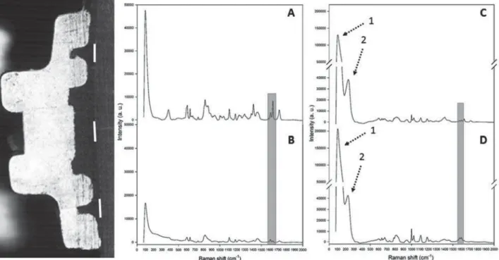
Orthodontic brackets were bonded to enamel (n=3) as previously described. The specimens remained in distilled water for 24 h and were cut in half by a cutting machine. Each half was positioned layer was evaluated at three sites (cervical, medium, and incisal) with micro-Raman spectroscopy (Figure 2) using Senterra equipment (Bruker Optik GmbH, Ettlingen, Baden-Württemberg, Germany). The unpolymerized spectrum of a primer sample was obtained before light activation. The average value of the measurements from each specimen was used to calculate the ratio of double bond content of monomer to polymer shown elsewhere13. The
characteristic Raman peaks for silver reference
compound were previously presented19.
Con t a ct a n gle ( CA) a n d su r fa ce fr e e e n e r gy ( SFE)
Three specimens of each group were submitted Stockholm, Sweden) to measure the mean CA
drops were: drop out size (2.0 μL), drop rate (2.0 μL/s), and displacement rate of water (20.0
μL/s). The test occurred for 20 s and the
Young-period of 10 s. The SFE calculation (in mN/m) occurred following OWRK/Fowkes* equation with Sweden).
Gr ow t h in h ibit or y a ct ivit y in liqu id m e dia
Growth inhibition assay occurred in a 96-well plate sterilized by ethylene oxide. Each well was
μ
broth supplemented with 0.5% of sucrose and 20
μL of inoculum, which was prepared after adjusting
St rept ococcus m ut ans (UA159) to 0.3 density at OD550nm. Twelve disk specimens (3.00 mmx2.00 mm) were made (n=3) and placed in contact with inoculum in BHI broth and incubated at 37°C for 24
*J
Ls=V
L+V
s¥
V
LD
.V
s
D
¥
V
LP
.V
sP
)
Figure 2- Half of a metal bracket used to analyze degree of conversion (DC) in situ (three lines) before (A) and after
% SRO\PHUL]DWLRQ RI $J13 DQG EHIRUH & DQG DIWHU ' SRO\PHUL]DWLRQ RI $J13 >KLJKOLJKW RI WKH SHDNV FRUUHVSRQGLQJWRDURPDWLFFPDQGDOLSKDWLFFPGRXEOHERQGV@$UURZVVKRZWKHFKDUDFWHULVWLF5DPDQ SHDNVRIVLOYHUFRPSRXQGV+LJKHULQWHQVLW\FRPSDUHGZLWK$J13LVGXHWRWKH$JODWWLFHYLEUDWLRQDOPRGH
140, 146, and 158 cm-1) and (2) Ag-O stretching/bending modes from AgO2 (230-248 cm-1) and AgCH
h. Hence, the disks were transferred to a microtube containing 900 μL of sterile saline solution (0.9% The bacterial suspensions were serially diluted (100
μL) in sterile saline solution. Two aliquots of 25 μL were plated onto BHI agar and incubated for 48 h anaerobically at 37°C, followed by enumeration of the CFU. Statistical analysis was performed by log10 CFU/mL.
D isk diffu sion a ssa y
Twelve disks were prepared (3.0 mmx2.00 mm) with the four concentrations of AgNP (n=3). Three disks of each group were placed on agar plates with 150 μL of grown S. m ut ans spread with glass balls. After 48 h of incubation at 37°C, the plates were visually inspected for the presence of inhibitory zones in the bacterial coat. The inhibitory halo of each disk was measured in millimeters.
St a t ist ica l a n a lysis
Statistical analysis was done on Sigma Plot version 12.0 for Windows (Systat Software Inc, San Jose, CA, USA). Normality test was performed with Shapiro-Wilk. One-way ANOVA and Tukey’s
post - hoc were performed for SBS, DC, SFE, and
log10CFU/mL. ARI score was evaluated by Kruskal-Wallis. The sample size calculation for each assay was based on previous studies3,11.
RESULTS
Data was normally distributed. The addition of AgNP particles statistically decreased the SBS (p<0.001) and DC in sit u (p<0.001) values of the commercial adhesive primer when compared with the control group (Table 1).
All groups with AgNP addition demonstrated similar antimicrobial activity against S. m ut ans. All groups statistically decreased (p<0.05) S. m ut ans
growth compared with control group (Table 1). After 48 h of disk-diffusion assay, no inhibitory halos were detected around disks of any silver concentration.
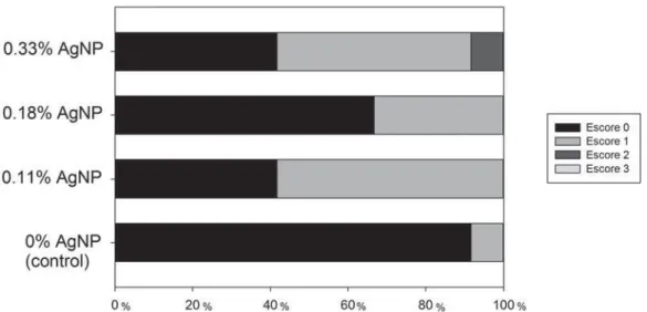
Figure 3 shows the ARI scores. The predominant mode of failure was adhesive failure (score 0), which occurred in control group. When AgNP was added to the adhesive primer, higher amount of cohesive failures occurred. No statistical difference was shown (p>0.05).
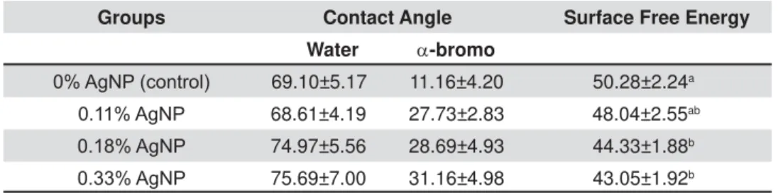
SFE mean values decreased (p<0.05) compared with control group after addition of 0.18% and 0.33% AgNP (Table 2).
Groups SBS* DC in situ* /RJ&)8ȉ
$J13FRQWURO 24.53±4.1a 89.50±0.58a 7.97±0.17a
$J13 17.63±3.2b 87.51±0.38b 5.90±0.66b
$J13 15.26±2.5b 87.44±0.03b 5.87±0.59b
$J13 16.29±2.9b 85.92±0.03c 5.62±0.71b
'LIIHUHQWOHWWHUVPHDQVWDWLVWLFDOGLIIHUHQFHLQWKHVDPHFROXPQS ȉ'LIIHUHQWOHWWHUVPHDQVWDWLVWLFDOGLIIHUHQFHLQWKHVDPHFROXPQS
Table 1- Mean and standard deviation of shear bond strength (SBS), degree of conversion (DC) in situ and antimicrobial activity against S. mutans of different silver concentrations
Figure 3-$GKHVLYHUHPQDQWLQGH[$5,VFRUH'HFUHDVHGDPRXQWRIDGKHVLYHUHPDLQHGRQWKHWRRWKZLWK$J131R
D I SCUSSI ON
O n e o f t h e m a i n r e a s o n s f o r e n a m e l demineralization during orthodontic treatment with biofilm on the enamel/adhesive interface. A large increase in antibiotic-resistant strains and vulnerable aspects of the antimicrobial agents present such as short-term antimicrobial activity and high toxicity, have led researchers to seek new alternative methods. Thus, because of their high reactivity due to their large surface-to-volume ratio, silver nanoparticles play a crucial role in inhibiting bacterial growth in aqueous and solid media18.
growth effect against S. m ut ans was achieved in liquid media (BHI broth) after incorporation of an aqueous solution with silver nanoparticles into an orthodontic adhesive. The bacterial inhibition growth in liquid media was successfully achieved with all AgNP concentrations.
Recently, a systematic review5 demonstrated
that the incorporation of antibacterial agents into orthodontic adhesives showed no difference in shear bond strength. One related study6 performed
antibacterial growth assay in liquid media with 250 and 500 ppm of silver nanoparticles against S. m ut ans without compromising shear bond strength; found after 24 h. Instead, in our study the inhibitory growth effect of silver nanoparticles was achieved with amounts of 500, 800, and 1500 ppm. The increase concentration of this aqueous solution of AgNP in the adhesive resin was probably the reason for this outcome. The addition of silver nanoparticles in water resulted in a homogenous dispersion through the adhesive. This enhanced distribution improved the antibacterial ability of the composite in spite of decreasing shear bond strength.
The inhibitory effect of silver was previously
determined against positive and
Gram-negative cells21,28. The DNA molecules become
condensed and lose their replication abilities due to a reaction against the denaturation effects of silver ions. Furthermore, silver ions interact with thiol groups in protein, which induce the inactivation of
the bacterial proteins15. Fan, et al.14 (2011) showed
inhibition halo against S. m ut ans after incorporation of 0.2 and 0.5% Ag benzoate (AgBz) on a PMMA-based resin blend. Based on the results of our study, the antimicrobial activity of Transbond™ XT primer after incorporating silver nanoparticles was due to direct contact with streptococci without silver ion release. In this study, no inhibition halo was observed around disks after 48 h incubation. The releasing property may harm bond strength longevity and induce adhesive weakness. Furthermore, non-releasing characteristics enable prolongation of the adhesive’s antibacterial effect.
DC in sit u decreased with higher concentrations of silver nanoparticles. Nonetheless, values of DC in a range between 85 and 90% are related to high cross-linking densities of dental polymers. A previous study20
obtained with its hardness results. The SFE values also decreased after incorporation of 0.18 and 0.33% AgNP. This might have occurred due to lower interaction between the dispersive liquid and the presence of water (polar liquid) in the specimens. The molecular interaction dipole x induced dipole forces between polar liquids and dispersive liquids is known to be weak. Lower SFE values can decrease the interface interaction between the enamel and the adhesive primer, resulting in lower values of SBS. On the other hand, as plaque accumulation around the bracket base has been associated with adhesive rough surface texture29, lower values of
SFE may prevent bacterial colonization as shown elsewhere7.
In accordance to a previous study25, water
incorporation over 5 vol% to dental adhesives may hinder the formation of an organized polymer network, consequently diminishing its physical properties. In this study, we incorporated an aqueous solution up to 3 vol% and, despite a decrease, the SBS values obtained after incorporation of AgNP solution were greater than the clinically acceptable values between 6-8 MPa27.
Considering that immediate bond strength to tooth substrate is related to the mechanical properties of the adhesive layer, higher mechanical properties are required to achieve more durable bonding to dental
Groups Contact Angle Surface Free Energy
Water D-bromo
$J13FRQWURO 69.10±5.17 11.16±4.20 50.28±2.24a
$J13 68.61±4.19 27.73±2.83 48.04±2.55ab $J13 74.97±5.56 28.69±4.93 44.33±1.88b $J13 75.69±7.00 31.16±4.98 43.05±1.92b
'LIIHUHQWOHWWHUVPHDQVWDWLVWLFDOGLIIHUHQFHLQWKHVDPHFROXPQS
substrate12. Hence, the values of SBS shown in this
study are consistent with data in the literature3,26.
The failure pattern shift after incorporation of AgNP may prevent enamel from possible damage. The adhesive failure between enamel and adhesive increases the chance of harming the enamel tissue’s surface.
Despite the fact that the prior application of an adhesive resin has been reported as a step that could be set aside during metal bracket bonding4,
a particular indication of adhesive resin should be used as an antimicrobial promoter to protect enamel against bacteria. The incorporation of antimicrobial agents into an adhesive resin may be considered a more suitable approach, since it comes into direct contact with the enamel surface1. A recent study
showed higher amounts of microleakage at the adhesive-enamel interface in different adhesive types2. Its lower viscosity and wettability compared
with orthodontic composites promote higher dispersion and penetration of antimicrobial agents into the enamel surface.
CON CLUSI ON
The incorporation of AgNP solutions into Transbond™ XT adhesive primer showed inhibition growth against S. m ut ans.
S h e a r b o n d s t r e n g t h d e c r e a s e d a f t e r incorporation of AgNP solution up to 0.33% without compromising the chemical and physical properties of the adhesive primer.
ACKN OW LED GM EN TS
The authors FWD and IMG gratefully acknowledge the support of CAPES and CNPq for the scholarship.
REFEREN CES
1- Akhavan A, Sodagar A, Motjahedzadeh F, Sodagar K. Investigating the effect of incorporation nanosilver/nanohydroxyapatite particles on the shear bond strength of orthodontic adhesives. Acta Odontol Scand. 2013;71(5):1038-42.
2- Alkis H, Turkkahraman H, Adanir N. Microleakage under orthodontic brackets bonded with different adhesive systems. Eur J Dent. 2015;9(1):117-21.
3- Altmann AS, Collares FM, Ogliari FA, Samuel SM. Effect of methacrylated-based antibacterial monomer on orthodontic adhesive system properties. Am J Orthod Dentofacial Orthop. 2015;147(4 Suppl):S82-7.
4- Altmann AS, Degrazia FW, Celeste RK, Leitune VC, Samuel SM, Collares FM. Orthodontic bracket bonding without previous adhesive priming: a meta-regression analysis. Angle Orthod. 2016;86(3):391-8.
5- Altmann AS, Collares FM, Leitune VC, Samuel SM. The effect of antimicrobial agents on bond strength of orthodontic adhesives: a meta-analysis of in vitro studies. Orthod Craniofac Res. 2016;19(1):1-9.
6- Ahn SJ, Lee SJ, Kook JK, Lim BS. Experimental antimicrobial
Dent Mater. 2009;25(2):206-13.
7- Ahn SJ, Lim BS, Lee SJ. Surface characteristics of orthodontic adhesives and effects on streptococcal adhesion. Am J Orthod Dentofacial Orthop. 2010;137(4):489-95.
8- Artun J, Bergland S. Clinical trials with crystal growth conditioning as an alternative to acid-etch enamel pretreatment. Am J Orthod. 1984;85(4):333-40.
9- Benson PE, Parkin N, Dyer F, Millett DT, Furness S, Germain P. Fluorides for the prevention of early tooth decay (demineralised
Syst Rev. 2013;(12):CD003809.
10- Blöcher S, Frankenberger R, Hellak A, Schauseil M, Roggendorf MJ, Korbmacher-Steiner HM. Effect on enamel shear bond strength of adding microsilver and nanosilver particles to the primer of an orthodontic adhesive. BMC Oral Health. 2015;15:42.
11- Centenaro CC, Rostirolla FV, Leitune VC, Parolo CF, Ogliari FA,
2-yl)-4-hydroxyphenyl)ethyl methacrylate to an experimental adhesive system. Acta Odontol Latinoam. 2015;28(1):72-8. 12- Collares FM, Ogliari FA, Zanchi CH, Petzhold CL, Piva E, Samuel
polymer network of adhesive resin. J Adhes Dent. 2011;13(2):125-9.
13- Collares FM, Portella FF, Leitune VC, Samuel SM. Discrepancies in degree of conversion measurements by FTIR. Braz Oral Res. 2014;28:9-15.
14- Fan C, Chu L, Rawls HR, Norling BK, Cardenas HL, Whang K. Development of an antimicrobial resin – a pilot study. Dent Mater. 2011;27(4):322-8.
15- Feng QL, Wu J, Chen GQ, Cui FZ, Kim TN, Kim JO. A mechanistic study of the antibacterial effect of silver ions on Escherichia coli and Staphylococcus aureus. J Biomed Mater Res. 2000;52(4):662-8.
16- Gajbhiye M, Kesharwani J, Ingle A, Gade A, Rai M. Fungus-mediated synthesis of silver nanoparticles and their activity against
2009;5(4):382-6.
17- Hernández-Sierra JF, Ruiz F, Pena DC, Martínez-Gutiérrez F, Martínez AE, Guillén AJ, et al. The antimicrobial sensitivity of Streptococcus mutans to nanoparticles of silver, zinc oxide, and gold. Nanomedicine. 2008;4(3):237-40.
Characterization and antimicrobial performance of nano silver coatings on leather materials. Braz J Microbiol. 2015;46(1):41-8. 19- Martina I, Wiesinger R, Jembrih-Simbürger D, Schreiner M. Micro-raman characterisation of silver corrosion products: instrumental set up and reference database. e-PS. 2012;9:1-8. 20- Moreira DM, Oei J, Rawls HR, Wagner J, Chu L, Li Y, et al. A novel antimicrobial orthodontic band cement with in situ-generated silver nanoparticles. Angle Orthod. 2015;85(2):175-83. 21- Morones JR, Elechiguerra JL, Camacho A, Holt K, Kouri JB, Ramírez JT, et al. The bactericidal effect of silver nanoparticles. Nanotechnology. 2005;16(10):2346-53.
RB. Transmission, diversity and virulence factors of Streptococcus mutans genotypes. J Oral Sci. 2005;47(2):59-64.
23- Øgaard B. White spot lesions during orthodontic treatment: mechanisms and fluoride preventive aspects. Sem Orthod. 2008;14(3):183-93.
24- Passariello C, Sannino G, Petti S, Gigola P. Intensity and duration of in-vitro antibacterial activity of different adhesives used in orthodontics. Eur J Oral Sci. 2014;122(2):154-60. 25- Paul SJ, Leach M, Rueggeberg FA, Pashley DH. Effect of water content on the physical properties of model dentine primer and bonding resins. J Dent. 1999;27(3):209-14.
26- Retamoso LB, Collares FM, Ferreira ES, Samuel SM. Shear bond
Appl Oral Sci. 2009;17(3):190-4.
28- Shrivastava S, Bera T, Roy A, Singh G, Ramachandrarao P, Dash D. Characterization of enhanced antibacterial effects of novel silver nanoparticles. Nanotechnology. 2007;18(28):1-9. 29- Sukontapatipark W, el-Agroudi MA, Selliseth NJ, Thunold K,
appliances. A scanning electron microscopy study. Eur J Orthod. 2001;23(5):475-84.