Original Article
414 Rev Bras Hematol Hemoter. 2013;35(6):414-6
Study of possible clinical and laboratory predictors of alloimmunization against red
blood cell antigens in cancer patients
Carla Luana Dinardo1 Gláucia Munemasa Ito2 Luciana Ribeiro Sampaio2 Alfredo Mendrone Júnior1
1Fundação Pró-Sangue – Hemocentro de São Paulo, São Paulo, SP, Brazil
2Instituto do Câncer do Estado de São Paulo – ICESP, São Paulo, SP, Brazil
Conlict-of-interest disclosure:
The authors declare no competing inancial interest
Submitted: 5/1/2013 Accepted: 6/24/2013
Corresponding author: Carla Luana Dinardo
Fundação Pró-Sangue - Hemocentro de São Paulo Av Dr. Enéas de Carvalho Aguiar, 155, Cerqueira César
05403-000 São Paulo, SP, Brazil [email protected]
www.rbhh.org or www.scielo.br/rbhh
DOI: 10.5581/1516-8484.20130123
Introduction
Alloimmunization against red blood cell (RBC) antigens is a late complication of blood transfusions that affects 8% to 12% of all recipients(1). This percentage increases signiicantly
if patients on chronic transfusion regimens, such as those with sickle cell anemia and myelodysplastic syndrome, are taken into account. In this scenario, approximately 15% of patients develop antibodies against one or more blood group antigen(1-3). The alloantibodies in
recipient’s sera are associated with transfusion delays due to the complexity of pre-transfusion tests, dificulty in inding compatible RBC units and late hemolytic transfusion reactions.
A classic stochastic modeling of the RBC alloimmunization process suggested that less than 15% of all blood recipients are prone to developing antibodies after an antigen-mismatched transfusion stimulus (immunologic responders)(4). The number of transfusions and
the presence of an hemoglobinopathy are associated with a higher risk of alloimmunization(5).
However, other clinical factors capable of accurately identifying those individuals before the appearance of the irst alloantibody are still lacking. Recent evidence suggests that patients with solid cancer may be at higher risk for developing alloantibodies against RBC antigens(6).
Development of an alloantibody after exposure to an external RBC antigen is inluenced by the recipients T cell function(7) and underlying disease status(8). In cancer patients, poor performance
status and a poor quality of life is associated with the patient’s systemic inlammatory background and with shorter survival(9). Moreover, cancer aggressiveness, represented by the presence of
metastasis and by undifferentiated histology, is associated with higher levels of inlammatory markers. This leads to worse performance in inlammation-based prognostic scores(10).
Providing phenotyped packed RBCs (comprising mostly of immunogenic antigens) to all solid cancer patients may be a useful strategy to prevent alloimmunization. On the other hand, it has a negative economic impact, since the price paid for a phenotyped red cell pack is higher than that paid for a regular unit. It would be useful to determine whether there are any clinical features capable of predicting alloimmunization in oncologic patients to justify the prescription of phenotyped RBC units.
The aim of this study was to evaluate whether factors related to disease severity (performance status/presence of metastasis/body mass index) and inlammatory background (C-reactive protein - CRP) can predict the risk of RBC alloimmunization in cancer patients.
Methods
All patients known to have become alloimmunized in a tertiary oncology service between 2009 and 2011 (Group 1) were selected for this case-control study. Patients were selected if they developed antibodies against any RBC antigen and if they had at least one negative Background: The inlammatory background of patients inluences the process of alloimmunization against red blood cell antigens. Proof of this statement to clinical practice is still lacking.
Objective:The aim of this study was to verify whether factors related to disease severity and inlammatory status of cancer patients can predict alloimmunization.
Methods: This was a case-control study in which alloimmunized oncologic patients treated between 2009 and 2012 were compared with a non-alloimmunized control group regarding the severity of the disease (metastasis/ performance status/body mass index) and C-reactive protein levels.
Results: The groups did not differ signiicantly in terms of C-reactive protein, Eastern Cooperative Oncology Group (ECOG)/Karnofsky performance status, presence of metastasis and body mass index.
Conclusion: It is not possible to predict alloimmunization in cancer patients based on severity of illness and inlammatory markers. Strategies of screening patients by phenotyping blood based on these criteria are not justiied.
415
Study of possible clinical and laboratory predictors of alloimmunization against red blood cell antigens in cancer patients
Rev Bras Hematol Hemoter. 2013;35(6):414-6
antibody result. Patients with hematologic malignancies were excluded as they present a higher rate of transfusion and different clinical behavior from solid tumors.
Every time an alloimmunized patient was included in the study, two control patients (Group 2) were selected amongst all patients that had been transfused in the hospital on the same day as long as they met the following criteria: 1) negative antibody screen, 2) same number of transfusions as the alloimmunized patient, 3) conirmed diagnosis of solid cancer and 4) same hospital loor or ambulatory as the case. All patients received bed side, leukodepleted RBC units and none received phenotyped units before the development of the irst alloantibody.
Groups were compared in terms of the Eastern Cooperative Oncology Group (ECOG) performance status scale, Karnofsky performance status scale, CRP, presence of liver, lung or bone metastasis and body mass index (BMI). The ECOG performance scale ranges from 0 to 5, with 0 denoting perfect execution of daily activities and 5, death. Similarly, the Karnofsky scale ranges from 100 to 0, where 100 represents perfect functional status and 0, death. Antibody screening and identiication were performed using Biorad®, Brazil RBC panels. CRP was dosed using an immunoturbidimetric method.
All scale variables were irst analyzed in terms of normality using the Kolmogorov-Smirnov test. The student t-test was used for variables with normal distribution, and the Mann-Whitney test for data with non-normal distribution. Categorical variables were compared using the chi-square test. A p-value < 0.05 was considered signiicant.
Results
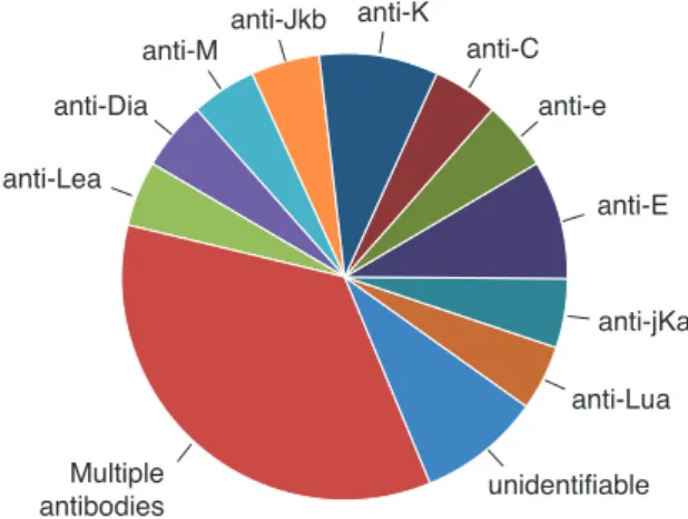
Twenty-two alloimmunized patients were allocated to Group 1 and 44 control patients to Group 2. Demographic characteristics of Group 1 and Group 2 are listed in Table 1. The mean age of patients in Group 2 was higher than those in Group 1. Both were homogeneous in respect to the remaining criteria. The number of transfusions was similar for both groups (a mean of 4.1 transfusions per patient). The antibodies identiied in the patients of Group 1 are shown in Figure 1. No patient with anti-D was included because all patients were previously sensitized to this antigen.
For Group 2 patients, some information is missing regarding smoking (3), alcohol consumption (4) and chemotherapy (2).
BMI: Body mass index; CRP: C-reactive protein; ECOG: Eastern Cooperative Oncology Group performance status scale
Table 1 - Demographic characteristics of alloimmunized (Group 1) and non-alloimmunized (Group 2) cancer patients
Table 2 - Comparison between alloimmunized (Group 1) and non-alloimmunized (Group 2) cancer patients in terms of possible clinical predictors of red blood cell alloimmunization
Group 1 Group 2 p-value
Number 22 44
Female/Male 14/8 27/17 0.858
Age (years - mean ± SD) 49.4 ± 13.65 57.9 ± 16.12 0.038
Diagnosis (n) 0.399
Esophagus cancer 1 0
Stomach cancer 1 5
Head and neck cancer 1 4
Lung cancer 0 1
Breast cancer 2 3
Prostate cancer 1 1
Bone cancer 0 1
Sarcoma 0 1
Colon cancer 3 11
Uterus cancer 5 2
Others 8 15
Smoking (n) 0.535
Yes 12 19
No 10 22
Alcohol consumption (n) 0.689
Yes 4 9
No 18 31
Need for Hospitalization (n) 0.705
Yes 16 30
No 6 14
Need for ICU (n) 0.848
Yes 6 13
No 16 31
Chemotherapy (n) 0.619
Yes 6 14
No 16 28
Variable Group 1 Group 2 p-value
Mean BMI
[kg/m2 - mean ± SD (n)] 23.4 ± 5.1 (20) 21.5 ± 4.1 (42) 0.608 CRP
[mg/L - mean ± SD (n)] 115 ± 115.3 (11) 157.3 ± 110.6 (24) 0.320
Presence of metastasis 0.741
Yes 11 19
No 11 18
Karnofsky score 0.964
≤ 70% 8 15
>70% 13 25
ECOG score 0.730
≥ 2 5 8
< 2 16 32
Figure 1 - Antibodies identiied amongst alloimmunized cancer
patients (Group 1)
416
Dinardo CL, Ito GM, Sampaio LR, Mendrone Júnior A
Rev Bras Hematol Hemoter. 2013;35(6):414-6 Conclusions
The severity of illness and inlammatory background do not predict RBC alloimmunization in oncologic patients. Strategies to provide phenotyped blood to recipients with cancer based on these variables should not be encouraged. Further studies assessing molecular immunological behavior of this speciic population may be helpful in understanding and preventing alloimmunization events.
References
1. Rosse WF, Gallagher D, Kinney TR, Castro O, Dosik H, Moohr J, et al.
Transfusion and alloimmunization in sickle cell disease. The Cooperative
Study of Sickle Cell Disease. Blood. 1990;76(7):1431-7.
2. Miller ST, Kim HY, Weiner DL, Wager CG, Gallagher D, Styles LA, Dampier CD, Roseff SD; Investigators of the Sickle Cell Disease Clinical Research Network. (SCDCRN). Red blood cell alloimmunization in sickle cell disease: prevalence in 2010. Transfusion. 2013;53(4):704-9 3. Sanz C, Nomdedeu M, Belkaid M, Martinez I, Nomdedeu B, Pereira
A. Red blood cell alloimmunization in transfused patients with
myelodysplastic syndrome or chronic myelomonocytic leukemia. Transfusion. 2013;53(4):710-5.
4. Higgins JM, Sloan SR. Stochastic modeling of human RBC alloimmunization: evidence for a distinct population of immunologic responders. Blood. 2008;112(6):2546-53. Comment in: Blood. 2010;115(21):4315; author reply 4315-6. Blood. 2008;112(6):2180-1. 5. Zalpuri S, Zwaginga JJ, le Cessie S, Elshuis J, Schonewille H, van der
Bom JG. Red-blood-cell alloimmunization and number of red-blood-cell transfusions. Vox Sang. 2012;102(2):144-9.
6. Bauer MP, Wiersum-Osselton J, Schipperus M, Vandenbroucke JP,
Briët E. Clinical predictors of alloimmunization after red blood cell
transfusion. Transfusion. 2007;47(11):2066-71.
7. Bao W, Yu J, Heck S, Yazdanbakhsh K. Regulatory T-cell status in
red cell alloimmunized responder and nonresponder mice. Blood.
2009;113(22):5624-7.
8. Hendrickson JE, Desmarets M, Deshpande SS, Chadwick TE, Hillyer CD, Roback JD, et al. Recipient inlammation affects the frequency and
magnitude of immunization to transfused red blood cells. Transfusion.
2006;46(9):1526-36.
9. Kao SC, Vardy J, Harvie R, Chatield M, van Zandwijk N, Clarke S, et al. Health-related quality of life and inlammatory markers in malignant pleural mesothelioma. Support Care Cancer. 2013;21(3):697-705. 10. Jeong JH, Lim SM, Yun JY, Rhee GW, Lim JY, Cho JY, et al. Comparison
of two inlammation-based prognostic scores in patients with unresectable advanced gastric cancer. Oncology. 2012;83(5):292-9.
11. Hendrickson JE, Roback JD, Hillyer CD, Easley KA, Zimring JC. Discrete
Toll-like receptor agonists have differential effects on alloimmunization
to transfused red blood cells. Transfusion. 2008;48(9):1869-77. 12. Hendrickson JE, Chadwick TE, Roback JD, Hillyer CD, Zimring JC.
Inlammation enhances consumption and presentation of transfused RBC antigens by dendritic cells. Blood. 2007;110(7):2736-43.
13. Noizat-Pirenne F, Tournamille C, Bierling P, Roudot-Thoraval F, Le
Pennec PY, Rouger P, et al. Relative immunogenicity of Fya and K antigens in a Caucasian population, based on HLA class II restriction analysis. Transfusion. 2006;46(8):1328-33.
14. Tyler LN, Harville TO, Blackall DP. Multiple alloantibodies after transfusion in an infant treated with inliximab. N Engl J Med. 2007;357(20):2092-3; discussion 2093.
15. Yazer MH, Triulzi DJ, Shaz B, Kraus T, Zimring JC. Does a
febrile reaction to platelets predispose recipients to red blood cell
alloimmunization? Transfusion. 2009;49(6):1070-5. Comment in: Transfusion. 2009;49(6):1032-6.
No statistically signiicant differences were found between Groups 1 and 2 in terms of CRP (p-value = 0.32), the ECOG performance status (p-value = 0.73) and Karnofsky performance status (p-value = 0.964). The presence of liver, lung or bone metastasis and the mean BMI also did not differ signiicantly between the two groups (p-value = 0.741 and p-value = 0.608, respectively). Table 2 summarizes data obtained from both groups.
Discussion
Our results show that neither the severity of the illness (ECOG performance scale, Karnofsky scale, presence of metastasis and BMI) nor inlammatory background (CRP) can predict the risk for RBC alloimmunization in solid cancer patients. To our knowledge, this is the irst study to assess alloimmunization phenomena in patients with non-hematologic malignancies.
Much of the current knowledge about alloimmunization is based on experiments performed on murine models. Those experiments demonstrated that the generation of an alloantibody against a RBC antigen after a transfusion stimulus relies on the regulatory function of Treg cells, which is declined in responders(7)
and also depends on the way a foreign RBC antigen is presented to T cells by spleen antigen presenting cells (APC)(8,11). In regards
to this last statement, recent evidence suggests that a viral-like inlammation stimulus leads to a higher rate of consumption of transfused RBCs by splenic dendritic cells and, consequently, higher rates of alloimmunization(12).
In humans, observational studies have been performed to search for an association between HLA molecules and risk for alloimmunization. An HLA restriction has been observed for the Fya and Jka antigens, but not for more promiscuous antigens such as K(13). In respect of a higher inlammatory background as
a predisposing factor for alloimmunization, a few case reports of patients with autoimmune diseases have shown that they are prone to alloimmunization(14). An association was found between
the development of RBC alloantibodies and acute febrile reactions during transfusion. The febrile reaction may act as an inlammatory stimulus(15).
Our results do not go against the inlammatory hypothesis presented above, but also do not reinforce it. Our indings were based on a small sample that may not have enough power to detect predictors. As in cancer patients, the aggressiveness of the disease is associated with its inlammatory background(10), it would be
plausible to ind an association between the presence of metastasis, poor performance status, CRP or BMI and alloimmunization. However, the lack of a clinical predictor between factors related to the severity of cancer and the inlammatory background of the recipient probably highlights that RBC alloimmunization depends on a combination of variables rather than on one isolated feature. Dosing suppressive cytokines (interleukin-10 and transforming growth factor-beta) in recipients may be a better parameter of Treg cell function compared to CRP however those assays are deinitely less widely available. Based on the indings of this study, neither the severity of cancer nor higher CRP levels clearly deine a group of cancer patients as ‘responders’.