Quantification of CD8
+CD38
+T lymphocytes by flow cytometry does not
represent a good biomarker to monitor the reactivation of cytomegalovirus
infection after allogeneic hematopoietic stem cell transplantation
1
Universidade Federal de Minas Gerais – UFMG, Belo Horizonte, MG, Brazil
2
Department of Complementary Propedeutics, Universidade Federal de Minas Gerais – UFMG, Belo Horizonte, MG, Brazil 3
Department of Clinical Medicine, Universidade Federal de Minas Gerais – UFMG, Belo Horizonte, MG, Brazil
Vânia Abadia Soares Lino1
Silvana Maria Eloi Santos2
Henrique Neves da Silva Bittencourt3
Maria Luiza Silva1
Tiago Spizziri1
Raquel Bretas1
Suzane Pretti Figueiredo Neves2
Background: Infection/reactivation of cytomegalovirus is a major cause of morbidity and mortality in immunocompromised transplant patients. It has already been observed in kidney and liver transplantation patients that cytomegalovirus disease is accompanied by significant increases in circulating CD8+CD38+ T lymphocytes. There are no reports that study CD8+CD38+ T lymphocytes
to monitor/diagnose cytomegalovirus disease in hematopoietic stem cell transplantation patients.
Objective: The aim of this study was to evaluate some cellular activation markers on circulating mononuclear cells (CD38 and HLA-DR) in patients submitted to hematopoietic stem cell transplantation and to establish any correlation with cytomegalovirus disease as diagnosed by antigenemia.
Methods: Blood samples of 15 transplant patients were analyzed by flow cytometry using anti-CD3, anti-CD4, anti-CD8, anti-CD38, CD16, CD56 and anti-HLA-DR monoclonal antibodies and the results were evaluated in respect to cytomegalovirus antigenemia measured by indirect immunofluorescence. Minitab for Windows was used for statistical analysis and a p-value < 0.05 was considered significant.
Results: Patients with positive antigenemia did not show any significant increase in the percentages of cells expressing the CD38 or HLADR activation markers when compared to patients with negative antigenemia. On the contrary, all patients showed high percentages of these cells independent of the presence of cytomegalovirus disease.
Conclusions: This study suggests that the investigation of these lymphocyte sub-populations in patients submitted to hematopoietic stem cell transplantation does not seem to contribute to the early identification of cytomegalovirus disease.
Keywords: Hematopoietic stem cell transplantation; Cytomegalovirus; Flow cytometry; Antigenemia, CD38
Introduction
Allogeneic hematopoietic stem cell transplantation (HSCT) is indicated as a therapeutic option to treat many bone marrow deficiencies, immunological diseases and congenital disorders of hematopoiesis.(1) Before transplant, the patient is subjected to a conditioning
regimen with high doses of chemotherapy with or without total body irradiation thus preparing him/her to receive the graft. In patients with nonmalignant diseases, the conditioning is intended to suppress the immunological system, while for patients with malignancies, it is associated to cytotoxic and immunosuppressant drugs.(2) Thus, these
patients become susceptible to infections including the reactivation of latent infections. Of these, infection by cytomegalovirus (CMV) is the leading cause of morbidity and mortality due to viral infections after HSCT.(3,4)
CMV, a virus that belongs to the Herpesviridae family, has the ability to remain latent in the human body and can be reactivated after immunosuppression.(5) Approximately 70% of
HSCT recipients are CMV-seropositive or will receive transplants from CMV-seropositive donors.(6) Due to this high prevalence of seropositivity, primary infection after transplant is
relatively rare. Reactivation, on the other hand, occurs in up to 60-70% of seropositive patients mainly between the second and seventh month after HSCT and, if not treated earlier, can cause serious impairment of several organs.(7) The diagnosis of CMV disease is usually
reached by identifying the CMV pp65 antigen in neutrophils (antigenemia test) or by real time polymerase chain reaction to identify viral DNA in plasma or blood.(8) However, these
techniques are time-consuming, laborious and expensive. Thus, flow cytometry has major advantages: it minimal manipulates cells and is a method widely used to define cell populations by immunophenotyping;(9) it is relatively fast and cheaper than PCR and antigenemia.
Conflict-of-interest disclosure: The authors declare no competing financial interest
Submitted: 1/25/2011 Accepted: 3/22/2011
Corresponding author:
Vânia Abadia Soares Lino Hospital das Clínicas, Faculdade de Medicina
Avenida Alfredo Balena, nº 190, sala 129, 1ºandar – Santa Efigênia
30130-100 – Belo Horizonte, MG, Brazil Phone: 55 31 3409-9607
www.rbhh.org or www.scielo.br/rbhh
In adults, immune activation correlates with an increase in the expression of the CD38 marker on CD4+ and CD8+
cells.(10) CD38 is a type II transmembrane glycoprotein with
enzyme activity expressed in lymphocytes, macrophages, endothelial cells, dendritic cells and in other cell types.(11) In
infections, like those caused by HIV, increased expressions of CD38 and HLA-DR in the cytotoxic T population indicate activation of the immune system and greater progression to AIDS.(10,12)
In kidney transplant patients, the identification of increased amounts of CD8+CD38+ T lymphocytes was found
to be highly sensitive (100%) and specific (91%) in the diagnosis of primary CMV infection. In all cases, increases in these subpopulations were detected early, before or together with the first signs of CMV infection.(13) In another study of
patients who received liver transplants, the increased expression of CD38 on CMV specific lymphocytes was a marker of active CMV infection or reactivation.(14) Another
molecule whose expression is increased in T lymphocytes after virus activation is the class II MHC antigen, HLA-DR; these events are either induced by pathological processes or by immunization.(15,16)
In patients who received HSCT, there are no studies that evaluate CD4 and CD8 lymphocyte subsets that express CD38 and HLA-DR to diagnose CMV infection or reactivation. Therefore, the purpose of this study was to evaluate CD38 and HLA-DR cell activation markers in circulating mononuclear cells by flow cytometry in the first two months after allogeneic HSCT and to correlate the findings with infection/reactivation by CMV.
Methods
This study was approved by the Research Ethics Committee of UFMG (COEP # ETIC 543/07). Fifteen patients submitted to HSCT at the Hospital das Clinicas, Federal University of Minas Gerais in the period from November 2007 to May 2010 were studied. This study included all patients submitted to allogeneic HSCT independent of gender, age and socioeconomic group (Table 1).
They were divided into two groups (antigenemia positive - Ag+ and antigenemia negative - Ag-) according to
the results of antigenemia; patients with negative antigenemia were considered the control group. Both groups were studied on two occasions: on day +30 and day +60 after transplant, the period in which infection/reactivation of CMV is most frequently observed.
Blood samples were collected in EDTA by venipuncture using the vacuum collection system (Vacutainer, Greiner, Brazil) to evaluate antigenemia. An extra 2 mL of blood was used for flow cytometry.
The CMV Brite Turbo kit was used (IQ Products, the Netherlands). The technique was performed according to the manufacturer's instructions using the following steps: preparation of the leukocyte suspension, cell count in a blood
cell counter (Sysmex, Roche); preparation of cytosmear; fixation, permeabilization and fluorescent staining of the slides. Reading of slides was by fluorescence microscopy (40x); positive cells showing homogeneous fluorescent green-yellow staining of core neutrophil were counted. Examinations were performed in duplicate with a positive result having at least two fluorescent cells per duplicate and negative results not having any stained cells.
incubation, the lysis of erythrocytes was performed with a commercial solution (FACSTM Lysing Solution - Becton
Dickinson, USA); the supernatant was discarded and the leucocytes washed with isotonic solution (Hemoton-Hemogram, Brazil). Then 250 µL of isotonic solution were added to each tube. Flow cytometry (FACScalibur- Becton Dickinson, USA) and the CELLQuestTM computer program
(Becton Dickinson, USA) were used to count the cells (50,000 events/tube).
Immunophenotyping data were analyzed using different strategies depending on the cellular phenotype using multiple resources of the CELLQuestTM computer program including
conventional analysis, combined analysis "gated" for the CD16 and CD56 expressions modified by to Sathler-Avelar,(17)
modified and semiquantitative analysis for the FcγR3 (CD16) expression on monocytes according to Martins-Filho.(18)
Statistical analysis
The Minitab program for Windows was used. The nonparametric Mann-Whitney test was used to compare groups. The correlation between variables was performed by calculating the Spearman correlation coefficient. For all statistical analyses a p-value < 0.05 was considered statistically significant.
Results
The fifteen patients were monitored for at least 60 days after HSCT. Only one patient had no complications until hospital discharge. No patient had primary CMV infection. Seven patients had reactivation of CMV (47%). Six patients (40%) had acute graft-versus-host disease (GvHD). Three of these patients had positive antigenemia test and three were negative. Four patients died during hospitalization (Table 1).
Thirteen of the 15 patients (86.6%) had low absolute (< 1.0 x 10⁹/L) and relative (≤ 15%) counts of total circulating
lymphocytes on days +30 and +60 after transplant. NK cells (CD3-CD16+ or CD56+) and monocytes showed normal counts
by day +30 (Table 4).
The number of B cells was very low, almost undetectable, on both occasions.
Three patients (two on day +30 and one on day +30 and day +60) did not have enough lymphocytes stained with monoclonal antibodies to be properly analyzed and so these data were excluded from the study. Two of these patients had positive antigenemia tests and one was negative.
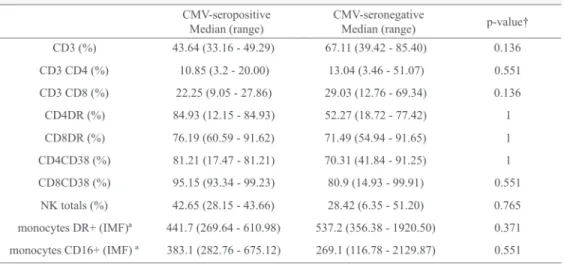
There were no significant differences in the percentages of CD4 and CD8 lymphocytes expressing CD38 and HLA-DR on comparing positive (Ag+) and negative (Ag-) antigenemia
groups on days +30 and +60 (Tables 4 and 5; Figure 1). Instead, regardless of whether infection/reactivation of CMV was present (antigenemia ≥ 2 cells/100,000 leukocytes) all patients had very high CD38+ and HLADR+ T lymphocyte
counts (Tables 4 and 5, Figure 1).
There were no significant differences in the expression of other markers of lymphocytes and monocytes between the two groups on both day +30 and day +60 (Tables 4 and 5).
Figure 1 - Percentages of T lymphocytes expressing CD38 and HLA-DR. Ag+ = positive antigenemia group; Ag - = negative antigenemia group; n = 12 (d+30), n = 8(d+60)
day +30 (r = 0.762, p-value < 0.004) and also on day +60 (r = 0.881, p-value < 0.004). There was no significant correlation for CD8 T lymphocytes.
Discussion
The aim of this study was to evaluate cell activation markers (CD38 and HLA-DR) on circulating mononuclear cells in allogeneic HSCT patients using flow cytometry and correlate the findings with infection/reactivation of CMV, the diagnosis of which was established by antigenemia. However, unlike as has been described in kidney and liver transplant patients,(13,14) we did not observe any increase in CD38+ or
HLADR+ T cells in individuals with reactivation/infection by
CMV compared to those without infection.
Belles-Isles et al. found high percentages of CD38+CD8+
cells in renal transplant recipients who had positive antigenemia tests compared to those who were negative. In this study, the percentage values > 20% corresponded to a 90% positive predictive value for primary CMV infection. In
another study on liver transplant recipients,(14) the authors
showed that the frequency of T CD38+ CMV-specific cells
in patients with seroconversion to IgG or increase in anti-CMV IgM, was significantly increased, suggesting that the analysis of these cells is a good parameter for early diagnosis of liver complications induced by viruses. The fact that we did not observe significant differences between the positive and negative antigenemia groups raises the following question: do HSCT patients in the early period post-transplant, as was studied here, when their immunity is being reconstituted (still under intense immuno-suppression) respond to viral infections in the same way as solid organ transplant patients in a situation where immunosuppression is less intense? It is known that in allogeneic HSCT, the T lymphocyte count takes months to one year to complete recovery, much longer than other leukocyte populations(19,20) as was also observed in our
study where, 60 days after transplant, the number of T lymphocytes was lower than normal. Another important point to consider is the small number of individuals that we studied which results in a small statistical power. A larger number of patients may confirm whether indeed there is no difference in the number of T CD8+CD38+ cells among
patients who presented or not positive antigenemia for CMV. A fact that drew attention was that, although there were no differences between the T CD8+CD38+ populations regardless of the result of antigenemia (that is, in patients with or without CMV disease), the percentages of these cells were greatly increased (> 80%) as were T CD4+CD38+
lymphocytes (> 67%). The results repeated when both subpopulations were evaluated, expressing HLADR (T CD8+HLADR+ > 71% and T CD4+HLADR+ > 46%) suggesting
that lymphocyte activation occurs in all patients after allogeneic HSCT. Faria et al.,(21) studying healthy individuals,
of 2.6% and 6.0%, respectively. The following percentages were found in another study to establish reference values for the different phenotypes of lymphocytes in peripheral blood:(22) T CD4+HKADR+ = 1.7%, CD8+HLADR+ = 1%,
CD4+CD38+ = 14.85% and CD8+CD38+ = 2.85%; all these
values were well below the values observed by our group. It was imagined, at first, that the increase in the T
CD8+CD38+ subpopulation would represent the
reconstitution of the immune system as, in the neonatal period, this marker is expressed in early hematopoietic cells, is lost during maturation and reappears during cell activation. Thus, newborns express high levels of CD38 on their T cells, which reduce with age.(23,11) Thus, our findings might
only be reflecting the generation of new T lymphocytes egress from the transplanted graft and selected in the thymus. However, proof of this would require the labeling of cells with other monoclonal antibodies characteristic of this period, which was not performed in this study. Storek et al.(23) showed that after allogeneic HSCT, the
reconstitution of the T repertoire is different from that which occurs in newborns. Literature data to date show that the T repertoire after bone marrow transplantation is basically constituted by T lymphocytes stimulated in the periphery and not from the graft. These peripheral lymphocytes suffer clonal expansion after antigen varied stimulation – the recipient's own antigens, viruses, bacteria, fungi – thus forming the new repertoire of receptors, but with less diversity.(20,24,25) Also the increase in the percentage of T
CD4+ and CD8+HLADR+, and especially the strong positive
correlation between the CD38 and HLADR markers in CD4+
T cells, reinforces the idea that they may be, in most cases, activated lymphocytes and not young, immature cells, coming straight out of the thymus.
Therefore, our findings suggest that the quantification of T CD8+CD38+ cells by flow cytometry is not useful in
detecting infection/reactivation of cytomegalovirus in HSCT patients, as all patients showed high percentages of these cells regardless of the outcome of antigenemia. Therefore, the diagnosis and monitoring of CMV infection should be performed by established and reliable testing methods, that is real time PCR (26,27) in blood or plasma and
antigenemia (where molecular methods are unavailable), or by promising methods currently under research, such as real time PCR using saliva.(28) Moreover, further studies
should be conducted to investigate these and other markers of immaturity, memory and lymphocyte activation and their relationships to long-term outcomes of transplantation because it is known that prolonged cell activation in other clinical situations, can lead to immune exhaustion.(29,30)
Acknowledgements
The authors wish to thank FAPEMIG – Fundação de Amparo à Pesquisa do Estado de Minas Gerais
References
1. Woo SB, Lee SJ, Schubert MM. Graft - host disease. Crit Rev Oral Biol Med. 1997;8(2):201-16.
2. Dahllöf G, Bagesund M, Ringden O. Impact of conditioning regimens on salivary function, caries-associated microorganisms and dental caries in children after bone marrow transplantation. A 4-year longitudinal study. Bone Marrow Transplant. 1997;20(6):479-83.
3. Detrick B, Hooks JJ, Keiser J, Tabbara I. Detections of cytomegalo-virus proteins by flow cytometry in the blood of patients undergoing hematopoietic stem cell transplantation. Exp Hematol. 1999;27 (3):569-75.
4. Bonon SH, Rossi CL, de Souza CA, Vigorito AC, Costa SC. Comparison of serology, antigenemia assay and the polymerase chain reaction for monitoring active cytomegalovirus infections in hematopoietic stem cell transplantation patients. Rev Inst Med Trop Sao Paulo. 2006;48(5):275-8.
5. Gerna G, Zavattoni M, Percivalle E, Zella D, Torsellini M, Revello MG. Diagnosis of human cytomegalovirus infections in the immunocompromised host. Clin Diagn Virol. 1996;5(2-3):181-6.
6. Hebart H, Jahn G, Sinzger C, Kanz L, Einsele H. CMV Infection in bone marrow and solid organ transplant patients in the era of antiviral prophylaxis. Herpes. 2000;7(1):13-7.
7. Boeckh M, Nichols WG, Papanicolaou G, Rubin R, Wingard JR, Zaia J. Cytomegalovirus in hematopoietic stem cell transplant recipients: current status, known challenges, and future strategies. Biol Blood Marrow Transplant. 2003;9(9):543-58.
8. Piiparinen H, Hockerstedt K, Gronhagen-Riska C, Lappalainen M, Suni J, Lautenschlager I. Comparison of plasma polymerase chain reaction and pp65-antigenemia assay in the quantification of cytomegalovirus in liver and kidney transplant patients. J Clin Virol. 2001;22(1):111-6.
9. D'hautcourt J-L, Éric M. Analysis of cellular antigens by flow cytometry. In: Marie-Christine B, Éric M. Immunophenotyping of blood and bone marrow leukocytes. Amsterdam: Harwood Academic Publishers; 1997. p. 21-7.
10. Liu Z, Cumberland WG, Hutlin LE, Prince HE, Detels R, Giorgi JV. Elevated CD38 antigen expression on CD8+ T cells is a stronger marker for the risk of chronic HIV disease progression to AIDS and death in the Multicenter for AIDS Cohort Study than CD4 cell count, soluble immune activation markers, or combinations of HLA-DR and CD38 expression. J Acquir Immune Defic Syndr Hum Retrovirol. 1997;16(2):83-92.
11. McCloskey TW, Cavaliere T, Bakshi S, Harper R, Fagin J, Kohn N, et al. Immunophenotyping of T lymphocytes by three-color flow cytometry in healthy newborns, children, and adults. Clin Immunol Immunopathol. 1997;84(1):46-55.
12. Rosenblatt HM, Stanley KE, Song LY, Johnson GM, Wiznia AA, Nachman SA, Krogstad PA. Pediatric AIDS Clinical Trials Group 377 Study Team. Immunological response to highly active antiretroviral therapy in children with clinically stable HIV-1 infection. J Infect Dis. 2005;192(3):445-55.
13. Belles-Isles M, Houde I, Lachance JG, Nöel R, Kingma I, Roy R. Monitoring of cytomegalovirus infections by the CD8+CD38+ T-cell subset in kidney transplant recipients. Transplantation. 1998; 65(2):279-82.
14. Benz C, Utermöhlen O, Wulf A, Villmow B, Dries V, Geoser T, et al. Activated Virus-Specific T cells are early indicators of anti-cmv immune reactions in liver transplant patients. Gastroentrology. 2002;122(5):1201-15.
16. Caruso A, Licenziati S, Corulli M, Canaris AD, De Francesco MA, Fiorentini S, et al. Flow cytometric analysis of activation markers on stimulated t cells and their correlation with cell proliferation. Cytometry.1997;27(1):71-6
17. Sathler-Avelar R, Lemos EM, Reis DD, Medrano-Mercado N, Araújo-Jorge TC, Antas PR, et al. Phenotypic features of peripheral blood leucocytes during early stages of human infection with Trypanosoma cruzi. Scand J Immunol. 2003;58(6):655-63.
18. Martins-Filho OA. Course on flow cytometry In XXV Meeting of the Brasilian Society of Immunology. Florianópolis(SC), Brasil; 2000. p.11-12.
19. Reis MA, Visentainer JE. Reconstituição imunológica após o transplante de medula óssea alogênico. Rev Bras Hematol Hemoter. 2004;26(3):212-7.
20. Williams KM, Hakim FT, Gress RE. T cell immune reconstitution following lymphodepletion. Semin Immunol. 2007;19(5):318-30. 21. Faria AM, de Moraes SM, Freitas LH, Speziali E, Soares TF, Figueiredo-Neves SP, et al. Variation rhythms of lymphocyte subsets during healty aging. Neuroimmunomodulation. 2008;15(4-6):365-79.
22. Bisset LR, Lung TL, Kaelin M, Ludwig E, Dubs RW. Reference values for peripheral blood lymphocyte phenotypes applicable to the healthyadult population in Switzerland. Eur J Haematol. 2004;72(3):203-12.
23. Storek J, Witherspoon RP, Storb R. T cell reconstitution after bone marrow transplantation into adult patients does not resemble
T cell development in early life. Bone Marrow Transplant. 1995; 16(3):413-25.
24. Geddes M, Storek J. Immune reconstitution following haematopoietic stem-cell transplantation. Best Pract Res Clin Haematol. 2007;20 (2):329-48.
25. Limave AP, Huang ML, Leisenring W, Stensland L, Corey L, Boeckh M. Cytomegalovirus (CMV) DNA load in plasma for the diagnosis of CMV disease before engraftment in hematopoietic stem-cell transplant recipients. J Infect Dis. 2001;183(3):377-82.
26. Williams KM, Gress RE. Immune reconstitution and implications for immunotherapy following haematopoietic stem-cell transplantation. Best Pract Res Clin Haematol. 2008;21(3):579-96.
27. Gault E, Micchel Y, Dehée A, Belabani C, Nicolas J-C, Garbarg-Chenon A. Quantification of human cytomegalovirus DNA by real-time PCR. J Clin Microbiol. 2001;39(2):772-5.
28. Correia-Silva JF, Bruna-Romero O, Resende RG, Miranda LP, Oliveira FE, Costa FO, et al. Saliva as a source of HCMV DNA in allogeneic stem cell transplantation patients. Oral Dis. 2010;16(2): 210-6.
29. Appay V, Almeida J R, Sauce D, Autran B, Papagno L. Accelerated immune senescence and HIV-1 infection. Exp Gerontol. 2007;42 (5):432-7.
30. Mueller SN, Ahmed R. High antigen levels are the cause of T cell exhaustion during chronic viral infection. Proc Natl Acad Sci U S A. 2009;106(21):8623-8.