ABSTRACT
Sao Paulo Med J. 2008;126(4):227-8.
C
A
SE REPOR
T
Daniel Sáenz-Abad
Santiago Letona-Carbajo
José Luis de Benito-Arévalo
Isabel Sanioaquín-Conde
Francisco José Ruiz-Ruiz
Prostatic tuberculosis: case report
Hospital Clínico Universitario “Lozano Blesa”, Zaragoza, Spain
CONTEXT: Tuberculosis of the prostate has mainly been described in immunocompromised patients. However, it can exceptionally be found as an isolated lesion in immunocompetent patients.
CASE REPORT: We report a case of prostatic tuberculosis in a young, healthy and immunocom-petent patient with unremarkable fi ndings from intravenous urographic examination. Computed tomography showed an abscess in the prostate and Mycobacterium tuberculosis was isolated in a urine culture. Treatment with isoniazid, rifampin and pyrazinamide was successful.
KEY WORDS: Tuberculosis, urogenital. Prostate. Immunocompetence. Diagnosis. Antitubercular agents.
INTRODUCTION Genitourinary tuberculosis is a common type of extrapulmonary tuberculosis. The kidneys, ureter, bladder or genital organs are usually involved. Tuberculosis of the prostate has mainly been described in im-munocompromised patients.1 However, it can
exceptionally be found as an isolated lesion in immunocompetent patients. We report a case of prostatic tuberculosis in a young, healthy and immunocompetent patient with unremarkable findings from intravenous urographic examination.
CASE REPORT A 36-year-old man who was born in Senegal was admitted to our hospital with a 12-month history of fever and fatigue and weight loss of 10 kg. He had no other medical problems and he reported not hav-ing traveled anywhere durhav-ing the last two years. On examination, he was febrile. The physical examination otherwise revealed no abnormalities. His white blood cell count was 4,100/mm3, hematocrit 34% and erythrocyte
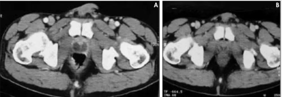
sedimentation rate 80 mm/h. Urinalysis showed pyuria and no abnormalities were found on a chest radiograph. The blood and urine cultures were negatives and so was the HIV-antibody test. A tuberculin skin test was positive. Thoracoabdominal computed tomography (CT) examination revealed a large prostatic abscess with necrosis, while the other structures of the genitourinary system were normal (Figure 1a). Several days later, we were able to isolate Mycobacterium tuberculosis by urine culture in Lowenstein-Jensen medium. Finally, we performed an intravenous urographic examination, without fi nding any abnormalities in other structures of the urinary tract.
The patient was treated with isoniazid, rifampin and pyrazinamide for two months
and he continued with isoniazid and rifampin for a further ten months. After this treatment, CT examination showed that the prostate had returned to its normal size and the necrotic ab-scess had decreased dramatically (Figure 1b).
DISCUSSION Changing patterns of population mi-gration and the development of large pools of immunocompromised individuals has reversed the downward trend of tuberculo-sis.2,3 Today, extrapulmonary tuberculosis is
becoming increasingly common, especially involving the lymphatic system, pleura and urogenital tract.3 Extrapulmonary sites are
involved in 50% to 70% of immunocom-promised patients, especially HIV patients. Genitourinary tuberculosis accounts for 5-10% of extrapulmonary cases in devel-oped countries and 15-20% in developing countries. Nevertheless, isolated tuberculous prostatic abscesses are uncommon, especially in immunocompetent patients. M. tuberculosis
is the most common pathogen involved, but others such as M. kansasii or fortuitum have been described. It is thought that tuberculous involvement of the prostate is usually the result of hematogenous spreading, although this can also occur as a result of descent of the organism from the kidneys or local spreading from the genital tract. Although sexual transmission of M. tuberculosis has been reported, it is extremely rare.1
228
Sao Paulo Med J. 2008;126(4):227-8.
are abnormal in most cases of genitourinary tuberculosis.4,5 In some patients,
prostate-spe-cific antigen (PSA) may be elevated. Imaging studies help to locate and determinate the presence of concurrent tuberculosis in other organs. Therefore, transrectal ultrasound, intravenous urography and chest X-ray should be considered. Ultrasound reveals
enlarge-1. Gebo KA. Prostatic tuberculosis in an HIV infected male. Sex Transm Infect. 2002;78(2):147-8
2. Wise GJ, Marella VK. Genitourinary manifestations of tuber-culosis. Urol Clin North Am. 2003;30(1):111-21. 3. Lenk S, Schroeder J. Genitourinary tuberculosis. Curr Opin
Urol. 2001;11(1):93-8.
4. Tazi K, Nouri M, Elkhadir K, et al. La tuberculose prostatique A propos de deux cas. [Prostatic tuberculosis. Report of 2 cases]. Ann Urol (Paris). 1999;33(4):274-6.
5. García-Rodríguez JA, García Sánchez JE, Muñoz Bellido JL, et al. Genitourinary tuberculosis in Spain: review of 81 cases. Clin Infect Dis. 1994;18(4):557-61.
Sources of funding: Without sources of funding
Conflict of interest: Without conflict of interest
Date of first submission: March 2, 2007
Last received: May 7, 2007
Accepted: May 27, 2008
REFERENCES
AUTHOR INFORMATION
Daniel Sáenz-Abad, MD. Internist. Emergency Department, Hospital Clínico Universitario “Lozano Blesa”, Zaragoza, Spain.
Santiago Letona-Carbajo, MD. Internist. Department of Infectious Disease, Hospital Clínico Universitario “Lozano Blesa”, Zaragoza, Spain.
José Luis de Benito-Arévalo, MD. Radiologist. Computed Tomography Section, Department of Radiology, Hospital Clínico Universitario “Lozano Blesa”, Zaragoza, Spain.
Isabel Sanjoaquín-Conde, MD. Internist. Department of Infectious Disease, Hospital Clínico Universitario “Lozano Blesa”, Zaragoza, Spain.
Francisco José Ruiz-Ruiz, MD. Internist. Emergency Depart-ment, Hospital Clínico Universitario “Lozano Blesa”, Zaragoza, Spain.
Address for correspondence: Daniel Sáenz Abad
Servicio de Urgencias Av. San Juan Bosco, 15 Zaragoza 50009. Spain. Phone +34 976 556 400 extn. 3915 Fax. +34 976 351 661
E-mail: [email protected] Copyright © 2007, Associação Paulista de Medicina
RESUMEN Tuberculosis prostática: informe del caso
CONTEXTO: La tuberculosis prostática ha sido principalmente descrita en pacientes inmunodeprimidos. Sin embargo, es excepcional como lesión aislada en pacientes inmunocompetentes.
CASO CLINICO: Describimos el caso de un varón sano e inmunocompetente con tuberculosis prostática y hallazgos irrelevantes en la urografía de eliminación realizada. La tomografía computerizada (TC) mostró un absceso en próstata, aislándose en el urocultivo Mycobacterium tuberculosis. El tratamiento con isoniazida, rifampicina y pirazinamida fue exitoso.
PALABRAS CLAVE: Tuberculosis urogenital. Próstata. Inmunocompetencia. Diagnóstico. Agentes antituber-culosos.
ment of the gland with solitary or multiple hypoechoic zones of varying sizes inside it. Irregularity of the outline of these hypoechoic areas may also be noted.
CT scans or magnetic resonance imaging may be useful for differential diagnosis, and some characteristic findings from prostate tuberculosis cases have been published. CT
provides direct viewing of intraprostatic le-sions and reveals them as low-density areas with irregular borders. Contrast-enhanced CT demonstrates these lesions more clearly. Magnetic resonance imaging (MRI) may re-veal low signal-intensity lesions suggestive of abscesses. Intravenous urographic examination is recommended because, in a high percentage of cases, renal tuberculosis is found in asso-ciation.2,3 In our case, CT scans showed an
abscess with central necrosis that improved after treatment. These characteristics may be considered diagnostic for tuberculosis. Nonetheless, the definitive diagnosis was given by microbiological findings. Faced with find-ings of genitourinary tuberculosis, physicians should ensure that pulmonary involvement can be ruled out.
In conclusion, in this patient, the symp-toms suggested the presence of tuberculosis. However, it is exceptional for the prostate alone to be affected, as an isolated lesion in the genitourinary tract of an immunocom-petent patient.
Figure 1. A. Computed tomography examination showing a large prostatic abscess with necrosis. B. Computed tomography examination after forty days of treatment, showing a normal size prostate gland with a minimal necrotic abscess.