Report on hearing loss in
oncology
Summary
Christiane Schultz 1, Maria Valéria Schmidt Goffi-Gomez 2, Patrícia Helena Pecora Liberman 3,
André Lopes Carvalho 4
1 Master in science, Fundação Antonio Prudente. Speech therapist, Audiology Sector, Hospital do Câncer A C Camargo.
2 Doctor in science of communication disorders, Universidade Federal de São Paulo. Speech therapist, Audiology Sector, Hospital do Câncer A C Camargo. 3 Master in sciences, Fundação Antonio Prudente. Speech therapist, Audiology Sector, Hospital do Câncer A C Camargo.
4 Livre-docente (habilitation) in oncology, FMUSP. Full surgeon, Head & Neck Surgery and Otorhinolaryngology Department, Hospital de Barretos - Hospital do Câncer.
Paper submitted to the BJORL-SGP (Publishing Management System – Brazilian Journal of Otorhinolaryngology) on May 2, 2008; and accepted on November 2, 2008. cod. 5829
C
isplatin is used frequently as an antineoplastic drug in the treatment of many different cancers. However, when used in doses over 360mg/m2, ototoxicity may ensue, resulting in lossof hearing. Criteria for identifying and quantifying hearing loss have been devised. Aim: To describe the features of different hearing loss classification systems and to identify their implications and use in oncologic patients. Method:
Hearing loss was classified in 31 patients before and after chemotherapy, according to different criteria, assessing the sensitivity and specificity of each classification system.
Results: Hearing loss results were highly variable (ranging from 29% to 61%). Only 4 of 31 subjects with post-therapy hearing loss were identified by all the methods. A few subjects with hearing loss were classified as normal hearing in some of the criteria. A normal PTA was found in 18 of 31 subjects in the post-treatment evaluation. Conclusion:
None of the criteria assesses the complaints of patients. The criteria described in this study were inadequate to identify hearing loss following chemotherapy, requiring additional information for physicians to better understand the hearing losses and their implications for the quality of life of patients.
Keywords: cisplatin/adverse effects, oncology, hearing loss/ classification, chemotherapy/adverse effects. ORIGINAL ARTICLE
INTRODUCTION
Cisplatin (CDDP) is an antineoplastic drug used often in the treatment of various tumors. Side effects include: nausea, vomiting, myelosuppression nephrotoxi-city, central and peripheral neuropathies, and ototoxicity
(Oliveira1; Rybak et al.2). Ototoxicity may ensue when the
drug is given at a cumulative dose over 360mg/m2 (Brock
et al.3; Pedalini et al.4, Simon et al.5, Knoll et al.6). Cisplatin
ototoxicity is the result of cochlear injury, initially in the vascular striae and the outer hair cells of the basal gyrus, which result in hearing loss at high frequencies
(Rade-maker et al.7; Rybak et al.8). Continued use of the drug
may result in hearing loss at low frequencies (Pedalini et al.4, Zuur et al.9).
Hearing loss may cause significant loss in the quality of life of patients; thus, a concern with ototoxicity should be present throughout oncological therapy. Detecting and monitoring ototoxicity to initiate preventive measures is one of the methods for avoiding hearing loss.
Oncologists are increasingly concerned with drug toxicity in oncological therapy, and have created objecti-ve criteria to measure the specific toxicity for each organ during chemotherapy cycles.
Many criteria have been published in the literature, with the aim of identifying, describing and even quanti-fying hearing loss. In this paper we apply four instruments
that will be described below (Davis and Silverman10;
Brock et al.3; ASHA-American Speech-language-Hearing
Association11; NCI12).
In clinical practice, there criteria are difficult to apply, since they do not define clearly the degree of he-aring loss or its impact - patient complaints are not taken into account - and do not include all types and grades of hearing alterations.
OBJECTIVE
The aim of this study was to assess and identify the characteristics of each classification method of hearing loss and to adapt them to oncological monitoring.
METHOD
A prospective study was proposed and approved by the Research Ethics Committee of the institution in which the investigation was to be conducted (acceptance number 549/03). All patients were consulted about their participation in the study, and signed a free informed consent form. Audiological assessments were carried out in 31 subjects seen at the Audiology unit; inclusion criteria were as follows: cisplatin chemotherapy only; absence of ontological complaints; no radiotherapy in the head and neck, and pre- and post-therapy audiological assessments.
There were 16 male and 15 female patients. Age ranged from 7 to 66 years; the mean age was 28 years.
All subjects in this study underwent full audiological testing before, during and after chemotherapy with CDDP; however, only conventional pure tone audiometry data were computed. A Madsen, Orbiter model version 922 audiometer was used for measuring air and bone conduc-tion pure tone auditory thresholds (in dBHL) (Redondo et al.13; Yantis14).
Results were tabulated according to classifications proposed by various authors and entities. The CTCAE
(Common Terminology Criteria for Adverse Events12) was
proposed by the NCI (National Cancer Institute). This ins-trument aims to describe the adverse events due to chemo-therapeutic drugs during oncological treatment; numerical values (1 to 4) were given to the reactions observed in each organ during each chemotherapy cycle (Annex 1).
Brock et al.3 attributed number values (0 to 4) to
va-rious types of hearing loss due to chemotherapy (Annex 2).
The ASHA11 considers ototoxicity as a 20dB threshold
elevation at a specific frequency, a 10dB threshold eleva-tion at two consecutive frequencies, or absent responses at three consecutive frequencies in post-therapy testing.
Davis and Silverman10 classify the degree of hearing
loss according to the mean value of thresholds at 500, 1000 and 2000Hz; normal values - 0 to 20 dB, mild hearing loss - 21 to 40 dBHL, moderate hearing loss - 41 to 70 dBHL, severe hearing loss - 71 to 90 dBHL, profound hearing loss - over 95 dBHL.
A comparison was done taking the 25 dBHL threshold as the limit between normal hearing and hearing loss to assess the sensitivity and specificity of each crite-rion, with the aim of analyzing hearing loss according to frequency (PPF). Fisher’s exact test was applied to study the significance of each method. The GraphPad Prism version 2.0 software was used. The significance level was < 0.05.
RESULTS
Table 1 shows that, according to NCI12 criteria, 12 of 31 patients (38%) presented hearing loss at the end of therapy. Of these, only one had grade I changes, 10 had grade II changes, and one had grade III changes.
According to Brock et al.’s3 criterion, 19 patients (61%)
presented hearing loss at the end of therapy. Of these, 5 had grade I changes, 5 had grade II changes, 5 had grade III changes, and 4 had grade IV changes. According to
ASHA11 criteria, 17 subjects (54%) presented hearing loss
Annex 1. Table of the NCI proposed classification
Common Terminology Criteria for Adverse Events (CTCAE) (2003)
Cancer Therapy Evaluation Program, Common Terminology Criteria for Adverse Events
Version 3.0 DCTD, NCI, NIH, DHHS. March31, 2003 (http:// ctep.cancer.gov) Publishing date, 10 June 2003
Hearing / Ear
Auditory Adverse
Event Name 1 2 3 4 5
patient with or with no baseline
audiogram and non-monitored patients Hearing: (monitoring pro-gram) Thresholds or 15 - 25dB de-crease relative to the baseline audiogram at two or more adjacent frequencies in at least one ear, or subjective change
in the absence of grade 1
Thresholds or 25 - 90 dB decrease in two or more adjacent
frequen-cies in at least one ear
ADULTS: thresholds over
25 - 90 dB at 3 adjacent
frequen-cies in at least one ear.
CHILDREN: Hearing loss sufficient to re-quire treatment
including AASI (loss > 20dB at speech frequen-cies, bilateral or > 30dB unilateral
and that require special phonoau-diological care) ADULTS: pro-found bilateral hearing loss (> 90dB) CHILDREN: Audiological indication for
co-chlear implants, requiring special
phonoaudiologi-cal care.
-OBS: pediatric recommendations are similar to those of adults unless otherwise specified. For children and teenagers (< or = 18 years) with no pre-treatment baseline audiogram, hearing is considered as below 5dB.
Hearing: patients with no baseline
audiogram and not included in a
monitoring pro-gram
Hearing: no moni-toring program
Hearing loss with no need to use AASI (no effect on
activities of daily life)
Hearing loss with no deed to use AASI (affecting activities of daily
life)
Profound bilateral hearing loss
( > 90dB )
-Otitis, outer ear
(non- infectious) Otitis media
Otitis external with erythema or dry desquamation
Otitis external with desquama-tion, ear wax or effusion, tympa-nic perforation, tympanoplas ty Otitis external with mastoiditis, stenosis or
oste-omyelitis
Necrosis of bone
or soft tissues Death
OBS: patients with or with no baseline audiogram, included or not in a monitoring program.
Otitis, middle ear
(non- infectious) Otitis media Otitis serous
Otitis serous with indication for me-dical intervention
Otitis with effu-sion, mastoiditis
Necrosis of canal, of bone or soft
tissues
Death
Tinnitus Tinnitus
-Tinnitus not affec-ting activities of
daily life
Tinnitus affecting activities of daily
life
Disability
Loss of function
-OBS: patients with or with no baseline audiogram, included or not in a monitoring program.
Hearing / Ear Others
Hearing / Ear
Others mild moderate severe
life risk / loss of
Davis and Silverman’s10 criteria revealed that 9
sub-jects (29%) had hearing loss at the end of therapy. The identification percentage of hearing loss varied conside-rably among criteria - from 29% to 61%. We also found that 61% had auditory thresholds over 25 dBHL in at least one frequency bilaterally.
Statistical analysis consisted of specificity and sen-sitivity calculations for each criterion; Fisher’s test was applied to calculate the p value, as shown on the Table below.
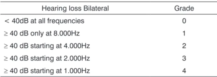
Annex 2. Descriptive table of Brock et al.’s proposed classification (1991)
Hearing loss Bilateral Grade
< 40dB at all frequencies 0 ≥ 40 dB only at 8.000Hz 1 ≥ 40 dB starting at 4.000Hz 2 ≥ 40 dB starting at 2.000Hz 3 ≥ 40 dB starting at 1.000Hz 4
Table 1. Distribution of hearing loss classifications in the study population according to the criteria applied.
Patient BROCK ASHA (2002) NCI SILVERMAN PPF
1 0 0 0 Normal Normal
2 0 0 0 Normal Normal
3 0 0 0 Normal Normal
4 0 0 0 Normal Normal
5 0 0 0 Normal Normal
6 0 0 0 Normal Normal
7 0 0 0 Normal Normal
8 0 1 0 Normal No
9 1 0 0 Normal No
10 0 1 0 Normal 8
11 1 1 2 Normal 8
12 0 0 0 Normal 6
13 0 0 0 Normal 6
14 0 1 0 Normal 250,500
15 1 1 0 Mild 250,8
16 2 0 0 Normal 4,6,8
17 3 0 2 Mild 6,8
18 2 1 2 Normal 4,6,8
19 2 1 2 Normal 4,6,8
20 2 1 2 Normal 4,6,8
21 3 1 0 Normal 4,6,8
22 3 1 2 Normal 4,6,8
23 3 1 2 Normal 4,6,8
24 3 1 2 Normal 4,6,8
25 1 1 0 Mild 2.4.6.8
26 4 1 2 Mild 2,3,4,6,8
27 2 0 0 Mild All
28 1 1 1 Mild All
29 4 1 2 Mild All
30 4 0 0 Moderate All
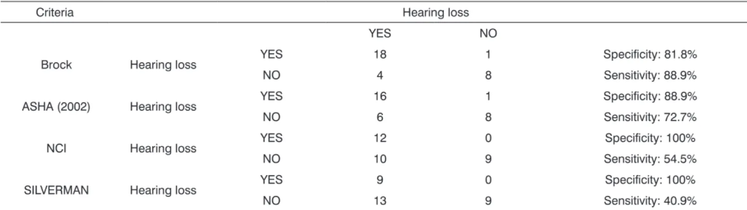
Table 2 shows Brock’s (1991)3 proposed criteria;
it had the highest sensitivity. The NCI’s12 and Davis and
Silverman’s10 criteria were the most specific.
DISCUSSION
The distribution of audiometric thresholds in the study sample showed that:
• Only 4 subjects with post-therapy hearing loss
were identified by all criteria; 7 subjects had no hearing loss in any of the criteria applied in this study.
• Thirteen of 18 subjects with normal pure tone
audiometry (according to Davis and Silverman10) after
therapy had some degree of hearing loss according to the other methods applied in this study.
Brock et al.’s3 criteria demonstrated the highest
number of significant hearing losses; the ASHA11 criteria
only revealed whether there was hearing loss or not, but provided no quantification.
Auditory losses due to cisplatin ototoxicity are generally symmetrical, bilateral, initially affecting high fre-quencies, followed by middle and low frequencies (Testa
et al.15; Rademaker et al.7).
Changes were not always detected at the beginning by the criteria described above. A major point about these criteria is that they do not take the complaints of patients into account, which would be extremely important. Addi-tionally, the impact on a patient’s life may not be propor-tional to the degree of hearing loss, since this impact de-pends on factors such as social and professional activities and personal aspects. The growing concern on the part of oncologists should lead to careful prevention of hearing loss, comprising periodic testing and close monitoring of hearing losses, not waiting for patients to complain before becoming concerned with hearing; this means preventing the onset of hearing loss and not only rehabilitation when hearing cannot be recovered any longer.
A relevant point was that NCI12 criteria were not
sufficiently specific for monitoring auditory function;
it does not define clearly which frequencies should be investigated.
Audiologically, there are monitoring proposals using
conventional frequencies (Testa et al.15, Kushner et al.16,
Marshall et al.17, Toral-Martinnon et al.18), monitoring
pro-posals using transient otoacoustic emissions (Liberman19)
or distortion products (Biro et al.20; Hyppolito et al.21),
and monitoring proposals using high frequencies (over
8000 Hz) (Garcia22), which may be first affects by ototoxic
drugs. In this case, patients with hearing loss normally do not complain and not always perceive loss of hearing
(Liberman23). Dhooge et al.,24 however, found
sympto-matic ototoxicity in 20% of cases - auditory complaints in 16 children treated with cisplatin and/or carboplatin. Monitoring with conventional frequencies (500 to 8000 Hz) shows that hearing loss leads to difficulties in diffe-rent situations; patients may complain only of tinnitus or difficulties to understand speech in noisy environments, or may present hearing loss at speech frequencies, no longer being able to follow a conversation. Still in the criterion for patients with auditory changes in pre-therapy testing, these subjects may be classified as not having post-therapy auditory changes; but therapy will have caused elevated audiometric thresholds, which are not taken into account in the final criterion.
A further discrepancy is the grade II in this criterion. This grade includes patients that had a 25 to 90 dB eleva-tion of the auditory threshold. A 25 dB threshold elevaeleva-tion may go unnoticed or may cause minimal difficulty; it may even be classified as mild hearing loss (Davis and
Silver-man10). On the other hand, a 90 dB elevation suggests that
subjects will probably not follow a conversation without using hearing aids, since voice during a conversation is issued at around 60 dBHL. Furthermore, hearing loss here is classified as severe, which brings significant restrictions on social life. Classifying these two extremes of threshold elevations in the same grade does not take into account the significant differences, complaints and limitations. Table 2. Sensitivity and Specificity, with their respective confidence intervals (CI), for each criterion.
Criteria Hearing loss
YES NO
Brock Hearing loss YES 18 1 Specificity: 81.8%
NO 4 8 Sensitivity: 88.9%
ASHA (2002) Hearing loss YES 16 1 Specificity: 88.9%
NO 6 8 Sensitivity: 72.7%
NCI Hearing loss YES 12 0 Specificity: 100%
NO 10 9 Sensitivity: 54.5%
SILVERMAN Hearing loss YES 9 0 Specificity: 100%
The 40 dB reference in Brock et al.’s10 proposed
criteria is debatable. A 40 dB loss characterizes mild hea-ring loss, with serious implications for the perception of
Portuguese consonants (Russo and Behlau25), especially
in children, for which this classification was proposed. This author does not take into account changes above 40 dB that may occur at single frequencies, as well as not considering small auditory alterations that may occur before the auditory threshold reaches 40 dB. Intensity is an important factor in hearing loss; it is also important in rehabilitation with hearing aids, and may be a limiting factor in choosing and using an appropriate aid.
The ASHA11 criteria do not account for affected
frequencies; from an audiological perspective this has implications for the follow-up of oncological therapy. A 10 dB threshold increase at 6000 and 8000 Hz may not result in minimal hearing loss, depending on age, while a 20 dB decrease at 1000 and 2000 Hz in a patient with a 30 dB pre-treatment threshold causes moderate hearing loss and may result in significant communication difficulties. Thus, the affected frequency should be taken into account.
Davis and Silverman10 proposed criteria is not
in-dicated for oncological patients, since it classifies hearing loss only at 500, 1000 and 2000 Hz thresholds, which are
not those commonly involved in ototoxicity.1,26-27 This is
a problem, because when ototoxic drug induced hearing loss affects these frequencies, loss is already significant and patients present major complaints; it is thus inadequate for monitoring patients at risk of hearing loss - when this criterion detects loss, it is already rather advanced.
The same subject with hearing loss was not always classified by all of the criteria above; thus, the sensitivity and/or specificity of each criterion needs to be known, as small changes in hearing are not always detected by these instruments. Important hearing losses are easily detected by any of these tools; however, a classification instrument should detect small changes so that oncologists and speech therapists may carefully monitor the hearing function of these patients, to avoid major loss of function.
The classification of hearing loss in audiological evaluations during oncological therapy aims firstly to iden-tify ototoxic effects, especially at high frequencies. This makes it possible for physicians to be alert and change therapy protocol measures. Secondly, the classification should indicate at which point patients will suffer the so-cial, educational and professional implications of hearing loss. Impact differs in adults and children, both in terms of the degree of hearing loss and the affected frequencies. A classification should be able to show the progression of hearing loss.
Knight et al.28 monitored ototoxicity in 67 children
with osteosarcoma, neuroblastoma and medulloblastoma, all of which were treated with cisplatin. These authors
compared the Brock et al.,3 ASHA,11 and NCI12 criteria, and
also found it difficult to adequately describe hearing losses. These authors believe that those criteria underestimate hearing loss resulting from oncological therapy; the result of this is that language development, learning and social/ emotional function may be compromised in these children. These authors also found that hearing loss may lead to low self-esteem, behavioral disorders, loss of energy and stress, compared to normal hearing children; these factors are not included in any of these classification systems.
Liberman29 studied patients with cancer treated
during childhood with cisplatin, and found a higher occur-rence of auditory complaints when hearing loss affected the 4000Hz frequency.
Marini et al.30 analyzed the predictive power,
sensi-tivity and specificity of auditory complaints in 795 patients and found that the sensitivity was high (80.9%) and the specificity was 60.4%. Audiometric test results should also be available; although more subjective, these results are less costly than new technologies.
Teles et al.31 compared data such as frequency,
proportion, agreement and consistency of responses in workers exposed to occupational noise; significant chan-ges were noted in the audibility threshold (MSL). These authors applied three Brazilian criteria and one interna-tional criterion to analyze threshold changes and found that these criteria in themselves were inadequate, given their subjective nature; prevention would be necessary not only in subjects presenting MSL but in all subjects in an auditory preservation program.
Gupta et al.32 found a small incidence of hearing
loss in children undergoing cisplatin chemotherapy by continuous infusion; these authors used Brock’s criterion and concluded that continuous drug administration is as-sociated with a lower incidence of ototoxicity. However, we believe that this criterion underestimates important losses in this population because it takes into account only losses over 40 dB.
According to the literature we cited, changes in hearing were not always detected at the beginning in onco-logical patients by the criteria described in this study. The main criticism of these criteria is that they do not include patient complaints, essential for understanding the impact of hearing loss on patient’s lives, which are not always proportional do the degree of loss, but depend on factors such as personal, social and professional activities. The growing concern of oncologists should lead to preventive care to avoid hearing loss; this includes periodic testing and close monitoring of auditory changes before patients complain to avoid hearing loss rather than just rehabilita-tion when reversion is no longer possible.
also to investigate eventual complains and the impact that hearing loss may cause on the quality of life of patients. It is impossible to define the degree of hearing loss that results in complaints and difficulties for patients using only the criteria mentioned above for classifying auditory losses. Thus, identifying and validating the complaints of patients is just as important as audiological evaluations and classification of losses according to charts.
It is extremely important to note that the criterion used for classifying hearing losses in oncological patients should be able to identify the beginning of loss, thus avoi-ding unnecessary adverse effects and preventing auditory losses due to oncological therapy.
CONCLUSION
We found that Davis and Silverman’s10 criteria
sho-wed hearing loss in 29% of subjects at the end of treatment;
the NCI12 criteria showed hearing loss in 38% of patients;
the ASHA11 criteria showed hearing loss in 54%; and Brock
et al.’s3 criteria showed hearing loss in 61% of patients
at the end of oncological therapy. Thus, Brock’s (1991)3
proposed criteria was the most sensitive, and the NCI’s12
and Davis and Silverman’s10 criteria were the most specific.
However, all of these criteria underestimated the description of identified auditory alterations; additional information was required to help physicians to understand the true implications of hearing loss in each case.
A common code is needed between audiologists and oncologists to increase our understanding and improve the therapy of these patients; not only should the grade and type of hearing loss be described, but also the impact on the lives of patients.
REFERENCES
1. Oliveira JAA. Ototoxicidade. In: Costa SS, Cruz OLM, Oliveira JAA. Otorrinolaringologia. Porto Alegre: Artes Medicas Sul; 1994. p.215-21. 2. LP, Whitworth CA. Ototoxicity: therapeutic opportunities. Drug Discov
Today. 2005; 10:1313-21.
3. Brock PR, Bellman SC, Yeomans EC, Pinkerton R, Pritchard J. Cisplatin ototoxicity in children: a practical grading system. Med Pediatr Oncol. 1991;19:295-300.
4. Pedalini MEB, Toniosso S, Goffi MVS. O papel do audiologista no tratamento do pacientes com câncer. In: Barros APB, editor. Fono-audiologia em cancerologia. São Paulo: Fundação Oncocentro de São Paulo; 2000. p.105-9.
5. Simon T, Hero B, Dupuis W, Selle B, Berthold F. The incidence of hearing impairment after successful treatment of neuroblastoma. Klin Padiatr. 2002;214:149-52.
6. Knoll C, Smith RJ, Shores C, Blatt J. Hearing genes and cisplatin deafness: a pilot study. Laryngoscope. 2006;116:72-4.
7. Rademaker-Lakhai JM, Crul M, Zuur L, Baas P, Beijnen JH, Simis YJ, et al. Relationship between cisplatin administration and the develo-pment of ototoxicity. J Clin Oncol. 2006;20:918-24.
8. Rybak LP, Whitworth CA, Mukherjea D, Ramkumar V. Mechanisms of cisplatin-induced ototoxicity and prevention. Hear Res. 2006;226:157-67.
9. Zurr CL, Simis YJ, Landsdaal PE, Rasch CR, Tange RA, Balm AJ, Dreschler WA. Audiometric Patterns in Ototoxicity of Intra-Arterial Cisplatin Chemoradiation in Patients with Locally Advanced Head and Neck Cancer. Audiol Neurootol. 2006;11:318-30.
10. Davis H, Silverman SR. Hearing and Deafness, 4th ed. New York, Holt, Rinehart & Winston, 1978.
11. American Speech-Language-Hearing Association (ASHA). Guidelines for the Audiologic Management of Individuals Receiving Cochleotoxic Drug Therapy. ASHA. 2002;2:81-92.
12. NCI. National Cancer Institute. Common Terminology Criteria for Adverse Events (CTCAE) version 3.0 (2003). Disponível em: < URL:http://ctep.cancer.gov/reporting/ctc.html. Acessado em 21 de maio de 2006.
13. Redondo MC, Lopes Filho OC. Testes básicos da avaliação auditiva. In: Lopes Filho OC. Tratado de fonoaudiologia. São Paulo: Roca; 1997. p.81-108.
14. Yantis PA. Pure air conditioning thresholds listing. In: Katz J, editor. Handbook of clinical audiology. 4th ed. Baltimore: Willian & Wilkins; 1994. p.97-108.
15. Testa JR, Liberman PHP, Goffi-Gomez MV S. Complicações otorrino-laringológicas do tratamento oncológico. In: Kowalski LP, Anelli A, Salvajoli JV, Lopes LF. Manual de Condutas diagnósticas e terapêuticas em oncologia. São Paulo: Ambito Editorial; 2002. p.395-7.
16. Kushner BH, Budnick A, Kramer K, Modak S, Cheung NK. Ototo-xicity from high-dose use of platinum compounds in patients with neuroblastoma. Cancer. 2006; 107: 417-22.
17. Marshall NE, Ballman KV, Michalak JC, Schomberg PJ, Burton GV, Sandler HM, et al. Ototoxicity of cisplatin plus standard radiation therapy vs. accelerated radiation therapy in glioblastoma patients. J Neurooncol. 2006;77:315-20.
18. Toral-Martinnon R, Collado-Corona MA, Mora-Magana I, Leal-Leal C, Gutierrez-Castrellon P, Gonzalez-De Leo S. Evaluation of cisplatin ototoxicity by the audiometric curve in retinoblastoma. Cir Cir. 2006; 74:79-82.
19. Liberman PHP. Monitorizaçäo auditiva em crianças portadoras de retinoblastoma: relato de dois casos. Acta Oncol Bras. 2002;22:250-4. 20. Biro K, Noszek L, Prekopp P, Nagyivanyi K, Geczi L, Gaudi I, et al.
Characteristics and risk factors of cisplatin-induced ototoxicity in testicular cancer patients detected by distortion product otoacoustic emission. Oncology. 2006;70:177-84.
21. Hyppolito MA, de Oliveira JA, Rossato M. Cisplatin ototoxicity and otoprotection with sodium salicylate. Eur Arch Otorhinolaryngol. 2006; 263:798-803.
22. Garcia AP, Irio MCM, Petrilli A S. Monitoramento da audição de pacientes expostos à cisplatina. Rev Bras Otorrinolaringol. 2003; 69:215-21.
23. Liberman PH, Schultz C, Gomez MV, Carvalho AL, Pellizzon AC, Testa JR, et al. Auditory effects after organ preservation protocol for laryngeal/ hypofaryngeal carcinomas. Arch Otolaryngol Head Neck Surg. 2004; 130:1265-8.
24. Dhooge I, Dhooge C, Geukens S, De Clerck B, De Vel E, Vinck BM. Distortion product otoacoustic emissions: an objective technique for the screening of hearing loss in children treated with platin derivatives. Int J Audiol. 2006;45:337-43.
25. Russo I, Behlau M. Percepçäo da fala: análise acústica do português brasileiro. Säo Paulo: Lovise. 1993. p.25-50.
26. Li Y, Womer RB, Silber JH. Predicting cisplatin ototoxicity in chil-dren: the influence of age and the cumulative dose.Eur J Cancer. 2004;40:2445-51.
27. Langer T, Stohr W, Bielack S, Palussen M, Treuner J, Beck JD. Late effects surveillance system for sarcoma patients. Pediatr Blood Cancer. 2004;42:373-9.
29. Liberman PHP. Avaliação Auditiva em pacientes tratados de câncer na infância. [Dissertação]. Fundação Antonio Prudente, São Paulo; 2005.
30. Marini ALS, Halpern R, Aetrs D. Sensibilidade, especificidade e valor preditivo da queixa auditiva. Rev Saude Publica 2005;39:982-4.
31. Teles RM, Sena APRC, Medeiros MPH. Estudo comparativo entre critérios de análise da mudança significativa do limiar (MSL) Fono Atual. 2002;5:53-9.