Cloning and Molecular Characterization of the
Schistosoma
mansoni
Genes
RbAp48 and Histone H4
Patrícia P Souza, Débora N Santos, Sérgio D J Pena, Glória R Franco
+Laboratório de Genética Bioquímica, Departamento de Bioquímica e Imunologia, ICB, Universidade Federal de Minas Gerais, Av. Antônio Carlos 6627, 31270-901 Belo Horizonte, M G, Brasil
The human nuclear protein RbAp48 is a member of the tryptophan/aspartate (WD) repeat family, which binds to the retinoblastoma (Rb) protein. It also corresponds to the smallest subunit of the chromatin assembly factor and is able to bind to the helix 1 of histone H4, taking it to the DNA in replication. A cDNA homologous to the human gene RbAp48 was isolated from a Schistosoma mansoni adult worm library and named SmRbAp48. The full length sequence of SmRbAp48 cDNA is 1036 bp long, encoding a protein of 308 amino acids. The transcript of SmRbAp48 was detected in egg, cercariae and schistosomulum stages. The protein shows 84% similarity with the human RbAp48, possessing four WD repeats on its C-terminus. A hypothetical tridimensional structure for the SmRbAp48 C-terminal domain was constructed by computational molecular modeling using the β-subunit of the G protein as a model. To further verify a possible interaction between SmRbAp48 and S. mansoni histone H4, the histone H4 gene was amplified from adult worm genomic DNA using degenerated primers. The gene fragment of SmH4 is 294 bp long, encoding a protein of 98 amino acids which is 100% identical to histone H4 from Drosophila melanogaster.
Key words: RbAp48 - histone H4 - Schistosoma mansoni - gene cloning
The human protein RbAp48 was first identified as one of the major polypeptides from HeLa cell lysates that binds specifically to a putative functional domain of the carboxy terminus of the Rb protein, a known cellular tumor sup-pressor (Lee et al. 1991). Human RbAp48 protein shares sequence homology with MSI1, a negative regulator of the Ras-cAMP pathway in the yeast Saccharomyces cerevisiae. Overexpression of MSI1 gene suppresses the heat-shock sensitivity of iraI and Ras2val19 mutant strains and reduces the cAMP levels in these mutants (Ruggieri et al. 1989). Furthermore, similarly to MSI1, the human RbAp48 supresses the heat-shock sensitivity of the same mutants (Qian et al. 1993). This finding demonstrates that there is a functional homology between both proteins (Qian et al. 1993). The yeast null mutant of MSI1 has been obtained and presents sensitivity to UV irradiation asso-ciated to a decrease in the silencing of telomere adjacent genes (Kaufman et al. 1997). RbAp48 is a nuclear protein and a member of the tryptophan/aspartate (WD) repeat family (Qian et al. 1995). Proteins constituted by at least four WD repeats can be clustered into this structural fam-ily of proteins, the members of which appear to perform regulatory functions in several cellular processes, such as cell division, cell fate determination, gene transcrip-tion, transmembrane signaling, mRNA modificatranscrip-tion, and vesicle fusion (Neer et al. 1994).
This work was supported by grants from Brazilian Research Council (CNPq) and Pró Reitoria de Pesquisa, UFMG.
+Corresponding author. Fax: +55-31-3499.2984. E-mail:
gfranco@icb.ufmg.br. Received
Accepted
Verreault and colleagues, in 1996, described a human chromatin assembly complex containing a chromatin as-sembly factor (CAF-1) and modified histones H4 and H3, acetylated in specific lysine residues. It was further veri-fied that RbAp48 corresponds to the smallest subunit of CAF-1 and is able to bind to the helix 1 of histone H4, taking the later to the DNA in replication (Krude 1999). A related p48 protein in Saccharomyces, named Hat2p, is a constituent of a subunit of histone H4 acetyltransferase B type (Parthurn et al. 1996). These findings suggest that a family of p48 proteins may be involved in diverse as-pects of histone functions in a variety of different organ-isms (Verreault et al. 1996).
Another function attributed to RbAp48 is its partici-pation on the assembly of a basal repression complex, formed by histone deacetylases (HDAC) 1 and 2 and also RbAp46, recruited by a variety of co-repressors and re-pression associated factors to strength the transcriptional repression during the cell cycle (Knoepler & Eisenman 1999). Nicholas and colleagues (2000) found that RbAp48 belongs to the HDAC complex that associates with the Rb protein to repress the E2F transcription factor during the cell cycle.
MATERIALS AND METHODS
cDNA libraries andDNA purification - S. mansoni egg, cercariae, 3h schistosomulum and adult worm cDNA libraries were constructed in λZAP as part of the Schisto-soma genome project (Franco et al. 2000). Genomic DNA was purified from S. mansoni LE strain adult worms as described previously (Simpson et al. 1982). Plasmids and Polymerase Chain Reaction (PCR) fragments were puri-fied with the Wizard DNA Purification SystemsTM (Promega).
DNA cloning - Two identical clones (MAAD0269 and MAAD0270) carrying cDNA fragments homologous to the human gene RbAp48 were isolated from a S. mansoni adult worm library after random clone selection. The his-tone H4 gene was amplified by PCR from S. mansoni LE strain adult worm genomic DNA using degenerated prim-ers. Several strategies were used to obtain the full-length sequence of both strands of SmRbAp48 and SmH4 genes. Both SmRbAp48 cDNA clones were digested with the RsaI restriction enzyme in internal sites of the insert. Spe-cific primers were also designed and used to amplify by PCR internal regions of the cDNA fragment. The initial portion of the cDNA containing part of the Open Reading Frame (ORF) and the 5’ untranslated region (5’UTR), that was not present in the original cDNA clones, was ob-tained by amplification of other cDNA libraries using an hemi-nested PCR strategy. The digestion fragments and all the PCR products were cloned into the SmaI site of pUC18 (Amersham Pharmacia Biotech) using the Sureclone Ligation kit (Amersham Pharmacia Biotech).
PCR and sequencing - Specific primers targeting re-gions of SmRbAp48 were used to amplify parts of the SmRbAp48 cDNA cloned into pBlueScript KS+ (Stratagene) and the four cDNA libraries (egg, cercariae, 3h schistosomulum and adult worm). Degenerated prim-ers designed based on histone H4 genes from different organisms were used to amplify the SmH4 gene from adult worm genomic DNA.
For the amplification of SmRbAp48, 100 µl reaction mixture was used containing approximately 4 µl of the clones, 10 mM Tris HCl pH 8.5, 50 mM KCl, 1.5 mM MgCl2, 200 µM each deoxynucleotide triphosphate, 200 nM each primer and 2.5 U of Taq polymerase. The conditions used for amplification were 96°C for 2 min, followed by a step cycle program set to denature at 96°C for 1 min, anneal at 54°C for 1 min, and extend at 72°C for 2 min for a total of 25 cycles. Amplifications of the cDNA libraries were per-formed in a 30 µl volume containing 1 µl of the cDNA library, 10 mM Tris HCl pH 8.5, 50 mM KCl, 1.5 mM MgCl2, 200 µM each deoxynucleotide triphosphate, 200 nM each primer and 2 U of Taq DNA polymerase. The conditions used for amplification were the same described above. PCR of genomic DNA was performed in a 30 µl volume containing 20 ng of genomic DNA, 10 mM Tris HCl pH 8.8, 75 mM KCl2, 3.5 mM MgCl2, 200 µM each deoxynucleotide triphosphate, 400 nM each primer and 2U of Taq DNA polymerase. The conditions used for the amplifications were 95°C for 2 min, followed by a step cycle program set to denature at 95°C for 1 min, anneal at 48°C for 1 min, and extend at 72°C for 2 min for a total of 35
cycles. The amplicons were analyzed in 1% agarose gel stained by ethidum bromide or in 6% polyacrylamide gels silver stained (Santos et al. 1993).
Sequencing reactions were performed using the Thermo Sequenase fluorescent labeled primer cycle se-quencing kitTM with 7-deaza-dGTP (Amersham Pharmacia Biotech). M13 fluorescent primers targeting the margins of the cloning sites were used for DNA sequencing of both strands, using the A.L.F. Automated DNA Sequencer (Amersham Pharmacia Biotech).
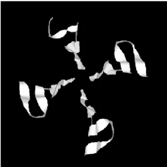
Sequence analysis - Search for homologous sequences was undertaken using the BLAST program (Altschul et al. 1997, http://www.ncbi.nlm.nih.gov). Open reading frame (OFR) search and DNA translation were performed using the DNAsis program. The PredictProtein server (http:// cubic.bioc.columbia.edu/predictprotein/) was used for prediction of secondary structure and post-translational modification sites. A BMERC “The WD repeat Familyof Proteins” Server (http://bmerc-www.bu.edu/ bioinformatics/wdrepeat.html) was used for prediction of the number and localization of the WD repeats in the pro-tein, as well as to produce a three-dimensional (3D) model for the SmRbAp48 gene product, based on its homology to the β-subunit of the G protein (PDB ID 1SCG). The 3D model visualization was performed using the RASMOL Program.
RESULTS
sequences were aligned and the collated sequence of both strands was obtained (Fig. 2).
Theoretical analysis and structural predictions - The full-length sequence of the SmRbAp48 cDNA is 1036 bp long, with an ORF of 927 bp which encodes a 308 aa pro-tein with a calculated molecular mass of 34,965Da and estimated pI of 5.34 (Figs 1, 2). The sequence revealed a 5’ UTR of 85 bp and a poly-A tail of 36 bp. The putative ATG initiation codon was chosen due to the absence of an-other ATG codon on previous positions in the same frame. However, the context where this codon is present is not in agreement with the Kosak consensus sequence, that sig-nals the translation initiation in eukaryotes (Kosak 1987). The amino acid sequence of the SmRbAp48 translation product is rich in leucine (8.4%), aspartate (8.1%) and serine (8.1%), but very poor in tyrosine and glutamine (2.9%), tryptophan (2.3%) and cysteine (1.6%).
Protein database searches revealed that the SmRbAp48 protein shows 84% identity with human RbAp48. Fifteen putative phosphorylation sites were iden-tified: one for cAMP- and cGMP-dependent protein ki-nases (residues 25-28), six for protein kinase C (residues 56-58, 99-101, 111-113, 117-119, 162-164 and 259-261), eight for casein kinase II (residues 6-9, 56-59, 71-74, 95-98, 160-163, 194-197, 227-230 and 244-247), suggesting that the protein can be regulated by phosphorylation. No nuclear localization signal, which is responsible for recognition of the protein by nuclear transport factors, is found on SmRbAp48. However, this protein is not very big (< 50 kDa) and its small size could make possible for it to cross through the nuclear pore by a passive diffusion process.
SmRbAp48 also has at its carboxy terminus four WD repeat motifs (residues 121-154, 174-205, 224-255 and 270-301), two of them starting with GH and ending in WD. This characteristic is seen in classical WD repeat motifs found in members of this protein family. The motifs fold as sheets composed each one by four antiparallel β-strands, forming the blades of a β-propeller (Neer et al. 1994; Garcia-Higuera et al. 1996). An hypothetical 3D struc-ture for the carboxy end of the SmRbAp48 protein was obtained by computational molecular modeling based on the homology to the G protein β-subunit, the archetypal of this structural family (Fig. 3). The model clearly demon-strated the presence of four blades forming a β-propeller. Each blade is a sheet formed by four antiparallel β-strands, three of them derived from one WD repeat and the fourth b-strand from the following WD repeat. It was not possible to obtain an hypothetical 3D model for the first 121 amino acids of SmRbAp48, once there is not any protein structure with enough similarity to this region of the parasite protein. Nevertheless, according to the sec-ondary structure prediction, using the PredictProtein server, this region could form two α-helices (results not shown). The helices might stabilize the β-propeller struc-ture of SmRbAp48, as has been shown for the G protein β-subunit, which needs to be associated with the two a-helices of the g-subunit to be crystallized (Sondek et al. 1996).
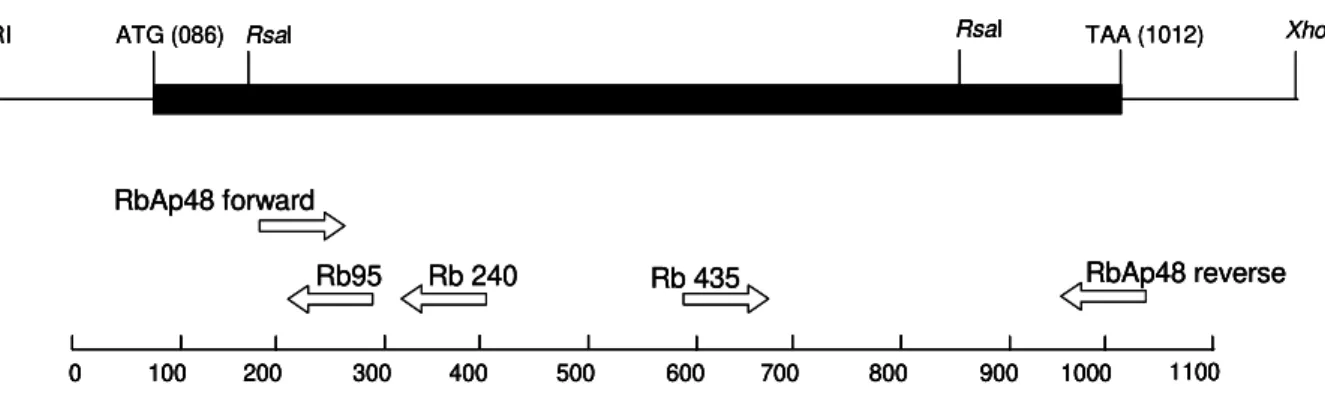
Expression of SmRbAp48 in S. mansoni life cycle stages - cDNA libraries of different stages of the parasite life cycle (egg, cercariae, 3h schistosomulum and adult worm) were amplified by PCR using the primers Rb435 XhoI
EcoRI ATG (086) RsaI RsaI TAA (1012)
Rb95 Rb 240 Rb 435 RbAp48 forward
RbAp48 reverse
0 100 200 300 400 500 600 700 800 900 1000 1100
XhoI
EcoRI ATG (086) RsaI RsaI TAA (1012) XhoI
EcoRI ATG (086) RsaI RsaI TAA (1012)
Rb95 Rb 240 Rb 435 RbAp48 forward
RbAp48 reverse
0 100 200 300 400 500 600 700 800 900 1000 1100
Rb95 Rb 240 Rb 435 RbAp48 forward
RbAp48 reverse
0 100 200 300 400 500 600 700 800 900 1000 1100
0 100 200 300 400 500 600 700 800 900 1000 1100
Fig. 1: schematic representation of the strategies used for cloning and sequencing the SmRbAp48 cDNA. Positions of start (ATG) and stop (TAA) codons, RsaI restriction sites used for digestion and primers used for amplification of the cDNA are indicated.
TABLE
Set of primers used for cloning and sequencing of SmRbAp48 and SmH4 genes
Primers Nucleotide sequence Nucleotide position
Rb 435 5’ CGTCTCAAGGGTCATCA 3’ 596 - 612
Rb 240 5’ TCACCTCGTTCACTATCA 3’ 376 - 393
Rb 95 5’ TAATCTCGCCCAGTCCT 3’ 248 - 264
RbAp48 forward 5’ GACGGATCCATGAGGAATACTCCGTTCTTG 3’ 161 - 178 RbAp48 reverse 5’ GAGCTGCAGGTTATTTGGTTATTTATGTG 3’ 1006 - 1025
Hist4 forward 5’ GGWMGWGGWAARGGWGGWAA 3’ 1 - 20
Hist4 reverse 5’ CCCRTAVAGVGTNCKKCCYTG 3’ 274 - 294
5' AAACTGACCGTTTACACTTAACAAAGCATCTGTGTAATTATTCAAAGGAGGTTACGCCGTTTCTGAAGCGTAT 73
TCTATAATAAAA ATG ATG TTG CAT CCT TCG GAT TCT GAA GAC ATT GTC GAA GAG AGA 130 Met Met Leu His Pro Ser Asp Ser Glu Asp Ile Val Glu Glu Arg 15
GTA ATA AAC GAA GAA TAC AAG ATA TGG AAG AGG AAT ACT CCG TTC TTG TAC GAT 184 Val Ile Asn Glu Glu Tyr Lys Ile Trp Lys Arg Asn Thr Pro Phe Leu Tyr Asp 33
ATG CTG ATG TCA CAC TGC TTG GAA TGG CCA AGT TTA ACT GCC CAA TGG TTG CCA 238 Met Leu Met Ser His Cys Leu Glu Trp Pro Ser Leu Thr Ala Gln Trp Leu Pro 51
TCT GTG GAA AGG ACT GGG CGA GAT TAC TCC GTT CAT CGT TTA ATA CTC GGG ACT 292 Ser Val Glu Arg Thr Gly Arg Asp Tyr Ser Val His Arg Leu Ile Leu Gly Thr 69
CAC ACA TCT GAT GAG CAA AAT CAC TTG TTG ATA GTT ACG GTT CAT CTA CCA AAT 346 His Thr Ser Asp Glu Gln Asn His Leu Leu Ile Val Thr Val His Leu Pro Asn 87
GAC CAG GCG GAG TTT GAT GCA AGT GCT TAT GAT AGT GAA CGA GGT GAT TTC GGG 400 Asp Gln Ala Glu Phe Asp Ala Ser Ala Tyr Asp Ser Glu Arg Gly Asp Phe Gly 105
GGA TTT TAT TTT CCA TCT GGG AAG TTG GAA ATA TCA ATG AAA ATA AAT CAT GAA 454 Gly Phe Tyr Phe Pro Ser Gly Lys Leu Glu Ile Ser Met Lys Ile Asn His Glu 123
GGC GAA GTC AAT CGT GCT AGG TTT ATG CCA CAG AAC CCA GAC ATA ATA GCT ACC 508 Gly Glu Val Asn Arg Ala Arg Phe Met Pro Gln Asn Pro Asp Ile Ile Ala Thr 141
AAA ACA CCA AGT GGT GAT GTT TTA ATA TTC AAT TAT CCA AGA CAT CCA CCG AAA 562 Lys Thr Pro Ser Gly Asp Val Leu Ile Phe Asn Tyr Pro Arg His Pro Pro Lys 159
ACC CCA TCA GAC CGT GGT TGC CAA CCT GAT CTA CGT CTC AAG GGT CAT CAA AAA 616 Thr Pro Ser Asp Arg Gly Cys Gln Pro Asp Leu Arg Leu Lys Gly His Gln Lys 177
GAA GGT TAT GGT CTT TCA TGG AAT GTG TCT CTT AAT GGT CAT CTT CTT TCA GCG 670 Glu Gly Tyr Gly Leu Ser Trp Asn Val Ser Leu Asn Gly His Leu Leu Ser Ala 195
TCT GAT GAT CAG ACA ATT TGT TTA TGG GAT GTT AAT GCT GCT CCT TTA GAT GGC 724 Ser Asp Asp Gln Thr Ile Cys Leu Trp Asp Val Asn Ala Ala Pro Leu Asp Gly 213
TGT GAT CTA GAT GCG ATG GCT ATC TTT ACG GGT CAT CAT TCA GTA GTT GAG GAC 778 Cys Asp Leu Asp Ala Met Ala Ile Phe Thr Gly His His Ser Val Val Glu Asp 231
GTT TCC TGG CAC CTT TTC CAT GGA CAT ATT TTT GGT TCA GTA GCA GAT GAT AAT 832 Val Ser Trp His Leu Phe His Gly His Ile Phe Gly Ser Val Ala Asp Asp Asn 249
AAA CTT ATG GTT TGG GAT ACA CGG AGT TCA AAT CGT ACA AAA CCT CAG CAC CAA 886 Lys Leu Met Val Trp Asp Thr Arg Ser Ser Asn Arg Thr Lys Pro Gln His Gln 267
GTG GAT GCT CAT ACA GCC GAA GTC AAT TGT CTT GCT TTT AAT CCA TTT TCT GAG 940 Val Asp Ala His Thr Ala Glu Val Asn Cys Leu Ala Phe Asn Pro Phe Ser Glu 285
TTT ATT ATT GCT ACA GGA AGT GCG GAC AAA GTA ATT AAG TAT TTT ACC CTC GTA 994 Phe Ile Ile Ala Thr Gly Ser Ala Asp Lys Val Ile Lys Tyr Phe Thr Leu Val 303
TCT TTT TTT TAC ACA TAA ATA ACC AAA TAA CAT TCA TGC AGT 3’ 1036 Ser Phe Phe Tyr Thr *** 308
Fig. 2: nucleotide sequence of the SmRbAp48 cDNA with its deduced amino acids. The nucleotide sequence obtained by the hemi-nested PCR strategy is in bold italics. The four tryptophan/aspartate repeat motifs in the protein are double underlined and the fifteen putative sites for phosphorylation are in gray. Conserved residues essential for stabilization of the β-propeller fold are boxed. The SmRbAp48 cDNA sequence is available in GenBankTM with acession number AF297468.
and RbAp48 reverse (Table). Amplicons of 429 bp were detected in all the stages of the life cycle evaluated, indi-cating that this gene is expressed throughout the parasite development (Fig. 4).
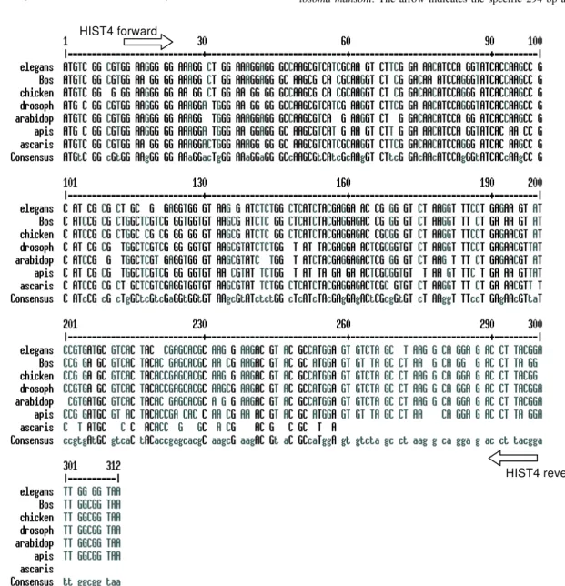
Hi-stone H4 protein, degenerated primers were designed to amplify the Histone H4 gene from S.mansoni. The prim-ers were designed based on a detailed inspection in the alignment of Histone H4 gene sequences from diverse organisms, using the Multalin Program (http://www. protein.toulose.inra.fr/multalin/multalin.html) (Fig. 5). The designed primers Hist4 forward and reverse (Table) target conserved regions of the gene, but the amplification prod-uct lacks the initial and final portions of the gene. The Histone H4 gene fragment was amplified from S. mansoni adult worm genomic DNA and from D. melanogaster and mice genomic DNA, which were used as controls in the
experiment (Fig. 6). It is noticeable that all amplified bands have the same size, since Histone H4 genes do not con-tain introns and are highly conserved during evolution. The amplified fragment of the SmH4 gene was cloned into pUC18 vector and fully sequenced. The partial sequence of SmH4 is 294 bp, encoding a putative protein of 98 aa, with a calculated molecular mass of 10,887 Da and an estimated pI of 11.36. The translation product does not contain the first two and the last three amino acids present in Histone H4 from other organisms (Fig. 7). Database homology search results show 100% identity between SmH4 and the D. melanogaster protein. Theoretical analy-sis reveals the presence of four probable phosphoryla-tion sites (residues 43-46, 70-73, 76-79 and 81-84), two nuclear localization signatures (residues 15-18 and 16-19) and four important lysine residues, conserved in all his-tones of this class (residues 4, 7, 11 and 15) (Fig. 7).
DISCUSSION
This work reports on the cloning and characterization of RbAp48 and histone H4 genes of S. mansoni. The SmRbAp48 gene encodes a putative nuclear protein, mem-ber of the WD repeat family, presenting four WD repeats on its carboxy end, which is probably regulated by phos-phorylation. RbAp48 proteins are very conserved, and this high degree of conservation during the course of evolution is indicative of their functional importance in biological processes. The predicted amino acid sequence of SmRbAp48 shows 84% of similarity and 72% of iden-tity to the human RbAp48.
The WD repeat was first described in the β subunit of heterotrimeric GTP-binding proteins, which transduce sig-nals across the plasma membrane (Fong et al. 1986). WD repeat proteins are constituted of highly conserved re-peating units, usually ending with Trp-Asp (WD) and have been found in all eukaryotes, but not in prokary-otes. The number of repeats vary from four to nine in different proteins (Garcia-Higuera et al. 1996). Detailed analysis of these repeats shows that there are four resi-dues almost totally invariant, occupying strategic posi-tions in loops and β-strand regions of the protein struc-ture which in turn, by a network of hydrogen bonds, link the blades of the propeller and are essential for stabiliza-tion of the correct fold (Branden & Tooze 1999). They are boxed in the SmRbAp48 sequence (Fig. 2). The β -propel-ler structure is unstable as a monomer, as seen for the β subunit of G protein which becomes stable when associ-ated with the γ subunit of this protein. The later one is unfolded in vitro and folds into two a-helices when form-ing a dimer with the β subunit. Their association is par-tially due to hydrophobic interactions between the long N-terminal helix of the β subunit and the N-terminal α-helix of the γ subunit of G protein (Branden & Tooze 1999). According to secondary structure predictions, the amino terminus of SmRbAp48 could form two a-helices. These helices might stabilize the β-propeller structure of SmRbAp48 after its association to another protein, as showed for the β-subunit of G protein, which needed to be associated to the γ-subunit to be crystallized (Sondek et al. 1996). The search for new SmRbAp48 partners and the understanding of the molecular mechanisms
govern-Fig. 3: threotical three-dimensional structure of the SmRbAp48 carboxy terminus obtained by molecular modelling using the β sub-unit of G protein as a model (PDB ID 1SCG). A β-propeller of four blades, each one formed by a β-sheet composed by four antiparallel
β-strands is shown.
ing the interactions between them is of great interest to help elucidate the biological function of this protein in the parasite.
There are relatively few papers on the characteriza-tion of transcripcharacteriza-tion factors and control of gene expres-sion in S. mansoni, and none of them deals with the study of repression of gene transcription and histone metabo-lism. Another function attributed to RbAp48 is to be part of a basal repression complex, composed by HDAC-1 and 2 and RbAp46 (Knoepler & Eisenman 1999). This complex is recruited by a variety of co-repressors and repression associated factors to strength the transcriptional repres-sion during the cell life cycle (Knoepler & Eisenman 1999). Thus, SmRbAp48 may consist of a factor participating in the repression of gene expression in the parasite. In a dbEST search, we found ESTs homologous to human HDAC3 (gi 5790729) and HDAC8 (gi 4224573, 4225199,
HIST4 forward
HIST4 reverse
Fig. 5: multiple alignment of histone gene sequences from diverse organisms using the Multalin program. Sites for annealing of the degenerated primers Hist4 forward and reverse are indicated by arrows. Elegans (Caenorhabditis elegans histone H4, gi 17541085); bos (Bos taurus histone H4, gi 2981287); chicken (chicken histone H4, gi 211905); drosoph (Drosophila melanogaster histone H4, gi 17975541); arabidop (Arabidopsis thaliana histone H4, gi 166741); apis (Apis mellifera histone H4, gi 1883000), ascaris (Ascaris lumbricoides histone H4, gi 1177237)
5' GGA AGA GGA AAG GGA GGA AAG GGC CTG GGG AAA GGG GGT GCC AAG CGC CAC CGC 54 Gly Arg Gly Lys Gly Gly Lys Gly Leu Gly Lys Gly Gly Ala Lys Arg His Arg 18
AAG GTC CTG CGC GAC AAC ATC CAG GGT ATC ACC AAG CCA GCC ATT CGG CGC CTT 108 Lys Val Leu Arg Asp Asn Ile Gln Gly Ile Thr Lys Pro Ala Ile Arg Arg Leu 36
GCT CGC CGC GGC GGC GTG AAG CGC ATT TCT GGC CTC ATC TAT GAG GAG ACC CGC 162 Ala Arg Arg Gly Gly Val Lys Arg Ile Ser Gly Leu Ile Tyr Glu Glu Thr Arg 54
GGA GTG CTG AAG GTG TTC CTG GAG AAC GTG ATT CGT GAT GCT GTG ACT TAC ACG 216 Gly Val Leu Lys Val Phe Leu Glu Asn Val Ile Arg Asp Ala Val Thr Tyr Thr 72
GAG CAC GCC AAA CGC AAG ACA GTG ACA GCG ATG GAT GTG GTC TAC GCG CTG AAG 270 Glu His Ala Lys Arg Lys Thr Val Thr Ala Met Asp Val Val Tyr Ala Leu Lys 90
AGA CAG GGC CGC ACT CTG TAT GGG 3' 294 Arg Gln Gly Arg Thr Leu Tyr Gly 98
5788601, 5790341, 5788859), demonstrating the existence of the two different types of HDACs in schistosomes. The verification of a possible interaction between HDAC and SmRbAp48 is of interest, once Ahmad and colleagues (1999) described that chicken p48 subunit of CAF-1 inter-acts directely with HDAC1 and 2 in vivothrough its WD repeat domain and Nicolas and colleagues (2001) found that HDAC3 interacts with human RbAp48, mediating its recruitment to the Rb protein.
Once human RbAp48 is homologous to the MSI1 gene of S. cerevisiae, a negative regulator of the Ras-cAMP pathway (Ruggieri et al. 1989) and SmRbAp48 is 84% similar to the human protein, it is also possible that SmRbAp48 may perform the same functions as human RbAp48 in the cell, and thus complement yeast mutant strains in which the gene MSI1 was deleted. It can be of interest to find other functions for RbAp48 in the parasite and to understand the polyvalent roles played by this protein in eukaryotic cells.
The second gene characterized was the histone H4 gene of S. mansoni, which shows 100% identity with his-tone H4 of D. melanogaster. It has four probable phos-phorylation sites, two nuclear localization signatures and four important lysines, conserved in all H4 histones. These lysines are part of acetyltransferases and deacetylases recognition signatures – GRGK5GGK, KGGK8GLG and GLGK12GGA – important on the process of recognition of recently modified histones for chromatin assembly (Ruiz-Carillo et al. 1975, Jackson et al. 1976). It is still un-clear how human RbAp48 recognizes the helix 1 of his-tone H4, but the high degree of conservation of the WD repeats in RbAp48 proteins from different organisms is indicative of the functional importance of this motif on the interaction between the two proteins.
To our knowledge, this is the first report on a gene encoding a S. mansoni WD repeat protein and histone H4, and certainly the verification of a possible interaction between these two proteins demands further studies. This
Fig. 7: nucleotide sequence of the SmH4 gene with its deduced amino acids. The four important lysines present in signatures for acetyltransferases and deacetylases recognition are in gray. The four probable sites for phosphorylation are double underlined. Genomic DNA sequence of SmH4 is available in GenBankTM with accession number AF297469.
paper describes the structural characterization of two im-portant genes for S. mansoni. Some conclusions were driven from theoretical predictions, and should be vali-dated by future experimental research aiming to elucidate the functional role of RbAp48 protein in S. mansoni.
ACKNOWLEDGEMENTS
To Neuza Antunes Rodrigues and Kátia Barroso (UFMG, Minas Gerais, Brazil) for technical support and automated DNA sequencing and to Dr Mohammed Saber (TRBI, Cairo, Egypt) for supplying the egg, cercariae, 3h schistosomulum, and adult worm cDNA libraries.
REFERENCES
Ahmad A, Takami Y, Nakayama T 1999. WD repeats of the p48 subunit of chicken chromatin assembly factor-1 re-quired for in vitro interaction with chicken histone deacetylase-2. J Biol Chem 274: 16646-16653.
Altschul SF, Thomas L, Madden AA, Schaffer JZ, Zheng Z, Webb M, David JL 1997. Gapped blast PSI-Blast; a new generation of protein database search programs. Nucl Acids Res25: 3389-3402.
Branden C, Tooze J 1999. Signal transduction. In C Branden & J Tooze (eds), Introduction to Protein Structure, Garland Publishing, INC, New York, p. 251-281.
Franco GR, Valadão AF, Azevedo V, Rabelo EML 2000. The Schistosoma gene discovery program; state of the art. Int J Parasitol30: 453-463.
Fong HK, Hurley JB, Hopkins RS, Miake-Lye R, Johnson MS, Doolittle RF, Simon MI 1986. Repetitive segmental struc-ture of the transducin beta subunit: homology with the CDC4 gene and identification of related mRNAs. Proc Natl Acad Sci USA 83: 2162-2166.
Garcia-Higuera I, Fenoglio J, Lewis C, Panchenko MP, Reiner O, Smith TF, Neer EJ 1996. Folding of proteins with WD-repeats: comparison of six members of the WD-repeat su-perfamily to the G protein beta subunit. Biochemistry 35: 13985-94.
Kaufman PD, Kobayashi R, Stillman B 1997. Ultraviolet radia-tion sensitivity and reducradia-tion of telomeric silencing in Sac-charomyces cerevisiae cells lacking chromatin assembly factor-I. Genes Dev 11: 345- 357.
Knoepfler PS, Eisenman RN 1999. Sin meets NuRD and other tails of repression. Cell99: 447-50.
Kosak M 1987. An analysis of 5'-coding region sequence from 699 vertebrate messenger RNAs. Nucl Acids Res15: 8125-8148.
Krude T 1999. Chromatin assembly during DNA replication in somatic cells. Eur J Biochem263: 1-5.
Lee WH, Hollingsworth RE Jr, Qian YW, Chen PL, Hong F, Lee EY 1991. RB protein as a cellular “corporal” for growth-promoting proteins. Cold Spring Harbor Symp Quant Biol 56: 211-217.
Neer EJ, Schmidt CJ, Nambudripad R, Smith TF 1994. The ancient regulatory-protein family of WD-repeat proteins. Nature371: 297-300.
Nicolas E, Ait-Si-Ali S, Trouche D 2001. The histone deacetylase HDAC3 targets RbAp48 to the retinoblastoma protein. Nucl Acids Res 29: 3131-3136.
Nicolas E, Morales V, Jaulin LM, Harel-Bellan A, Richard-Foy H, Trouche D 2000. RbAp48 belongs to the histone deacetylase complex that associates with the retinoblas-toma protein. J Biol Chem 275: 9797-9804.
Parthun MR, Widom J, Gottschling DE 1996. The major cyto-plasmic histone acetyltransferase in yeast: links to
chroma-tin replication and histone metabolism. Cell 87: 85-94. Qian YW, Lee EYHP 1995. Dual retinoblastoma-binding
pro-teins with properties related to a negative regulator of Ras in yeast. J Biol Chem270: 25507-25513.
Qian YW, Wang YCJ, Hollingsworth RE, Jones D, Ling N, Lee EYHP 1993. A retinoblastoma-binding protein related to a negative regulator of Ras in yeast. Nature364: 648-652. Ruggieri R, Tanaka K, Nakafuku M, Kaziro Y, Toh-e A,
Matsumoto K 1989. MSI1, a negative regulator os Ras-cAMP pathway in Saccharomyces cerevisiae. Proc Nat Acad Sci USA86:8778-8782.
Ruiz-Carrillo A, Wangh LJ, Allfrey VG 1975. Processing of newly synthesized histone molecules. Science 190: 117-128. Santos FR, Pena SDJ, Epplen JT 1993. Genetic and population
study of a Y-linked tetranucleotide repeat DNA polymor-phism with a simple non-isotopic technique. Hum Genet 90: 655-656.
Simpson AJG, Sher A, McCutchan TF 1982. The genome of Schistosoma mansoni: isolation of DNA, its size, bases and repetitive sequences. Mol Biochem Parasitol22: 169-176.
Sondek J, Bohm A, Lambright DG, Hamm HE, Sigler PB 1996. Crystal structure of a G-protein beta gamma dimer at 2.1A resolution. Nature 379: 369-375.