UNIVERSIDADE FEDERAL DO CEARÁ
FACULDADE DE FARMÁCIA, ODONTOLOGIA E ENFERMAGEM PROGRAMA DE PÓS-GRADUAÇÃO EM ODONTOLOGIA
THÂMARA MANOELA MARINHO BEZERRA
AVALIAÇÃO DA SINALIZAÇÃO DE c-MET E SEUS
EFEITOS EM LINHAGENS CELULARES DE CARCINOMA
MUCOEPIDERMÓIDE
THÂMARA MANOELA MARINHO BEZERRA
AVALIAÇÃO DA SINALIZAÇÃO DE c-MET E SEUS
EFEITOS EM LINHAGENS CELULARES DE CARCINOMA
MUCOEPIDERMÓIDE
Tese apresentada ao Programa de Pós-Graduação em Odontologia da Faculdade de Farmácia, Odontologia e Enfermagem da Universidade Federal do Ceará, como um dos requisitos para obtenção do título de doutor em Odontologia.
Área de Concentração: Clínica Odontológica
Orientadora: Profa. Dra. Karuza Maria Alves Pereira Co-orientadora: Profa. Dra. Cristiane Helena Squarize
THÂMARA MANOELA MARINHO BEZERRA
AVALIAÇÃO DA SINALIZAÇÃO DE c-MET E SEUS EFEITOS EM LINHAGENS CELULARES DE CARCINOMA MUCOEPIDERMÓIDE
Tese de doutorado submetida à Coordenação do Programa de Pós-Graduação em Odontologia, da Universidade Federal do Ceará, como requisito parcial para a obtenção do título de Doutor em Odontologia; Área de Concentração: Clínica Odontológica.
Aprovada em: _____/_____/_______
BANCA EXAMINADORA
________________________________________________ Profa. Dra. Karuza Maria Alves Pereira (Orientadora)
Universidade Federal do Ceará – UFC
________________________________________________ Profa. Dra. Cristiane Helena Squarize (Co-Orientadora)
University of Michigan - UMICH
________________________________________________ Prof. Dr. Fábio Wildson Gurgel Costa
Universidade Federal do Ceará – UFC
_________________________________________________ Profa. Dra. Éricka Janine Dantas da Silveira
Universidade Federal do Rio Grande do Norte – UFRN _________________________________________________
Prof. Dra. Renata Ferreira de Carvalho Leitão Universidade Federal do Ceará – UFC
A minha amada avó Simone, na certeza de que
AGRADECIMENTOS ESPECIAIS
Profa. Dra. Karuza Alves, minha orientadora, por ter me olhado devagar, quando muita gente já me olhou tão depressa, por enxergar em mim o que eu não vejo e me mostrar sempre qual melhor caminho a seguir. Por saber mais do que eu mesma o que é melhor para mim, por muitas vezes ter me levantado quando eu caí, me motivado quando eu queria desistir, por arrancar o melhor de mim e por me acolher tão maternalmente tantas vezes. Obrigada por todos os ensinamentos sobre patologia oral e técnicas de laboratório. Obrigada por ter abraçado comigo o meu sonho de fazer doutorado sanduíche e por sempre fazer questão de me lembrar da minha essência, me fazendo ver o melhor em mim. Obrigada por me ajudar a achar o meu lugar no mundo. Levarei seus ensinamentos por toda a minha prática docente.
A Profa. Dra. Cristiane Squarize, minha co-orientadora, pelo voto de confiança, por ter me aceito no seu laboratório e por me mostrar como realmente é fazer pesquisa laboratorial. O que levarei dela será muito mais do que artigos publicados e conhecimentos repassados, mas sim um carinho, que aflorou apesar do curto período de tempo. Obrigada por nunca ter desistido de mim, por entender minhas profundas limitações e por me ajudar a transpô-las com tanta leveza e sabedoria. Obrigada pelos sorrisos e abraços compartilhados, por me proibir de pipetar, quando eu não sabia mais parar, por sentar tantas vezes na bancada comigo e pelos cookies e chocolates durante as infinitas análises estatísticas de scratch.
Ao Prof. Dr. Rogério Castilho, uma pessoa que cativa pelo seu jeito de ser, de ensinar e que me motivou a buscar sempre mais conhecimento. Obrigada pelas horas sentadas em frente ao FACs, me ensinado a analisar dados e pelas proveitosas contribuições nesse trabalho. Obrigada por comprar minhas ideias, pelas conversas informais no corredor e por me ensinar a como sobreviver ao frio de Ann Arbor.
AGRADECIMENTOS
Agradeço a Deus e à Virgem Maria que sempre caminharam ao meu lado, me iluminando, confortando e acalmando meu coração. Obrigada Senhor por me permitir chegar até aqui.
Aos professores do Programa de Pós-Graduação em Odontologia da UFC, Profa. Dra. Ana Paula Negreiros Nunes Alves, um exemplo de patologista, pelos ensinamentos compartilhados, por sentar comigo tantas vezes na frente do microscópio me mostrando o que os meus olhos não treinados não queriam ver, pelos momentos de descontração e alegria no laboratório de histopatologia e pelas conversas pessoais nos corredores da faculdade. A ela meu profundo respeito e admiração. Ao Prof. Dr. Mário Rogério Lima Mota pelas pertinentes colocações no meu exame de qualificação, pelos ensinamentos de histopatologia e pelo agradável convívio no laboratório. Ao Prof. Dr. Fabrício Bittu pelos ensinamentos compartilhados na Clínica de Pacientes Especiais e pelos momentos de alegria compartilhados.
Ao Prof. Dr. Fábio Wildson Gurgel por torcer tanto por mim e por tantas vezes ter verbalizado isso. Por achar que o meu melhor é suficiente e por me lembrar que com dedicação e muita fé podemos conquistar nossos sonhos. Obrigada por me inspirar.
Às minhas primeiras professoras de Patologia Oral, Profa. Dra. Eveline Turatti e Profa. Dra. Roberta Barroso, pelas quais tenho profundo respeito e admiração. As aulas e a forma de lecionar delas me fizeram enxergar a Patologia com outros olhos e querer tomá-la também minha especialidade. Foi graças a vocês que minha vida tomou um rumo diferente (e para melhor). Muito obrigada pelo carinho, por incentivarem tanto que eu seguisse a carreira acadêmica e por acreditarem que esse sonho seria possível. Essa conquista também é de vocês! À minha professora de iniciação científica, Profa. Dra. Maria Vieira de Lima Saintrain, por me apresentar um novo horizonte: o da pesquisa científica.
Ao meu marido Hegel Jorge. Sou muito grata a Deus por ter achado, tão cedo, o que muita gente passa a vida inteira procurando: o grande amor da sua vida. Sua ausência, nos últimos meses, me fez ter certeza, todos os dias, da escolha que tomamos, ainda tão novos.
Aos meus pais amados (Manoel e Nadja), pelo seu amor incondicional, por nunca pouparem esforços para a minha educação, por me darem todo o suporte e apoio necessários.
A minha família UFC Ealber, Carolina Maia, Filipe, Samuel, Sthefane e Thais. Meus amigos de alma e o presente que a UFC me deu. Obrigado por vocês terem sido minha família, quando eu precisava de uma. A felicidade de vocês é a minha. Os levarei no meu coração onde quer que eu vá.
Obrigada aos demais colegas e amigos da PPPGO Artur, Breno, Carol, Camila, Clarissa, Ernando, Gerardo, Isabelly, Mariana Araújo, Mariana Canuto, Thales, Malena, Karine, Eliza, Paulinho e Ronildo pela amizade, companhia e troca de experiências.
Obrigada a minha família UMICH Ana Elizia, Carlos Henrique, Eduardo, profa. Éricka, Gabriell, Gláucia, Jeff, Justin, Karina, Leonardo, Liana, Renata, Tobias e Verônica. A amizade de vocês me aqueceu o coração. Sou grata a Deus pelos laços que construímos.
Ao Alceu e Júnior pela disponibilidade e momentos de descontração e alegria no laboratório de histopatologia.
RESUMO
Introdução: Os tumores malignos de glândula salivar (TMGSs) representam aproximadamente 2% a 6% de todas as neoplasias de cabeça e pescoço, sendo o carcinoma mucoepidermóide (CME) a mais frequente delas. Os TMGSs possuem um ruim prognóstico, respondem inesperadamente as terapias disponíveis e apresentam altas taxas de recorrência, levando aos pacientes portadores dessas lesões a apresentarem uma pobre taxa de sobrevida. A patogênese do CME ainda é desconhecida, o que limita a existência de marcadores moleculares de prognóstico que proporcione uma melhor abordagem terapêutica, dessa forma, ferramentas como a cultura de células são essenciais no processo de estudo do comportamento celular. Estudos imunoistoquímicos mostram que o Fator de Crescimento de Hepatócito (HGF, o único ligando de c-MET) e c-MET (Tirosina-Proteína Quinase MET) estão presentes em amostras de TMGSs humanos. Embora esses marcadores estejam presentes nesses tumores malignos, sua contribuição para a patobiologia dos TMGSs é desconhecida. Objetivo: Entender e investigar o papel da via de sinalização HGF/c-MET e seus efeitos em linhagens celulares de CME. Materiais e Métodos: As linhagens celulares de CME usadas (UM-HMC-1, UM-HMC-3A e UM-HMC-3B) foram estabelecidas na Universidade de Michigan. Imunfluorescência e citometria de fluxo avaliaram a presença e quantificação, respectivamente, do receptor c-MET nas linhagens celulares estudadas. A ativação e sinalização da via foi testada usando HGF e Western blot para MET, via PI3K/AKT, via MAPK e histona 3. O impacto biológico da ativação da via HGF/c-MET foi avaliada usando ensaio de migração celular, ensaio de invasão e avaliação da geração de células-tronco cancerígenas por meio da marcação celular com ALDH/CD44. Resultados: A presença e a ativação de c-MET foram detectadas em todas as linhagens celulares de CME. A ativação de c-MET, induzida por HGF, promoveu maior invasividade e migração celular, além de aumentar a quantidade de células-tronco cancerígenas nas linhagens celulares UM-HMC-1 e UM-HMC-3A. Essa ativação também produziu mudanças globais na cromatina (acetilação de histona 3). Conclusões: Nossos achados trazem evidências de que a via de sinalização HGF/c-MET está ativa no CME e contribui principalmente para sua invasão, migração e geração de células-tronco cancerígenas.
ABSTRACT
Introduction: Malignant salivary gland tumors (MSGTs) account for approximately 2% to 6% of all head and neck neoplasms, with mucoepidermoid carcinoma (MEC) being the most frequent. MSGTs have a terrible prognosis, respond unexpectedly to the available therapies and present high rates of recurrence, leading to patients with these lesions to present a poor survival rate. The pathogenesis of MEC is still unknown, which limits the existence of molecular markers of prognosis that provides a better therapeutic approach, so tools such as cell culture are essential in the process of studying cell behavior. Immunohistochemical studies show that the Hepatocyte Growth Factor (HGF, the sole c-MET ligand) and c-MET (Tyrosine Protein Kinase MET) are present in human MSGT samples. Although these markers are present in these malignant tumors, their contribution to the pathobiology of MSGTs is unknown. Objective: To understand and investigate the role of the HGF/c-MET signaling pathway and its effects on MEC cell lines. Materials and Methods: The used MEC cell lines (1, UM-HMC-3A and UM-HMC-3B) were established at the University of Michigan. Immunofluorescence and flow cytometry evaluated the presence and quantification, respectively, of the c-MET receptor in the cell lines studied. Pathway activation and signaling was tested using HGF and western blot for MET via PI3K/AKT via MAPK and histone 3. The biological impact of the activation of the HGF/c-MET pathway was assessed using cell migration assay, invasion assay and evaluation of the generation of cancer stem cells through ALDH / CD44 cell labeling. Results: The presence and activation of c-MET were detected in all MEC cell lines. The activation of c-MET, induced by HGF, promoted more invasiveness and cell migration, besides increasing the amount of cancer stem cells in the cell lines UM-HMC-1 and UM-HMC-3A. This activation also produced global changes in chromatin (histone acetylation 3). Conclusions: Our findings provide evidence that the HGF / c-MET signaling pathway is active in MEC and contributes mainly to its invasion, migration and generation of cancer stem cells.
LISTAS DE FIGURAS
Figura 1 Representação esquemática dos genes CRTC1 e MAML2 tipo selvagem e do oncogenes de fusão (CRTC1-MAML2) resultado da t(11;19).
19
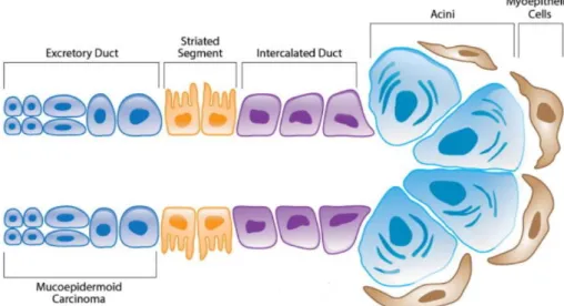
Figura 2 Representação esquemática de uma glândula salivar indicando áreas putativas de origem do CME.
20
Figura 3 Estrutura esquemática de c-MET. 24
Figura 4 Estrutura esquemática de HGF. 25
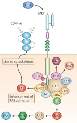
Figura 5 Estrutura esquemática da via de sinalização HGF/c-MET. 26
Figura 1 Presence of c-MET in MEC Cell Lines (A) Immunofluorescence was performed in order to verify the presence of c-MET in cell lines examined. The subcellular distribution of c-MET is show in green, Pan-keratin in red and the degree of overlap in orange. Note that HMC-1 and UM-HMC-3B have c-MET predominantly in the cytoplasm. (B) Accumulation of c-MET in MEC cell lines was determined using flow cytometry assay. The assay was performed in triplicate and the percentage of c-MET + was plotted in the graphs.
47
Figura 2 Effects of HGF on MEC cell lines. The treatment of MEC cell lines with HGF up-regulation of PI3K / AKT and MAPK cascade in UM-HMC-1 and UM-HMC3A. In the metastatic cell line there was a greater accumulation of p-STAT3 and p-PTEN.
48
Figura 3 Migration and invasion of MEC cell lines in vitro is increased under HGF stimulation. (A) Scratch were generated after cell confluence. In vitro cell migration and wound closure were assessed every 8 hours for cell lines 3A and 3B and every hour for cell line UM-HMC-1. Areas of migration were measured in triplicates wells (* p <0.05; ** p <0.01; *** p <0.001; **** p <0.0001). (B) Boyden chamber assay. Medium containing 1μl / ml HGF was added into the lower chamber. Cells that migrated through fibronectin and attached to the under surface of the filter were counted. The mean values of triplicate experiments are presented. Compared with control group, treated cells show significant invasion after 12h for UM-HMC-1, 48h for UM-HMC-3A and 72h for UM-HMC-3B (**** p <0.0001).
Figura 1 Even without the HGF stimulus, all MEC cell lines have a small CSC population. The HMC-1 cell line exhibits more CSCs than UM-HMC-3B and UM-HMC-3A have more CSCs than the UM-UM-HMC-3B lineage. Therefore, the metastatic cell line is the one with the lowest amount of CSCs. **p < 0.01.
62
Figura 2 (a,b) HGF increases the population of CSC cell line in HMC-1 and UM-HMC-3A. Cells were stimulated with HGF for 48h in culture medium containing 2% FBS and 1% HEPES. Cells were collected and processed for ALDH enzymatic activity and anti-CD44 using flow cytometry. (c). Note that even in the presence of HGF there were no statistically significant differences with respect to CSC in the metastatic cell line. *p < 0.05; **p < 0.01 and NS p > 0.05.
LISTAS DE TABELAS
Tabela 1 SGHM de acordo com AFIP (2008) e Brandwein et al. (2001) para CME
LISTA DE ABREVIATURAS E SIGLAS
AFIP Instituto de Patologia das Forças Armadas
CCECP Carcinomas de Células Escamosas de Cabeça e Pescoço
CME Carcinoma Mucoepidermoide
C-MET Proteína Tirosina Quinase MET
CTRC1 Do inglês Regulated transcription coactivator 1 CUPs Do inglês Cancers of Unknown Primary Origin ECS Do inglês Extracapsular Spread
EGFR Receptor de Crescimento Epidérmico
EMT Transição Epitélio-Mesênquima
GAB1 Do inglês GRB2-Associated Binding protein 1 GRB2 Do inglês Growth Factor Receptor-Bound protein 2 HER2 Do inglês Human Epidermal Growth Factor Receptor 2
HGF Fator de Crescimento de Hepatócito
HPV Papiloma Vírus Humano
IPT Do inglês Immunoglobulin Plexins Transcription
MAML2 Do inglês Mastermind-like protein 2
MAPK Do inglês Mitogen-activated Protein Kinases
MHGS Do inglês Malignant Histologic Gradation System
NF-kB Factor nuclear kappa B
PI3K Do inglês Phosphatidylinositol-3-Kinase
PSI Do inglês Plexin, Semaphorin and Integrin cysteine-rich PTEN Do inglês Phosphatase and Tensin homolog
RON Do inglês Receptor Originated from Nantes RTKs Do inglês Receptor tyrosine kinase
SGHM Sistema de Gradação Histológica de Malignidade
STAT Do inglês Signal Transducer and Activator of Transcription TMGSs Tumores Malignos de Glândulas Salivares
TNM Classificação OMS para estaiamento tumoral
SUMÁRIO
1 INTRODUÇÃO... 16
2 REVISÃO DE LITERATURA... 18
2.1 Carcinoma Mucoepidermoide ... 18
2.2 C-MET e seu ligante HGF ... 24
3 CAPÍTULOS... 29
3.1 Capítulo 1... 30
3.2 Capítulo 2... 50
4 CONCLUSÃO GERAL... 66
REFERÊNCIAS... 67
1 INTRODUÇÃO
O carcinoma mucoepidermoide (CME) é o tumor maligno de glândula salivar mais comum, representando 30% a 40% de todas as malignidades das glândulas salivares maiores (MCHUGH; VISSCHER; BARNES, 2009). Clinicamente, apresenta-se como tumor de crescimento lento, indolor, com ampla infiltração local (ANDISHEH-TADBIR et al., 2015; COCA-PELAZ et al., 2015), acometendo preferencialmente o sexo feminino com pico de ocorrência na 5ª década de vida (ELLIS; AUCLAIR, 2008; BRANDWEIN et al., 2001). O comportamento do CME é variável, havendo lesões mais indolentes, que se apresentam com crescimento lento, e outras localmente agressivas, recidivantes e altamente metastáticas (BYRD et al., 2003; ANDISHEH-TADBIR et al., 2015). A patogênese do CME é ainda desconhecida (O’NEILL, 2008; ADAMS, WARNER; NOR, 2013; WARNER et al., 2013; LIU et al., 2015), o que leva ao desconhecimento de marcadores moleculares que prevejam, com precisão, o prognóstico do CME, precise seu diagnóstico e melhore a abordagem terapêutica (OTA et al., 2010; LIU et al., 2014; SHIGEISHI et al., 2014; LIU et al., 2015) já que, mesmo atualmente, o tratamento do tumor se encontra limitado, principalmente, a procedimentos cirúrgicos que possuem significativa morbidade e que são bastante mutilantes (ADAMS, WARNER; NOR, 2013; CLAUDITZ et al., 2013; SHIGEISHI et al., 2014). Dessa forma, o acesso a certas ferramentas de pesquisa, como linhagens celulares, é fundamental para o entendimento da biologia do CME (WARNER et al., 2013).
Recentemente, pesquisadores da Universidade de Michigan, nos Estados Unidos, isolaram quatro linhagens celulares de CME (UM-HMC1, UM-HMC2, UM-HMC3A, UM-HMC3B), as quais constituem as únicas que podem ser facilmente expandidas em cultura, sendo as linhagens UM-HMC-3A e UM-HMC-3B viáveis quando transplantadas em camundongos imunodeficientes e capazes de mimetizar a histologia do tumor primário (WARNER et al., 2013). O uso dessas células será de importante valia para estudos translacionais que possam contribuir para o conhecimento da fisiopatologia desse tumor e de seus processos de invasão locorregional e metástase (WARNER et al., 2013).
biológicos como o desenvolvimento do feto, onde exerce um importante papel na formação do fígado, placenta e dos músculos, e o desenvolvimento do sistema nervoso (COMOGLIO; GIORDANO; TRUSOLINO, 2008; GARAJOVÁ et al., 2015). No entanto, a via HGF/c-MET pode ser reativada nas células cancerígenas, levando a disseminação tumoral. Tanto HGF quanto c-MET encontram-se superexpressos em mais de 80% dos Carcinomas de Células Escamosas de Cabeça e Pescoço (CCECP), estando presente nos tumores de comportamento mais agressivos, uma vez que promove a Transição Epitélio-Mesênquima (EMT), responsável pelo desenvolvimento de metástases, atua no mecanismo de resistência tumoral contra inibidores do Receptor de Crescimento Epidérmico (EGFR) e promove maior invasão e proliferação celular (LAU; CHAN, 2011; ROTHENBERGER; STABILE, 2017).
Em Tumores Malignos de Glândula Salivar (TMGSs) a via HGF/c-MET é estudada principalmente por meio da técnica imunoistoquímica. Tsukinoki et al. (2004) encontrou que a imunoexpressão de HGF nas células estromais e c-MET no parênquima de TMGSs estavam correlacionados com metátase linfonodal e a distância, bem como com piores taxas de sobrevida. Estudo realida por Ach et al. (2013) constatou que neoplasias de glândula salivar com perda genômica de PTEN apresentam significativa aberração genômica de MET. Vasconcelos et al. (2015) demonstrou que HGF e c-METestavam presentes em TMGSs humanos. Esses eventos em conjunto levam a limitadas conjecturas sobre como a ativação da via HGF/c-MET influencia no comportamento biológico dos TMGSs.
Com base no exposto, o objetivo desta tese foi entender e investigar o papel bem como os efeitos da ativação da sinalização HGF/c-MET em linhagens celulares de CME.
2 REVISÃO DE LITERATURA
2.1 Carcinoma Mucoepidermoide
O sistema de glândulas salivares é formado pelas glândulas salivares maiores e menores (ELLIS; AUCLAIR, 2008). Cada glândula salivar contém um parênquima com vários ductos que confluem para as unidades secretoras terminais. As células secretoras são chamadas de ácinos e podem ser serosas, mucosas ou mistas. As células mioepiteliais encontram-se entre as células ductais e a membrana basal dos ácinos (DARDICK et al., 1996). Conhecer a histologia das glândulas salivares é de grande importância para o diagnóstico de suas neoplasias devido à grande similaridade, em termos histológicos, entre o normal e o patológico (DARDICK et al., 1996; ELLIS; AUCLAIR, 2008).
A neoplasia maligna mais frequente das glândulas salivares é o CME (SPIRO et al., 1986; GRÉNMAN et al., 1992; QUEIMADO et al., 1999; TONON et al., 2003; ADAMS; WARNER; NOR, 2013; LIU et al., 2014; KATABI et al., 2014; SHIGEISHI et al., 2014; LIU et al., 2015), perfazendo cerca de 30% a 35% de todos os tumores malignos de glândula salivar (MCHUGH; VISSCHER; BARNES, 2009).
Figura 1 – Representação esquemática dos genes CRTC1 e MAML2 tipo selvagem e do oncogenes de fusão (CRTC1-MAML2) resultado da t(11;19). O domínio de ligação Notch do gene MAML2 é trocado pelo domínio de ligação CREB do gene CRTC1, o qual se fusiona aos éxons 2-5 do gene MAML2. Esse oncogenes de fusão fica sob o controle do promotor do CRTC1.
Fonte: Adaptado de O’Neill (2009).
O CME tem como principal sítio primário de acometimento a glândula parótida seguida do palato duro e mole, língua e assoalho de boca (NANCE et al., 2008; ALI et al., 2013; LIU et al., 2014). Quando subjacente ao palato ou área retromolar, o osso cortical pode ser reabsorvido (COCA-PELAZ et al., 2015). Acomete preferencialmente o sexo feminino e apresenta o pico de ocorrência na 5ª década de vida (ELLIS; AUCLAIR, 2008; BRANDWEIN et al., 2001). Clinicamente, apresenta-se como um tumor de crescimento lento, indolor, com ampla infiltração local, podendo ser variavelmente firme, elástico ou mole à palpação (ANDISHEH-TADBIR et al., 2015; COCA-PELAZ et al., 2015). 2015). Frequentemente são descobertos em um estágio avançado, o que contraindica o tratamento cirúrgico (CROS et al., 2013). Devido a sua superficial localização, tumores intraorais podem apresentar-se como um aumento de volume de coloração azul-avermelhada, simulando um tumor vascular ou uma mucocele (COCA-PELAZ et al., 2015). Frequentemente são descobertos em um estágio avançado, o que contraindica o tratamento cirúrgico (CROS et al., 2013).
cístico (com formação de estruturas císticas e estruturas glandulares ductais) e sólido (com formação de ilhas tumorais) (KATABI et al., 2014). Sua organização histológica lembra o ducto excretor das glândulas salivares, ao qual tem sido atribuída sua origem (Figura 2) (DARDICK, 1996; AZEVEDO et al., 2008; AKRISH et al., 2009).
Figura 2 – Representação esquemática de uma glândula salivar indicando áreas putativas de origem do CME. Fonte: Adaptado de ADAMS; WARNER; NOR (2013).
Parâmetros clínico-patológicos como idade, sexo, tamanho do tumor, estágio, estadiamento TNM, disseminação extracapsular, terapia adjuvante e o status da margem têm mostrado valor preditivo na sobrevida de pacientes com CME, embora tenha sido sugerido que o mais relevante seja o SGHM e o estágio clínico (BYRD et al., 2013). Este é realizado com base no sistema TNM proposto pelo American Joint Commitee on Cancer. Este sistema
classifica os tumores em 4 estádios de acordo com o tamanho da lesão (T), presença de metástases regionais em linfonodos (N) e metástase à distancia (M). Apesar de apresentar limitações no seu potencial prognóstico, esse sistema permanece mundialmente reconhecido para descrever a extensão do tumor (PATEL; LYDIATT, 2008). Os critérios para o estadiamento são os seguintes (EL-NAGGAR et al., 2017):
• T (tamanho):
o TX - Tumor primário não pode ser avaliado o T0 - Sem evidências do tumor
primário. o Tis – Carcinoma in situ o T1 - O tumor tem até 2 cm de diâmetro e não invade tecidos adjacentes clinicamente.
o T2 - O tumor tem mais do que 2 cm e menos do que 4 cm de diâmetro não invade
o T3 - O tumor tem mais do que 4 cm de diâmetro e está invadindo tecidos moles
adjacentes.
o T4a (moderadamente avançado) - O tumor invade estruturas próximas, como o
osso da mandíbula, pele, canal auditivo ou nervo facial.
o T4b (muito avançado) – o tumor invade estruturas próximas, como a base do
crânio ou outros ossos nas proximidades ou que circundam a artéria carótida.
• Linfonodo regional (N):
o NX - Linfonodo regional não pode ser avaliado. o N0 - Ausência de metástase em linfonodo.
o N1 – Metástase em apenas um linfonodo ipsilateral, medindo até 3cm na sua
maior extensão.
o N2a - Metástase em apenas um linfonodo ipsilateral medindo entre 3 e 6 cm de
diâmetro.
o N2b - Metástase em múltiplos linfonodos ipsilaterais nenhum medindo mais que
6 cm de diâmetro.
o o N2c - Metástase em linfonodos bilaterais ou contralaterais nenhum medindo
mais que 6 cm de diâmetro.
o N3 - Metástase em linfonodo medindo mais que 6 cm de diâmetro.
• Metástase à distância:
o Ausência de metástase à distância. o Metástase à distância.
Após realizada a análise das características acima citadas (T, N, M) o tumor é classificado de acordo com os seguintes critérios:
• Estágio I - T1, N0, M0.
• Estágio II - T2, N0, M0.
• Estágio III - T3, N0, M0; T1 a T3, N1, M0.
• Estágio IVA - T4a, N0 ou N1, M0; T1 a T4a, N2, M0.
• Estágio IVC - Qualquer T, qualquer N, M1.
Para Coca-Pelaz et al. (2015), o SGHM deve ser aliado também a testes moleculares. O SGHM mais utilizado na atualidade para gradações de CME é o da AFIP (Instituto de Patologia das Forças Armadas) (ELLIS; AUCLAIR, 2008) e o de Brandwein et al. (2001), os quais utilizam informações relativas ao padrão de crescimento, infiltração e achados citológicos (KATABI et al., 2014; COCA-PELAZ et al., 2015). O sistema de gradação da AFIP é baseado em pontos, que têm como parâmetros características histológicas úteis em predizer o prognóstico (Tabela 1). Ao fim da análise, os pontos devem ser somados e obtido um escore, que corresponderá ao grau de malignidade do tumor. Brandwein et al. (2001) adicionaram alguns parâmetros histológicos ao SGHM da AFIP (Tabela 1).
Tabela 1 – SGHM de acordo com AFIP (2008) e Brandwein et al. (2001) para CME
AFIP BRANDWEIN et al. (2001)
Componente cístico < 20% = 2 pontos Componente cístico < 25% = 2 pontos Invasão neural = 2 pontos Tumor invadindo em pequenos ninhos e ilhas
= 2 pontos
Necrose = 3 pontos Atipia nuclear pronunciada = 2 pontos ≥ 4 mitoses por campo (aumento de 10x) =
3 pontos
Invasão linfovascular = 3 pontos
Anaplasia = 4 pontos Invasão óssea = 3 pontos
>4 mitoses por campo (aumento de 10x) = 3
pontos
Invasão perineural = 3 pontos Necrose = 3 pontos
Baixo grau: 0-4 pontos
Grau intermediário: 5-6 pontos Alto grau: 7 a 14 pontos
Baixo grau: 0 pontos
Grau intermediário: 2-3 pontos Alto grau: ≥ 4 pontos
Fonte: Adaptado de Ellis e Auclair (2008) e Brandwein et al. (2001).
portadores de CME de grau intermediário possuem sobrevida de 62% a 92%, e o seu tratamento é controverso por refletir o duvidoso SGHM de tumores de glândula salivar (BYRD et al., 2013; KATABI et al., 2014). De acordo com vários pesquisadores, não há um SGHM uniformemente aceito para CME (BYRD et al., 2013; KATABI et al., 2014; COCA-PELAZ et al., 2015).
As formas de tratamento para o CME atualmente encontram-se estagnadas e dependentes de sistemas de gradações histológicas de malignidade que não possuem uma linguagem uniforme. Ainda nos dias atuais, não existe nenhum indicador que possa ser usado no diagnóstico preciso ou que preveja o prognóstico não só do CME mas também de outras neoplasias malignas de glândula salivar (OTA et al., 2010). O maior obstáculo das pesquisas que procuram estudar a efetividade e a segurança de terapias para o tratamento do CME é o pobre entendimento da sua fisiopatologia (O’NEILL, 2009; WARNER et al., 2013; ANDISHEH-TADBIR et al., 2015; LIU et al., 2015). Os mecanismos envolvidos no processo de migração, invasão locorregional e metástase das células do CME são desconhecidos (WARNER et al., 2013). Ainda não é conhecido nem mesmo o seu mecanismo molecular de desenvolvimento (ANDISHEH-TADBIR et al., 2015). O acesso a certas ferramentas de pesquisa como linhagens celulares e modelos de xenoenxertos é fundamental não só para o melhor entendimento da biologia do CME como também para o desenvolvimento de terapias mais eficazes, isto é, que sejam baseadas no mecanismo da doença (WARNER et al, 2013).
Escassas linhagens celulares de CME têm sido estabelecidas até o momento (WARNER et al., 2013). Grénman et al. (1992) estabeleceram uma linhagem celular (denominada de UT-MUC-1) a partir de um CME pobremente diferenciado e constataram resistência a radioterapia. Queimado et al. (1999) conseguiram estabelecer e caracterizar linhagem de CME (UTSW-MEC-49) por infecção com genes E6 e E7 do HPV 16, o que proporcionou maior estabilidade nas células em cultura. Tonon et al. (2003) estabeleceram duas linhagens de CME (NCl-H292 e H3118) e constataram translocação recíproca (t 11;19) que desfaz a linha de sinalização Notch.
Células explantadas de tumores de glândula salivar são particularmente difíceis de se propagar in vitro (QUEIMADO et al., 1999). Recentemente, a Universidade de Michigan,
fisiopatologia da doença levará ao provável desenvolvimento futuro de terapias alvo mais racionais, além de contribuir para uma melhor qualidade de vida do paciente (ADAMS; WARNER; NOR, 2013; SHIGEISHI et al., 2014).
2.2 C-MET e seu ligante HGF
O proto-oncogene MET é localizado no cromossomo 7q21-31 e codifica c-MET. O receptor MET é uma glicoproteína heterodimérica, formada pela subunidade extracelular alfa ligada a subunidade transmembrana beta através de uma ponte disulfídeo. A porção extracelular inclui os domínios Sema (Semaphorin), PSI (Plexin, Semaphorin and Integrin cysteine-rich) e quatro IPT (Immunoglobulin Plexins Transcription). O domínio
intracelular inclui uma sequência justamembrana, uma região catalítica e um local de ancoragem carboxiterminal multifuncional. O domínio justamembrana contém os resíduos Ser975 e Tyr1003, que são envolvidos na regulação negativa de MET, a região catalítica é responsável por modular a atividade quinase e o local de ancoragem carboxiterminal multifuncional é responsável pelo recrutamento de várias moléculas adaptadoras e transdutoras de sinal (Figura 3) (OWSU et al., 2017).
Figura 3 – Estrutura esquemática de c-MET.
Fonte: Adaptado de COMOGLIO; GIORDANO; TRUSOLINO (2008).
de que se converta na sua forma madura. Sua forma biologicamente ativa consiste em um heterodímero ligador por pontes dissulfitos, contendo uma cadeia alfa e outra beta. A cadeia alfa contém um loop N-terminal (HL) seguinda por quatro domínios conhecidos como Domínios kringle (K) (Figura 4). A cadeia β é homóloga às serinas proteases da cascata de coagulação do sangue, mas carece de atividade proteolítica devido a substituições de aminoácidos no local catalítico (ORGAN; TSAO, 2011).
Figura 4 – Estrutura esquemática de HGF.
Fonte: Adaptado de COMOGLIO; GIORDANO; TRUSOLINO (2008).
A iniciação da via de sinalização de MET começa com a ligação de HGF a c-MET na membrana plasmática celular, levando a dimerização e estabilidade do receptor (BARROW-MCGEE; KERMORGANT, 2014). A subsequente ativação do seu domínio intracellular acontece através da fosforilação dos dois resíduos de tirosina na porção catalítica Y1234 e Y1235, seguida pela fosforilação das duas tirosinas de ancoragem Y1349 e Y1356 na cauda terminal. Estas duas tirosinas formam o local de ancoragem multifunctional, o qual é particular aos membros da subfamília MET e essencial para a sua sinalização (BARROW-MCGEE; KERMORGANT, 2014). Após a fosforilação desses domínios, c-MET está apto a se ligar a múltiplos substratos e ativar uma variedade de vias de sinalização, que ocorrerão através da interação direta com esse receptor por meio dos adaptadores GRB2 (Growth Factor Receptor-Bound protein 2) e GAB1 (GRB2-Associated Binding protein 1)
Figura 5 – Estrutura esquemática da via de sinalização HGF/c-MET. Fonte: Adaptado de COMOGLIO; GIORDANO; TRUSOLINO (2008).
O conjunto das alterações celulares desencadeadas por c-MET (proliferação, sobrevida celular, migração e invasividade) é chamado de crescimento invasivo (BOCCACCIO; COMOGLIO, 2014). Este está envolvido nos processos morfogenéticos, como gastrulação (processo pelo qual as três camadas germinativas, que são precursoras de todos os tecidos embrionários, são estabelecidos nos embriões), desenvolvimento dos múculos e durante a angiogênese, sendo mediado pela sinalização HGF/c-MET (BOCCACCIO; COMOGLIO, 2014; MOORE; PERSAUD; TORCHIA, 2016). Na idade adulta, o crescimento invasivo se torna quiescente, mas pode ser reativado durante, por exemplo, a cicatrização de feridas em que células residuais podem proliferar e migrar a fim de reconstituir a integridade dos tecidos que sofreram injúria. Além disso, a via HGF/c-MET é importante na manutenção da homeostasia dos tecidos (BARROW-MCGEE; KERMORGANT, 2014). Estudos mostram que os níveis plasmáticos de HGF aumentam depois que órgãos como o coração, rim e fígado sofrem algum tipo de injúria, mostrando que níveis elevados de HGF e a ativação subsequente de c-MET pode ser parte de uma resposta fisiológica protetora.
tem sido mostrado que sinais de crescimento invasivo provenientes de c-MET representam um achado marcante em tumores altamente agressivos (BOCCACCIO; COMOGLIO, 2014). Em câncer, os mecanismos genéticos responsáveis pela sinalização aberrante de MET são lesões genéticas específicas (rearranjo cromossômico e mutação), aumento da transcrição e ligante exercendo ação autócrina ou parácrina (COMOGLIO; GIORDANO; TRUSOLINO, 2008). Mutações de MET podem gerar um tipo de autonomia celular responsável por CUPs (Cancers of Unknown Primary Origin), os quais mostram disseminação inicial, exibem um fenótipo
altamente indiferenciado, não apresentam marcadores moleculares do tecido de origem e possuem um fenótipo de célula-tronco (BOCCACCIO; COMOGLIO, 2014). No entanto, as alterações genéticas de MET são raras (1% a 3% dos tumores), sendo mais frequente a superexpressão do gene selvagem em cânceres (BOCCACCIO; COMOGLIO, 2014). Nesse contexto, diversas neoplasias maligas, tais como cânceres de cabeça e pescoço, mama, colorretal, gástrico, tireóide e de pulmão tem evidenciado a expressão de c-MET aumentada.
A biologia do câncer pode ser melhor compreendida pelo modelo da Heterogeneidade tumoral, o qual postula que as populações de células neoplásicas tumorais compreendem populações heterogêneas, nos quais células com diferentes potenciais tumorigênicos habitam dentro do mesmo tumor (MARJANOVIC; WEINBERG; CHAFFER, 2013). Dessa forma, terapias que visam debelar um único receptor pode facilmente resultar na seleção positiva de células que não possuem o receptor alvo da terapia (BOCCACCIO; COMOGLIO, 2014). Pesquisa com câncer de pulmão mostrou que as lesões recidivantes após a terapia anti-EGFR exibem uma amplificação de novo de c-MET, indicando que o tratamento
selecionou positivamente uma pequena população tumoral detentora de c-MET amplificado (TURKE et al., 2010). Apesar das evidências indicarem que a mesma célula tumoral possui concomitantemente múltiplos receptores, a fim de enviar sinais mais robustos para a proliferação, a idéia contrária está atualmente em foco, uma vez que o tipo selvagem de MET e os receptores da família do EGFR podem ser expressos de maneira mutuamente exclusivas. Paulson et al. (2013) mostrou haver expressão de MET ou HER2 (Human Epidermal Growth
Factor Receptor 2) em câncer de mama. Boccaccio e Comoglio (2013) encontrou que
e intracelulares que interagem com c-MET e são fundamentais para sustentar tumores que possuem vício nesse oncogene. Dentre esses parceiros destaca-se RON (Receptor Originated from Nantes), o mais conhecido deles, que é requerido para desencadear completamente o potencial oncogênico de células com amplificação de MET (BENVENUTI et al., 2011) e STAT3 (Signal Transducer and Activator of Transcription 3), que antes era conhecido por
exercer sinais de crescimento invasivo oriundos da ativação fisiológica de c-MET, emerge agora como peça fundamental na proliferação celular mediada por tumores que possuem MET amplificado (LAI et al., 2016).
A expressão de HGF e c-MET tem sido observada em células progenitoras. Estudo com células embrionárias de glândula salivar mostrou forte expressão de c-MET no tecido glandular e concluiu que essa molécula é envolvida no desenvolvimento das glândulas salivares (LORETO et al., 2010). Um recente estudo com epitélio de glândula mamária de camundongo revelou que c-MET está expresso especificamente em células luminais progenitoras e que HGF é capaz de reter células no estado tronco, prevenindo a sua diferenciação (GASTALDI et al., 2013). Dessa forma, c-MET pode guiar o tumor para um fenótipo mais tronco e ser um marcador do crescimento numérico das células luminais progenitoras, que são impedidas de se diferenciar. Com base no exposto, c-MET possui um papel dual no câncer: na sua forma geneticamente alterada é capaz de gerar e manter o fenótipo celular transformado, guiando a evolução clonal; já na sua forma selvagem, c-METcontribui para manter o fenótipo inerte das células-tronco cancerígenas, conferindo imortalidade replicativa ao tumor.
3 CAPÍTULOS
Esta tese está baseada no Artigo 46 do Regimento Interno do Programa de Pós-Graduação em Odontologia da Universidade Federal do Ceará, que regulamenta o formato alternativo para dissertações de Mestrado e teses de Doutorado e permite a inserção de artigos científicos de autoria ou coautoria do candidato e exige certificação de línguas. Assim sendo, esta tese é composta de dois capítulos contendo um artigo científico em processo de submissão no periódico “Scientific Reports” e “Stem Cell Research & Therapy”, respectivamente, conforme descrito abaixo:
AVALIAÇÃO DA SINALIZAÇÃO DE c-MET E SEUS EFEITOS EM LINHAGENS CELULARES DE CARCINOMA MUCOEPIDERMOIDE
Thamara Manoela M Bezerra, DDS, MsC, PhD Student; Liana Preto Webber DDS, MsC, PhD Student; Gabriell Bonifácio Borgato DDS, MsC, PhD Student; Rogério Moraes Castilho, DDS, MsC, PhD; Cristiane Helena Squarize, DDS, MsC, PhD; Karuza Maria Alves Pereira, DDS, MsC, PhD. Scientific Reports. Status: Processo de Submissão iniciado em março de 2018.
HGF PROMOVE O DESENVOLVIMENTO DE CÉLULAS-TRONCO
CANCERÍGENAS EM LINHAGENS CELULARES DE CARCINOMA
MUCOEPIDERMOIDE.
Thamara Manoela M Bezerra, DDS, MsC, PhD Student; Liana Preto Webber DDS, MsC, PhD Student; Gabriell Bonifácio Borgato DDS, MsC, PhD Student; Rogério Moraes Castilho, DDS, MsC, PhD; Cristiane Helena Squarize, DDS, MsC, PhD; Karuza Maria Alves Pereira, DDS, MsC, PhD. Stem Cell Research & Therapy. Status: Processo de Submissão iniciado em março
3.1 Capítulo 01: AVALIAÇÃO DA SINALIZAÇÃO DE c-MET E SEUS EFEITOS EM LINHAGENS CELULARES DE CARCINOMA MUCOEPIDERMOIDE.
Tittle Page Evaluation of c-MET signaling and its effects on mucoepidermoid carcinoma cell lines
Original Article
Running Head
HGF/c-MET signalling promotes aggressive behavior in MEC
Authors and name affiliations
Thâmara Manoela Bezerra Marinho1, Liana Preto Webber2, Gabriel Bonifácio Borgato3, Rogério Moraes Castilho2, Cristiane Helena Squarize2, Karuza Maria Alves Pereira4*
1Department of Dental Clinic, Division of Oral Pathology, Faculty of Pharmacy, Dentistry and Nursing, Federal University of Ceará, Fortaleza, Ceará, Brazil
2Division of Oral Pathology/Medicine/Radiology, Department of Periodontics and Oral Medicine University of Michigan School of Dentistry, Ann Arbor, Michigan, USA
3Department of Morphology, Piracicaba Dental School, University of Campinas, Piracicaba, São Paulo, Brazil
4Department of Morphology, School of Medicine, Federal University of Ceará, Fortaleza, Ceará, Brazil.
*Correspondence author: PhD. MSc. DDS. Karuza Maria Alves Pereira Department of Morphology
School of Medicine
Federal University of Ceará
Rua Delmiro Farias, s/n, Rodolfo Teófilo, 60430-170, Fortaleza, CE, Brazil.
Phone: +55.85.33668471.
Abstract
Mucoepidermoid carcinoma (MEC) is an infrequent malignant neoplasm that originates most commonly in the salivary glands. Its variable biological behavior is not well understood due to lack of studies on its pathobiology. Hepatocyte Growth Factor (HGF, MET ligand) and c-MET are immunoexpressed in human Salivary Gland Malignant Tumors (SGMTs) tissues samples using immunohistochemistry. Herein, we sought to understand and investigate the role of HGF/c-MET signaling and its effects in MEC cell lines. Our finding shows that the activation of PI3K/AKT signaling and MAPK cascade, via HGF/c-MET signaling, is an effective strategy used by MEC to promote increased cell migration and invasiveness. We have achieved an important step towards a better understanding of MEC pathobiology.
Introduction
Malignant Salivary Gland Tumors (MSGTs) are relatively rare but deadly. An average of 3300 new cases are diagnosed each year in the USA1. Among the MSGTs, the
Mucoepidermoid Carcinoma (MEC) is the most frequently reported pathology2,3,4.
Histologically, these tumors are characterized by the presence of mucous, epidermoid, and intermediate cell types1,4. The clinical and pathological behavior of ECM is highly variable, since it may be indolent and slow-growing or locally aggressive and highly metastatic5. In order to better predict patient survival and the highly variable behavior of these tumors, a variety of prognostic factors have been studied, including age, sex, tumor site, stage, TNM status, extracapsular spread (ECS), adjuvant therapy, margin status5,2. However, for MEC, the most prognostically relevant of these is histological tumor grade2. The Malignant Histologic Gradation System (MHGS) has shown strong correlations with the clinical behavior of the tumor5. In the present study, the majority of MHGSs were classified as MEC in three tiers: MECs of low, intermediate and high degree of malignancy5,1,4. However, these parameters may vary according to the MHGS adopted by the pathologist and, despite the strong clinical correlations, the lack of consensus and ambiguity of the existing MHGSs for grading MECs is
a problem because some gradation systems upgrade MEC and other downgrade MEC5,6. Thus,
research focused on the understanding of the pathobiology of CME may help to clarify the highly variable clinicopathological characteristics of this tumor, identify molecular biomarkers that will help better predict the clinical outcomes of the disease and improve the survival and quality of life of the affected patient by MEC1,7,5,2,4.
Among the research tools that can help in the better understanding of the pathobiology of the MEC, cell lines and xenograft models stand out. We recently established 5 new mucoepidermoid carcinoma cell lines, two of which (UM-HMC-3A and UM-HMC-3B) are able to recapitulate the histology of the primary tumor when transplanted into immunodeficient
mice7. In this study we used three of these cell lines (HMC-1, HMC-3A and
in organ formation during embryogenesis and in tissue homeostasis in the adult and is converted into its bioactive form through extracellular proteases10,9. The initiation of HGF/c-MET signaling occurs when HGF binds to the c-MET receptor at the plasma membrane, there being the receptor homodimerization and phosphorylation of two tyrosine residues (Y1234 and Y1235), located within the catalytic loop of the tyrosine kinase domain, followed by phosphorylation of two docking tyrosines (Y1349 and Y1356) in the carboxy-terminal site. After phosphorylation, there is recruitment of the adaptor proteins GRB2 (Growth Factor Receptor Bound Protein 2), which binds directly to c-MET, and Gab1 (Grb2-Associated Binder 1), which can bind either directly to c-MET or indirectly, through GRB2. Subsequently, it occurs the activation of different intracellular signaling pathways (MAPK, PI3K-AKT cascades, STAT and NF-κB signaling pathways) which are responsible for driving the cellular activities of proliferation, cell survival, migration and invasiveness10,9,11,12.
The high-affinity HGF receptor/c-Met system is overexpressed in human cancers. The inappropriate activation of this pathway in Head and Neck Squamous Cell Carcinoma (HNSCC) promotes induction of Epithelial-Mesenchymal Transition (EMT), lymph node metastasis, poor prognosis, higher tumor staging, local recurrence and EGFR resistance12. Although limited, c-MET studies in SGMTs indicate that aberrations of MET are
associated with EGFR and PTEN signaling13 HGF/c-MET immunoreactivity might be
associated with poor prognosis in patients with high grade salivary gland carcinomas14 and HGF may play differentiation of ductal structures of SGMTs15.
Taking into consideration the scarce studies on the understanding of the pathobiology of MEC, we decided to investigate, for the first time, the presence of c-MET as well as the effects of its activation by HGF in three different MEC cell lines, recently established at the University of Michigan School of Dentistry. We found that all MEC cell lines have the constitutively activated c-MET receptor and that it can be in both the membrane (sometimes distributed asymmetrically and punctate) and in the cytoplasm of cells. We have seen that HGF stimulation provides increased migration and invasiveness in all lineages examined by the activation of PI3K/AKT and ERK1/2 signaling pathways. The metastatic lineage showed to be quiescent, requiring a long time of exposure to HGF to promote change to the proliferative and migratory cell state.
Materials and Methods
The cell lines used in this study were firstly described by Warner et al.7. All cell lines examined were derived from tumors located in the minor salivary gland (UM-HMC-1 - no previous treatment; UM-HMC3A - local recurrence and UM-HMC3B - lymph node metastasis) (WAGNER et al., 2016). The cell lines were grown in Dulbecco's Modified Eagle's Medium supplement (DMEM/High Glucose, Life Sciences, Utah, USA) with 10% Fetal Bovine Serum (FBS) (Sigma-Aldrich Corp., St. Louis, MO, USA), 1% penicillin/streptomycin (Life Technologies, Grand Island, NY, USA), 1% L-glutamine (Life Technologies, Grand Island, NY, USA), 20 ng/ml Epidermal Growth Factor (PeproTechUS, Rockey Hill, NJ), 400 μg/mL hydrocortisone (Sigma-Aldrich Corp., St. Louis, MO, USA), 10 mg/mL insulin (Sigma-Aldrich Corp., St. Louis, MO, USA) and maintained in incubators under controlled temperature (37oC), humidity and CO2 concentration (5%). Cells were passaged using 0.05% trypsin/EDTA (Life Technologies, Grand Island, NY, USA).
Immunofluorescence
Cells were seeded on glass coverslips in 6-well plates. After reaching the ideal confluence, non-adherent cells were washed away by Phosphate Buffer Saline (PBS), whereas adherent cells were fixed with 4% paraformaldehyde for 20 minutes at room temperature and permeabilized with 0.1% Triton for 5 minutes. Blocking was performed with 3% Bovine Serum Albumin (BSA) in PBS for 45 minutes at 37oC. After the incubation, cells were rinsed once with PBS for 5 minutes and then incubated with c-MET (1:50, R&D Systems, Minneapolis, MN, USA) and Pan-keratin (C11) (1:700, Cell Signaling, Danvers, MA, USA). The cells were washed three times, incubated with FITC-conjugated secondary antibody and co-stained with Hoechst 33342 (Sigma-Aldrich Corp., St. Louis, MO, USA) for visualization of DNA content. Images were taken using a QImaging ExiAqua monochrome digital camera attached to a Nikon Eclipse 80i Microscope (Nikon, Melville, NY, USA) and visualized with QCapturePro software.
Western Blotting
electro-transferred to an Immobilon-FL polyvinyl difluoride membrane (Millipore, Billerica, MA, USA). Nonspecific binding was blocked in 5% nonfat dry milk (non-phosphorylated antibodies) or Bovine Serum Albumin (BSA) (phosphorylated antibodies) both containing 0.1 M Tris (pH 7.5), 0.9% NaCl and 0.05% Tween-20 for 1 hour at room temperature. The membranes were then incubated overnight at 4°C with the following primary antibodies: c-MET (D1C2) XP(R) (1:500, Cell Signaling, Danvers, MA, USA), phospho-c-MET (Tyr1234/1235) (1:500, Cell Signaling, Danvers, MA, USA), phospho-MET (Tyr1349) (1:500, Cell Signaling, Danvers, MA, USA), pan AKT (C67E7) (1:1000, Cell Signaling, Danvers, MA, USA), phospho-AKT (Ser473) (1:1000, Cell Signaling, Danvers, MA, USA), p44/42 MAPK (Erk1/2) (1:1000, Cell Signaling, Danvers, MA, USA), phospho-p44/42 MAPK (Erk1/2) (1:1000, Cell Signaling, Danvers, MA, USA), phospho-GAB1 (1:1000, Cell Signaling, Danvers, MA, USA), phospho-S6 (Ser235/336) (1:1000, Cell Signaling, Danvers, MA, USA), PTEN (138G6) (1:1000, Cell Signaling, Danvers, MA, USA), histone H3 (1:1000, Cell Signaling, Danvers, MA, USA), acetyl-histone H3 (Lys9) (1:10.000, Cell Signaling, Danvers, MA, USA) and phosphor-STAT3 (Tyr705) (3E2) (1:1000 1:1000, Cell Signaling, Danvers, MA, USA). GAPDH (1:20.000, Calbiochem, Gibbstown, NJ, USA) served as a loading control. The reaction was visualized using ECL reagent (Thermo Scientific,
Rockford, IL).
Scratch assay
Cells were seeded into six-well plates to create a confluent monolayer. The plates were appropriately incubated for approximately 6h at 37oC, using 10% FBS culture medium. After the cell adhesion to the cultivation plate, scratches were made with a P200 pipette tip across the diameter of each well. Then, the dishes were washed with PBS two times before adding the starving medium (2% FBS) to the control group. HGF (50ng/ml) was added only to the test group. Scratch area was photographed every 8 hours for the cell lines UM-HMC-3A and UM-HMC-3B and every hour for the cell line UM-HMC-1 using Axiovert 200M microscope (Carl Zeiss, Germany) with x40 magnification. The quantification of the evolution of the scratch area was analyzed with Imaging Processing and Analysis in Java program (ImageJ®, National Institute of Mental Health, Bethesda, Maryland, USA). Results from two independent experiments with three replicates per experiment were pooled.
Invasion assays were carried out 24-well Boyden chambers (Greiner Bio-One,
Frickenhausen, Germany) containing polycarbonate filter membranes 8µm pores precoated with homogeneous thin layer of fibronectin (Haematologic Technologies, Inc). We determined that the invasion time and ideal number of cells for each MEC cell line would be 60-70% of cells/total area in the bottom of the polycarbonate filter membrane. The upper chamber was loaded with the solution of MEC cell lines (UM-HMC-1, 80x102; HMC-3A and UM-HMC-3B 80x103) and 2% FBS. In the experimental group, the bottom chamber was filled with 2% FBS and HGF (50ng/ml) as a chemoattractant. The control group was maintained in DMEM/High Glucose supplemented with 2% FBS. After planting, the cells were incubated according to the ideal invasion time of each cell line (UM-HMC-1, 12h; UM-HMC3A, 48h and UM-HMC-3B, 72h) at 37°C in a humidified atmosphere of 5% CO2. At the end of the experiment, cells were fixed with methanol for 10 minutes and stained with hematoxylin and eosin (H&E). Cells on the upper side of the membrane were then removed using a cotton swab. Images de 10 randomly selected fields at 100x magnifications were taken using a QImaging ExiAqua monochrome digital camera attached to a Nikon Eclipse 80i Microscope (Nikon, Melville, NY, USA) and visualized using QCapturePro software. Each assay was performed in triplicate.
Flow Cytometry - cell surface staining for c-MET
To quantify c-MET, MEC cell lines were maintained in their standard cell cultivation medium. Cells were trypsinized and resuspended in cold FACS buffer (PBS + 0.5% BSA) at a density of 4x10⁴ cells/mL. The experimental group was stained with c-MET (1:400, R&D Systems, Minneapolis, MN, USA) and incubated for 30 minutes at 4oC under agitation. A sample without c-MET was the reaction`s negative control. The cells were resuspended in 500µL of cold FACS buffer and analyzed using Accuri™ C6 flow cytometer (BD Biosciences, USA). The experiment was carried out in quintuplicate.
Statistical Analysis
All statistical analysis was performed using GraphPad Prism (GraphPad Software, San Diego, CA). Statistical analysis of the scratch assay, invasion assay and flow cytometry were performed by unpaired t test. Asterisks denote statistical significance (*p < 0.05; **p < 0.01; ***p < 0.001; ****p < 0.0001; and NS p > 0.05).
c-MET is constitutively expressed in MEC cell lines.
Under normal physiological conditions, c-MET is crucial in the control of tissue homeostasis, embryonic development, organogenesis and wound healing10. However, the physiological functions of this signaling pathway are usurped by cancer cells, facilitating invasion and metastasis. It has been found to be over activated mainly in solid cancers16, and in adenocarcinomas its deregulation is greater when compared than a squamous cell tumor11. In CME, very little is known about the presence of c-MET and its effects. Existing studies are mainly immunohistochemical assays8,15,17. In addition, the availability of c-MET and phospho-MET in formalin-fixed, paraffin embedded samples have limited the development of clinical trials using archived tumor specimens18. For the first time it was evaluated the presence of c-MET in three different MEC cell lines recently established at the University of Michigan School of Dentistry7 using immunofluorescence (Fig.1A) and Western blotting (Fig. 2) and we found that c-MET was present in all cell lines examined. Further, we have explored the localization of c-MET in our MEC cell lines. Cell sub-localization of c-MET was evident in plasma membrane and cytoplasm of cells, showing different expression patterns between cell lines (Fig 1A). UM-HMC-1 and UM-HMC-3B showed predominance of c-MET in the cytoplasm. In UM-HMC-3A cell line, c-MET was mainly seen on the plasma membrane. Although the internalization of c-MET is part of the process of signal attenuation, recently, it has become evident that c-MET trafficking within endosomes compartments, under protein kinase C control, results in full activation of signaling pathways involved in cell survival, invasion and metastasis such as Gab1, ERK1/2, STAT3 and Rac119,20. Flow cytometry assay was performed to quantify the expression of c-MET (Fig. 1B), which showed similar for UM-HMC-3A and UM-HMC-3B. The UM-HMC-1 cell line showed slightly lower c-MET expression when compared to the other cell lines examined. Although c-MET is constitutively expressed in MEC cell lines, it is unknown which signaling pathways are activated after its phosphorylation by HGF.
HGF activates c-MET and triggers signaling modulators common to many RTKs in MEC cell
lines
c-MET in MEC had never been explored until now. We performed western blotting in order to investigate and understand HGF/c-MET pathway signaling in MEC cells. Three MEC cell lines underwent 18 hours of starving and we treated only the experimental group with HGF. We observed that the treatment of cell lines led to the phosphorylation of c-MET in different motifs (p-MET1234/1235 and p-MET1349) in all MEC cell lines (Fig. 2). Interestingly there appears to be ligand-independent MET activation, since we observed the phosphorylation of c-MET in the control group with consequent activation of downstream proteins, although less intensely when compared to the experimental group. The ligand-independent activation of MET signaling occurs due to overexpression or amplification of MET or due to mutational activation of c-MET21 and are rare in primary human cancers11,22.
We observed the activation of different signaling pathways as well as differences in the expression patterns of certain proteins between the MEC cell lines (Fig.2). UM-HMC-1 cell line showed high levels of p-GAB1, p-ERK1/2, p-AKTSer473 and PS6. Cell line UM-HMC-3A, similarly to UM-HMC-1, showed higher levels of p-ERK1/2, p-AKTSer473 and PS6, but similar levels of p-GAB1 among the control and experimental groups. These findings led us to believe that the presence of p-GAB1 is important to extend the duration of p-AKT and p-ERK1/2 phosphorylation, which explains the significant expressions of these proteins in UM-HMC-1 cell line. However, p-GAB1 is not essential to keep AKT and ERK signaling active after c-MET phosphorylation, since p85 subunit of PI3K binds directly to c-c-MET and the oncogenic Ras/Raf signaling, which can subsequently activate the MAPK, may be activated by the phosphorylation of another c-MET-like adapter protein, similar to p-GAB1, termed phosphorylated GRB2. We recently demonstrated that cell proliferation and activation of EMT during oral carcinogenesis23, as well as the accumulation of CSC in MEC cell lines3 is associated with the reduction of H3K9ac. In this study the treatment of cells with HGF led to a decrease in histone 3 (Lys9) acetylation levels in both UM-HMC-1 and UM-HMC-3A cell lines. However, it promoted increased histone 3 acetylation (Lys9) in UM-HMC3B. Similarly, to the HNSCC cells24, MEC cell lines respond differently to environmental stimuli by modulating chromatin acetylation.
actively dividing cells. Recent studies have shown that imbalances in ERK signaling pathways activity may determine the fate of cancer cells (tumorigenicity or cellular dormancy)26, 27.
Because PTEN can interact with c-Met-dependent signaling, we evaluated the expression of PTEN and p-PTEN in MEC cell lines treated and not treated with HGF. The PTEN tumor suppressor gene was shown to be less expressed in the UM-HMC-1 cell line and p-PTEN was present in all MEC cell lines, being more expressive in the metastatic lineage after treatment with HGF. In salivary gland cancer PTEN loss is associated with MET aberration and amplification17. Thus, the inactivation of PTEN and the activation of c-MET is a molecular dysfunction associated with MEC malignancy and contributes to the metastatic signature phenotype.
The biological impact of the activation of these pathways through the HGF/c-MET signaling is unknown.
HGF increases the migration and invasion capacity of MEC cell lines
The migratory ability and invasiveness of MEC cell lines, after treatment with HGF, was evaluated by means of scratch assay and Boyden chamber assay, respectively. We observed that the presence of HGF accelerates the migration and invasion of all MEC cell lines into the denuded area (Fig. 3A and B). However, the onset of the HGF response as well as the time for complete scratch closure ranged between cell lines. The UM-HMC-1 and UM-HMC-3A cell lines showed less time for complete closure of the scratch and to promote invasion. We attributed this finding to the ERK1/2 signaling pathway activation (known to be involved in the migration and invasion process) as can be observed through western blotting (Fig. 2). The lineage of metastatic origin (UM-HMC-3B) presented a slower HGF-dependent cell migration and invasion (Fig. 3A and B). Metastatic cells are usually present in a quiescent and non-proliferative state28, which explains why this cell line requires a greater HGF stimulation to promote cell state change and thus lead to migration and invasion. The switch between dormant and proliferative cells is largely regulated by factors present in cell microenvironment, which directly interacts with tumor cells. These factors are able to affect growth, survival, motility and angiogenesis of tumor cells25. Thus, HGF appears to be one of these cellular factors responsible for inducing a state change in MEC cell lines.
Discussion
improvement in overall survival and in the treatment of SGC patients29. The most important prognostic factor currently available for MEC is the histological grade2. The classical histopathological diagnosis is still important to evaluate the degree of aggressiveness of the tumor, but a molecular diagnosis is needed to understand whether the tumor of a particular patient carries some particular genetic alteration that can be used as a target in its treatment10 The lack of molecular biomarkers in MEC that could help predict the clinical outcomes and improve long-term survival of patients is probably due to the lack of research aimed at studying the pathogenesis of this lesion2. Using three MEC cell lines, we provided initial evidence on the biology of MEC through the presence of c-MET and the molecular response triggered by HGF. We have shown that c-MET is constitutively expressed in MEC cell lines and that it may be present in both the membrane and the cytoplasm of cells. In the absence of ligand, c-Met is predominantly distributed around the plasma membrane. The endocytosis of c-MET was considered the mechanism responsible for its inactivation, however, recent studies have pointed out that its removal from the plasma membrane may not cause signal attenuation19,20,30,31,32. In fact, there is great evidence that the internalization not only of c-MET, but also of other receptors allows them to remain active and that signaling related to cell migration, anchorage independent growth and tumorigenesis can occur from endosomes20. The presence of active c-MET in the cytoplasm of cells (intracellular vesicles) was seen in HeLa cells30, NIH3T3 cells32 and breast cancer33.
However, this type of study has been performed in few cell lines, and more work is needed to evaluate the relevance of these mechanisms in a wide variety of cell lines.
being the most common in human tumors10. Our findings provide important hints for future research that seeks genetic identification of MEC as being c-MET addicted tumor. Furthermore, even in the absence of genetic alterations, c-MET can act as a "expedient oncogene", when its activation occurs secondary in already transformed cells, exacerbating the malignant properties of these cells by potentiating the effect of other oncogenes and promoting the tumor progression10,31. Inappropriate activation of c-MET, without genetic alterations, resulting in expedient oncogenes can occur by upregulation by others oncogenes, hypoxia and substances secreted by the tumor reactive stroma such as inflammatory cytokines, proangiogenic factors and HGF itself9. We also show that HGF is required for invasion and migration of MEC cells. This biological activity is best understood through the identification of active molecular pathways. Thus, we show that HGF/c-MET pathway activates the MAPK cascade, the PI3K / AKT signaling pathway in nonmetallic cell lines, contributing to its shorter invasion and migration times. Although we stimulated all MEC cell lines with HGF for 10 minutes, this time was not necessary to effectively PI3K/AKT pathway and MASPK cascade in the metastatic cell line. This is shown, for example, quiescent, requiring a large time of presentation of a HGF to promote the change to the invasive and migratory state. However, a presence of HGF phosphorylates STAT3, which promotes activation of the cell cycle de novo. Thus, combined
multiple pathway activation is necessary to instruct the complete implementation of MET dependent invasive growth in cancer cells31.
Drugs that target histone deacetylation and suppress cell differentiation lead to stem-cell phenotype. We have demonstrated that HNSCC have low levels of Ac.H3, which may account for the accumulation and maintenance of CSC24. In addition, we showed that compacted chromatin in HNSCCs leads to chemoresistance35 and that histone acetylation decreases during oral carcinogenesis23. We observed that the stimulation of HGF in non-metastatic MEC cell lines decreased chromatin accessibility. Based on these facts, we hypothesized that histone deacetylation in MEC cell lines selectively activates stem cell-associated genes. It is interesting to note that in MEC cell line metastatic the stimulation with HGF was able to enhance chromatin accessibility. We have demonstrated that HDAC (histone deacetylase) inhibitors can promote differentiation of CSC and enhance chromatin accessibility in MEC36 and in HNSCC24, besides inducing an EMT phenotype, which corroborates with the metastatic phenotype of this study.
Acknowledgment
This work was supported by grants from Coordenação de Aperfeiçoamento de Pessoal de Nível Superior (CAPES), Brazil and Robert Wood Johnson Foundation. The authors would like to acknowledge Programa de Pós-Graduação em Odontologia da Universidade Federal do Ceará, Brazil and University of Michigan.
Author Contributions
C.H.S. conceived the idea and guided experiments. T.M.M.B, L.P.W and G.B.B. performed the experiments. C.H.S. and R.M.C. analyzed and interpreted the results. T.M.M.B and K.M.A.P. wrote the manuscript with inputs from all authors. All authors discussed the results and gave final approval of the manuscript.
Additional Information
Competing Interests: The authors declare that they have no competing interests.
References
1. ADAMS, A. et al. Salivary gland cancer stem cells. Oral Oncology. 49, 845–853 (2013). 2. LIU, S. et al. Prognostic factors in primary salivary gland mucoepidermoid carcinoma: an analysis of 376 cases in an Eastern Chinese population. Int. J. Oral Maxillofac. Surg. 43, 667–673 (2014).
3. GUIMARAES, D. M. et al. Sensitizing mucoepidermoid carcinomas to chemotherapy by targeted disruption of cancer stem cells. Oncotarget. 7, 42447-60 (2016).
4. ADAMS, A. et al. ALDH/CD44 identifies uniquely tumorigenic cancer stem cells in salivary gland mucoepidermoid carcinomas. Oncotarget. 6, 26633-50 (2015).
5. BYRD, S. A. Predictors of recurrence and survival for head and neck mucoepidermoid carcinoma. Otolaryngol Head Neck Surg. 149, 402-408 (2013).
6. KATABI, N. et al. Prognostic features in mucoepidermoid carcinoma of major salivary glands with emphasis on tumour histologic grading. Histopathology. 65, 793–804 (2014). 7. WARNER, K. A. et al. Characterization of tumorigenic cell lines from the recurrence and lymph node metastasis of a human salivary mucoepidermoid carcinoma. Oral Oncology. 49, 1059–1066 (2013).