Cytoadhesion of
Plasmodium
falciparum
-infected erythrocytes
and the infected placenta: a two-way
pathway
1Departamento de Microbiologia e Imunologia, 2Departamento de Parasitologia, Instituto de Biologia,
Universidade Estadual de Campinas, Campinas, SP, Brasil
3Unité de Parasitologie Expérimentale, URA Institut Pasteur,
Université de la Méditerranée, Marseille, France
4Centro de Pesquisa em Medicina Tropical, Porto Velho, RO, Brasil
F.T.M. Costa1,2, M. Avril3,
P.A. Nogueira4
and J. Gysin3
Abstract
Malaria is undoubtedly the world’s most devastating parasitic disease, affecting 300 to 500 million people every year. Some cases of Plasmo-dium falciparum infection progress to the deadly forms of the disease responsible for 1 to 3 million deaths annually. P. falciparum-infected erythrocytes adhere to host receptors in the deep microvasculature of several organs. The cytoadhesion of infected erythrocytes to placental syncytiotrophoblast receptors leads to pregnancy-associated malaria (PAM). This specific maternal-fetal syndrome causes maternal ane-mia, low birth weight and the death of 62,000 to 363,000 infants per year in sub-Saharan Africa, and thus has a poor outcome for both mother and fetus. However, PAM and non-PAM parasites have been shown to differ antigenically and genetically. After multiple pregnan-cies, women from different geographical areas develop adhesion-blocking antibodies that protect against placental parasitemia and clinical symptoms of PAM. The recent description of a new parasite ligand encoded by the var2CSA gene as the only gene up-regulated in
PAM parasites renders the development of an anti-PAM vaccine more feasible. The search for a vaccine to prevent P. falciparum sequestra-tion in the placenta by eliciting adhesion-blocking antibodies and a cellular immune response, and the development of new methods for evaluating such antibodies should be key priorities in mother-child health programs in areas of endemic malaria. This review summarizes the main molecular, immunological and physiopathological aspects of PAM, including findings related to new targets in the P. falciparum var gene family. Finally, we focus on a new methodology for mimick-ing cytoadhesion under blood flow conditions in human placental tissue.
Correspondence
F.T.M. Costa
Departamento de Microbiologia e Imunologia
Instituto de Biologia, UNICAMP 13083-862 Campinas, SP Brasil
Fax: +55-19-3788-6276 E-mail: [email protected]
F.T.M. Costa is supported by FAPESP and CNPq-Foundation.
Received January 18, 2006 Accepted August 18, 2006
Key words
•Plasmodium falciparum
•Cytoadhesion •Pregnancy-associated
malaria
Introduction
Globally, malaria is the most widespread human parasitic disease, affecting 300 to 500 million people per year. Four species of
Plasmodium can infect humans: P. falcipa-rum, P. vivax, P. malariae, and P. ovale. No complications are observed in most malaria cases, but some P. falciparum infections develop into severe forms of the disease, such as cerebral malaria and pregnancy-as-sociated malaria (PAM), which cause more than two million deaths annually. It is esti-mated that 2.4 billion people, almost half the world’s population, are at risk of contracting malaria. In subtropical regions, and sub-Saharan African countries in particular, this disease limits economic development. The control of this disease has been hampered by the alarming spread of drug-resistant para-sites, insecticide-resistant mosquitoes, and the lack of an effective vaccine.
The situation has been aggravated by the deterioration of socioeconomic conditions in rural areas and disordered human migra-tion in countries in which malaria is en-demic. These factors have contributed to the re-emergence of malaria. As a result, much of the current research into malaria contin-ues to focus on attempts to develop a vaccine capable of controlling parasite transmission. Some promising results have been obtained, but it seems unlikely that a vaccine confer-ring significant levels of immune protection, particularly against severe infection, will be developed in the near future.
Severe malaria and Plasmodium falciparum cytoadhesion
Severe malaria is a multifactorial phe-nomenon involving the sequestration of P. falciparum-infected erythrocytes (IE) in deep vascular beds and the production of inflam-matory cytokines, such as TNF-α and IFN-γ
(1). IE adhere directly to various host endo-thelial receptors, including CD36,
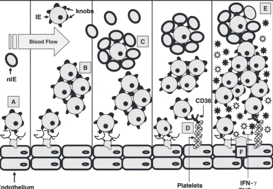
intracel-lular adhesion molecule-1 (ICAM-1), vas-cular cellular adhesion molecule-1 (VCAM-1), E-selectin, P-selectin, hyaluronic acid (HA), and chondroitin sulfate-A (CSA), or to other IE. They may also form rosettes by adhering to non-infected erythrocytes (Fig-ure 1B) (2). It has been suggested that adhe-sion to host receptors expressed on the sur-face of endothelial cells enables IE to avoid spleen-mediated filtration and host immune attack, potentially implicating cytoadhesion in parasite survival (3). In addition to direct parasite adhesion to host receptors, platelets can act as a bridge between IE and endothe-lial cells, providing additional CD36 recep-tors for cytoadhesion (Figure 1A).
bind-Figure 1. Sequestration mechan-isms involved in Plasmodium fal-ciparum infections. P. falcipa-rum-infected erythrocytes (IE) adhere directly to different re-ceptors on the host endothelium via knobs (A); to other IE by auto-agglutination (B); to non-in-fected erythrocytes (nIE), form-ing rosettes (C); to platelets, which act as a bridge in IE cytoadhesion via the CD36 re-ceptor (D). All these phenomena are thought to contribute to blood flow occlusion (E) and produc-tion of the inflammatory cyto-kines, TNF-α and IFN-γ (F); thus leading to the poor clinical out-comes observed in severe ma-laria.
ing sites arranged in tandem at the N-termi-nal end of the molecule (Figure 2A). These motifs are known as Duffy binding-like (DBL) domains, as they were first identified in P. vivax Duffy binding protein, interca-lated by cysteine-rich interdomain region domains (CIDR). Both CIDR and DBL re-gions can be identified on the basis of their amino-acid sequences (2,4).
Different PfEMP-1 molecules have bind-ing sites for adhesion to different host recep-tors (Figure 2A), such as CD36, ICAM-1, VCAM-1, E-selectin, P-selectin, CSA, and others dependent on multiple functional bind-ing domains within PfEMP-1. For example, adhesion to CD36, ICAM-1 and CSA is mediated by different PfEMP-1 variants, as
var genes are expressed in a mutually exclu-sive manner, with only one PfEMP-1 ex-pressed on the surface of an IE at a given time (5). Thus, placental parasites can bind CSA, but not the CD36 receptor (6). This dichotomous behavior may result from dif-ferences in gene location and transcription orientations between CSA-binding and non-binding parasites (7-8).
General aspects of PAM
After years of exposure to the parasite, individuals living in areas of endemic ma-laria acquire high levels of immunity, limit-ing parasitemia and attenuatlimit-ing the clinical outcome of malaria. However, pregnant women remain susceptible, especially in their first pregnancy, in which case the risk of contracting malaria is two to ten times higher than that in non-pregnant women living in the same area. Until recently, it was thought that this particular susceptibility of women to malaria during pregnancy was due to preg-nancy-related immune suppression and hor-monal alterations. However, it has been re-cently shown that the placenta provides an ideal environment for the development of a subpopulation of malaria parasites that ad-here to receptors in the placental
syncy-tiotrophoblast. In most cases, the parasites remain on the maternal side of the placenta, but this maternal-fetal syndrome, known as PAM, has adverse effects on both mother and unborn child, causing maternal anemia and low-birth weight (LBW) babies (9). PAM is thought to be responsible for 62,000 to 363,000 infant deaths in sub-Saharan Africa annually (10). Unfortunately, these figures are probably underestimates since peripher-al parasitemia is not peripher-always observed and the symptoms are not well characterized in some cases.
parasitemia than infants born to multi-gravidae (12). The precise reasons for this remain unclear, but the marked change in cytokine profile and the timing of cytokine production in primigravidae may be involved (12).
Most studies of maternal malaria have been carried out in P. falciparum-infected women. However, pregnant women are sus-ceptible to all four human malaria parasites, including P. vivax, the most prevalent para-site in Brazil and elsewhere outside sub-Saharan Africa. A study in Thailand revealed that primigravidae had a significantly higher risk of P. vivax infection than multigravidae. Moreover, P. vivax infection has also been shown to be significantly associated with maternal anemia and risk of LBW, although these outcomes were more marked in multi-gravidae (13). The deposition of malaria pigment in the placenta has been observed in
P. vivax-infected women (14), and variant antigens encoded by a specific P. vivax mul-tigene family have been identified (15). How-ever, hard data on P. vivax cytoadhesion to the placental syncytiotrophoblast or endo-thelial cells remain scarce.
In Brazil, where malaria transmission is unstable, P. falciparum and P. vivax infec-tions account for 15.1 and 84.4% of cases among non-pregnant women (16). However, the corresponding proportions for pregnant women are 29.7% for falciparum and 67.7% for vivax malaria (16). This corresponds to a significant, 2.5 times increase in the fre-quency of P. falciparum infection for the 195 cases of malaria in pregnant women analyzed (16). The precise reason for this shift in prevalence is unclear and further studies with a larger number of patients are required.
Ligands and receptors involved in PAM
The DBL-γ3 domain of PfEMP-1, en-coded by the var1CSA gene, was initially
thought to be the ligand responsible for para-site cytoadhesion in PAM (17). However, several recent studies have suggested that the protein encoded by the var2CSA gene may be the principal ligand involved in pla-cental cytoadhesion. Unlike var1CSA, var2CSA is up-regulated in placental parasites after selection for adhesion to the CSA receptor in vitro (18-20); indeed it is the only gene shown to be transcriptionally up-regulated following such selection. var2CSA knockout parasites are unable to recover their initial CSA binding, even after repeated exposure to CSA (21,22). The PfEMP-1 encoded by the var2CSA gene has been shown to have a structure different from that of other var
genes, in that it lacks the CIDR, DBL-γ and N-terminal DBL-α domains (Figure 2B). The PfEMP-1 encoded by the 3D7 var2CSA gene (3D7gDNA-PFL0030cvar) has six DBL motifs: DBL4-ε, DBL5-ε and DBL6-ε, and a further three motifs that do not fit into the current classification: DBL1-X, DBL2-X and DBL3-DBL2-X. The DBL2-DBL2-X and DBL6-ε
domains are able to bind CSA (Figure 2B) (18).
A recent mass spectrometry-based prote-omics study identified novel parasite anti-gens, which might be expressed on the IE surface, exclusively in CSA-binding or pla-cental parasites, but did not evaluate the binding of these antigens to host receptors (23).
adhesion to placental sections was signifi-cantly abolished in all placental parasites tested. In contrast, soluble HA, non-immune IgG and protein A failed to inhibit parasite binding to placental cryosections. These find-ings strongly suggest that CSA is the major placental receptor, and support the develop-ment of vaccines targeting parasite ligands to CSA (24).
The CSA or chondroitin 4-sulfate (C4S) receptor is a glycosaminoglycan present in the extracellular matrix, and was first identi-fied as a receptor for parasite binding to
Saimiri monkey endothelial brain cells and to Chinese hamster ovary cells (25,26). Gysin et al. (27) showed that thrombomodulin with a CSA chain was the dominant proteoglycan involved in the sequestration of CSA-bind-ing parasites in the placenta and that a CSA chain at least 9 kDa in size was required to sustain the adhesion of CSA-binding para-sites (28). Achur et al. (29) subsequently purified and identified several types of chon-droitin sulfate proteoglycan (CSPG) from the human placenta, showing that these natu-ral CSPGs present in the intervillous space contained unusually low levels of sulfate and served as receptors for PAM parasite adhesion. The same group went on to define the structures required for parasite cyto-adhesion as C4S dodecasaccharides, and showed that these placental CSPGs have a unique distribution of sulfate groups through-out the second and third semesters of preg-nancy (30-32). It was recently shown that the ability of antiadhesive molecules to in-hibit C4S-specific binding also depends on the sulfation partner of these CSPGs (33).
Does the antibody-mediated immune response play a role in PAM?
Despite pregnancy-related immunosup-pression, pregnant women with malaria de-velop antibodies that inhibit the binding of IE to CSA, and these antibodies are
associ-ated with protection against placental infec-tion. Primigravidae have a much higher sus-ceptibility to maternal malaria than multi-gravidae, because the antibodies acquired after multiple pregnancies are associated with a reduction in the number of IE in the pla-centa (34). In addition, higher levels of these CSA adhesion-blocking antibodies are cor-related with less pronounced maternal ane-mia and with higher birth weight for babies born at term (35,36).
For var2CSA parasites, a recent study showed that rabbit antibodies raised against two VAR2CSA recombinant proteins, cor-responding to the DBL1-X and DBL5-ε do-mains, recognize only the surface proteins of PAM parasites (20). Plasma samples from Ghanaian individuals recognized these re-combinant proteins in a sex- and parity-dependent manner in ELISA tests; this was particularly true for the recombinant protein based on the DBL5-ε domain (20). High plasma levels of anti-VAR2CSA antibodies in women were also found to be correlated with a lower risk of LBW (20). Finally, monoclonal antibodies (mAbs) that recog-nize VAR2CSA DBL domains inhibit, to various extents, the adhesion of a placental isolate to placental cryosections under flow conditions. Moreover, sera from mice im-munized with native VAR2CSA domain complexes with specific mAbs strongly in-hibit PAM parasite cytoadhesion to CSA on the surface of endothelial cells (37).
af-ter just 12 weeks of gestation, whereas primi-gravidae took eight weeks longer to develop such adhesion-blocking antibodies. How-ever, as cytophilic antibodies have been col-lected from infected pregnant women (39), it should be borne in mind that other antibody-dependent mechanisms, such as phagocyto-sis and complement activation, may also help to protect against PAM, in addition to blocking adhesion. Consistent with the ex-istence of additional antibody mechanisms, Megnekou et al. (40) showed that IgG1 and IgG3 were the most prevalent subclasses of PAM antibodies in Cameroonian women, and that larger amounts of these antibodies were found in pregnant multiparous women.
Is the cell-mediated immune response involved in PAM pathogenesis or protection?
The precise mechanism by which PAM parasites evade the immune system and the possible involvement of a cell-mediated im-mune response in protection remains unre-solved. However, it has been shown that massive sequestration of the parasite in the placenta leads to a switch in the cell-medi-ated immune response, typically from TH2 to TH1, resulting in the clinical manifesta-tion of PAM, characterized by an increase in the level of inflammatory cytokine pro-duction (10). Several studies have shown that high levels of IFN-γ and TNF-α, mainly secreted by placental macrophages, are as-sociated with poor clinical outcome in pa-tients with PAM and with the concentration of hemozoin in the placenta (41). Placental infection increases the levels of α- and ß-chemokines, which, in turn, increase im-mune cell recruitment to the placenta (10,42). In contrast, IFN-γ levels have been re-ported to be higher following in vitro stimu-lation of blood mononuclear cells from mul-tigravidae than following the stimulation of such cells from women in their first or sec-ond pregnancy. The cells collected from
women in their second pregnancy secreted high levels of IL-4 and IL-10 (43). Multi-gravidae have been shown to present higher levels of lymphocyte proliferation and natu-ral killer cell cytotoxic activity in response to CSA-binding parasites than primigravidae women (10). These observations suggest that IFN-γ is involved in immunity to PAM. Further evidence for the protective role of IFN-γ is provided by the higher susceptibil-ity to PAM of women with both malaria and HIV infection (10). Indeed, it has been re-cently shown that neonates born to mothers with active placental infection have lower levels of PAM-parasite antigen-specific
IFN-γ T cells and higher levels of IL-10 CD4 T cells than do pregnant infected women treated for malaria (44). However, TGF-ß, an anti-inflammatory cytokine, is produced in larger amounts in multigravidae than in primi-gravidae, suggesting a possible role in con-trolling the manifestation of PAM clinical symptoms.
A recent study in monkeys showed that infection with P. coatneyi did not result in higher levels of CD4 and CD8 T lympho-cytes than observed in infected non-preg-nant monkeys. Indeed, the pregnon-preg-nant infected monkeys had lower levels of monocytes and macrophages in peripheral blood than did the non-pregnant infected monkeys (45). Conversely, high levels of mononuclear cell accumulation have been associated with poor PAM outcomes (46). It should be noted that, in these studies, cells were counted in pe-ripheral blood, so we cannot exclude the possibility that this modulation may alter the levels of these cells in placental compart-ments. In PAM, T cells collected from pe-ripheral blood proliferate more efficiently than those collected from the intervillous blood, whereas intervillous and peripheral monocytes are equally able to present anti-gens (47).
pro-and anti-inflammatory cytokines dictates whether an individual will manage to con-trol PAM or whether the clinical symptoms associated with the disease will develop. This dual effect may depend on regulation of the macrophage migration inhibitory factor, a specific cytokine that counter-regulates the immunosuppressive effects of pregnancy. Placental infection was recently shown to increase macrophage migration inhibitory factor production in the presence of cytotro-phoblast-adherent IE (48).
Is an anti-PAM vaccine feasible?
The first evidence that it might be pos-sible to develop an anti-PAM vaccine was provided by the study of Fried and Duffy (9,35) and Staalsoe et al. (34,36) showing that multiparous women were less suscep-tible to PAM than women in their first preg-nancy, that infected women developed high levels of adhesion-blocking antibodies against PAM parasites after several preg-nancies (9,34) and that these antibodies were associated with attenuation of the clinical outcome of PAM. Antibodies against var2CSA parasites have also been shown to cross-react with genetically different P. falcipa-rum strains (49). Cross-reactivity between the DBL-γ3CSA domain and var2CSA-encoded antigens has been observed (50). Further-more, mAbs raised against var2CSA parasite surface antigens have also proved to be pan-reactive with CSA-binding parasites from different geographical origins (37). More-over, molecular analysis of the var1CSA
DBL-γ3 minimal binding domain revealed 37% sequence identity to the var2CSA DBL3-X domain (51). These somewhat surprising pan-reactivity results are probably related to con-formational similarities.
In light of the antibody-mediated im-mune response in PAM, an efficient vaccine would probably elicit large amounts of ad-hesion-blocking antibodies, mainly against conserved binding motifs. However, as the
cell-mediated immune response also seems to be involved in immune protection against PAM, immunization regimes and adjuvants should aim to induce high levels of IFN-γ -secreting T cells. Immunization regimens based on naked DNA for priming and re-combinant viral vectors or proteins for boost-ing have been shown to elicit high levels of CD4- and CD8-producing T cells able to induce immune protection against several viral and protozoan diseases (52). Finally, anti-PAM vaccines may contain other para-site antigens since sera collected from Cam-eroonian women showed a significant corre-lation between low or null levels of anti-bodies against the carboxyl-terminal 19-kDa segment of the P. falciparum merozoite sur-face protein-1 and the risk of PAM (53).
Is it possible to model PAM?
Reliable in vitro adhesion models are required for the evaluation of potential vac-cine candidates and the antibody-mediated immune response directed against them. Most of the existing in vitro models of IE seques-tration were developed for studying IE adhe-sion in parasites thought to be involved in cerebral malaria. Many knob+ laboratory-adapted P. falciparum strains adhere in vitro
to various cell types, including human um-bilical vein endothelial cells, C32 amelanotic melanoma cells, human dermal microvascu-lar endothelial cells, human brain capilmicrovascu-lary endothelial cells, human monocytes and platelets, and transfected COS cells (54). However, in 1995, Gay et al. (55) described the use of Saimiri microvascular brain endo-thelial cell clones differing in terms of the expression of several combined surface mol-ecules such as CD36, ICAM-1, E-selectin, and CSA, permitting for the first time the selection by cytoadhesion of distinct mono-morphic adhesion phenotypes.
ideal. The Saimiri cell model has some ad-vantages in that it allows in vitro cytoadhesion studies, the results of which can be con-firmed or rejected using the homologous
Saimiri/P. falciparum monkey model (56). This model is also considered to be more relevant than non-primate cell models due to the phylogenetic proximity to humans. The use of organ-specific endothelial cells ap-pears to be particularly useful for structure-function studies in which the native confor-mation of a receptor is critical. A placental BeWo-derived cell line has been success-fully used to select monomorphic CSA-bind-ing parasites (57). Since placental CSPGs have an unusual sulfation pattern, BeWo cells provide an alternative to cells of non-placental origin in CSA-binding studies.
In all the parasite-binding assays de-scribed above, cytoadhesion was investi-gated under static conditions, in which sus-pensions of IE were allowed to settle on a confluent monolayer of cultured cells. This method is technically simple, making it pos-sible to carry out a large number of assays simultaneously. However, static assays do not model the dynamic blood flow condi-tions encountered by IE in vivo.
In 1995, Cooke and Coppel (54) devel-oped an in vitro assay for visualizing and quantifying the adhesion of IE to endothelial cells or to immobilized adhesion receptors under flow conditions. The flow assembly used consisted of a parallel-plate flow cham-ber or a glass microcapillary tube (microslide) on which a monolayer of endothelial cells, such as human umbilical vein endothelial cells, C32 melanoma cells, or Saimiri micro-vascular brain endothelial cell clones (58), can be cultured, or a plastic slide coated with purified proteins, such as CD36, ICAM-1 or thrombospondin, by adsorption. Adhesion under dynamic flow conditions can be quan-tified by counting adherent IE directly under the microscope.
Nevertheless, with the exception of this flow adhesion model using endothelial cells
and immobilized proteins, very few models have been developed for studying placental malaria. One example is the Saimiri (squir-rel monkey) microvascular endothelial cell line Sc17, which expresses a thrombomodu-lin bearing only a CSA chain and a CD44-csa isoform. The presence of the chondroitin sulfate of thrombomodulin, or a CD44 isoform, on endothelial cells mimics, to some extent, the presence of CSA on the surface of the syncytiotrophoblast, thereby providing a clear advantage over previous cell models or commercial CSA preparations from various non-placental sources (59). Conversely, one major disadvantage of the use of cell lines as models for CSA binding is the presence of unidentified or unknown adhesion receptors in addition to CSA on the surface of endo-thelial cells. Another disadvantage of these assays is that CSA preparations from vari-ous sources, used by different laboratories, can generate conflicting results. Thus, the conformational modification of CSA by add-ing dipalmitoyl-diphosphatidylethanolamine may bias results, especially when this sys-tem is used to select CSA-binding IE by panning. The addition of charged groups seems to be problematic for the specific selection of CSA-binding IE from labora-tory strains and field isolates (28). Further-more, cytoadhesion inhibition assays with
Saimiri brain endothelial or Chinese ham-ster ovary cells cannot distinguish between subpopulations of CSA-binding parasites in field samples.
con-ditions revealed significant differences in the presence of soluble CSA, or inhibitor mAbs in serum samples from primi- and multigravidae (61). Static parasite adhesion assays are subject to considerable inter-ex-perimental variation, due primarily to differ-ences in washing procedures.
The use of placental cryosections made it possible, for the first time, to measure the shear-stress resistance of CSA-binding IE. Distinct subpopulations within the CSA-binding phenotype were identified by in-creasing the flow rate gradually from 0.2 to 3.2 Pascal (Pa). For example, at 0.6 Pa, which exceeds the normal shear stress in the placenta (0.05 Pa), 70% of IE remained ad-herent for the laboratory strain FCR3CSA, and 25% of IE resisted a shear stress >3.2 Pa (60).
These results strongly suggest that the initial FCR3CSA strain was composed of a mixture of strong (≥3.2 Pa) and weak (≤0.8 Pa) CSA-binding parasites, confirming the existence of distinct adhesion subpopula-tions among the CSA phenotypes of various strains (58; Nogueira PA, Costa FT and Gysin J, unpublished data), and supporting the hy-pothesis that only some subpopulations of CSA-binding IE have the potential for se-questration in the microvasculature (58). This hypothesis is based on the notion that IE in the placenta are not normally exposed to shear stresses exceeding 0.05 Pa, whereas shear stresses in the postcapillary venules vary from 0.1 to 1 Pa (58).
Conclusions
There is now considerable evidence that PAM is a particularly severe form of ma-laria, and that primigravidae and their off-spring have a higher risk of developing PAM than do multigravidae and their children. These observations, and others, provide an
impetus for the development of an anti-PAM vaccine. However, several issues concern-ing the expression of antigens by PAM para-sites and the precise immunological mechan-isms involved in protection remain unre-solved in the context of PAM. First, does the unique set of hormones and cytokines in-duced during gestation play a role in antigen or placental host receptor expression? Sec-ond, why is CSA the major PAM receptor, given that this glycosaminoglycan is found in various organs other than the placenta? Third, how many antigenically different PAM parasites exist, and what are their rela-tive prevalences in infected pregnant women? Fourth, what is the evolutionary importance of this pan-reactivity and the presence of multiple CSA-binding domains? Can these domains be ordered into a hierarchy? Fi-nally, as some of the poor placental out-comes observed in falciparum malaria are also observed in vivax malaria, is IE cytoadhesion in the placenta an exclusive feature of P. falciparum parasites? Or, like the inhabitants of “Plato’s Cave”, are we merely watching the “theater of shadows” of real PAM parasite interactions and mechan-isms reflected on the cave wall?
Acknowledgments
References
1. Schofield L, Grau GE. Immunological processes in malaria patho-genesis. Nat Rev Immunol 2005; 5: 722-735.
2. Wahlgren M, Treutiger CJ, Gysin J. Cytoadherence and rosetting in pathogenesis of severe malaria. In: Wahlgren M, Perlmann P (Edi-tors), Malaria molecular and clinical aspects. The Netherlands: Harwood Academic Publishers; 1999. p 289-328.
3. David PH, Hommel M, Miller LH, Udeinya IJ, Oligino LD. Parasite sequestration in Plasmodium falciparum malaria: spleen and anti-body modulation of cytoadherence of infected erythrocytes. Proc Natl Acad Sci U S A 1983; 80: 5075-5079.
4. Winter G, Chen Q, Wahlgren M. Meeting report: the molecular background of severe and complicated malaria. Mol Biochem Parasitol 2004; 134: 37-41.
5. Freitas-Junior LH, Bottius E, Pirrit LA, Deitsch KW, Scheidig C, Guinet F, et al. Frequent ectopic recombination of virulence factor genes in telomeric chromosome clusters of P. falciparum. Nature 2000; 407: 1018-1022.
6. Gamain B, Gratepanche S, Miller LH, Baruch DI. Molecular basis for the dichotomy in Plasmodium falciparum adhesion to CD36 and chondroitin sulfate A. Proc Natl Acad Sci U S A 2002; 99: 10020-10024.
7. Robinson BA, Welch TL, Smith JD. Widespread functional special-ization of Plasmodium falciparum erythrocyte membrane protein 1 family members to bind CD36 analysed across a parasite genome. Mol Microbiol 2003; 47: 1265-1278.
8. Kraemer SM, Smith JD. Evidence for the importance of genetic structuring to the structural and functional specialization of the Plas-modium falciparum var gene family. Mol Microbiol 2003; 50: 1527-1538.
9. Duffy PE, Fried M. Malaria in the pregnant woman. Curr Top Micro-biol Immunol 2005; 295: 169-200.
10. Beeson JG, Duffy PE. The immunology and pathogenesis of malaria during pregnancy. Curr Top Microbiol Immunol 2005; 297: 187-227. 11. Le Hesran JY, Cot M, Personne P, Fievet N, Dubois B, Beyeme M, et al. Maternal placental infection with Plasmodium falciparum and malaria morbidity during the first 2 years of life. Am J Epidemiol 1997; 146: 826-831.
12. Mutabingwa TK, Bolla MC, Li JL, Domingo GJ, Li X, Fried M, et al. Maternal malaria and gravidity interact to modify infant susceptibility to malaria. PLoS Med 2005; 2: e407.
13. Nosten F, McGready R, Simpson JA, Thwai KL, Balkan S, Cho T, et al. Effects of Plasmodium vivax malaria in pregnancy. Lancet 1999; 354: 546-549.
14. McGready R, Davison BB, Stepniewska K, Cho T, Shee H, Brockman A, et al. The effects of Plasmodium falciparum and P. vivax infections on placental histopathology in an area of low ma-laria transmission. Am J Trop Med Hyg 2004; 70: 398-407. 15. Del Portillo HA, Fernandez-Becerra C, Bowman S, Oliver K, Preuss
M, Sanchez CP, et al. A superfamily of variant genes encoded in the subtelomeric region of Plasmodium vivax. Nature 2001; 410: 839-842.
16. Martinez-Espinosa FE, David PH, Daniel-Ribeiro CT, Alecrim WD. Malaria during pregnancy in a reference centre from the Brazilian Amazon: unexpected increase in the frequency of Plasmodium falci-parum infections. Mem Inst Oswaldo Cruz 2004; 99: 19-21. 17. Buffet PA, Gamain B, Scheidig C, Baruch D, Smith JD,
Hernandez-Rivas R, et al. Plasmodium falciparum domain mediating adhesion to chondroitin sulfate A: a receptor for human placental infection. Proc Natl Acad Sci U S A 1999; 96: 12743-12748.
18. Gamain B, Trimnell AR, Scheidig C, Scherf A, Miller LH, Smith JD. Identification of multiple chondroitin sulfate A (CSA)-binding do-mains in the var2CSA gene transcribed in CSA-binding parasites. J Infect Dis 2005; 191: 1010-1013.
19. Salanti A, Staalsoe T, Lavstsen T, Jensen AT, Sowa MP, Arnot DE, et al. Selective upregulation of a single distinctly structured var gene in chondroitin sulphate A-adhering Plasmodium falciparum involved in pregnancy-associated malaria. Mol Microbiol 2003; 49: 179-191. 20. Salanti A, Dahlback M, Turner L, Nielsen MA, Barfod L, Magistrado P, et al. Evidence for the involvement of VAR2CSA in pregnancy-associated malaria. J Exp Med 2004; 200: 1197-1203.
21. Andrews KT, Pirrit LA, Przyborski JM, Sanchez CP, Sterkers Y, Ricken S, et al. Recovery of adhesion to chondroitin-4-sulphate in Plasmodium falciparum varCSA disruption mutants by antigenically similar PfEMP1 variants. Mol Microbiol 2003; 49: 655-669. 22. Viebig NK, Gamain B, Scheidig C, Lepolard C, Przyborski J, Lanzer
M, et al. A single member of the Plasmodium falciparum var multi-gene family determines cytoadhesion to the placental receptor chon-droitin sulphate A. EMBO Rep 2005; 6: 775-781.
23. Fried M, Wendler JP, Mutabingwa TK, Duffy PE. Mass spectromet-ric analysis of Plasmodium falciparum erythrocyte membrane pro-tein-1 variants expressed by placental malaria parasites. Proteomics 2004; 4: 1086-1093.
24. Fried M, Domingo GJ, Gowda CD, Mutabingwa TK, Duffy PE. Plas-modium falciparum: chondroitin sulfate A is the major receptor for adhesion of parasitized erythrocytes in the placenta. Exp Parasitol 2006; 113: 36-42.
25. Rogerson SJ, Chaiyaroj SC, Ng K, Reeder JC, Brown GV. Chon-droitin sulfate A is a cell surface receptor for Plasmodium falcipa-rum-infected erythrocytes. J Exp Med 1995; 182: 15-20.
26. Pouvelle B, Meyer P, Robert C, Bardel L, Gysin J. Chondroitin-4-sulfate impairs in vitro and in vivo cytoadherence of Plasmodium falciparum infected erythrocytes. Mol Med 1997; 3: 508-518. 27. Gysin J, Pouvelle B, Le Tonqueze M, Edelman L, Boffa MC.
Chon-droitin sulfate of thrombomodulin is an adhesion receptor for Plas-modium falciparum-infected erythrocytes. Mol Biochem Parasitol 1997; 88: 267-271.
28. Pouvelle B, Fusai T, Lepolard C, Gysin J. Biological and biochemi-cal characteristics of cytoadhesion of Plasmodium falciparum-in-fected erythrocytes to chondroitin-4-sulfate. Infect Immun 1998; 66: 4950-4956.
29. Achur RN, Valiyaveettil M, Alkhalil A, Ockenhouse CF, Gowda DC. Characterization of proteoglycans of human placenta and identifica-tion of unique chondroitin sulfate proteoglycans of the intervillous spaces that mediate the adherence of Plasmodium falciparum-in-fected erythrocytes to the placenta. J Biol Chem 2000; 275: 40344-40356.
30. Alkhalil A, Achur RN, Valiyaveettil M, Ockenhouse CF, Gowda DC. Structural requirements for the adherence of Plasmodium falcipa-rum-infected erythrocytes to chondroitin sulfate proteoglycans of human placenta. J Biol Chem 2000; 275: 40357-40364.
31. Achur RN, Valiyaveettil M, Gowda DC. The low sulfated chondroitin sulfate proteoglycans of human placenta have sulfate group-clus-tered domains that can efficiently bind Plasmodium falciparum-infected erythrocytes. J Biol Chem 2003; 278: 11705-11713. 32. Agbor-Enoh ST, Achur RN, Valiyaveettil M, Leke R, Taylor DW,
33. Andrews KT, Klatt N, Adams Y, Mischnick P, Schwartz-Albiez R. Inhibition of chondroitin-4-sulfate-specific adhesion of Plasmodium falciparum-infected erythrocytes by sulfated polysaccharides. Infect Immun 2005; 73: 4288-4294.
34. Staalsoe T, Megnekou R, Fievet N, Ricke CH, Zornig HD, Leke R, et al. Acquisition and decay of antibodies to pregnancy-associated variant antigens on the surface of Plasmodium falciparum-infected erythrocytes that protect against placental parasitemia. J Infect Dis 2001; 184: 618-626.
35. Duffy PE, Fried M. Antibodies that inhibit Plasmodium falciparum adhesion to chondroitin sulfate A are associated with increased birth weight and the gestational age of newborns. Infect Immun 2003; 71: 6620-6623.
36. Staalsoe T, Shulman CE, Bulmer JN, Kawuondo K, Marsh K, Hviid L. Variant surface antigen-specific IgG and protection against clini-cal consequences of pregnancy-associated Plasmodium falciparum malaria. Lancet 2004; 363: 283-289.
37. Avril M, Gamain B, Lépolard C, Viaud N, Scherf A, Gysin J. Mapping of var2CSA-DBL domains recognized by mouse monoclonal anti-bodies that inhibit adhesion to chondroitin sulfate A. Microbes Infect 2006 (in press).
38. O’Neil-Dunne I, Achur RN, Agbor-Enoh ST, Valiyaveettil M, Naik RS, Ockenhouse CF, et al. Gravidity-dependent production of anti-bodies that inhibit binding of Plasmodium falciparum-infected eryth-rocytes to placental chondroitin sulfate proteoglycan during preg-nancy. Infect Immun 2001; 69: 7487-7492.
39. Gysin J, Pouvelle B, Fievet N, Scherf A, Lepolard C. Ex vivo desequestration of Plasmodium falciparum-infected erythrocytes from human placenta by chondroitin sulfate A. Infect Immun 1999; 67: 6596-6602.
40. Megnekou R, Staalsoe T, Taylor DW, Leke R, Hviid L. Effects of pregnancy and intensity of Plasmodium falciparum transmission on immunoglobulin G subclass responses to variant surface antigens. Infect Immun 2005; 73: 4112-4118.
41. Fried M, Muga RO, Misore AO, Duffy PE. Malaria elicits type 1 cytokines in the human placenta: IFN-gamma and TNF-alpha asso-ciated with pregnancy outcomes. J Immunol 1998; 160: 2523-2530. 42. Suguitan AL Jr, Leke RG, Fouda G, Zhou A, Thuita L, Metenou S, et al. Changes in the levels of chemokines and cytokines in the placen-tas of women with Plasmodium falciparum malaria. J Infect Dis 2003; 188: 1074-1082.
43. Moore JM, Nahlen BL, Misore A, Lal AA, Udhayakumar V. Immunity to placental malaria. I. Elevated production of interferon-gamma by placental blood mononuclear cells is associated with protection in an area with high transmission of malaria. J Infect Dis 1999; 179: 1218-1225.
44. Brustoski K, Moller U, Kramer M, Petelski A, Brenner S, Palmer DR, et al. IFN-gamma and IL-10 mediate parasite-specific immune re-sponses of cord blood cells induced by pregnancy-associated Plas-modium falciparum malaria. J Immunol 2005; 174: 1738-1745. 45. Davison BB, Kaack MB, Rogers LB, Rasmussen KK, Rasmussen T,
Henson EW, et al. Alterations in the profile of blood cell types during malaria in previously unexposed primigravid monkeys. J Infect Dis 2005; 191: 1940-1952.
46. Ordi J, Ismail MR, Ventura PJ, Kahigwa E, Hirt R, Cardesa A, et al. Massive chronic intervillositis of the placenta associated with ma-laria infection. Am J Surg Pathol 1998; 22: 1006-1011.
47. Diouf I, Fievet N, Doucoure S, Ngom M, Gaye A, Dumont A, et al.
Monocyte activation and T cell inhibition in Plasmodium falciparum-infected placenta. J Infect Dis 2004; 189: 2235-2242.
48. Chaisavaneeyakorn S, Lucchi N, Abramowsky C, Othoro C, Chaiyaroj SC, Shi YP, et al. Immunohistological characterization of macrophage migration inhibitory factor expression in Plasmodium falciparum-infected placentas. Infect Immun 2005; 73: 3287-3293. 49. Elliott SR, Duffy MF, Byrne TJ, Beeson JG, Mann EJ, Wilson DW, et
al. Cross-reactive surface epitopes on chondroitin sulfate A-adher-ent Plasmodium falciparum-infected erythrocytes are associated with transcription of var2csa. Infect Immun 2005; 73: 2848-2856. 50. Bir N., Yazdani S.S., Avril M., Layez C., Gysin J., Chitnis C.
Immu-nogenicity of Duffy binding-like domains that bind chondroitin sulfate and protection against pregnancy-associated malaria. Infct Immn. 2006; 74; 5955-5963.
51. Gamain B, Smith JD, Avril M, Baruch DI, Scherf A, Gysin J, et al. Identification of a 67-amino-acid region of the Plasmodium falcipa-rum variant surface antigen that binds chondroitin sulphate A and elicits antibodies reactive with the surface of placental isolates. Mol Microbiol 2004; 53: 445-455.
52. Todryk SM, Walther M. Building better T-cell-inducing malaria vac-cines. Immunology 2005; 115: 163-169.
53. Taylor DW, Zhou A, Marsillio LE, Thuita LW, Leke EB, Branch O, et al. Antibodies that inhibit binding of Plasmodium falciparum-infected erythrocytes to chondroitin sulfate A and to the C terminus of mero-zoite surface protein 1 correlate with reduced placental malaria in Cameroonian women. Infect Immun 2004; 72: 1603-1607. 54. Cooke BM, Coppel RL. Cytoadhesion and falciparum malaria: going
with the flow. Parasitol Today 1995; 11: 282-287.
55. Gay F, Robert C, Pouvelle B, Peyrol S, Scherf A, Gysin J. Isolation and characterization of brain microvascular endothelial cells from Saimiri monkeys. An in vitro model for sequestration of Plasmodium falciparum-infected erythrocytes. J Immunol Methods 1995; 184: 15-28.
56. Gysin J, Aikawa M, Tourneur N, Tegoshi T. Experimental Plasmo-dium falciparum cerebral malaria in the squirrel monkey Saimiri sciureus. Exp Parasitol 1992; 75: 390-398.
57. Viebig NK, Nunes MC, Scherf A, Gamain B. The human placental derived BeWo cell line: a useful model for selecting Plasmodium falciparum CSA-binding parasites. Exp Parasitol 2006; 112: 121-125.
58. Pouvelle B, Traore B, Nogueira PA, Pradines B, Lepolard C, Gysin J. Modeling of Plasmodium falciparum-infected erythrocyte cytoad-hesion in microvascular conditions: chondroitin-4-sulfate binding, A competitive phenotype. J Infect Dis 2003; 187: 292-302.
59. Fusai T, Parzy D, Spillmann D, Eustacchio F, Pouvelle B, Lepolard C, et al. Characterisation of the chondroitin sulphate of Saimiri brain microvascular endothelial cells involved in Plasmodium falciparum cytoadhesion. Mol Biochem Parasitol 2000; 108: 25-37.
60. Avril M, Traore B, Costa FT, Lepolard C, Gysin J. Placenta cryosections for study of the adhesion of Plasmodium falciparum-infected erythrocytes to chondroitin sulfate A in flow conditions. Microbes Infect 2004; 6: 249-255.