LncRNA
SNHG12
promotes cell growth and inhibits
cell apoptosis in colorectal cancer cells
J.Z. Wang, C.L. Xu, H. Wu and S.J. Shen
Department of Gastroenterology, The Second Affiliated Hospital of Wenzhou Medical University, Wenzhou, China
Abstract
Several long non-coding RNA (lncRNA) might be correlated with the prognosis of colorectal cancer (CRC) and serve as a diagnostic and prognostic biomarker. However, the exact expression pattern of small nucleolar RNA host gene 12 (SNHG12) in colorectal cancer and its clinical significance remains unclear. The level ofSNHG12was detected by qRT-PCR in CRC tissues and CRC cells. MTT assay and colony formation assay were performed to examine the cell proliferation of CRC cells transfected with pcDNA-SNHG12or si-SNHG12. Flow cytometry technology was used to detect cell cycle and cell apoptosis of CRC cells transfected with pcDNA-SNHG12or si-SNHG12. The protein level of cell cycle progression-related molecules, including cyclin-dependent kinases (CDK4, CDK6), cyclin D1 (CCND1) and cell apoptosis-related molecule caspase 3 was detected by western blot. The effect ofSNHG12knockdown was examinedin vivo. Increased levels ofSNHG12were observed in CRC tissues and in CRC cells.SNHG12promoted the cell proliferation of CRC cells. In addition,SNHG12overexpression boosted the cell cycle progression of SW480 cells transfected with pcDNA-SNHG12andSNHG12knockdown inhibited the cell cycle progression of HT29 cells transfected with si-SNHG12.SNHG12also inhibited the cell apoptosis of CRC cells. We also found thatSNHG12 increased the expression of cell cycle-related proteins and suppressed the expression of caspase 3. Our results suggest that SNHG12promoted cell growth and inhibited cell apoptosis in CRC cells, indicating thatSNHG12might be a useful biomarker for colorectal cancer.
Key words: SNHG12; Cell growth; biomarker; Cell apoptosis; Colorectal cancer
Introduction
Colorectal cancer (CRC) is the third most common malignant tumor globally (1,2). The high incidence of CRC and CRC-related deaths has been a great threat to public health. In spite of the improvement of diagnosis and treat-ment of CRC, it remains a severe disease. Progression of CRC is a multi-step process involving the deregulation of several oncogenes and tumor suppressor gene, which might be used as diagnostic and therapeutic targets (3). However, the precise mechanisms of these genes in CRC are poorly understood and novel diagnostic and prognos-tic biomarkers need to be discovered.
With the advances in next generation sequencing tech-nologies, long non-coding RNAs (lncRNAs) were identified as a new class of non-coding RNA. LncRNA is longer than 200 nucleotides with no protein-coding capacity (4). Mount-ing evidence suggests that aberrant lncRNA expression participate in a variety of biological processes, such as em-bryogenesis, stem cell pluripotency, cell growth and the occurrence of malignancies, through regulating gene ex-pression at several levels (5,6). Notably, lncRNA might be correlated with patients’ prognosis and serve as a diagnostic and prognostic biomarker for disease. In CRC,
various lncRNAs have been identified to be abnormally expressed and related to disease progression (7). For example, Niu et al. (8) found that lncRNAAK027294 was strongly expressed in CRC and closely correlated with cell proliferation, migration, and apoptosis. TUG1 was found to indicate a poor prognosis for CRC and promote metas-tasis by regulating epithelial-mesenchymal transition (9). In addition, Xie et al. reviewed the CRC-associated lncRNAs published recently, including CCAT1,H19,HOTAIR,UCA1 andPTENP1(10). However, no robust tumor markers have been yet identified.
Long non-coding RNA small nucleolar RNA host gene 12 (SNHG12) was a novel lncRNA identified to be up-regulated in several cancer cells, such as human osteo-sarcoma cell, nasopharyngeal carcinoma cell, and human endometrial carcinoma (11–13). Moreover,SNHG12played
important roles in cancer cell proliferation and migration. However, the exact expression pattern ofSNHG12in CRC and its clinical significance remains unclear. In the present research, we discovered that SNHG12 was up-regulated in CRC tissues and cells for the first time. We further detected the effect of SNHG12 on cell proliferation,
Correspondence: S.J. Shen:<sujian_shen63@163.com>
cell cycle, apoptosis and the related proteins expres-sion in CRC cells.
Material and Methods
Patients and specimens
Human primary CRC tissues and their paired adjacent tissue were obtained from 60 patients at the Second Affiliated Hospital, Wenzhou Medical University. These patients did not receive local or systemic treatment before the operation. All of the tissues were stored at–80°C. An
experienced pathologist assessed the differentiation grade, pathological stage, grade and nodal status. All subjects submitted the written informed consent. The study protocol was approved by the Ethics Committee of the Second Affiliated Hospital of Wenzhou Medical University.
Cell culture and transfection
All human colonic cancer cell lines including SW480, LOVO, HCT116, HT29 and the human colonic epithelial cells HCoEpiC were obtained from the American Type Cul-ture Collection. Cells were culCul-tured in RPMI-1640 supple-mented with 10% fetal bovine serum at 37°C in a 5% CO2 incubator.
TheSNHG12expression vector, pcDNA-SNHG12, was synthetized and constructed by Ribobio (China). siRNA-SNHG12(si-SNHG12) which targetsSNHG12was ob-tained from Sigma-Aldrich (USA). Cells were transfected with pcDNA-SNHG12 or siRNAs using Lipofectamine2000 (Life Technologies, USA) following the manufacturer’s instructions.
Quantitative real-time PCR
Total RNA was extracted from tumor tissue samples or cultured cells using Trizol reagent (Invitrogen Inc., USA). Two micrograms of total RNA was reverse transcribed to obtain cDNA using Moloney Murine Leukemia Virus Reverse Transcriptase (M-MLVRT; Promega, USA). Quan-titative real-time PCR was performed with 1 mL of cDNA using SYBR green real-time Master Mix (Takara, Japan) on Applied Biosystems 7500 Sequence Detection system (ABI, USA). Glyceraldehyde 3-phosphate dehydrogenase (GAPDH) was used as an internal control to normalize the data. The primers for SNHG12 and GAPDH were as followed: for SNHG12, (forward) 50-TCTGGTGATCGAG GACTTCC-30, and (reverse) 50-ACCTCCTCAGTATCAC ACACT-30; forGAPDH, (forward) 50-ACACCCACTCCTC CACCTTT-30 and (reverse) 50-TTACTCCTTGGAGGCCA TGT-30. Real time PCR was performed in triplicate, and the relative expression ofSNHG12was calculated using
2-DDCTmethod.
Western blot analysis
Total proteins were extracted from cells and pro-tein concentrations were determined using the BCA Pro-tein Assay kit (Takara). ProPro-teins were separated on 12%
sodium lauryl sulfate-polyacrylamide gels (SDS-PAGE) and transferred to polyvinylidene difluoride membranes (PVDF; Millipore, USA). After blocked with 5% non-fat skimmed milk powder at 37°C for 2 h, the membranes were incubated with primary antibodies: anti-cyclin-dependent kinase 4 (anti-CDK4) antibody (1:5000, Abcam, UK), CDK6 antibody (1:5000, Abcam), CCND1 antibody (1:5000, Abcam), Caspase 3 anti-body (1:5000, Abcam), anti-p-AKT antianti-body (1:500, Abcam) and GAPDH diluted at 1:2000 (Abcam) for 1 h at 37°C. The second antibody was anti-rabbit IgG-horseradish peroxi-dase (HRP, 1:4000; Santa Cruz, USA). Proteins were detect-ed by enhancdetect-ed chemiluminescence as describdetect-ed by the manufacturer (Beyotime, China).
MTT assay and soft agar colony formation assay The 3-[4,5-dimethylthiazol-2-yl]-2,5-diphenyltetrazolium bromide (MTT) assay was carried out to detect the cell viability of SW480 cells with pcDNA-SNHG12or HT29 cells with si-SNHG12at 0, 12, 24, 36, 48, 60 and 72 h of the transfection. The transfected CRC cells (2104cells) were seeded on 6-well plates and were washed with PBS, then incubated in MTT solution (5 mg/mL, 100mL; Invitrogen Inc., USA) for 3 h. After 3 h, 100mL of solubilization buffer was added to each well. The absorbance of samples at 450 nm was measured using the Thermo Plate microplate reader (Rayto Life and Analytical Science Co. Ltd., Germany).
For the colony formation assay, 800–1500 cells were
placed in a 6-well plate and maintained in complete culture medium containing 0.3% agar layered on top of 0.6% agar at 37°C in the presence of 5% CO2 for 16 days. We evaluated the colonies containing at least 50 cells. The data offive randomly scoredfields were used for statistics.
Flow cytometry technology to detect cell cycle and cell apoptosis
For the detection of cell cycle, SW480 cells with pcDNA-SNHG12 or HT29 cells with si-SNHG12were harvested after 48 h of transfection. Propidium oxide was used to stain cells with the BD Cycletest Plus DNA Reagent Kit (BD Biosciences, USA). The quantitation of cell cycle distribution was performed with FACScan cytometry (Becton Dickinson, USA). The percentage of the cells in G0-G1, S, and G2-M phases were counted and compared.
An Annexin V-fluorescein isothiocyanate propidium iodide (FITC/PI) apoptosis detection kit (Life Technolo-gies, USA) was used to detect cell apoptosis. SW480 cells with pcDNA-SNHG12or HT29 cells with si-SNHG12were harvested after 48 h of transfection and washed twice with 1 PBS. Cells were incubated with Annexin-V and PI at
Animal experiments
All animal experiments were performed according to the guidelines approved by the China Association of Labo-ratory Animal Care. Twelve female 6-week-old BALB/c nude mice were obtained from Vital River Co. Ltd. (China) and maintained under specific pathogen-free conditions. HT29 cells were transfected si-NC or si-SNHG12 and then injected subcutaneously into the hind limb of BALB/c nude mice (5106cells/mouse; n=6 for each group). The
tumor size was measured every 7 days for a total period of 28 days and estimated using the equation length
(width)20.5. The weight of the tumor on the 28th day was measured.
Statistical analysis
All statistical analyses were performed using SPSS 20.0 software (SPSS, USA). The measured parameters are reported as means±SD. The Fisher’s exact test was used to compare categorical data and the Kruskal-Wallis method was used to analyze continuous data. The dif-ferences between groups were estimated by the Student’s t-test. A P value of o0.05 was considered to be statis-tically significant.
Results
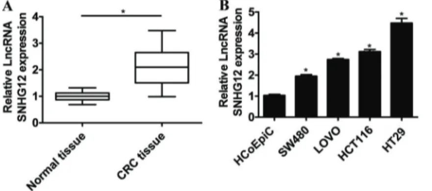
SNHG12was upregulated in CRC tissues and cell
lines
The level ofSNHG12in CRC tissues and the adjacent normal tissues were detected by qRT-PCR. As shown in Figure 1A, higher level ofSNHG12was observed in CRC tissues compared to that in normal tissues. qRT-PCR was also conducted to determine the level ofSNHG12in CRC cells (SW480, LOVO, HCT116, HT29) and the control cells HCoEpiC.SNHG12was significantly up-regulated in these CRC cells compared to that in the control (Figure 1B). In addition, the highest level ofSNHG12was found in HT29 cells and SW480 cells expressed the lowestSNHG12.
To detect the clinical significance ofSNHG12 expres-sion in CRC, 60 patients were divided intoSNHG12high expression group (n=30) and SNHG12 low expression group (n=30) according to the cutoff value, which was
defined as the median of the cohort. As demonstrated in Table 1, high expression of SNHG12 in CRC patients was significantly correlated with advanced tumor stage (P=0.007) and large tumor size (P=0.004). In addition, tumors with high expression ofSNHG12were associated with worse overall survival in CRC patients (Po0.001; Figure 2). The 5-year survival rate ofSNHG12high expres-sion group reached 33.3% and the rate in low expresexpres-sion group was 70%. These data indicated that SNHG12may act as a potent biomarker for predicting prognosis in CRC patients.
SNHG12promoted proliferation of CRC cells
To investigate the function of SNHG12 on the cell proliferation of CRC cells, SW480 cells or HT29 cells were transfected with pcDNA-SNHG12or si-SNHG12, respec-tively. The result of qRT-PCR showed that the expression of SNHG12 was greatly up-regulated in SW480 cells transfected with pcDNA-SNHG12 and down-regulated in HT29 cells transfected with si-SNHG12(Figure 3A and B). Cell viability of pretreated SW480 cells and HT29 cells were detected by MTT assay. The results showed that overexpression ofSNHG12significantly promoted the cell viability of SW480 cells andSNHG12knockdown inhibited the cell viability of HT29 cells (Figure 3C and D). The result of colony formation assays indicated that colony formation was significantly promoted bySNHG12overexpression and remarkably inhibited by SNHG12 knockdown (Figure 3E and F). These data indicated that SNHG12promoted the proliferation of colorectal cancer cells.
SNHG12promoted cell cycle of CRC cells
To explore the effect of SNHG12on the cell cycle of CRC cells, flow cytometric analysis was performed. As shown in Figure 4A and B,SNHG12overexpression signifi -cantly reduced the number of SW480 cells in the G0/G1 phase and increased the number of SW480 cells in the S phase, which suggested that the cell cycle progression of SW480 cells transfected with pcDNA-SNHG12was pro-moted. On the other hand,SNHG12knockdown inhibited the cell cycle progression of HT29 cells transfected with si-SNHG12(Figure 4C and D).
SNHG12inhibited apoptosis of CRC cells
To investigate the effect ofSNHG12on the cell apop-tosis of CRC cells, SW480 cells were transfected with pcDNA-SNHG12 or pcDNA for 48 h, then stained with annexin V and propidium iodide (PI), followed by detection using flow cytometry. The results showed thatSNHG12 overexpression can obviously suppress cell apoptosis of
SW480 cells (Figure 5A and B). In addition, SNHG12 knockdown induced cell apoptosis of HT29 cells trans-fected with si-SNHG12(Figure 5C and D).
SNHG12promoted the expression of cell cycle-related
proteins and suppressed the expression of caspase 3 To explore the molecular mechanisms by whichSNHG12 contributes to cell cycle and cell apoptosis of CRC cells, we performed western blot to detect the protein level of cell cycle progression-related molecules, including CDK4, CDK6), cyclin D1 (CCND1) and cell apoptosis-related mole-cule caspase 3. CDK4, CDK6 and CCND1 were found to be up-regulated in SW480 cells transfected with pcDNA-SNHG12 (Figure 6A) and down-regulated in HT29 cells transfected with si-SNHG12 (Figure 6C). The result of western blot showed that lower protein level of caspase 3 was observed in SW480 cells transfected with pcDNA-SNHG12 (Figure 6B). SNHG12 knockdown enhanced the protein level of caspase 3 in HT29 cells (Figure 6D). As it is well known that PI3K/AKT signaling pathway plays vital roles in regulating cell proliferation, cycle and apoptosis, we asked whether SNHG12 could also regulate PI3K/AKT signaling pathway. As shown in Figure 6E and F, overexpression of SNHG12significantly promoted the expression of phosphory-lated AKT, while SNHG12knockdown inhibited the expres-sion of phosphorylated AKT.
Figure 2.Prognostic significance of high and low expression of small nucleolar RNA host gene 12 (SNHG12) in colorectal cancer patients.
Table 1.Clinical characteristics of the 60 colorectal cancer patients and the correlation with high and low level expression of small nucleolar RNA host gene 12 (SNHG12) in colorectal cancer tissues.
Clinicopathologic characteristics
Total SNHG12expression P value
High (n=30) Low (n=30)
Age 0.772
p60 28 15 13
460 32 15 17
Gender 0.481
Female 25 11 14
Male 35 19 16
Tumor differentiation 0.232
Well/moderately 32 14 18
Poorly 28 16 12
TNM stages 0.007
I-II 27 7 20
III-IV 33 23 10
Tumor size 0.004
o5 cm 31 8 23
45 cm 29 22 7
Distant metastasis 0.137
No 22 8 14
Yes 38 22 16
SNHG12knockdown inhibited tumor growthin vivo To detect the effect of SNHG12 knockdown in vivo, a nude mouse xenograft model of HT29 cells was es-tablished. HT29 cells were transfected with si-NC or si-SNHG12and subcutaneously inoculated into the nude mice (n=6 for each group). As shown in Figure 7A and B, SNHG12 knockdown inhibited tumor growth significantly after inoculation for 28 days. In addition, the tumor weight on 28th day in mice inoculated with HT29 cells transfected with si-SNHG12 was significantly smaller than that of the controls (Figure 7C). Results in vivo indicated that SNHG12knockdown inhibited tumor growth.
Discussion
In this study, we observed increased levels ofSNHG12 in CRC tissues and in CRC cells including SW480, LOVO, HCT116, HT29. High expression of SNHG12 in CRC
patients was significantly correlated with advanced tumor stage and large tumor size. In addition, tumors with high expression ofSNHG12were associated with worse overall survival in CRC patients. The results of MTT assay and colony formation assay indicated that SNHG12 promot-ed proliferation of CRC cells. In addition, SNHG12 over-expression boosted cell cycle progression of SW480 cells transfected with pcDNA-SNHG12 and SNHG12 knock-down inhibited the cell cycle progression of HT29 cells transfected with si-SNHG12. SNHG12 also inhibited cell apoptosis of CRC. We further found that SNHG12 in-creased the expression of cell cycle-related proteins and suppressed the expression of caspase 3. SNHG12 knockdown inhibited the tumor growth in vivo. Taken together, our results suggest that SNHG12 promoted cell growth and inhibited cell apoptosis in CRC cells and might be a potent biomarker for predicting prognosis in CRC patients.
Figure 4.Flow cytometric analysis for cell cycle progression (A) and the corresponding statistical result (B) in SW480 cells transfected with pcDNA-SNHG12and in HT29 cells transfected with si-SNHG12(CandD, respectively). Data are reported as means±SD. *Po0.01vscontrol (Student’st-test).
To date, accumulating evidence has suggested that the dysfunctional activities of lncRNAs might be associated with
tumor initiation and progression of CRC, which further promotes the application of lncRNAs as cancer diagnostic Figure 6.Protein level of CDK4, CDK6, CCND1 (A) and of caspase 3 (C) in SW480 cells transfected with pcDNA-SNHG12. Protein level of CDK4, CDK6, CCND1(B)and of caspase 3 (D) in HT29 cells transfected with si-SNHG12. Protein level of p-AKT in SW480 cells transfected with pcDNA-SNHG12 (E) and in HT29 cells transfected with si-SNHG12(F).
or prognostic biomarkers (14,15). Yuan et al. (16) demon-strated that lncRNA-CTD903 was an independent pre-dictive factor of favorable prognosis for CRC and acted as a tumor suppressor to inhibit cell invasion and migration. In addition, the extensively studied lncRNAMALAT-1 was also expressed at a high level in CRC tissues (17). Apart from the abnormal expression of lncRNA, some genetic variants of lncRNA are also correlated with the risk of CRC. For example, rs2839689 in lncRNA H19 was reported to contribute to the susceptibility to CRC in a Chinese popula-tion study by Li et al. (18).
In this study, we found that a novel lncRNASNHG12was up-regulated in CRC tissues and cells. SNHG12was also related to the prognosis of CRC patients. Ruan et al. reported thatSNHG12contributed to cell proliferation and migration by upregulating the expression of angiomotin in human osteo-sarcoma cells (12). According to Peri et al. (19),SNHG12 was confirmed to be differentially expressed in nulliparous and parous breast tissues. In low-passage human naso-pharyngeal carcinoma HNE2 cells, the results of lncRNA microarray analysis and qRT-PCR confirmed thatSNHG12 was overexpressed following TP53 overexpression.
To further investigate the mechanism by which SNHG12contributed to cellular proliferation and apoptosis of CRC cells, we explored the expression of CDK4, CDK6, CCND1 and caspase 3. In SW480 cells transfected with pcDNA-SNHG12, CDK4, CDK6 and CCND1 were found to be up-regulated and Caspase 3 was down-regulated.
It is known that cyclins, CDKs, and cyclin-dependent kinase inhibitors play important roles in the functioning mechan-ism of the cell cycle (20). The activation of CDK4 or CDK6 is required for cell cycle G1/S transition. CDK4 or CDK6 can interact with tumor suppressor protein Rb. In cancer cells, CCND1 is upregulated and activates CDK4/6, whereas Rb is deactivated, resulting in the dysregulation of the DNA-damage repair system and acceleration of the cell cycle (21). Caspase 3 is the most important of the "executioner caspases" in the process of apoptosis, which could begin to disassemble the cell by activating DNA degrading enzymes and degrading the cellular architec-ture (22). Previous studies have shown that some lncRNAs could regulate cell proliferation and cycle via PI3K/AKT signaling pathway (23–25). We found that
overexpression of SNHG12 significantly promoted the phosphorylation of AKT, whileSNHG12knockdown inhib-ited the phosphorylation of AKT. It has been shown that the active Akt induced by PI3K could regulate the expression of its target genes, such as Bad, caspase-9, NF-kB, mTOR and p21, participating in the cell prolifera-tion and cell apoptosis (26). Therefore, SNHG12 may promote colorectal cell proliferation and cycle by activating PI3K/AKT signaling pathway. However, the underlying mechanism needs to be further investigated.
In conclusion, our data indicated thatSNHG12 might be a valuable diagnostic and prognostic biomarker for CRC and a potential target for gene therapy.
References
1. Booth RA. Minimally invasive biomarkers for detection and staging of colorectal cancer. Cancer Letters 2007; 249: 87–96, doi: 10.1016/j.canlet.2006.12.021.
2. Jemal A, Bray F, Center MM, Ferlay J, Ward E, Forman D. Global cancer statistics.CA Cancer J Clin2011; 61: 69–90,
doi: 10.3322/caac.20107.
3. Arends MJ. Pathways of colorectal carcinogenesis. Appl Immunohistochem Mol Morphol2013; 21: 97–102.
4. Gutschner T, Diederichs S. The hallmarks of cancer: a long non-coding RNA point of view.RNA Biol2012; 9: 703–719,
doi: 10.4161/rna.20481.
5. Saxena A, Carninci P. Long non-coding RNA modifies chromatin: epigenetic silencing by long non-coding RNAs. Bioessays2011; 33: 830–839, doi: 10.1002/bies.201100084.
6. Wang K, Chang H. Molecular Mechanisms of Long Non-coding RNAs. Mol Cell 2011; 43: 904–914, doi: 10.1016/
j.molcel.2011.08.018.
7. Dou J, Ni Y, He X, Wu D, Li M, Wu S, et al. Decreasing lncRNA HOTAIR expression inhibits human colorectal cancer stem cells.Am J Transl Res2016; 8: 98–108. 8. Niu H, Hu Z, Liu H, Hu G, Yang B, Wu S, et al. Long
non-coding RNA AK027294 involves in the process of prolifera-tion, migraprolifera-tion, and apoptosis of colorectal cancer cells. Tumour Biol2016; 37: 10097–10105, doi:
10.1007/s13277-015-4350-x.
9. Sun J, Ding C, Yang Z, Liu T, Zhang X, Zhao C, et al. The long non-coding RNA TUG1 indicates a poor prognosis for
colorectal cancer and promotes metastasis by affecting epithelial-mesenchymal transition.J Transl Med 2016; 14: 42, doi: 10.1186/s12967-016-0786-z.
10. Xie X, Tang B, Xiao YF, Xie R, Li BS, Dong H, et al. Long non-coding RNAs in colorectal cancer.Oncotarget2015; 22: 901–906.
11. Gong Z, Zhang S, Zeng Z, Wu H, Yang Q, Xiong F, et al. LOC401317, a p53-regulated long non-coding RNA, inhi-bits cell proliferation and induces apoptosis in the naso-pharyngeal carcinoma cell line HNE2. Plos One2014; 9: e110674, doi: 10.1371/journal.pone.0110674.
12. Ruan W, Pei W, Feng S, Yuan X, Li Y. Long non-coding RNA small nucleolar RNA host gene 12 (SNHG12) pro-motes cell proliferation and migration by upregulating angiomotin gene expression in human osteosarcoma cells. Tumour Biol2015; 37: 4065–4073, doi:
10.1007/s13277-015-4256-7.
13. Wen Z, Xu L, Shouzhen W, Yan Z, Huan P, Wei C. Micro-array expression profile of lncRNAs and the upregulated ASLNC04080 lncRNA in human endometrial carcinoma.Int J Oncol2015; 46: 2125–2137.
14. Prensner JR, Chinnaiyan AM. The emergence of lncRNAs in cancer biology. Cancer Discov 2011; 1: 391–407,
doi: 10.1158/2159-8290.CD-11-0209.
16. Yuan Z, Yu X, Ni B, Chen D, Yang Z, Huang J, et al. Overexpression of long non-coding RNA-CTD903 inhibits colorectal cancer invasion and migration by repressing Wnt/ beta-catenin signaling and predicts favorable prognosis.Int J Oncol2016: 48: 2675–2685.
17. Zheng HT, Shi DB, Wang YW, Li XX, Xu Y, Tripathi P, et al. High expression of lncRNA MALAT1 suggests a biomarker of poor prognosis in colorectal cancer.Int J Clin Exp Pathol 2014; 7: 3174–3181.
18. Li S, Hua Y, Jin J, Wang H, Du M, Zhu L, e al. Association of genetic variants in lncRNA H19 with risk of colorectal cancer in a Chinese population.Oncotarget2016; 7: 25470–25477.
19. Peri S, de Cicco RL, Santucci-Pereira J, Slifker M, Ross EA, Russo IH, et al. Defining the genomic signature of the parous breast.BMC Med Genomics2012; 5: 46, doi: 10.1186/1755-8794-5-46.
20. O’Leary B, Finn RS, Turner NC. Treating cancer with selective CDK4/6 inhibitors.Nat Rev Clin Oncol 2016;13: 417–430, doi: 10.1038/nrclinonc.2016.26.
21. Wikman H, Kettunen E. Regulation of the G1/S phase of the cell cycle and alterations in the RB pathway in human lung
cancer. Expert Rev Anticancer Ther 2006; 6: 515–530,
doi: 10.1586/14737140.6.4.515.
22. Wall DM, McCormick BA. Bacterial secreted effectors and caspase-3 interactions.Cell Microbiol2014; 16: 1746–1756,
doi: 10.1111/cmi.12368.
23. Dong Y, Liang G, Yuan B, C Yang, Gao R, Zhou X. MALAT1 promotes the proliferation and metastasis of osteosarcoma cells by activating the PI3K/Akt pathway.Tumour Biol2015; 36: 1477–1486, doi: 10.1007/s13277-014-2631-4.
24. Liu G, Xiang T, Wu QF, Wang WX. Long noncoding RNA H19-derived miR-675 enhances proliferation and invasion via RUNX1 in gastric cancer cells. Oncol Res 2016; 23: 99-1070, doi: 10.3727/096504015X14496932933575. 25. Zhu Y, Bo D, Zhang H, Shi G, Y Shen, Ye D. Long non-coding
RNA LOC572558 inhibits bladder cancer cell proliferation and tumor growth by regulating the AKT–MDM2–p53 signaling
axis.Cancer Letters2016, 380: 369–374.