KAINATE-INDUCED OXIDATIVE STRESS
AND NEUROTOXICITY IN THE RAT BRAIN
Lidija Radenović1 and vesna seLaković2
1Department of Physiology and Biochemistry, Faculty of Biology, University of Belgrade, 11000 Belgrade, Serbia and Montenegro; 2Institute for Medical Research, Military Medical Academy, 11000 Belgrade, Serbia and Montenegro
Abstract - We investigated superoxide production and MnSOD activity after kainate injection into the CA3 region of the rat hippocampus. The measurements took place at different times in hippocampus, forebrain cortex, striatum, and cer-ebellum homogenates. Free radicals including superoxide are responsible for post-lesional cytotoxicity. Neuronal cells responded to oxidative stress in kainate-induced neurotoxicity and caused the protective mechanism to increase MnSOD levels. The increase of MnSOD in distinct brain regions functionally connected via afferents and efferents suggests that these regions are affected by the injury. It implies that MnSOD protects the cells in these regions from superoxide-induced damage and therefore may limit the retrograde and anterograde spread of neurotoxicity.
uDC 612.001:599
INTrODuCTION
Glutamate neurotoxicity has been hypothesized to un-derlie several types of acute brain injury. Stimulation of glutamate receptors induces superoxide production, which may be one of the mediators of excitotoxic neuro-nal injury in CNS. Free radical reactions are implicated in a variety of physiological and pathological processes and abnormalities associated with superoxide dismutase (SOD) have been recently documented in several neuro-degenerative processes (C o y l e et al. 1993). However, the precise cellular mechanisms that lead to neurotoxicity under these conditions still remain unclear. Among the neurobiological conditions in which oxidative stress is believed to be involved are adult neurodegenerative dis-eases (Parkinson’s disease, Huntington’s disease, ALS, stroke, trauma, and seizures).
We were interested in changes induced by intra-hippocampal kainate treatment. Kainic acid (KA) is an endogenous excitotoxin acting on glutamate receptors that leads to neurotoxic damage resembling the altera-tions observed in some neurological disorders. While ex-citotoxic and oxidative injury may occur independently, growing evidence indicates that reactive oxygen species formation may also be a speciic consequence of
gluta-mate receptor-mediated neurotoxicity (D u g a n et al.
1995). Glutamate neurotoxicity is partly mediated by re-active oxygen species formed as a consequence of several processes, including nitric oxide (A l a b a d i et al. 1999; N a k a k i et al. 2000; R a d e n o v i ć et al. 2003) and superoxide production (L i et al. 2001) production. Su-peroxide radicals react rapidly with nitric oxide to form highly cytotoxic peroxynitrite, which acts through lipid peroxidation (L e e et al. 2001). Although there are a number of intracellular sources of free radicals, the mito-chondria are thought to be the most important (C i r i o l o
et al. 2001).
Superoxide dismutases (SOD) are the most effec-tive endogenous scavengers of superoxide radicals, and changes in both CuZnSOD and MnSOD affect processes of aging and learning (N i c h o l l s et al. 1999). Further-more, mutations within the CuZnSOD gene leading to alternations in SOD activity contribute to several neuro-pathological conditions (P i n t e a u x et al. 1998), where-as SODs coupled to polyethylen glycol, lecithinized SOD, and SOD entrapped in liposomes act neuroprotectively (C a s s a r i n o et al. 1999).
Superoxide dismutases are considered to be a major factor in protection of nervous tissue against excitotoxic
259
Key words: Brain, glutamate neurotoxicity, kainic acid, mitochondria, MnSOD, oxidative stress, superoxide
SOD immunoreactivity was found to be located predomi-nantly in astrocytes throughout the CNS. In contrast, the Mn-containing isoenzyme was predominantly conined to neurons and their processes throughout the brain and the spinal cord (L i n d e n a u et al. 2000). It seems rea-sonable to conclude that differences in the basal content
of SOD-isoenzymes may contribute to different cellular susceptibilities in neurodegenerative processes that are accompanied by oxidative stress.
Most of the studies cited above focused on
CuZn-MnSOD suffer from early neurodegeneration (K i m et al. 2000). Also, L i et al. (1998) reported that cultured cortical neurons of heterozygous knockout mutant mice with reduced Mnsod activity are signiicantly more sen-sitive to glutamate-induced toxicity than the neurons of wild type mice. Kainate-induced increases of mitochon-drial superoxide production and hippocampal neuronal loss were attenuated in transgenic mice overexpressing mitochondrial MnSOD (Liang et al. 2000).
Previous studies suggested that mitochondrial Mn-SOD is important for the resistance to toxic cellular in-sults and plays a major protective role. It is known that MnSOD has a heterogeneous distribution in the brain (G o n z a l e z -Z u l u e t a et al. 1999). In the striatum, cholinergic neurons and somatostatin neurons are en-riched with MnSOD. Cholinergic neurons of the basal forebrain are also highly MnSOD-enriched. In the hip-pocampus, enrichment with MnSOD mainly occurred in parvalbumin-containing neurons and MnSOD-posi-tive interneurons were present in the stratum piramidale, with the highest packing densities being recorded in the subiculum and CA3. The highest packing density within
Fig. 1. Low-power light micrograph of both hippocampal hemispheres of a rat following unilateral kainic acid injection into the CA3 region (ipsi), stained with hematoxylin.The rat was allowed to survive for 2 h. The ipsilateral pyramidal cells of the CA3 region were slightly damaged around the injection site (white marks). Damage to CA1 pyramidal cells was not observed, nor was damage to dentate granule cells. There were no morphological signs of neuronal degeneration in the
contralateral hippocampus. scale bar = 500 μm.
Figure 2. The effect of intrahippocampal kainate injection on superoxide produc-tion (O2-, μM nBT/mg prot.) in the rat hippocampus (ipsilateral) at different sur
-vival times. data are means ± s.d. *indicates a statistically signiicant difference
between kainate treated and control (sham-operated) animals (p<0.05).
**Indi-cates a statistically very signiicant difference between kainate-treated and control
(sham-operated) animals (p<0.01).
Figure 3. The effect of intrahippocampal kainate injection on superoxide produc-tion (O2
-, μM nBT/mg prot.) in the rat forebrain cortex (ipsilateral) at different
survival times. data are means ± s.d. *indicates a statistically signiicant differ -ence between kainate treated and sham-operated animals (p<0.05). **Indicates a
statistically very signiicant difference between kainate treated and control
(sham-operated) animals (p<0.01).
the hippocampal formation occurred in the polymorphic cell layer of the dentate gyrus (B i d m o n et al. 1997).
In view of the above considerations, we studied su-peroxide anion production and activity of mitochondrial MnSOD within the rat hippocampus, forebrain cortex, striatum, and cerebellum after intrahippocampal kainate injection.
MATerIALS AND MeTHODS
Animals
Adult rats of the Wistar strain (Rattus norvegicus) of both sexes, with body weight 200 ± 30 g, were used for experiments. Groups of two or three rats per cage (erath,
FrG), were housed in an air-conditioned room at room temperature of 23 ± 2oC with 55 ± 10% humidity and with lights on 12 h/day (07.00-19.00). The animals were given a commercial rat food and tap water ad libitum.
These animals were anesthetized by giving them intra-peritoneal injections of sodium pentobarbital (0.0405 g/ kg b.w.) and were placed in a stereotaxic frame.
Experimental procedure and intracerebral injection of drugs
The rats were divided into two basic groups (ac-cording to drug treatment), each basic group consisting of ive different subgroups (according to survival times) and each subgroup consisting of eight animals. Animals of the irst group received a unilateral injection of ka (Sigma Chemical Co., u.S.A., 0.5 mg/ml, dissolved in 0.1 M saline, pH 7.2; 1 μL total volume) into the Ca3 region of the hippocampus (coordinates from bregma; antero-posterior: -3.3 mm, dorsoventral: 3.2 mm, and lateral: 3.0 mm) using a Hamilton microsyringe with a beveled tip. The second group received the same volume (1 μL) but only saline solution and served as a control (sham-oper-ated). The animals were allowed to survive for 5 min up to seven days (5 min, 15 min, 2 h, 48 h, and 7 days). All animals were anesthetized, decapitated, and the brains immediately removed. The ipsi- and contralateral hippo-campus, forebrain cortex, striatum, and cerebellum from individual animals were quickly isolated and homog-enized in ice-cold buffer containing 0.25 M sucrose, 0.1 mM eDTA, and 50 mM K-Na phosphate buffer, pH 7.2. Homogenates were centrifuged twice at 1580g for 15 min at 4oC. The supernatant (crude mitochondrial fraction) obtained by this procedure was then frozen and stored at -70oC.
Light microscopy
Brains were removed from the skull and ixed in 4% paraformaldehyde for at least 24 h before dehydration through a range of alcohols and embedding in parafin wax. Frozen (-70oC) 50 μm-thick hippocampus brain sec-tions were cut in a cryostat, thaw-mounted on gelatin-coated glass slides, and stained with hematoxylin and eosin.
Superoxide production and measurement
In these experiments superoxide was measured from reduction of nitro blue tetrazolium (NBT) as previously
Figure 4. The effect of intrahippocampal kainate injection on superoxide produc-tion (O2΄, μM nBT/mg prot.) in the rat striatum (ipsilateral) at different survival
times. data are means ± s.d. *indicates a statistically signiicant difference be -tween kainate treated and sham-operated animals (p<0.05). **Indicates a
statisti-cally very signiicant difference between kainate treated and control (sham-oper -ated) animals (p<0.01).
Figure 5. The effect of intrahippocampal kainate injection on superoxide produc-tion (O2-, μM nBT/mg prot.) in the rat cerebellum (ipsilateral) at different survival
times. data are means ± s.d. *indicates a statistically signiicant difference be -tween kainate treated and sham-operated animals (p<0.05). **Indicates a
statisti-cally very signiicant difference between kainate treated and control (sham-oper -ated) animals (p<0.01).
described (S p i t z et al. 1989). Detection of this product was by spectrophotometric quantiication of a colored formazan product formed from blue tetrazolium. reduc-tion of NBT was measured at 560 nm.
Superoxide dismutase assay
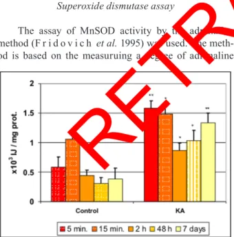
The assay of MnSOD activity by the adrenaline method (F r i d o v i c h et al. 1995) was used. The meth-od is based on the measuruing a degree of adrenaline
autooxidation inhibition by MnSOD contained in the ex-amined samples in 50 mM sodium carbonate buffer, pH
Data presentation and analysis
All experiments were done with n = 8. each assay was performed at least twice under identical conditions. Data are expressed as means ± SD. The statistical sig-niicance of differences between groups was assessed by Student’s t-test (paired and unpaired) for individual comparisons and regression analysis for overall signii-cance (with p < 0.05 as signiicant and p < 0.01 as very signiicant).
Materials
Chemicals were purchased from Sigma (St. Louis, MO, u.S.A.). Other chemicals were of analytical grade. All drug solutions were prepared on the day of the ex-periment.
Animals used for procedures were treated in strict accordance with the NIH Guide for Care and use of Lab-oratory Animals (1985).
reSuLTS
The injection site
kainate was injected unilaterally into the Ca3 subield of the hippocampus of rats. The ipsilateral pyramidal cells of the CA3 region were slightly damaged 2 h after KA injection (Fig. 1). Pyramidal cells in the CA1 and CA3 subields, granule cells in the dentate gyrus and hilar neurons which are known to be vulnerable in this mod-el, show a pattern of damage around the injection site. That was found to be similar at any of the survival times tested, only the size of the lesion varied. Damage to CA1 pyramidal cells was not observed, non was damage to dentate granule cells. There were no morphological signs of neuronal degeneration in the contralateral
hippocam-Figure 6. The effect of intrahippocampal kainate injection on MnSOD activity (x103 IJ/mg prot.) in the ipsilateral hippocampus at different survival times. Data
are means ± s.d. *indicates a statistically signiicant difference between kainate
treated and control (sham-operated) animals (p<0.05). **Indicates a statistically
very signiicant difference between kainate treated and control (sham-operated)
animals (p<0.01).
Figure 7. The effect of intrahippocampal kainate injection on MnSOD activity (x103 IJ/mg prot.) in the ipsilateral forebrain cortex at different survival times.
data are means ± s.d. *indicates a statistically signiicant difference between kai -nate treated and sham-operated animals (p<0.05). **Indicates a statistically very
signiicant difference between kainate treated and control (sham-operated) animals
(p<0.01).
pus at this early stage or at any other survival time tested. under conditions of normal behavior in the rat, the dam-age was localized mainly in the CA3 region of hippocam-pus, where neuronal loss occurred. The purpose of these micrographs was to verify the injection site.
Superoxide production in the rat brain after intrahippocampal kainate injection
The results presented in Fig. 2-5 show the superox-ide levels (O2-, μM nBT/mg proteins) in ipsilateral hippo-campal, cortical, striatal, and cerebellar homogenates, re-spectively. Superoxide levels in the brains of control rats showed no signiicant differences between the left and right hemispheres in any of the tested structures (data not shown). also, there was no statistically signiicant differ-ence between mean superoxide levels obtained from each hemisphere after KA treatment in any of the tested brain structures, although the injection site was in the ipsilat-eral hippocampus (not presented).
Kainate injection resulted in generally higher su-peroxide levels (p<0.05) in all tested brain structures. The obtained superoxide levels were highest in the hip-pocampus (Fig. 1). rapid increase in superoxide pro-duction at 5 min after KA injection and propro-duction levels continued to be above normal at all subsequent times (at 7 days inally) in all tested brain structures. Measurement at 15 min after KA injection in the hip-pocampus, in the forebrain cortex, and in the striatum showed statistically very signiicant differences (p<0.01) compared with the equivalent control group (Figs. 2, 3, 4). The results obtained for the contralateral
hippo-campus, forebrain cortex and striatum were similar.
Activity of MnSOD in the rat brain after intrahippocampal kainate injection
The results presented in Figs. 6-9 show the MnSOD levels (x103 IJ/mg protein) in ipsilateral hippocampal, cortical, striatal and cerebellar homogenates respectively. There was no statistically signiicant difference between mean MnSOD levels obtained from each hemisphere af-ter KA treatment, although the injection site was in the ipsilateral hippocampus. Activity of MnSOD in the brains of control rats showed no signiicant differences between
the left and right hemispheres in any of the tested struc-tures (not shown).
Levels of MnSOD at the injection site (control or KA treated) were highest in the hippocampus (Fig. 6). Intrahippocampal KA-induced neurotoxicity caused sig-niicant increase of Mnsod activity at 5 and 15 min, fol-lowed by further signiicant increase at 48 h and 7 days, in the hippocampus, in the forebrain cortex, and in the cerebellum (Figs. 6 , 7, 9). in the striatum, a signiicant increase of MnSOD activity was found only at the earliest tested time, at 5 min (Fig. 8). Neuronal cells responded to oxidative stress in KA-induced neurotoxicity by increas-ing MnSOD levels.
DISCuSSION
Mitochondria are the main cellular source of
super-Figure 8. The effect of intrahippocampal kainate injection on MnSOD activity (x103 IJ/mg prot.) in the ipsilateral striatum at different survival times. Data are
means ± s.d. *indicates a statistically signiicant difference between kainate treat -ed and sham-operat-ed animals (p<0.05).
Figure 9. The effect of intrahippocampal kainate injection on MnSOD activity (x103 IJ/mg prot.) in the ipsilateral cerebellum at different survival times. Data are
means ± s.d. *indicates a statistically signiicant difference between kainate-treat -ed and sham-operat-ed animals (p<0.05). **indicates a statistically very signiicant
difference between kainate treated and control (sham-operated) animals (p<0.01).
thereby destroying cells. The superoxide radical is much less reactive and can cross cell membranes and act at a distance. as the irst protective mechanism, sod reacts with superoxide to produce hydrogen peroxide and mo-lecular oxygen. The induction of mitochondrial MnSOD under pathological conditions is variable and related mainly to the type of injury (B i d m o n et al. 1997).
Mitochondria from different tissue sources display differential susceptibility to oxidizing species (H a e a l e s
et al. 1999). However, it is now becoming apparent that there is within the brain a differential susceptibility of various brain cell types to oxidizing species. In contrast to astrocytes, neurons appear to be particularly vulner-able to the action of free radicals. Such vulnerability may arise from an inability to sustain cellular energy demands by glycolysis and an inferior capacity to handle oxidizing species (S e n g p i e l et al. 1998).
regional distribution of KA receptors of the rat brain was found to be highest in deep layers (layer 5) of the forebrain cortex, the cerebellar granule cell layer, and the caudate putamen (C a r r o l l et al. 1998; B a i l e y et al. 2001), which is why we tested these particular brain regions: hippocampus, forebrain cortex, striatum, and cerebellum.
Neuronal superoxide production varies with meta-bolic activity and age. Developmental increase in mi-tochondrial superoxide production and oxidative DNA damage following KA seizures suggests that mitochon-drial oxidative stress may be a key factor that renders the developing brain resistant to seizure-induced brain dam-age (P a t e l et al. 2003).
In the present study, an appropriate dose of KA (0.5 mg/ml) was used to cause small brain damage in the ip-silateral, but not contralateral, hippocampus; there were
main endogenous protective systems against such reper-fusion injury. Moreover MnSOD may also be involved in limiting the damage in remote brain areas that were not ischemic by scavenging radicals formed in response to deafferentitation (B i d m o n et al. 1997).
The immunocytochemical distribution of MnSOD was determined in the rat hippocampus by A k a i et al. (1990), who reported that CA1 pyramidal cells were weakly immunostained, whereas CA3 pyramidal cells were strongly reactive. These differences in the intensity of the MnSOD immunostaining reactions may relate to variations in the sensitivity of subields of the hippocam-pus to ischemia. The hippocamhippocam-pus is especially vulner-able to ischemic damage. Neurons in the CA3 region and dentate hilus demonstrate fast progressive damage, while CA1 pyramidal cells demonstrate delayed neuronal dam-age. The delayed CA1 pyramidal cell loss could be caused by post-ischemic neuronal hyperactivity if hippocampal interactions are lost after ischemia. Because CA3 neu-rons constitute the main input to CA1 pyramidal cells, decreased activity of CA3 neurons indicates less excit-atory input to CA1 neurons. Also, MnSOD was localized in the cerebral cortex and hippocampus of patients with A l z h e i m e r -type senile dementia (M a e d a et al.
1997).
Our results show that there is a clear, transient in-crease of inducible MnSOD in all tested brain regions after intrahippocampal KA treatment. A rapid increase, within 5 min, was found in the ipsilateral and contralater-al areas, which possibly receive a few direct connections from the lesioned area. The data indicate that the onset of MnSOD upregulation was better in the hippocampus, which may be intrinsically more protected by MnSOD from toxic effects than other areas. The irst increase in MnSOD occurred between 5 min and 2 h. The delayed increases at 48 h and 7 days suggest a rapid de novo
thesis involving transcription of the gene and translation of its mrNA. It seems that the mechanisms or time points of the induction may be different. Most probably, direct injury leads to an instant induction of MnSOD expres-sion, whereas more time is needed to transfer the signal via afferents or efferents to the remote regions.
This inding indicates that protection against super -oxide radicals not only takes place around the lesioned area, but also may exist in more remote brain areas-re-gions that are affected by the injury. It suggests that Mn-SOD protects the cells in these regions from superoxide-induced damage and therefore may limit the retrograde and anterograde spread of neurotoxicity.
Acknowledgements. The present work was supported by the Government of the republic of Serbia.
reFereNCeS
Akai, F., Maeda, M., Suzuki, K., Inagaki, S., Takagi, H., Taniguchi, N. (1990). Immunocytochemical localization of MnSOD in the hippocampus of the rat. Neurosci Lett,115(1), 19-23.
Alabadi, J., Thibault, J.L., Pinard, E., Seylaz, J., Lasbennes, F.
(1999). 7-Nitroindazole, a selective inhibitor of nNOS, increases hippocampal extracellular glutamate concentration in status epilepticus induced by kainic acid in rats. Brain Res.839, 305-312.
Bailey, A., Kelland, E.E., Thomas, A., Biggs, J., Creawford, D., Kitchen, I., Toms, N.J. (2001). Regionalmapping of low afinity
kainate receptors in mouse brain using [3H] (2S, 4r)-4-methyl-glutamate autoradiography. Eur. J. Pharmacol. 431, 305-310.
Bidmon, H.J., Kato, K., Schleicher, A., Witte, O.W., Zilles, K. (1997). Transient increase of Mn-superoxide dismutase in remote brain areas after focal photothrombotic cortical lesion. Stroke 29, 203-211.
Carroll, F., Finkelstein, D.I., Horne, M.K., Lawrence, A.J., Creaw ford, D., Paxinos, G., Beart, F.M. (1998). regional distribution of low afinity kainate receptors in brain of Macaca fascicu-laris determined by autoradiography using [3H] (2S, 4r)-4-methylglutamate. Neurosci. Lett. 255, 71-74.
Cassarino, D.S., Bennett, J.P. (1999). An evaluation of the role of mitochondria in neurodegenerative diseases: mitochondrial mu-tations and oxidative pathology, protective nuclear responses, and cell death in neurodegeneration. Brain Res Rev,29, 1-25.
Ciriolo, M., Aquilano, K., De Martino, A., Carri, M.T., Rotilio, G.
(2001). Differential role of superoxide and glutathione in S-nitrosoglutathione-mediated apoptosis: a rationale for mild forms of familial amyotrophic lateral sclerosis associated with less active Cu,Zn superoxide dismutase mutants. J. Neurochem. 77, 1433-1443.
Coyle, J.T., Puttfarcken, P. (1993). The effects of oxidative stress on the brain. Science,262, 689-695.
Dugan, L.L., Sensi, S.L., Canzoniero, L.M.T., Handran, S.D., Roth
man, S.M., Lin, T.S., Goldberg, M.P., Choi, D.W. (1995). Mitochondrial production of reactive oxygen species in cortical neurons following exposure to N-methyl-D-aspartate. J Neuro-sci,15, 6377-6388.
Fridovich I. (1995). Superoxide radical and superoxide dismutases.
Annu Rev Biochem,64, 97-112.
Gonzalez-Zulueta, M., Ensz, L.M., Mukhina, G., Lebovitz, R.M., Zwacka, R.M., Engelhardt, J.F., Oberlay, L.W., Dawson, V.L., Dawson, T.M. (1999). Mn-superoxide dismutase protects nNOS neurons from NMDA and nitric oxide-mediated neurotoxicity.
J Neurosci,18(6), 2040-2055.
Heales, S., Bolanos, J.P., Land, J.M., Clark, J.B. (1999). Nitric oxide, mitochondria and neurological disease. Biochim. Biophys. Acta,
1410, 215-228.
Kim, H.C., Jhoo, W.K., Kim, W.K., Suh, J.H., Shin, E.J., Kato, K., Ho Ko, K. (2000). An immunocy to chemical study of mitochon-drial MnSOD in the rat hippocampus after kainate administra-tion. Neurosci Lett, 281(1), 65-68.
Lee, M.H., Hyun, D.H., Halliwell, B., Jenner, P. (2001). effect of overexpression of wild-type and mutant Cu/Zn-superoxide dismutase on oxidative stress and cell death induced by hydro-gen peroxide, 4-hydroxynonenal or serum deprivation: poten-tiation of injury by ALS-related mutant superoxide dismutases and protection by Bcl-2. J Neurochem,78, 209-220.
Li, Q.Y., Pedersen, C., Day, B.J., Patel, M. (2001). Dependence of excitotoxic neurodegeneration on mitochondrial aconitase inactivation. J Neurochem, 78, 746-755.
Li, Y., Copin, J.C., Reola, L.F., Calagui, B., Gobbel, G.T., Chen, S.F., Sato, S., Epstein, C.J., Chan, P.H. (1998). reduced mitochon-drial Mn-superoxide dismutase activity exacerbates glutamate toxicity in cultured mouse cortical neurons. Brain Res, 814, 164-170.
Liang, L.P., Ho, Y.S., Patel, M. (2000). Mitochondrial superoxide production in kainate-induced hippocampal damage. Neurosci-ence,101(3), 563-570.
Lindenau, J., Noack, H., Possel, H., Asayama, K., Wolf, G. (2000). Cellular distribution of superoxide dismutases in the rat CNS.
Glia, 29(1), 25-34.
Lowry, O., Rosebrough, N.J., Farr, A.L., Randall, R.J. (1951). Protein measurement with the Folin phenol reagent. J. Biol. Chem.193, 265-275.
Maeda, M., Takagi, H., Hattori, H., Matsuzaki, T. (1997). Localiza-tion of MnSOD in the cerebral cortex and hippocampus of Alzheimer-type senile dementia. Osaka City Med. J. 43(1), 1-5.
Nakaki, T., Mishima, A., Suzuki, E., Shintani, F., Fujii, T. (2000). Glufosinate ammonium stimulates nitric oxide production through N-methyl D-aspartate receptors in rat cerebellum. Neu-rosci. Lett.290, 209-212.
Nicholls, D.G., Budd, S.L. (1999). Mitochondria and neuronal gluta-mate excitotoxicity. Biochim Biophys Acta, 1366(1-2), 97-112.
Patel, M., Li, Q.Y. (2003). Age dependence of seizure-induced oxida-tive stress. Neuroscience, 118(2), 431-437.
лидија раденовић1и весна селаковић2
1Институт за физиологију и биохемију, Биолошки факултет, 11000Београд, србија и Црна Гора;
2Институт за медицинска истраживања, ВМА, 11000Београд, србија и Црна Гора
Претпоставља се да је присутна количина Mnsod у организму довољна за неутралисање физиолошке брзине стварања супероксида радикала. Показали смо да излагање организма деловању виших концентрација супероксидног радикала доводи до повећане продукције Mnsod. интерцеребрална апликација количине киселине, агонисте глутаматних рецептора, у селективно осетљиви Ca3 регион хипокампуса пацова доводи до неуротоксичног оштећења неурона у овој структури, посредовано стварањем слободних радикала као медијатора оштећења. с обзиром да неуротоксичност доводи до оксидационог оштећења и повећањаног стварања супероксидног радикала, пратили смо ниво активности Mnsod у различитим можданим структурама (ипси- и контралатерални хипокампус, кортекс, стриатум и церебелум), у различитим временским интервалима у односу на тренутак изазивања неуротоксичног ефекта (5 min,
15 min, 2 h, 48 h и 7 дана). детектовали смо врло брзо повећање нивоа супероксид анјон радикала које се задржава током целог експеримента закључно са 7 даном. статистички најзначајније повећање продукције забележено је у 15 min након апликације каиничне киселине у свим тестираним структурама. У хипокампусу пацова (место апликације) измерене су највише концентрације супероксидног анјон радикала, док се церебелум (физички најудаљенија структура) показао као најрезистентнија тестирана мождана структура на изазвану ексцитотоксичност. Такође, детектовали смо врло брзо повећање нивоа Mnsod већ после 5 min до 2 h у свим испитиваним можданим структурама. накнадно повећање активности ензима 48 h и 7 дана по апликацији каината објашњавамо поновном de novo синтезом овог индуцибилног
ензима који има протективни ефекат на иницирану неуротоксичност и оксидативни стрес.