UNIVERSITY OF SÃO PAULO
CHEMISTRY INSTITUTE
Graduate Program in Biological Sciences (Biochemistry)
LUCIANE SCHONS DA FONSECA
Characterization of the SOS response in
Leptospira interrogans
serovar Copenhageni
São Paulo
Deposit date at SPG:
3
LUCIANE SCHONS DA FONSECA
Characterization of the SOS response in
Leptospira interrogans
serovar Copenhageni
Thesis presented to the Chemistry Institute of the
University of São Paulo to obtain the title of
Philosophical Doctor in Sciences (Biochemistry)
Supervisor: Dr. Paulo Lee Ho
São Paulo
5
ACKNOWLEDGMENTS / AGRADECIMENTOS
Agradeço aos vários mentores que tive ao longo desses 5 anos: ao Paulo Lee Ho,
pela orientação e por me dar a liberdade necessária para desenvolver o projeto e deixá-lo
com a minha cara; à Renata Costa, minha coorientadora de coração, pela amizade e
empolgação científica compartilhada; ao Fredy Gueiros, por acompanhar meu doutorado e
me ajudar mesmo sem ter nenhuma obrigação. Ao Alan Grossman, por abrir as portas do
seu laboratório e me receber como uma das suas, sendo um verdadeiro mentor e exemplo
de cientista.
A todas as pessoas que fazem do Grossman's lab um lugar que dá vontade de
voltar: Janet (pelos scripts e por não me deixar congelar no inverno), C. Lee (pela
competência, paciência e vídeos de gatinho), Charlotte, Laurel e Monica (pela amizade e
festas do brigadeiro).
O Instituto Butantan é puro amor, e conheci muitas pessoas incríveis lá. Aos
pesquisadores do Lab de Biotecnologia Molecular I (Jô, Enéas, Léo, Eliane, Alessandra),
agradeço não só pelo apoio científico, mas pelos papos-cabeça, ensinamentos e por
aguentarem meu mau-humor enquanto escrevia essa tese. Às técnicas Nídia e Aline, pois
sem elas eu não encontraria nem minhas pipetas na bancada. A todas as amigas que fiz,
parceiras de turno da noite, mimimi, comilanças e/ou Augusta: Carol Mancini, Lili Fernandes,
Dri Moreno, Re Fávaro, Swiany, Ágatha, Fabi Lauretti, Ju Branco. Às butantetes da nova
geração, por manter o lab com clima leve e divertido.
Agradeço aos amigos que fiz no IQ, que colaboraram muito com a minha formação
através de discussões científicas, experimentos, brigadeiro, lesatinas e churrascos na praça
de integração (afinal, ciência também é feita de ócio): Liã, Gabi, Pererê, Ana Paula, Ana
Laura, Carlos Fufu, Batata, Bisson, e por aí vai...
Às volúveis Re Pires e Gi Victor, pela companhia, amizade, cuidado e puxões de
6
Às minhas eternas chuchuzinhas Sug, Bi e Amanda, minhas amigas mais antigas
(do Champa pro mundo!); ao Rodrigo Scharnberg, por acompanhar minha trajetória por mais
de uma década.
Agradeço à minha família, sensacional, barulhenta, e cheia de mentores que me
fizeram essa nerd traça de livros que eu sou: meu pai, tio Ari (ainda indignado por eu não
entender de metabolismo de carboidratos complexos e ácidos graxos), padrinhos Auri e
Nica, tia Maria.
Agradeço especialmente aos meus pais Henrique e Nilde, maiores exemplos de
amor, cumplicidade e integridade, por sempre apoiarem as minhas ideias malucas (como
fazer Biologia e querer ser cientista); à Juli, amor da minha vida, pela amizade e pelo colo
quando precisei; mesmo nunca se interessando pelas várias informações científicas super
legais que eu tinha pra contar, e discordando que tardígrados são fofuchos.
Ao Helder, por me deixar menos medrosa para encarar mares gelados, a bioinfo, o
pós-doc; pelas histórias de zumbis e pela perspectiva dos capítulos que nos esperam.
Por fim, agradeço à FAPESP e ao Programa Ciência sem Fronteiras (CNPq/Capes),
7
“The scientist does not study nature because it is useful; he studies it because he
delights in it, and he delights in it because it is beautiful. If nature was not beautiful it
would not be worth knowing, and life would not be worth living.”
8
ABSTRACT
Fonseca, L.S. Characterization of the SOS response in Leptospira interrogans serovar Copenhageni. 2014. 116p. Thesis – Graduate Program in Biological Sciences (Biochemistry). Chemistry Institute, University of São Paulo, São Paulo.
Leptospira is a basal genus in an ancient group of bacteria, the spirochetes. The pathogenic species are responsible for leptospirosis, a disease with worldwide distribution and of public health importance in developed tropical countries. L. interrogans serovar Copenhageni is the agent for the majority of human leptospirosis in Brazil. In this work, we used a great variety of experimental approaches to characterize the SOS system in this serovar, to identify its impact in general DNA damage response, as well as to assess the DNA repair toolbox owned by pathogenic and saprophytic leptospires. We identified an additional repressor LexA, acquired by lateral gene transfer, exclusively in serovar Copenhageni. We also observed that UV-C irradiation led to massive death of cells and blockage of cell division in the survivors. Both repressors were active and we identified the sequences responsible for binding to promoters. However, the LexA1 SOS box was redefined after a de novo motif search on LexA1 ChIP-seq enriched sequences. This regulator was able to bind to at least 25 loci in the genome. DNA damage also caused a massive rearrangement of metabolism: increase in expression was observed in transposon and prophage genes, in addition to DNA repair pathways and mutagenesis inducers; on the other hand, motility, general metabolism and almost all virulence genes were repressed. Two induced prophages provided several proteins with useful functions. We also assessed the DNA repair-related genes presented by the three species of Leptospira: the saprophytic L. biflexa, the facultative pathogen L. interrogans and the obligatory pathogen L. borgpetersenii. There are more diversity and redundancy of repair genes in L. interrogans in comparison with the other species. Lateral gene transfer seems to be an important supplier of DNA repair functions. In addition, leptospires share characteristics of both positives and Gram-negatives bacteria. Representative genes from several different pathways were induced during infection of susceptible mice kidneys, suggesting DNA repair genes are active while causing disease. All these data suggest mobile genetic elements are the major forces in leptospiral evolution. Moreover, during DNA damage response, several SOS-dependent and independent mechanisms are employed to decrease cell growth and virulence in favor of controlled induction of mechanisms involved in genetic variability.
9
RESUMO
Fonseca, L.S. Caracterização da resposta SOS em Leptospira interrogans sorovar Copenhageni. 2014. 116p. Tese – Programa de Pós-Graduação em Ciências Biológicas (Bioquímica). Instituto de Química, Universidade de São Paulo, São Paulo.
Leptospira é um gênero basal em um grupo já considerado um dos mais ancestrais, as espiroquetas. As espécies patogênicas são responsáveis pela leptospirose, uma doença presente em todo o mundo e de principal importância em países tropicais em desenvolvimento. L. interrogans sorovar Copenhageni é o agente da maior parte dos casos no Brasil. Nesse trabalho, utilizamos diversas abordagens experimentais para caracterizar o sistema SOS nesse sorovar, identificar seu impacto na resposta geral a danos no DNA, assim como avaliar as funções de reparo de DNA disponíveis em leptospiras patogênicas e saprofíticas. Identificamos um repressor LexA adicional, adquirido por transferência horizontal e exclusivo do sorovar Copenhageni. Observamos também que irradiação por UV-C causou significativa morte celular e bloqueio da divisão celular dos sobreviventes. Ambos os repressores são ativos e identificamos as sequências que utilizam para se ligar aos promotores dos genes regulados. Entretanto, o SOS box de LexA1 foi redefinido após uma busca de novo por motivos enriquecidos nas sequências recuperadas por ChIP-seq. Esse regulador ligou-se ao menos a 25 locais do genoma. A maioria desses alvos teve aumento de expressão após UV-C. Danos no DNA também causaram um importante rearranjo metabólico: houve aumento de expressão em transposons e profagos, além de indutores de mutagênese e vias de reparo; por outro lado, mobilidade, crescimento celular e quase todos os fatores de virulência foram reprimidos. Dois profagos induzidos durante essa resposta, possivelmente proporcionam algumas proteínas de funções importantes. Nós também avaliamos a presença de genes envolvidos no reparo de DNA em três espécies de leptospira: L. biflexa, L. interrogans e L. borgpetersenii. L. interrogans é a espécie com maior diversidade e redundância de genes de reparo. Além disso, transferência horizontal parece ser um importante fornecedor de funções de reparo nesse gênero. Leptospiras também apresentam genes característicos tanto de bactérias positivas quanto Gram-negativas. Genes representando diferentes vias de reparo foram induzidos durante infecção em modelo animal, sugerindo que essas vias estão ativas no curso da doença. Todos esses dados, em conjunto, sugerem que elementos genéticos móveis são de extrema importância na evolução do gênero e das vias de reparo. Assim, durante a resposta a danos no DNA, diversos mecanismos dependentes e independentes de SOS são empregados para frear o crescimento celular e virulência em favor da indução controlada de mecanismos para aumentar variabilidade genética.
10
ABBREVIATIONS
AP apurinic/apyrimidinic ATL alkyltransferase-like BER base excision repair Cas CRISPR-associated
cDNA complementary DNA
ChIP-seq chromosome immunoprecipitation coupled with massive parallel sequencing CPD cyclobutane pyrimidine dimer
CRISPR clustered regularly interspaced short palindromic repeats dRpases DNA deoxyribophosphodiesterases
EMJH Ellinghausen-McCullough-Johnson-Harris medium EMSA electrophoretic migration shift assay
gDNA genomic DNA
HR homologous recombination
LGT lateral gene transfer LPS lipopolysaccharide
MMC mitomycin C
MMR mismatch repair
NER nucleotide excision repair NHEJ non-homologous end joining ORF open reading frame
PP1 prophage 1
PP2 prophage 2
SDS-PAGE polyacrylamide gel electrophoresis with Sodium Dodecil Sulfate SMC structural maintenance of the chromosome
SSB single stranded DNA binding protein ssDNA single stranded DNA
TLS translesion
11
SUMMARY
1. INTRODUCTION ... 12
1.1. The pathogen: L. interrogans ... 12
1.1.1. Biology of spirochetes and leptospires ... 12
1.1.2. Classification inside genus Leptospira ... 14
1.2. The disease: leptospirosis ... 15
1.2.1. Symptoms, treatment and prevention ... 16
1.3. Molecular biology of leptospires ... 17
1.3.1. Genomics ... 17
1.3.2. Genetic manipulation ... 18
1.3.3. Omics to understand virulence ... 19
1.4. DNA damage responses ... 21
1.4.1. DNA repair mechanisms ... 21
1.4.2. SOS induction cycle ... 23
1.4.3. LexA repressor ... 25
1.4.4. SOS response consequences ... 25
1.4.5. DNA damage response in leptospires ... 26
1.5. Objectives ... 27
1.6. Structure of the thesis ... 27
2. CHAPTER 1: Leptospira interrogans serovar Copenhageni harbors two lexA genes involved in SOS response ... 29
3. CHAPTER 2: Integrated analysis of Leptospira interrogans expression reveals induction of mobile genetic elements and repression of virulence genes during genotoxic stress ... 30
4. CHAPTER 3: Genomic survey and expression analysis of DNA repair genes in the genus Leptospira ... 57
5. GENERAL CONCLUSIONS... 80
6. Appendices ... 82
6.1. Appendices – Chapter 1 ... 82
6.2. Appendices – Chapter 2 ... 84
6.3. Appendices – Chapter 3 ... 89
7. GENERAL REFERENCES ... 92
12
1. INTRODUCTION
Traditional evolutionary biology would consider mutations as randomly occurring
phenomena, happening constantly and gradually, with vertical transmission of genetic
information. However, the study of molecular mechanisms of mutagenesis shows that
mutations do not occur randomly in space or time. There are hotspots for mutations and
strand breaks in the DNA. Moreover, bacteria activate mutagenesis mechanisms in response
to environmental stresses, increasing their ability to evolve in specific windows of time
(Rosenberg and Queitsch, 2014). Even when there is no direct influence in virulence, stress
responses can allow faster adaptation and increased diversity in the pathogen population. In
the long term, understanding the processes that generate variation and provide adaptability
invites the possibility of new ways to approach the rise of virulent strains and development of
antimicrobial therapies.
Some findings are presented here on how the human pathogen Leptospira
interrogans serovar Copenhageni deals with DNA damage. This characterization goes from general phenotypic and survival traits to genetic expression control dependent of these
lesions. There is also a comparison of DNA repair pathways existent in the genus Leptospira.
Possible consequences for virulence, pathogenicity and evolution are presented. In
conjunction, these are the most extensive studies on DNA repair in the genus.
1.1. The pathogen: L. interrogans
1.1.1. Biology of spirochetes and leptospires
Spirochetes are mobile helicoidal bacteria, considered one of the oldest phylogenetic
eubacterial group (Paster et al., 1991). Apart from the helicoidal shape, they possess a
typical Gram-negative structure, with a peptidoglycan cell wall between an internal and an
external membrane. The periplasmatic space is defined as the interval between the cell wall
and the external membrane (Fig. 1). Motility of spirochetes is result of endoflagella function,
a variable number of flagella anchored in the extremities of the cell that lie within the
13
They are widely spread in nature, as free-living or parasitic bacteria. Some important human
diseases caused by spirochetes are periodontitis (Treponema denticola), syphilis (T.
pallidum pallidum), Lyme’s disease (Borrelia bourgdorferi) and leptospirosis (Leptospira). Leptospires are deeply branched in the Spirochaetales order (Paster et al., 1991).
The genus Leptospira is composed by pathogenic and saprophytic bacteria, presenting two
axial endoflagella and characteristic low guanine-cytosine content, between 33.5 to 43.4%.
They are usually 0.1µm thick by 6-20µm long, depending on the subgroup and growth
conditions (Faine et al., 1999; Levett, 2001). Growth is slow and it requires a rich medium,
containing their preferable source of carbon, long-chained fatty acids (Ellinghausen and
McCullough, 1967; Faine et al., 1999). As a result of doubling times from 8-18h, leptospires
can take up to four weeks to form colonies in solidified medium and 5-10 days to grow in
liquid medium (Faine et al., 1999). As pathogenic leptospires are continuously cultivated in
vitro (each cycle being named passage), they start growing faster, up to 6h of doubling time. However, virulence drops with increased passages until a point where it cannot cause
disease anymore. To keep the virulence of cultivated leptospires, after three or four in vitro
passages they are again passed in hamsters and reisolated (da Silva et al., 2012).
Figure 1. Morphology and cell structure of L. interrogans. Electron microscopy of a L. interrogans
14
1.1.2. Classification inside genus Leptospira
The basic systematic unit for Leptospira is the serovar, defined by cross agglutination
absorption test. Using this parameter, about 250 pathogenic serovars are recognized. They
are clustered in 24 serogroups, containing antigenically related serovars (Cerqueira and
Picardeau, 2009). All this diversity is caused by the heterogeneity of sugars contained in the
lipopolysaccharides (LPS) on the membrane (Bharti et al., 2003). However, the serogroup
classification has poor correlation with the molecular one, possibly as a result of LPS
determinants being exchanged between species by lateral gene transfer (LGT) (de la
Peña-Moctezuma et al., 1999). Molecular classification is based on homology and DNA
hybridization, and divides the genus into 20 defined species (Cerqueira and Picardeau,
2009) (Fig. 2).
15
Phylogeny based on the 16S rRNA sequence separates leptospires into three clades
reflecting their pathogenicity status: non-pathogenic (as L. biflexa, L. wolbachii), pathogenic
(as L. interrogans, L. borgpetersenii) and intermediate (as L. licerasiae, L. inadai).
Intermediates are, in general, strains isolated from patients but unable to reproducibly cause
disease in animal models (Levett et al., 2006; Matthias et al., 2008). Ancestral leptospires
were probably free-living, and the ability to colonize and infect hosts was acquired during
evolution of the genus (Faine et al., 1999).
1.2. The disease: leptospirosis
Pathogenic leptospires are causative agents of leptospirosis, a zoonotic disease of
worldwide occurrence (Faine et al., 1999). Several mammals serve as reservoirs of bacteria,
which colonize the kidneys and chronically infect their renal tubules, usually not causing any
important symptoms (Levett, 2001). Reservoirs can shed leptospires in the urine throughout
their lives. Even after weeks in mud or water, L. interrogans maintains the ability to colonize
a new host, invading it by skin abrasions or contact with mucous membranes (Faine et al.,
1999). The different species and serovars of leptospires are adapted to a relatively specific
group of reservoir, but capable to infect other species, the accidental hosts. In this situation,
bacteria colonize kidneys, liver, lungs, eyes and meninges, causing disease (Faine et al.,
1999). Humans can get infected by contact with contaminated water (Fig. 3). In developed
countries, leptospirosis is considered an occupational and recreational disease, as people
can be infected through work with animals, hunting, camping, or swimming (Levett, 2001).
However, in tropical developing countries, leptospirosis is considered an important public
health concern. Frequent flooding and poor sanitary conditions in urban areas is a powerful
combination for spread of the disease, which is mainly maintained in this environment by rats
16
Figure 3. Transmission cycle of leptospirosis in urban environments. Pathogenic bacteria colonize rats chronically, being shed in their urine and contaminating water and soil. Humans can get infected through mucous membranes or abrasions on the skin. Leptospires can infect several organs, but mainly kidneys, livers and lungs.
1.2.1. Symptoms, treatment and prevention
As accidental hosts, humans can present a wide range of symptoms. The majority of
cases has mild severity: fever, chills, headache and abdominal pain. These general
symptoms, similar to dengue or malaria, make diagnosis difficult (Faine et al., 1999; Ko et al.,
1999). However, up to 10% of patients develop the icteric manifestation, with a mortality rate
of 15%. The Weil’s syndrome, as it is called, presents acute renal failure, thrombocytopenia,
uveitis and pulmonary hemorrhage (Levett, 2001). Leptospirosis-associated pulmonary
hemorrhagic syndrome can be responsible for more than 50% lethality (Gouveia et al.,
2008). These severe manifestations are usually associated with the serovars
Icterohaemorrhagiae, Copenhageni or Lai (Adler and de la Peña Moctezuma, 2010). In
Brazil, the most prevalent serovar in human leptospirosis is Copenhageni (Gouveia et al.,
2008; Ko et al., 1999). In dairy cattle, serovars Hardjo and Pomona are responsible for
17
Treatment depends on the specific symptoms, but use of doxycycline reduces the
duration and severity of the illness (McClain et al., 1984). Immunity against leptospires is
humoral, predominantly targeting LPS, and after infection there is protection against same
and similar serovars only for a limited amount of time (Adler and de la Peña Moctezuma,
2010; Felzemburgh et al., 2014; Levett, 2001). Accordingly, immunization against leptospires
achieves limited success, since it is relatively serovar-specific and requires annual boosters.
Veterinary vaccines composed of killed or attenuated bacteria are available, although human
vaccines are not licensed in Western countries (Levett, 2001; Palaniappan et al., 2007). Until
now, the best way to prevent further cases of leptospirosis is to decrease the possibility of
infection, mainly by improving sanitary conditions in urban areas (Faine et al., 1999;
Felzemburgh et al., 2014).
1.3. Molecular biology of leptospires
1.3.1. Genomics
Genome sequencing of four species, representing the whole range of leptospiral
life-styles, provided tools to a whole new chapter in the study of these organisms: the
saprophytic L. biflexa (strains Patoc and Ames) (Picardeau et al., 2008), the intermediate
pathogenic L. licerasiae (strains VAR010 and MMD0835, draft sequences) (Ricaldi et al.,
2012), the facultative pathogen L. interrogans (serovar Copenhageni strain FioCruz L1-130
and serovar Lai strain 56601) (Nascimento et al., 2004; Nascimento et al., 2004; Ren et al.,
2003) and the obligate pathogen L. borgpetersenii (serovar Hardjo, strains L550 and JB197)
(Bulach et al., 2006). The genome size of leptospires varies from approximately 3.6 to
4.7Mb, being L. borgpetersenii the smaller and, L. interrogans, the largest. They all have at
least two circular replicons, containing essential genes, called chromosome I (CI) and
chromosome II (CII), approximately 12-fold smaller than CI. However, L. biflexa has a third
replicon, p74 and a plasmid, LE1 (Bourhy et al., 2007).
The most interesting feature from an evolutive point of view is the role of insertion
18
possibly closer to the common ancestor, possesses only five ISs (Picardeau et al., 2008),
while L. borgpetersenii has 167 ISs, with huge impact on coding sequences and the synteny
between close strains (Bulach et al., 2006). This IS-mediated genomic deterioration is
centered on genes involved in environmental sensing and metabolite transport and
utilization. On the other hand, L. interrogans presents 36 ISs in serovar Copenhageni and 69
in serovar Lai, with drastic differences in distribution (Nascimento et al., 2004). IS elements
were even responsible for a large CI inversion that took place in Lai.
The molecular mechanisms by which leptospires cause disease are still not clear.
However, comparison between saprophytic and pathogenic-specific genes can facilitate the
search for vaccine candidates, as well as to help understand the biology and pathogenesis of
leptospires, through reverse vaccinology (Rappuoli, 2001) and pathogenomic (Lehmann et
al., 2014; Lehmann et al., 2013; Pallen and Wren, 2007) approaches. Genomics also
provided the knowledge for developing genetic manipulation tools (Girons et al., 2000;
Kameni et al., 2002; Picardeau, 2008; Picardeau et al., 2001).
1.3.2. Genetic manipulation
Transformation efficiencies in leptospires are extremely low, being lower for
pathogenic bacteria. Identification of LE1 plasmid in L. biflexa supplied an important start for
genetic manipulation in leptospires, allowing genetic replacement through conjugative
transfer (Girons et al., 2000; Louvel and Picardeau, 2007; Picardeau, 2008). However,
LE1-based plasmids do not replicate in pathogenic strains. Gene transfers in those strains were
only validated by random insertion of Himar1 transposon, at very low frequencies (Bourhy et
al., 2005; Murray et al., 2009). Some serovars, as Copenhageni, are even more difficult to transform (Murray et al., 2009). However, some serovars are able to achieve sufficient
efficiency in transposon insertion to create libraries, as Lai and Manilae. The use of such
mutant libraries provided tools for the discovery of virulence factors in L. interrogans (Bourhy
19
examples (Liao et al., 2009; Zhang et al., 2012). Likewise, serovar Copenhageni has only
one targeted mutant, ligB- (Croda et al., 2008).
1.3.3. Omics to understand virulence
Several advances from the last decade in genomics and mutagenesis are helping to
elucidate the key molecular players in as adhesion, colonization, evasion and toxicity. There
are currently nine genes considered virulence factors in L. interrogans, due to loss of
capacity to cause disease in the correspondent mutants, and several putative
virulence-associated factors, involved in adhesion and toxicity (Fig. 4).
The first step in the establishment of infection is attachment to host cells, and
leptospires have the ability to enter phagocytic and non-phagocytic cells (Merien et al., 1997;
Thomas and Higbie, 1990). Consequently, OMPs are obvious targets for testing adhesion
and protective potential (Cullen et al., 2004). Based on the concept of reverse vaccinology to
look for host-interacting proteins (Rappuoli, 2001), several hypothetical proteins containing
signal peptides were tested. These searches revealed a myriad of potential adhesins, mainly
lipoproteins, able to bind to extracellular matrix components (Ching et al., 2012; Lima et al.,
2013; Lin and Chang, 2007; Longhi et al., 2009; Mendes et al., 2011; Merien et al., 2000;
Souza et al., 2012). The redundancy of adhesins can indicate the importance of adhesion to
the bacteria. However, the knockout of some important adhesins such as LipL32 and LigB
does not affect colonization or virulence (Croda et al., 2008; Murray et al., 2009). On the
other hand, the OMP Loa22 was shown to be essential for virulence (Ristow et al., 2007), as
well as Mce (mammalian cell entry). The mce gene has high expression in leptospires in
contact with macrophages, and a mce- mutant was less efficient invading them (Zhang et al.,
2012). The same is true for LPS: disruption of the LPS-locus abolish colonization of the
20
Figure 4. Virulence factors and virulence-associated proteins of L. interrogans. The potential
mechanisms in which these factors influence pathogenesis are indicated, as well as the level of requirement of the corresponding genes to virulence.
Some interesting factors involved in immune system evasion and host cell binding
were also identified applying pathogenomics (Pallen and Wren, 2007). Two prophage
structural genes possibly important in pathogenesis were identified after a search for genes
carrying mutations in a attenuated strain of serovar Lai in comparison to the ancestral, highly
virulent strain (Lehmann et al., 2013): one putatively involved in the onset of
leptospirosis-associated thrombocytopenia (LIC12611), and a toxin (LIC10172). In addition, comparison
between gene expression profiles of low and high passage strains in contact with
macrophages recognized the role of a hypothetical lipoprotein and LigB in enhancing
invasion of macrophages (Toma et al., 2014). Finally, Marcsisin et al. identified five genes
not necessarily essential for virulence in the susceptible host (hamster), but required for
colonization of the reservoir host (mouse), like LIC11235, LIC12986 and LIC20153
21
Motility is also usually involved with virulence. For L. interrogans, flagellum protein
FliY (Liao et al., 2009), and flagellin FlaA-2 (Lambert et al., 2012), are absolutely required for
pathogenesis. A sensor protein involved in regulation of motility was needed as well (Eshghi
et al., 2014). Finally, the transition from the environment to the host induces several stress
responses, and some of the induced genes are also vital for infection, as catalase katE
(Eshghi et al., 2012) and chaperones clpB (Lourdault et al., 2011) and htpG (King et al.,
2014). The hemO gene, involved in the iron acquisition, is necessary to cause disease, but
not for colonization of the kidney (Murray et al., 2008; Murray et al., 2009).
1.4. DNA damage responses
The DNA is constantly subjected to a myriad of damaging agents, from intracellular
and external origin. In face of that, all organisms have the challenge of maintaining its
integrity, essential for survival and correct transmission of genetic information for the next
generation. Some agents used to induced DNA damage and to study its consequences are:
(i) UV-C, which causes cyclobutane pyrimidine dimers (CPD) and other photoproducts that
cause severe distortions in the DNA (van Steeg and Kraemer, 1999); (ii) mitomycin C
(MMC), an alkylating agent which also forms DNA crosslinks (Dusre et al., 1989); (iii) reactive
species, responsible for innumerous base alterations modifying their properties of pairing
(Cooke et al., 2003). This list is far from exhaustive, but all these lesions have the capacity to
generate mutations or block duplication and transcription of DNA (Tornaletti et al., 1997;
Tornaletti and Hanawalt, 1999; Tornaletti et al., 2004).
1.4.1. DNA repair mechanisms
Cells apply distinct repair pathways for different kinds of lesions, in several levels and
with important superposition between them. Even when these barriers fail, the cells still have
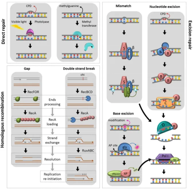
mechanisms to tolerate lesions (Friedberg et al., 2006). The main DNA repair systems are
direct, excision and recombinational repair, with extensive reviews in the literature (Alberts et
22
The direct repair is a simple and error-free way to reverse damage caused by UV-C
or alkylation (Goosen and Moolenaar, 2008). CPD lesions are repaired by photolyases
through photo reactivation, a mechanism that uses energy derived from visible light to break
the covalent bond between pyrimidines. Alkylated guanines can be reversed by the transfer
of methyl groups from the base to a cysteine region in the methyltransferases active site.
Repair of a great variety of DNA lesions can be achieved by base or nucleotide
excision, including deamination, alkylation, CPDs and mismatches. The usually bulky lesions
are detected and removed, either as free bases or nucleotides (David et al., 2007; Smith et
al., 2001; Van Houten et al., 2005). The formed gap is then filled using the undamaged
strand as template. The excision systems are nucleotide excision repair (NER, composed by
uvrABC), base excision repair (BER, carried out by DNA glycosylases, AP endonucleases and dRpases) and mismatch repair (MMR, composed by mutS and mutL, with an additional
mutH in E. coli (Radman and Wagner, 1986)). All of them are finished by the actions of UvrD, DNA polymerase II and DNA ligase (Fig 5).
Finally, the recombinational repair can deal with broken chromosomes and
single-stranded gaps. Homologous recombination permits the use of an undamaged homologous
chromosome as template to repair the gap. The search for this template and the strand
invasion are catalyzed by RecA, loaded in the damaged site by one of the classical (RecBCD
and RecFOR) or alternative (RecQ, RecS, RecJ) pathways. When a homologous
chromosome is not available, double strand breaks can be repaired by the highly mutagenic
non-homologous end joining (NHEJ), carried out by homologous of the eukaryotic Ku protein
and ATP-dependent DNA ligases (Pitcher et al., 2007; Schwartz et al., 2005).
Several transcriptional regulators can modulate the response of DNA damage and
induce these repair systems, through mechanisms not always determined (Hong et al., 2009)
and connections to oxidative and general stress responses (Storz and Hengge, 2010). The
most extensively studied DNA damage response network is the SOS system, which relies in
23
Figure 5. Main DNA repair pathways. Direct repair is carried out with photolyase or methyltransferases; classical pathways of homologous recombination are catalyzed by RecA, loaded onto DNA by RecFOR at ssDNA gaps and by RecBCD at double strand breaks; after strand invasion, RuvABC resolve the Holliday junctions and replication initiation occurs at former double strand break sites. The enzymes responsible for mismatch repair are MutS and MutL, which interacts with the β
clamp of the replisome. Nucleotide repair is carried out by UvrABC. Both excision repair pathways use also UvrD. Finally, base excision repair is catalyzed by specific DNA glycosylases (gly), AP endonucleases (AP) and dRpases. The repair is finalized by DNA polymerase I and DNA ligase.
1.4.2. SOS induction cycle
The mechanisms of SOS activation have been extensively studied in Escherichia coli
and Bacillus subtilis (Cheo et al., 1993; Friedberg et al., 2006; Janion, 2001; Little et al.,
1981; Winterling et al., 1997). During normal bacterial growth, without any significant level of
24
located at the promoter of the regulated genes, the SOS box. Binding of LexA represses
expression by physically blocking the binding of RNA polymerase (Fig. 6). However, a basal
level of expression is allowed, since binding is intermittent. Occurrence of several kinds of
damages results in single stranded DNA (ssDNA), either directly or through processing of
lesions or strand breaks. Several monomers of RecA are recruited, forming filaments with
ssDNA and turning to an active form. Interaction of the ssDNA-RecA complex with LexA
induces the auto-cleavage of the repressor. The resulting polypeptides lose capacity of DNA
binding, allowing RNA polymerase to have access to the promoters. Two classical genes
controlled by SOS is recA and the repressor itself, providing an off-switch to the system: as
soon as the lesions are repaired, levels of ssDNA-RecA filaments start to drop; without
interaction with activated RecA, the level of intact LexA protein rises; and promoters once
again are occluded by LexA dimer binding, eventually shutting the whole system down.
Figure 6. SOS induction as characterized in E. coli. While the system is inactive, the LexA repressor is bound to the promoter of several genes, including itself and recA (1). Once there is any damage in the
25
1.4.3. LexA repressor
The SOS regulator belongs to Family S24 of serine-proteases, which mechanism
involves a catalytic dyad Ser119-Lys156 in E. coli (Slilaty and Little, 1987). The structure of the
protein is divided in two regions. The amino-terminal domain, formed by three alpha-helices
and two beta-barrels, is responsible for DNA binding. The carboxy-terminal domain,
composed only by beta-barrels, is where dimerization and catalysis occur. The
carboxy-terminal domain exists in two distinct conformations, cleavable and non-cleavable. In the
non-cleavable form, Lys156 is protonated and exposed to the solvent. In addition, the targeted
peptide bond Ala84-Gly85 is distant from the active site. On the cleavable conformation the
loop containing this specific peptide bond moves closer to the active site (Luo et al., 2001).
The new hydrophobic environment created around Lys156 drops the pK of its lateral group,
allowing the residue to lose a proton and act as a general base to Ser119. The transfer of the
positive charged Lys from the solvent to a hydrophobic cleft is an important energetic barrier.
As so, LexA only undergoes self-cleavage when the cleavable form is stabilized by the
interaction with activated RecA (Butala et al., 2009). The same mechanism hold true for
other members of the Family S24, as phage lambda regulators cI/Cro, which are also
cleaved in contact with activated RecA (Little, 1984). Binding to DNA is mediated by helices
2 and 3 from the amino-terminal region, which form a helix-turn-helix domain. LexA dimers
interact with imperfect palindromes (sometimes direct repeats) in the DNA major groove.
SOS boxes are greatly variable throughout the bacterial taxa due to the lack of conservation
in the LexA DNA-binding domain from different groups (Mazon et al., 2004).
1.4.4. SOS response consequences
Even with high diversity of SOS regulons among bacteria, they usually play a role in
aspects of DNA repair, replication, recombination and regulation of cell cycle events (Storz
and Hengge, 2010). Cell division inhibition activated by SOS leads to a classical filamented
phenotype (Friedberg et al., 2006; Trusca et al., 1998). The main DNA repair systems
26
tolerance mechanisms, such as alternative polymerases. These are part of the Y family of
polymerases, capable of translesion synthesis of DNA on damaged sites where the
replicative polymerase gets stalled. DNA polymerases V (umuCD) and IV (dinP) have low
fidelity (Goodman, 2002; Neeley et al., 2007; Ohmori et al., 2001), and the last is associated
with increasing occurrences of -1 frameshifts (McKenzie et al., 2001).
These mutator phenotypes associated with the up-regulation of dinP and umuCD can
lead to important consequences for pathogens, due to the potential for developing antibiotic
resistance and increased persistence inside the host. Other relevant aspect in the medical
point of view is the induction of prophages and integrons directly through LexA or cI/Cro.
Some classical examples of SOS-dependent induction of toxins are the expression of Shiga
toxin in E. coli during treatment with beta-lactams (Wagner et al., 2002; Zhang et al., 2000)
and cholera toxin in Vibrio cholera (Quinones et al., 2005). An additional advantage of the
SOS system for pathogens is the dissemination of virulence factors and antibiotic resistance
genes, through the increased mobility of prophages (Abella et al., 2007; Dibbens et al., 1992;
Harrison and Gabriel, 1983; Singletary et al., 2009), integrons (Cambray et al., 2010; Guerin
et al., 2009) and pathogenicity islands (Hacker and Kaper, 2000; Schmidt and Hensel, 2004;
Ubeda et al., 2007).
1.4.5. DNA damage response in leptospires
Despite the significance of DNA damage response for adaptation, virulence and
diversity of pathogens, little is known about how leptospires handle genotoxic stress. A
pioneer work by Stamm et al. (Stamm and Charon, 1988) evaluated the sensitivity of several
strains of leptospires to UV-C and MMC. All bacteria had the capacity of photo reactivation,
although L. interrogans strains were extremely sensitive to both agents when compared to
free-living L. biflexa and L. illini. The reasons for this difference were never addressed.
However, regulation of DNA damage repair pathways was shown to be important for
saprophytic and pathogenic species. Knocking out recA from L. biflexa resulted in a
27
repair systems seem to be essential for maintenance of virulence in L. interrogans even
during normal growth. The most remarkable difference in gene expression profile of a virulent
strain of serovar Lai in comparison to a attenuated derivative is up-regulation of genes in the
DNA replication and repair functional category (Zhong et al., 2011). The first superficial study
on SOS response in leptospires was a characterization of LexA binding to the recA promoter
in L. interrogans serovar Lai (Cuñé et al., 2005). A SOS box was determined, and a SOS
response composed solely by recA proposed.
1.5. Objectives
The aim of this thesis was to characterize the SOS system of L. interrogans serovar
Copenhageni. Specifically to: (i) analyze the behavior of this bacterium to UV-C irradiation;
(ii) assess the functionality of a putative second LexA repressor; (iii) identify the genes
controlled by SOS and the sequence responsible for LexA recognition; (iv) evaluate the
global response to genotoxic stress; (v) identify the DNA repair pathways present in L.
interrogans serovar Copenhageni and their role during SOS activation.
1.6. Structure of the thesis
Our findings investigating the SOS and general DNA damage response in L.
interrogans will be presented in the three following chapters.
Chapter 1 (“Leptospira interrogans serovar Copenhageni harbors two lexA genes
involved in SOS response”) presents the overall response to UV-C-induced damages in the
leptospiral genome and a brief characterization of the lexA-like gene LIC12654, exclusive of
the serovar Copenhageni. UV-C irradiation of serovar Copenhageni led to massive death
and filamentation of the survival cells. After 12h, it was observed a peak in expression of
some common SOS genes: lexA1, lexA2, recA, recN and dinP. This was concomitant with
LexA1 and LexA2 intact protein levels decrease. LexA1 was able to bind to the promoter
regions of these five genes, while LexA2 would bind to its own promoter region. LexA1
binding was though to happen through versions of the TTTGN5CAAA palindrome, while
28
Chapter 2 (“Integrated analysis of Leptospira interrogans expression reveals induction
of mobile genetic elements and repression of virulence genes during genotoxic stress”)
explores further the response of serovar Copenhageni to genotoxic stress and the role of
LexA1 in regulation of expression. LexA1 was found to bind to 25 sites in the genome using
chromatin immunoprecipitation coupled with massive parallel sequencing (ChIP-seq). These
enriched sequences were used to redefine the leptospiral SOS box to an asymmetrical
imperfect palindrome CTNARYAYYTGTNTAG. Site-associated genes were involved in
various processes, including prophage mobilization, cell growth, motility, mutagenesis and
some putative virulence factors, in addition to previously identified targets (shown in Chapter
1). Genotoxic stress also influenced the expression of 18% of leptospiral genes, as assessed
by microarrays. Increase of expression was observed in transposon and prophage genes, in
addition to DNA repair pathways. On the other hand, motility, general metabolism and almost
all virulence genes were repressed. These findings are discussed as mechanisms to adapt
and evolve potential new characteristics, including virulence.
Intrigued by the sensitivity of serovar Copenhageni to UV-C and conscious of the
complexity of the DNA damage response in leptospires, we performed a comparison of DNA
repair-related genes of leptospira species with genomes available in public databases. In
addition, we assessed the induction of several DNA repair pathways during infection of
susceptible animals with L. interrogans serovar Copenhageni. The Chapter 3 (“Genomic
survey and expression analysis of DNA repair genes in the genus Leptospira”) shows more
diversity and redundancy of repair genes in L. interrogans than in saprophytic (L. biflexa) or
obligatory pathogens (L. borgpetersenii). Direct repair, nucleotide excision repair (NER) and
homologous recombination in leptospires have several characteristics typical of
Gram-positive, naturally transforming bacteria. Other interesting finding was the huge impact of
LGT in providing tools for repair, as seen in direct and excision repair, non-homologous end
joining (NHEJ) and also the SOS response. Finally, key players from excision repair, NHEJ
29
2. CHAPTER 1
Leptospira interrogans
serovar Copenhageni harbors two
lexA
genes involved in SOS response
Luciane S. Fonseca1,2, Josefa B. da Silva1, Juliana S. Milanez1, Claudia B. Monteiro-Vitorello3, Leonardo Momo4, Zenaide M. de Morais5, Silvio A. Vasconcellos5, Marilis V. Marques6, Paulo L. Ho1,2, Renata M. A.da Costa4
1 Centro de Biotecnologia, Instituto Butantan, São Paulo, Brazil
2 Departamento de Bioquímica, Instituto de Química, Universidade de São Paulo, São Paulo, Brazil 3 Departamento de Genética, Escola Superior de Agricultura Luiz de Queiroz, Piracicaba, Brazil 4 Centro de Ciências Naturais e Humanas, Universidade Federal do ABC, Santo André, Brazil 5 Departamento de Medicina Veterinária Preventiva e Saúde Animal, Universidade de São Paulo,
Brazil
6 Departamento de Microbiologia, Instituto de Ciências Biomédicas, Universidade de São Paulo, Brazil
Presented as published.
Leptospira interrogans
serovar Copenhageni Harbors
Two
lexA
Genes Involved in SOS Response
Luciane S. Fonseca1,2, Josefa B. da Silva1, Juliana S. Milanez1, Claudia B. Monteiro-Vitorello3, Leonardo Momo4, Zenaide M. de Morais5, Silvio A. Vasconcellos5, Marilis V. Marques6, Paulo L. Ho1,2*, Renata M. A. da Costa4*
1 Centro de Biotecnologia, Instituto Butantan, São Paulo, Brazil, 2 Departamento de Bioquímica, Instituto de Química, Universidade de São Paulo, São Paulo, Brazil, 3 Departamento de Genética, Escola Superior de Agricultura Luiz de Queiroz, Piracicaba, Brazil, 4 Centro de Ciências Naturais e Humanas, Universidade Federal do ABC, Santo André, Brazil, 5 Departamento de Medicina Veterinária Preventiva e Saúde Animal, Universidade de São Paulo, Brazil, 6 Departamento de Microbiologia, Instituto de Ciências Biomédicas, Universidade de São Paulo, Brazil
Abstract
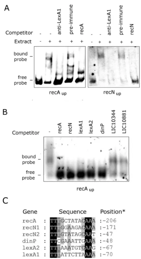
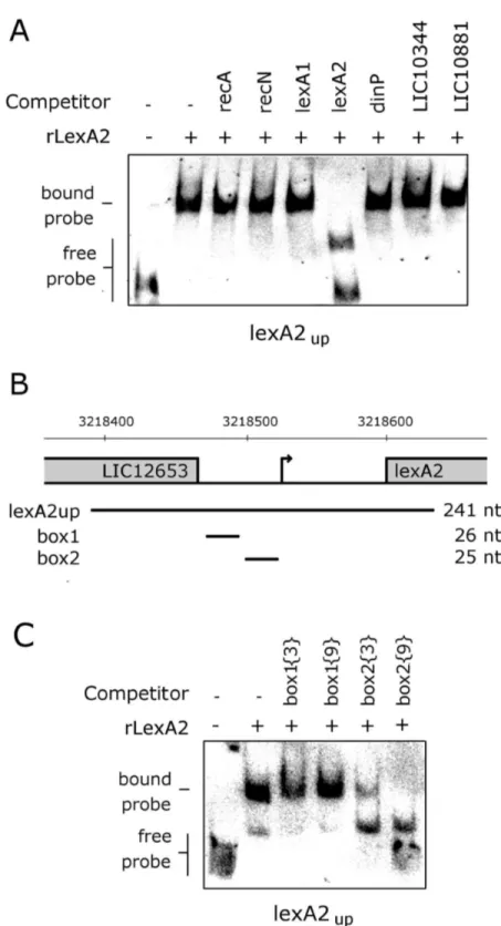
Bacteria activate a regulatory network in response to the challenges imposed by DNA damage to genetic material, known as the SOS response. This system is regulated by the RecA recombinase and by the transcriptional repressor lexA. Leptospira interrogans is a pathogen capable of surviving in the environment for weeks, being exposed to a great variety of stress agents and yet retaining its ability to infect the host. This study aims to investigate the behavior of L. interrogans serovar Copenhageni after the stress induced by DNA damage. We show that L. interrogans serovar Copenhageni genome contains two genes encoding putative LexA proteins (lexA1 and lexA2) one of them being potentially acquired by lateral gene transfer. Both genes are induced after DNA damage, but the steady state levels of both LexA proteins drop, probably due to auto-proteolytic activity triggered in this condition. In addition, seven other genes were up-regulated following UV-C irradiation, recA, recN, dinP, and four genes encoding hypothetical proteins. This set of genes is potentially regulated by LexA1, as it showed binding to their promoter regions. All these regions contain degenerated sequences in relation to the previously described SOS box, TTTGN
5CAAA. On the other hand, LexAβ was able to bind to the palindrome TTGTAN 10TACAA, found in its own promoter
region, but not in the others. Therefore, the L. interrogans serovar Copenhageni SOS regulon may be even more complex, as a result of LexA1 and LexAβ binding to divergent motifs. New possibilities for DNA damage response in Leptospira are expected, with potential influence in other biological responses such as virulence.
Citation: Fonseca LS, da Silva JB, Milanez JS, Monteiro-Vitorello CB, Momo L, et al. (β01γ) Leptospira interrogans serovar Copenhageni Harbors Two lexA Genes Involved in SOS Response. PLoS ONE 8(10): e76419. doi:10.1γ71/journal.pone.0076419
Editor: Fenfei Leng, Florida International University, United States of America Received November 1γ, β01β; Accepted August β8, β01γ; Published October γ, β01γ
Copyright: © β01γ Fonseca et al. This is an open-access article distributed under the terms of the Creative Commons Attribution License, which permits unrestricted use, distribution, and reproduction in any medium, provided the original author and source are credited.
Funding: This research was funded by Fundação Instituto Butantan (www.butantan.gov.br), FAPESP (projects β010/51γ65-0; β009/171βγ-β, www.fapesp.br) and CNPq (project 48β04γ/β009-1), www.cnpq.br). The funders had no role in study design, data collection and analysis, decision to publish, or preparation of the manuscript.
Competing interests: The authors have declared that no competing interests exist. * E-mail: hoplee@butantan.gov.br (PLH); renata.costa@ufabc.edu.br (RMAC)
Introduction
Leptospira interrogans is one of the etiologic agents of leptospirosis, a worldwide disease with important economic and public health consequences, in particular to developing tropical countries [1,β]. The leptospires can infect a wide range of mammalian species that compose their natural reservoir, colonizing the kidneys, and being shed in the urine during the whole life of these animals [γ]. There are nine pathogenic species of Leptospira, divided in more than β60 serovars [4]. In Brazil, the majority of the leptospirosis cases in humans is the result of infection with serovar Copenhageni [1]. In spite of its social and economic impact, the molecular mechanisms of
Leptospira pathogenesis are still poorly understood, as a
consequence of the difficulties in their genetic manipulation. Particularly, L. interrogans serovar Copenhageni remains one of the serovars most refractory to genetic transformation and only two mutants were so far obtained by targeted mutagenesis [5,6].
L. interrogans can survive in water or mud for weeks, after which they are still able to infect the host. These leptospires are exposed to a wide spectrum of DNA-damaging agents, from sun radiation and heavy metals to oxidative stress and antibiotics [7,8]. One of the most important mechanisms employed by bacteria to deal with stress induced by DNA damage is the SOS response. This regulatory network controls DNA repair, error prone DNA replication, cell division and mobilization of phages and transposable elements in E. coli
[9-1β]. The expression of these genes is repressed by LexA, which dimerizes and binds to operators in their promoters at regions called SOS boxes [1γ,14]. The induction of the SOS response is triggered by genomic structure alterations that generate single-stranded DNA (ssDNA), which is sensed by the cells during replication. RecA recognizes and interacts with these damaged regions, acquiring an active conformation and playing a role as a co-factor in the self-cleavage reaction of LexA. The cleavage generally occurs in a peptide bond flanked by Ala-Gly residues near the center of the protein sequence, disrupting LexA dimerization, which in turn, reduces DNA binding and allows transcription initiation [15-18]. Once the damage is repaired, the level of activated RecA drops, and newly synthesized LexA (whose expression is usually under its own regulation) binds to SOS boxes again, returning the system to the non-induced state [11].
The lexA gene is found in most bacterial taxa, with few exceptions. However, the set of genes directly repressed by LexA diverges substantially. This is in part consequence of the great degree of sequence heterogeneity among SOS boxes. The sequence variation is a result of the low conservation in amino acid residues in the DNA binding domain of LexA among different groups of bacteria [19]. Although the majority of bacteria has only one copy of lexA, genomic sequences of some organisms underwent duplication or lateral gene transfer, resulting in two genes coding LexA proteins regulating different sets of genes [β0-ββ]. As a consequence, the SOS response is a very unique and complex regulatory network, with a remarkable flexibility of LexA-regulated genes.
The SOS response has important consequences for bacterial physiology and for virulence mechanisms in pathogenic organisms [βγ,β4]. All leptospiras sequenced to date harbor one copy of the lexA gene. Previous work reported that the LexA protein from L. interrogans serovar Lai (LA1447) [β5] has activity of a transcriptional repressor, acting only on recA gene expression. Moreover, the identified SOS box palindrome (TTTGCTATACAAA) was found only upstream of the recA
gene. As such, L. interrogans, along with Thermotoga maritima, would be among the rare organisms in which LexA does not regulate its own transcription [β6]
In this study we show that the DNA damage induced by UV-C irradiation triggered the SOS response in L. interrogans
serovar Copenhageni. Analyzing the bacterium genome, we found a second lexA gene (lexA2) within a prophage-like region rich in genes encoding hypothetical proteins. Following the stress induced by UV-C irradiation, L. interrogans displayed filamentation and both LexA repressors were depleted, presumably as a consequence of self-cleavage. The expression levels of both lexA genes, as well as those of other seven genes, were increased from eight up to 1β hours after the UV-C treatment. LexA1 was able to bind to the promoter sequences of recA and recN, and competition assays indicate its binding to the promoters of the remaining UV-C induced transcripts, including the one containing lexA1. Not all the genes showing UV-C induction do have the exact previously described SOS box in their promoter regions, but alignment of these sequences showed the presence of imperfect palindromic dyads that could be LexA1 binding sites. On the
other hand, LexAβ showed specific binding only to a sequence upstream of its own transcriptional unit. Therefore, the physiological response of L. interrogans serovar Copenhageni may be even more complex than in other bacteria, as LexA1 appears to have some flexibility to recognize degenerated SOS sequences.
Material and Methods
In silico analysis
All sequences used were obtained from Kyoto Encyclopedia of Genes and Genomes (KEGG) and National Center for Biotechnological Information (NCBI) databases. The secondary structure predictions were made by PsiPred [β7], and search for structural domains in hypothetical protein sequences by HHPred (http://hhpred.tuebingen.mpg.de). Finally, the search for putative SOS box motifs was carried out by using the tool “genome scale DNA-pattern”, available from RSAT (http:// rsat.ccb.sickkids.ca). The output returned all palindromes present in the upstream region of the genes (nucleotides +β0 to -β50 from the start codon).
Phylogenetic analysis
A total of 48 protein sequences (Table S1) were obtained through BLAST searches using either LexA1 or LexAβ as query. MUSCLE [β8,β9] alignments were used to infer phylogenetic trees, constructed using maximum likelihood analysis with WAG substitution model in PhyML [γ0]. The robustness of the trees was assessed by aLRT [γ1]. Node support was assessed as the posterior probability from two independent runs, with four chains of β00,000 generations each (sampled at intervals of 100 generations with a burn-in of 1000 trees).
Bacterial strains and growth conditions
L. interrogans serovar Copenhageni Strain FioCruz L1-1γ0 and other serovars (Australis, Autumnalis, Bataviae, Canicola, Pomona, Pyrogenes, Hardjo) and L. borgpetersenii serovar Hardjobovis were obtained from Faculdade de Medicina Veterinária e Zootecnia (Universidade de São Paulo, Brazil), while the genomic DNA from serovars Smithi and Naam were obtained as described by da Silva et al. [γβ]. The growth and virulence maintenance were carried out according to da Silva et al. [γγ]. E. coli DH5α and BLβ1(DEγ) Star pLysS were used for cloning and expression procedures, respectively. E. coli
cells were grown at γ7°C in LB medium containing the appropriate antibiotics.
UV-C irradiation, survival curves and visualization
Virulent L. interrogans serovar Copenhageni L1-1γ0 was cultivated until density of approximately 4x10⁸ cells/ml. Bacteria
were transferred to 140mm diameter Petri dishes, conserving a thin layer of culture, and exposed for increasing times to a germicidal lamp (β54 nm, rate 1 J.m-². s-1). After treatment, the
same volume of fresh medium was added to stimulate cellular division and the culture was incubated at γ0°C in the dark. Surviving bacteria were counted β4 hours post-treatment using
Leptospira interrogans Harbors Two lexA Repressors
a Petroff-Hausser counting chamber and survival frequency was calculated as the ratio of irradiated to non-irradiated cells. Cells were visualized by fluorescence microscopy after labeling with DAPI (4’,6-diamidino-β-phenylindole) [γ4] and measured using ImageJ software (http://rsbweb.nih.gov/ij). For RNA extraction, cells were collected 4, 8, 1β and β8h following UV-C exposure, as well as their non-treated counterparts, immediately frozen in liquid nitrogen and stored at -80°C until use.
Recombinant protein expression and purification
All enzymes cited in this section were obtained from Fermentas (USA), and used according to the manufacturer instructions. The coding regions of lexA1 (LIC1βγ05) and lexA2
(LIC1β654) were codon-optimized for expression in E. coli
(Genscript, USA). These sequences were cloned in the expression vector pAE [γ5] and the recombinant proteins were over-expressed in E. coli BLβ1(DEγ) Star pLysS as described elsewhere [γ6]. The cells were harvested by centrifugation, ressuspended in 50 mM Tris-HCl (pH 6.γ), 150 mM NaCl and lysed in a French press (Thermo Spectronic). The supernatant was applied to a 1 cm-diameter column containing γ ml Niβ+
-charged chelating Sepharose (GE Healthcare Life Sciences, USA). The proteins were eluted with 400 mM imidazole, which was removed by dialysis. Purified proteins were visualized by Coomassie blue staining after separation by 15% SDS-PAGE.
Western blot
For immunoblotting, β0 ng of purified proteins and 40 µg or 150 µg of leptospiral extracts were separated by 15% SDS-PAGE and transferred to Hybond-P Polyvinylidene Difluoride (GE Healthcare Life Sciences, USA) membranes. Incubations and detection were carried out as described elsewhere [γ6], using anti-LexA1 in 1:5000 dilution, anti-LexAβ in 1:1000 and anti-LipLγβ [γ7] in 1:5000. Anti-LexAβ serum was incubated with β00ng/µl purified LexA1 for βh prior to use, to decrease cross-reactivity.
DNA purification and PCR
Genomic DNA (gDNA) of Leptospira was isolated using DNAzol (Invitrogen), following the manufacturer instructions, and quantified by NanoDrop (Thermo Scientific, USA) spectrophotometer. It was used as positive control in RT-PCR experiments (see below).
RNA manipulation and quantitative PCR
Total RNA was prepared using Trizol (Invitrogen, USA) according to manufacturer instructions and treated with DNaseI (Fermentas, USA) to avoid gDNA contamination. Purified RNA was quantified by NanoDrop. Next, 1 µg of RNA was used as template for the complementary DNA (cDNA) synthesis by the reverse transcriptase M-MuLV (New England Biolabs, USA), using random hexamers. Reverse transcriptase-PCR (RT-PCR) to assess the transcription organization of lexA1 and
lexA2 vicinities were carried out using 1 µl of 1:5 cDNA as template, for 40 cycles. Quantitative PCR (qPCR) was performed with SYBR Green Master Mix (Applied Biosystems,
USA), using 1 µl of 1:100 cDNA in 1β µl reactions. Reactions were set at the default profile of Applied Biosystems 7γ00 Real-Time PCR System (βmin at 50°C and 10min at 95°C, followed by 40 cycles of 15s at 90°C and 1min at 60°C). A posterior dissociation cycle (15s at 96°C, β0s at 60°C, 15s at 90°C and 15s at 60°C) was added to discard the existence of any contaminating product. Fold change was calculated by the β-∆∆Ct method, using the 16S as internal control. Each
experiment was repeated three times, with biological replica. Data were analyzed through GraphPad Prism5, where the variance was assessed by one way ANOVA and significance of differences by Dunnett post-test. Oligonucleotides used in these experiments are compiled in Table Sβ.
Electrophoretic Mobility Shift Assay (EMSA)
EMSA was performed using DIG Gel Shift Kit (Roche), following the manufacturer instructions. Probes amplified by PCR (Table Sβ) were purified by GFX™ PCR DNA and Gel Band Purification Kit (GE Healthcare, USA), quantified by NanoDrop and labeled with a terminal DIG. Alternatively, probes were labeled with [ γβP ATP] by T4 Polynucleotide
Kinase (Fermentas) and column purified (GenElute PCR Clean-Up Kit, Qiagen). Binding reactions were carried out in ice, in binding buffer provided in the kit, using poly[d(A-T)] as unspecific competitor. Solutions containing 1.55 fmol of labeled probes were incubated with 40 µg leptospiral extracts or 80 ng purified LexAβ for β0min. In antibody blockade assay, extracts were incubated with 1 µl of anti-LexA1 or preimmune sera for γ0min in ice prior to the addition of the probe. In competition assays, non-labeled probes were added to the binding reaction after the labeled one, in β00 fold excess (γ10 fmol). Mixtures were loaded onto a 5% non-denaturing 0.5x TBE gel pre-run at 80 V for 90min. DNA-protein complexes were separated at 80 V for 150min at 4°C and transferred to a Hybond-N (GE Healthcare, USA) nylon membrane using a Trans-Blot Semi-Dry Electrophoretic Transfer Cell (Bio-Rad, Germany) in TBE 0.5x for 90min at 5 V. Detection followed the manufacturer instructions, and the membranes were exposed to photographic films (Hyperfilm, ECL GE Healthcare, USA).
Results
L. interrogansserovar Copenhageni genome harbors a second lexA gene
Analysis of the genome of L. interrogans serovar Copenhageni [γ8] revealed the presence of a second homologous gene for lexA, LIC1β654. For clarity, we named it
lexA2, whereas the gene identical to lexA from L. interrogans
serovar Lai [β5] was named lexA1 (LIC1βγ05). The LexAβ predicted amino acid sequence exhibits very low similarity with the known LexA proteins, sharing β8% of amino acid identity to LexA1. Nevertheless, the predicted secondary structure shows both DNA-binding and serine-protease domains compatible with LexA-like protein structure [16,γ9] (Figure 1). The catalytic residues Ser and Lys (indicated with arrowheads in Figure 1), located at the carboxy-terminal domain and typically γ7 amino acids apart, are at positions 1γ0 and 166 in LexAβ, respectively, while the scissile peptide bond is probably located
Leptospira interrogans Harbors Two lexA Repressors
in Cys9β-Gly9γ (indicated with a bar in Figure 1). The
helix-turn-helix structure of the amino-terminal domain of LexAβ is conserved. However, from the β1 amino acid residues potentially involved in DNA binding in this domain (labeled with Δ in Figure 1), 16 are different between LexA1 and LexAβ. Since this is the domain responsible for the SOS box recognition, it is conceivable that both proteins must regulate different sets of genes.
LexA2 coding sequence was possibly acquired through lateral gene transfer
The remarkable differences in amino acid sequence of LexAβ raised the question if it was acquired through lateral gene transfer. This hypothesis was tested through phylogenetic analyses (Figure β). The multiple alignments were used to construct phylogenetic trees with maximum-likelihood algorithm. The distribution of the evolutionary distances and the tree topology reveals a long phylogenetic distance among the two L. interrogans serovar Copenhageni LexA repressors. The LexAβ protein grouped in a distinct clade with sequences from marine metagenomes, while LexA1 clustered with orthologous from other leptospires. Therefore, both coding sequences did not evolve together.
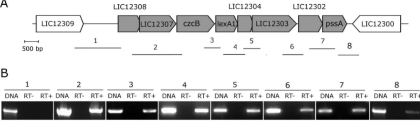
Comparative genomic organization of genes lexA1 and lexA2 vicinities
The lexA1 and lexAβ-containing regions are located within the large inversion which differentiates serovars Lai and Copenhageni genomes [40]. The lexA1 gene vicinities of both genomes are identical, sharing 99% of nucleotide identity. This region is enriched with genes encoding peptidases and stress response proteins (Figure γA). Besides the Sβ4 peptidase
lexA1, LIC1βγ0γ is annotated as a S41 peptidase and LIC1βγ0β as a Mββ peptidase. The hypothetical protein LIC1βγ04 shows a structure similar to the xenobiotic response element (XRE) family of transcriptional regulators; czcB is a heavy metal efflux pump; and LIC1βγ07 is a TolC superfamily transporter protein. The genomic organization, exhibiting less than 4γ bp intergenic spaces, indicates a structure of an operon (Figure γA) [41]. To investigate this hypothesis, primers were designed to amplify across intergenic regions of L. interrogans serovar Copenhageni cDNA. The result showed amplicons for all gene pairs, from LIC1βγ08 to pssA (Figure γB), suggesting the occurrence of an mRNA spanning this entire region. The lexAβ gene lies within a prophage-like region rich in genes encoding hypothetical proteins. The genome context of lexAβ resembles the remnant of an ancient phage infection, which has been subject of mutational decay and rearrangements leading to losses of most of the prophage Figure 1. Comparison of LexA amino acid sequences. Amino acid sequence alignment and secondary structure prediction were carried out using E. coli LexA as reference. The amino-terminal region is composed of three helixes (striped rectangles) and strands 1 and β (striped arrows), while the carboxy-terminal is composed of nine strands (grey arrows). Arrowheads indicate the catalytic residues, and the bar indicates the residues flanking the scissile peptide bond. Open triangles represent residues that interact with DNA. The percentage of identity of each sequence to either L. interrogans serovar Copenhageni LexA proteins is indicated.
doi: 10.1γ71/journal.pone.0076419.g001
Leptospira interrogans Harbors Two lexA Repressors
Figure 2. Phylogenetic analysis of LexA. Phylogenetic analysis was performed using LexA amino acid sequences from several bacteria. The leptospiras clade is highlighted by a grey box, and the sequences of the LexA proteins present in L. interrogans
serovar Copenhageni are indicated with arrows. Local bootstrap values are attached to the internal nodes. Species code description and sequences used are compiled in Table S1.
doi: 10.1γ71/journal.pone.0076419.g00β
Leptospira interrogans Harbors Two lexA Repressors