Oral motor and electromyographic characterization
of adults with facial fractures: a comparison between
different fracture severities
Amanda Pagliotto da Silva,IFernanda Chiarion Sassi,IIEndrigo Bastos,III Nivaldo Alonso,III Claudia Regina Furquim de AndradeII,*
IDivisao de Miologia Orofacial, Hospital das Clinicas HCFMUSP, Faculdade de Medicina, Universidade de Sao Paulo, Sao Paulo, SP, BR.IIDepartamento de
Fisioterapia, Fonoaudiologia e Terapia Ocupacional, Faculdade de Medicina (FMUSP), Universidade de Sao Paulo, Sao Paulo, SP, BR.IIIDivisao de Cirurgia Plastica, Hospital das Clinicas HCFMUSP, Faculdade de Medicina, Universidade de Sao Paulo, Sa˜o Paulo, SP, BR.
OBJECTIVES:To characterize the oral motor system of adults with facial injuries and to compare the oral motor performance/function between two different groups.
METHODS:An observational, descriptive, cross-sectional study was conducted in 38 patients presenting with facial trauma who were assigned to the Division of Orofacial Myology of a Brazilian School Hospital. Patients were divided into two groups: Group 1 (G1) consisted of 19 patients who were submitted to open reduction of at least one facial fracture, and Group 2 (G2) consisted of 19 individuals who were submitted to closed fracture reduction with maxillomandibular fixation. For comparison purposes, a group of 19 healthy volunteers was recruited. All participants underwent a clinical assessment that included an oral motor evaluation, assessment of the mandibular range of motions, and electromyographic assessment of the masticatory muscles.
RESULTS:Clinical assessment of the oral motor organs indicated that G1 and G2 presented deficits related to the posture, position, and mobility of the oral motor organs. Patients also presented limited mandibular ranges of movement. Deficits were greater for individuals in G1, especially for maximal incisor opening. Additionally, patients in G1 and G2 presented a similar electromyographic profile of the masticatory muscles (i.e., patients with facial fractures presented lower overall muscle activity and significant asymmetrical activity of the masseter muscle during maximum voluntary teeth clenching).
CONCLUSION:Patients in G1 and G2 presented similar functional deficits after fracture treatment. The severity of facial fractures did not influence muscle function/performance 4 months after the correction of fractures.
KEYWORDS: Mandibular Fractures; Condylar Fractures; Open Reduction; Closed Reduction; Orofacial Functions.
Silva AP, Sassi FC, Bastos E, Alonso N, de Andrade CR. Oral motor and electromyographic characterization of adults with facial fractures: a comparison between different fracture severities. Clinics. 2017;72(5):276-283
Received for publication onAugust 26, 2016;First review completed onNovember 30, 2016;Accepted for publication onFebruary 13, 2017 *Corresponding author. E-mail: [email protected]
’ INTRODUCTION
The human face constitutes the first contact point in seve-ral human interactions; thus, injuries and/or mutilation of the facial structures may have a disastrous influence on the affected person (1). The literature has reported an increasing occurrence of facial injuries over the last four decades, espe-cially as a consequence of the increase in motor vehicle acci-dents as well as in urban violence (2,3).
Facial fractures can have long-term consequences, both functionally and aesthetically. The management of fractures
to the face remains a challenge to all health professionals involved in treatment and rehabilitation (i.e., oral and maxi-llofacial surgeons and multidisciplinary teams), demanding a high level of both skill and expertise (4). According to the literature, mandibular fractures are the most common type of facial bone fractures and are frequently accompanied by condylar fracture, with a frequency that ranges from 26% to 57% (2,5,6). The high incidence of mandibular condylar frac-ture is attributable to the binding of the mandibular ramus, which has a high stiffness, to the mandibular condyle head, which has a low stiffness (7).
The functions executed by the oral myofunctional system depend highly on the mandibular movements. These func-tions are responsible for intraoral space modificafunc-tions and have a strong impact on masticatory function, swallowing, and speech patterns because they are responsible for enabl-ing adequate movements of the tongue and other soft tissues (i.e., amplitude) inside the oral cavity (8). Post-traumatic anky-losis, for example, can arise from intra-capsular condylar DOI:10.6061/clinics/2017(05)04
Copyright&2017CLINICS–This is an Open Access article distributed under the terms of the Creative Commons License (http://creativecommons.org/licenses/by/ 4.0/) which permits unrestricted use, distribution, and reproduction in any medium or format, provided the original work is properly cited.
fractures. Temporomandibular joint (TMJ) ankylosis is an extremely disabling affliction that causes problems with masti-cation, swallowing, speech, appearance, and hygiene (9,10). Studies that describe and evaluate the impact of condylar fractures on the functions performed by the oral myofunc-tional system are limited. However, it is known that mandi-bular movements are necessary to maintain the production of synovial fluid (9,10). Therefore, one would expect the limitation, or even the impossibility, of mouth opening to have severe consequences for the TMJ, not only with respect to maintenance of joint lubrication but also to the overall mandi-bular range of motion (11).
Studies that describe the functional characteristics of post-facial injuries are very scarce. While mandibular fractures have been extensively documented, little information has been presented regarding fractures of the maxilla and zygoma, even though these fractures can also have severe functional consequences (12-14). Salentijn et al. (13) reported that patients with zygoma fractures present better functional outcomes when not submitted to surgical procedures. Tabrizi et al. (14) found that patients with condylar fractures asso-ciated with contralateral mandibular fractures present more signs of temporomandibular disorders compared with patients with isolated unilateral fractures. The authors emphasize that the indirect impact of facial fractures on the TMJ should be considered as an etiological factor in the development of temporomandibular disorders.
The restoration of normal function and long-term stability are indispensable for the successful recovery of facial injuries. Both function and stability, however, may be compromi-sed by inadequate or incorrect postoperative muscular rehabilitation after fracture reduction. Muscle atrophy, denervation, alteration of fiber types, myofibrosis, decreased muscle mass, morphological alterations of the condyle, and facial nerve disorders are biological consequences of fracture reduction, which can have profound clinical consequences (5,6,15).
Despite the adopted medical treatment (i.e., opened or closed fracture reduction), the literature recommends oral motor rehabilitation in patients who have suffered facial injuries (16). An efficient oral motor assessment, including assessment of the masticatory muscles, can shed light on the best rehabilitation procedure to restore oral motor functions (17). A clearer understanding of the patterns of facial injuries will also assist health care providers in planning and man-aging the treatment of traumatic facial injuries. Such infor-mation can also be used to guide the future funding of public health programs, as considerable resources are needed for rehabilitation (i.e., multidisciplinary teams), thus placing a large burden on the health care system (3).
This study was designed to characterize the oral motor system of adults with facial injuries and to compare the oral motor performance/function between two distinct groups: one submitted to open reduction of at least one facial fracture and the other submitted to closed reduction of the facial fracture with maxillomandibular fixation.
’ METHODS
An observational, descriptive, cross-sectional study was performed in patients presenting with facial trauma who were assigned to the Division of Orofacial Myology of a large Brazilian School Hospital (Hospital das Clínicas) between December 2010 and September 2014 for assessment and
treatment. The study design was approved by the Ethics Committee for the Analysis of Research Projects of the Institution (CAPPesq HCFMUSP no. 495.639). Prior to their enrollment, all participants were informed of the purpose and procedures of the study, after which time they all pro-vided written informed consent.
Thirty-eight adults (i.e., age 418 years) with facial
frac-tures were divided into two groups: Group 1 (G1) consisted of 19 patients who were submitted to open reduction of at least one facial fracture, and Group 2 (G2) consisted of 19 individuals who were submitted to closed fracture reduction with maxillomandibular fixation.
The presence of facial fractures was confirmed by the medical team of the Division of Cranio-Maxillofacial Surgery of the same hospital. In line with the basic principles of trauma surgery regarding the open or closed reduction of fractures, open reduction has been recognized to be best for patients presenting with post-fracture malocclusion, an angulation of fracture displacement above 30o
, translocation greater than 4 mm, or lateral overriding (18). However, for moderately displaced condylar fractures or for fractures with no evident displacement or no risk of displacement, closed reduction with rigid or elastic maxillomandibular fixation is used. In this case, an arch bar is used for approximately two weeks.
For comparison purposes, a group of 19 healthy volunteers was recruited (Control Group [CG]). The inclusion criteria for this group were the following: age418 years, absence of
oral motor disorders/deficits, absence of alterations in the scapular region, complete permanent dentition (absence/ extraction of the third molar was accepted), Skeletal Angle Class I facial pattern, and absence of malocclusion. Indivi-duals were excluded if they reported having previous ortho-dontic treatment.
All groups were paired by age. Individuals with pre-existing surgical procedures involving the head and neck, communication or hearing deficits, neurological disorders of any type, and/or cognitive deficits were excluded.
Oral motor clinical assessment
Participants underwent clinical oral motor assessment. Individuals were examined while sitting on a chair in a room with appropriate lighting. The Expanded Protocol of Oro-facial Myofunctional Evaluation with Scores (OMES-E) was used for this assessment (19). This protocol was constructed based on previous models of evaluation, with the addition of numerical scales that reflect the physical characteristics and orofacial behaviors of the subjects. Its purpose is to evaluate the components of the stomatognathic system (lips, tongue, mandible, and cheeks) in terms of aspect/posture, mobility, and performance during swallowing and mastication. The maximum score is 230.
Participants were evaluated individually by visual inspection, and the evaluation was later complemented by the analysis of images recorded on a digital camera Sony DSC-W120 (Sony, Manaus, Brazil).
Mandibular range of movement
The technique used to measure the mandibular range of movement was based on the methodology already publis-hed in the literature (20). With the use of a digital caliper (Digimess Pró-Fono Digital Caliper), the following measure-ments (in millimeters) were collected:
1) maximal incisor distance - we measured the distance between the incisive faces of the mandibular and maxillary central incisors;
2) right lateral excursion - we measured the horizontal distance from the mandibular central incisor to the maxillary cen-tral incisor after asking the individual to glide his/her man-dible to the right. When there was a midline deviation (i.e., between the mandibular and maxillary central incisors), we used the pertinent adjustment;
3) left lateral excursion - the same procedure described above was performed to measure mandibular lateraliza-tion to the left;
4) protrusion - for this measurement, the patient was asked to glide the mandible forward. We then measured the hori-zontal overlap between the mandibular central incisors and the maxillary central incisors;
5) horizontal dental occlusion overlap - we measured the distance between the occlusal face of the maxillary central incisors and the distal face of the mandibular central incisors.
Surface electromyography (sEMG) evaluation
Two muscle groups were examined in this study: the anterior temporal muscles and the masseter muscles.
All EMG recordings were performed using standard sur-face sensors (SDS500). We used the Miotool 400 (Miotecs
Biomedical Equipment, Brazil) 4-channel computer-based system and disposable double electrodes (SDS 500 Ag/AgCl, contact surfaces with a 10-mm diameter). This EMG system has a wide bandpass filter, a bandwidth (RMS) of 20 to 500 Hz, and a 60-Hz notch filter. The system uses the Active Electrode technology, which is a compact sensor assembly that includes a miniaturized instrument preamplifier. Locat-ing the amplifier at the electrode site allows artifacts to be canceled and the signal to be boosted before being trans-ferred down the electrode cable (noise level o5 mV RMS).
Each EMG record was full-wave and low-pass filtered. The computer program indicates the mean, standard deviation (SD), minimum, maximum, and range of muscle activity during each trial. Muscle activity (EMG) was quantified in microvolts (mV).
The interelectrode distance was 10 mm. Four sets of two bipolar pre-gelled stick-on surface electrodes were applied to the skin on each side of the face over the anterior temporal muscles and masseter muscles to record myoelectrical acti-vity during specific tasks involving the masticatory muscles. This electrode arrangement included a third ground elec-trode positioned on the right wrist. Electrical impedance at the sites of electrode contact was reduced because the skin was scrubbed with alcohol gauze pads.
The electric activity of the above-mentioned muscles was assessed at the following times (21,22):
Rest - Participants remained seated with their heads positioned horizontally according to the Frankfort plane. After the pairs of EMG electrodes were placed over the skin, each participant was instructed to remain quiet and
relax for a period of 1 minute. Three separate recordings of the resting condition were made, with a duration of 30 seconds each;
Maximum voluntary tooth clenching on cotton rolls (CR) -two, 10-mm-thick cotton rolls were positioned on the mandibular second premolars/first molars of each subject. Individuals were asked to clench as hard as possible and to maintain the same level of contraction for 5 s. During the test, individuals were verbally encouraged to perform at their best. All individuals repeated the test three times; Maximum voluntary tooth clenching (MVC) - Individuals
were asked to clench as hard as possible and to maintain the same level of contraction for 5 s; during the test, indi-viduals were verbally encouraged to perform at their best. All individuals repeated the test three times.
To avoid any fatigue effect, a rest period of at least 3 minutes was allowed between each test. Subjects were instructed that, during clenching, they should not feel additional muscular/ articular pain.
sEMG data analysis
Surface EMG traces were evaluated for onset of, peak, and offset of activity during clenching events. Onset was identified as the point of upward excursion of the sEMG trace from resting baseline that led to the clenching event. The peak was the highest amplitude point of the sEMG clenching trace. The offset was the point at which sEMG activity returned to baseline. Computer software calculated the mean value of the action potential during the movements (onset-peak-offset). To compare the results between partici-pants, sEMG amplitude values were normalized relative to rest to provide evidence of possible differences.
To perform intragroup and multiple comparisons between the groups, the average sEMG amplitude obtained in all tooth clenching tasks was standardized against the activity of the resting task.
sEMG data reliability
Because subjective judgment was used for sEMG measure-ments, interjudge reliability was estimated. To establish the interjudge reliability of the measurements used in the study, a second experienced staff member who was blinded to the original results measured the same parameters of 35 randomly selected samples from the 342 total clenching events. Intraclass correlation coefficients were high for all comparisons (range of lower 95% confidence interval [CI]= 0.9157-0.9728), suggesting strong consistency between examiners.
Data analysis
The collected data were analyzed using IBM SPSS Sta-tistics 22.0. The distribution of the data was non-normal for all variables. For this reason, the analysis was performed using non-parametric tests. In addition to the descriptive analysis, paired comparisons between groups were performed using the Mann-Whitney U test and multiple comparisons between the groups were performed using the Kruskal-Wallis test. Dunn’s test was used for the post hoc pairwise analysis. The adopted significance level was 5% for all analyses.
’ RESULTS
2 females and 17 males, with a mean age of 32.2 years (±11.19); and the participants in the control group included 5 females and 14 males, with a mean age of 32.9 years (±13.15). The groups did not present a significant difference in age (p=0.961). Apart from age, we also controlled the time
between the facial fracture reduction and the oral motor assessment. The statistical analysis indicated that G1 and G2 were assessed within the same time period (Table 1).
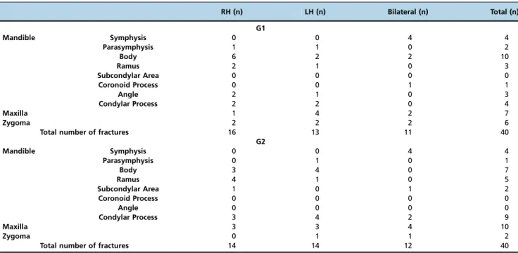
A classification of the location of the fractures was perfor-med based on computed tomogram records obtained from patients’medical files. Most of the patients presented with bilateral fractures in more than one location of the face. Patients in G1 presented more fractures of the mandible body, whereas patients in G2 presented more condylar and maxilla fractures. The mean number of fractures per patient was 2.22 in G1 and 2.05 in G2 (Table 2).
Table 3 presents the results of the multiple and pairwise comparisons for the scores obtained on the OMES-E. As expected, patients with facial fractures differed from the control group in all of the assessed parameters, with the exception of the static posture and position of the oral motor organs. Although not significant, the median obtai-ned for the assessed parameters indicated that G1 pre-sented higher scores for the mobility of the oral motor organs and higher overall scores on the clinical protocol compared with G2.
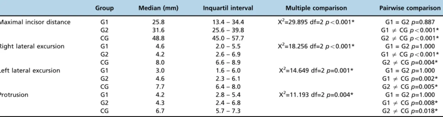
The three groups were compared with respect to differences in the mandibular ranges of motion (Table 4). Differences were observed for all of the parameters when comparing the groups with facial fractures with the control group. No significant differences were observed between G1 and G2. However, when considering the medians obtained for the assessed mea-surements, G2 presented a better mandibular range of motion compared with G1, especially regarding the maximal incisor distance.
The descriptive data related to the electromyographic asses-sment of the masticatory muscles are presented in Table 5. Individuals in the control group presented higher muscle acti-vity in both tooth clenching tasks.
For the purpose of comparing the electromyographic data obtained for each group, the coefficient of asymmetry between both sides of the face during the clenching tasks was calcu-lated for each pair of muscles: ratio of the side with lower temporal muscle activity/side with higher temporal muscle activity, and ratio of the side with lower masseter muscle activity/side with higher masseter muscle activity. The closer the ratio is to 1, the greater the symmetry in the muscle acti-vity in that specific task.
Data analyses indicated that the groups differed only in maximum voluntary tooth clenching (i.e., MVC–see Table 6).
A significant difference was observed between G1 and the control group for the masseter muscle. Looking closely at our results, it is important to highlight that the comparison between G2 and the control group for the same muscle during MVC yielded results near the threshold of statistical significance.
’ DISCUSSION
The treatment of facial fractures, especially those involv-ing the condyle, depends greatly on individual biological characteristics and on adaptations made by the masticatory
Table 1-Time between fracture reduction and oral motor assessment (in days).
Group Median Interquartil interval U Z p-value
G1 51.0 22.00 – 78.0 168.000 -0.365 0.715 G2 57.0 30.00 – 101.0
Legend: G1=patients submitted to open fracture reduction; G2=patients submitted to closed fracture reduction with maxillomandibular fixation; Mann-Whitney test.
Table 2-Classification of fractures locations.
RH (n) LH (n) Bilateral (n) Total (n)
G1
Mandible Symphysis 0 0 4 4
Parasymphysis 1 1 0 2
Body 6 2 2 10
Ramus 2 1 0 3
Subcondylar Area 0 0 0 0
Coronoid Process 0 0 1 1
Angle 2 1 0 3
Condylar Process 2 2 0 4
Maxilla 1 4 2 7
Zygoma 2 2 2 6
Total number of fractures 16 13 11 40
G2
Mandible Symphysis 0 0 4 4
Parasymphysis 0 1 0 1
Body 3 4 0 7
Ramus 4 1 0 5
Subcondylar Area 1 0 1 2
Coronoid Process 0 0 0 0
Angle 0 0 0 0
Condylar Process 3 4 2 9
Maxilla 3 3 4 10
Zygoma 0 1 1 2
Total number of fractures 14 14 12 40
Table 3-Comparisons among groups for the results of the Expanded Protocol of Orofacial Myofunctional Evaluation with Scores (OMES-E).
Group Median Inquartil interval Multiple comparison Pairwise comparison
Aspect/Posture G1 53.0 49.0 – 54.0 X2=20.803 df=2
po0.001* G1 = G2p=0.068
G2 55.0 53.0 – 57.0 G1aCGpo0.001*
CG 58.0 55.0 – 60.0 G2 = CGp=0.068
Mobility G1 80.0 69.0 – 91.0 X2=16.997 gl=2
po0.001* G1 = G2p=1.000
G2 74.0 67.0 – 90.0 G1aCGp=0.002*
CG 99.0 86.0 – 108.0 G2aCGp=0.001*
Performance
swallowing/mastication
G1 34.0 29.0 – 40.0 X2=24.407 gl=2
po0.001* G1 = G2p=1.000
G2 36.0 30.0 – 39.0 G1aCGpo0.001*
CG 45.0 43.0 – 48.0 G2aCGpo0.001*
Total Score G1 172.0 148.0 – 176.0 X2=24.467 gl=2
po0.001* G1 = G2p=1.000
G2 168.0 153.0 – 181.0 G1aCGpo0.001*
CG 204.0 183.0 – 214.0 G2aCGpo0.001*
Legend: OMES-E= Expanded Protocol of Orofacial Myofunctional Evaluation with Scores; G1= patients submitted to open fracture reduction; G2= patients submitted to closed fracture reduction with maxillomandibular fixation; CG= control group; df= degrees of freedom; * = significant results (po0.05); Kruskal-Wallis Dunn’s test.
Table 4-Comparisons among groups for the mandibular range of movement in millimeters.
Group Median (mm) Inquartil interval Multiple comparison Pairwise comparison
Maximal incisor distance G1 25.8 13.4 – 34.4 X2=29.895 df=2
po0.001* G1 = G2p=0.887
G2 31.6 25.6 – 39.8 G1aCGpo0.001*
CG 48.8 45.0 – 57.7 G2aCGpo0.001*
Right lateral excursion G1 4.6 2.0 – 5.5 X2=18.256 df=2
po0.001* G1 = G2p=1.000
G2 4.2 2.6 – 6.9 G1aCGpo0.001*
CG 8.0 6.6 – 8.9 G2aCGp=0.004*
Left lateral excursion G1 3.0 1.6 – 6.0 X2=14.649 df=2
p=0.001* G1 = G2p=1.000
G2 4.6 2.3 – 6.1 G1aCGp=0.002*
CG 7.7 6.4 – 8.0 G2aCGp=0.005*
Protrusion G1 4.2 2.8 – 5.4 X2=11.193 df=2
p=0.004* G1 = G2p=1.000
G2 4.3 2.4 – 6.8 G1aCGp=0.008*
CG 6.7 5.7 – 7.3 G2aCGp=0.018*
Legend: mm=millimeters; G1= patients submitted to open fracture reduction; G2= patients submitted to closed fracture reduction with maxillomandibular fixation; CG= control group; df= degrees of freedom; * = significant results (po0.05); Kruskal-Wallis Dunn’s test.
Table 5-Electromyographic characterization of the temporal and masseter muscles.
Task Group Median (lV) Interquartil Interval
MVC – left temporal muscle G1 8.5 4.5 – 15.8
G2 13.1 6.4 – 22.3
CG 23.4 13.0 – 36.8
MVC – right temporal muscle G1 8.5 5.7 – 14.8
G2 15.0 6.9 – 29.2
CG 22.6 14.4 – 30.3
MVC – left masseter muscle G1 7.2 3.6 – 15.3
G2 13.7 6.6 – 22.3
CG 28.3 14.4 – 39.0
MVC – right masseter muscle G1 7.2 3.9 – 11.1
G2 9.6 4.1 – 21.7
CG 28.2 17.2 – 39.7
CR – left temporal muscle G1 7.5 2.8 – 10.5
G2 11.1 6.1 – 18.5
CG 25.4 15.9 – 31.0
CR – right temporal muscle G1 6.5 4.7 – 10.5
G2 12.7 6.6 – 23.8
CG 17.6 10.0 – 24.8
CR – left masseter muscle G1 6.6 3.7 – 11.1
G2 11.1 7.0 – 21.7
CG 27.3 19.1 – 34.9
CR – right masseter muscle G1 5.6 3.7 – 9.3
G2 11.6 5.1 – 17.9
CG 27.7 16.9 – 30.9
system (11). Although these characteristics differ by a wide range among patients, understanding the mechanisms under-lying the recovery of oral motor functions is essential for a successful rehabilitation program.
As expected, our results confirmed that patients with facial fractures present a poorer performance when executing move-ments with the oral motor organs and in swallowing and mastication. Although no studies have specifically investi-gated the oral myofunctional system in individuals with facial fractures, we can correlate our findings with the functional outcomes of patients submitted to orthognathic surgery. Stu-dies related to orthognathic surgery have documented post-surgical decreases in muscular extensibility and strength, increases in muscular fatigability, hypomobility, and altera-tions of biomechanical efficiency and the length of the masti-catory muscles as clinical consequences (23). Moreover, these signs are still reported 6 months after surgery in patients submitted to mandibular distraction, most likely as a result of muscle fiber regeneration reduction due to stretching during surgical procedures (24). All of these findings are possible explanations for our results.
Skeletal muscles are systematically targeted by growth promoters, such as growth hormone, and produce their own growth factors that act as regulators of muscle fiber hyper-trophy and muscle volume (12). However, these mechanisms are still not completely understood for masticatory muscles in humans. The hypothesis is that certain types of facial fractures can overstretch the muscles and trigger processes that lead to muscle atrophy, thus causing alterations in the masseter muscle fibers. Sciote et al. (12) found a decrease in muscle activity and muscle fiber recruitment associated with a reduction in muscle volume in individuals with few occlusal contacts. In our study, patients in G1 showed altera-tions in dental occlusion that were corrected through surgery, but this was not true for patients in G2. Overall, although patients in G1 presented with more severe facial fractures, their oral motor performance in the clinical assessment was very similar to that of patients in G2.
Our study also revealed deficits in the mandibular range of motion. Again, both groups of patients with facial fractures presented similar performances (i.e., limited range of motion). According to previous studies (20), the expected values for mandibular movements in healthy individuals are the fol-lowing: maximal incisor opening, between 40 and 60 mm; mandibular lateral excursions - between 7 and 11 mm (i.e., to each side); and mandibular protrusion, between 7 and
11 mm, with no distinction between gender and age group. When looking more closely at our results, patients submit-ted to open fracture reduction presensubmit-ted a greater limitation in all mandibular movements, particularly maximal incisor opening.
Mandibular function requires adaptation to a wide variety of factors associated with the stomatognathic system (25). Studies that have investigated the mobility of the mandible after different facial fracture treatments present conflict-ing results. A few reports have indicated that patients who were submitted to open reduction exhibit less discomfort, better regeneration of the condyle, and better mandibular movement during mouth opening compared with patients submitted to closed reduction (13,26). Nogami et al. (17) reported better functional results in patients with condylar fractures who had been submitted to arthrocentesis com-pared with those who underwent conventional closed redu-ction with maxillomandibular fixation. According to the study, the first group presented measurements of maxi-mal incisor opening of approximately 40 mm 3 months after fracture treatment, and the second group reached the same result only after 6 months. Additionally, other stu-dies have reported variations in maximal incisor opening, i.e., between 32 and 64 mm, in the presence of mandibular deviations and discomfort in patients submitted to open reduction (27).
Although maximal incisor opening movements have tradi-tionally been used to evaluate the function of the TMJ, Schneider et al. (26) argued that mouth opening must be considered a less sensitive parameter than other mandibular movements because a higher rotational component may compensate for a deficit in the translational movement of the condyle in the glenoid fossa. In contrast, according to the authors, the active protrusion of the mandible seems to be a more sensitive marker for the translational movement of the condyle, which may have more significance for the func-tional abilities of the TMJ.
Studies on the electromyographic assessment of patients with facial fractures were not found in the current literature. Again, a few correlations can be made with the data obtained from patients who were submitted to orthognathic surgery. For more than two decades, surgeons have relied on parameters of mastication to evaluate the functional success of orthognathic surgery (28,29). Several parameters to quantify the function of the masticatory system have already been reported and include chewing efficiency, maximum bite
Table 6-Comparisons among groups for the coefficient of asymmetry.
Coefficient of asymmetry Group Median (lV) Interquartil interval Multiple comparison Pairwise comparison
MVC – temporal muscle G1 0.58 0.31 – 0.75 X2=10.223 gl=2
p=0.006* G1 = G2p=1.000
G2 0.59 0.29 – 0.77 G1aCGp=0.010*
CG 0.8 0.7 – 0.9 G2aCGp=0.031*
MVC – masseter muscle G1 0.66 0.58 – 0.81 X2=18.221 gl=2
po0.001* G1 = G2p=0.183
G2 0.50 0.36 – 0.63 G1aCGpo0.001*
CG 0.9 0.8 – 1.0 G2 = CGp=0.051**
CR – temporal muscle G1 0.61 0.23 – 0.77 X2=3.775 gl=2
p=0.151
-G2 0.53 0.25 – 0.74
CG 0.7 0.6 – 0.8
CR – masseter muscle G1 0.53 0.41 – 0.86 X2=4.293 gl=2
p=0.117
-G2 0.6 0.4 – 0.8
CG 0.85 0.52 – 0.89
force, electromyographic activity of the masticatory muscles, and the maximum range of mandibular motion (29,30).
Several studies have reported that individuals with dento-facial deformities, compared with individuals with normal dental occlusion, have lower EMG activity in the masticatory muscles (28,21,32), lower occlusal force (28,31), few occlusal contacts (28,32), and lower masticatory efficiency (32,33). In some studies, chewing efficiency improved after surgical correction but did not reach control values (33), while in others, improvement could not be shown (34,35). Long-term increases in maximum voluntary bite forces after surgery have been reported, but the effects of orthognathic surgery on the occlusal forces generated during mastication remain unknown (31).
We found that our groups with facial fractures presented a similar electromyographic profile for the masticatory mus-cles (i.e., patients with facial fractures presented lower overall muscle activity and significant asymmetrical activity of the masseter muscle during maximum voluntary tooth clenching). Thus, our results suggest that fracture severity had no influence on the recovery of muscle activation as measured by the electrical muscle potential. This finding is in agreement with studies that investigated the outcomes of orthognathic surgery. These studies detected no significant changes in maximum voluntary bite forces 12-18 months after surgery (28,33). In our study, muscle overstretching and postsurgical dental occlusion alterations are possible expla-nations for the asymmetrical activation of the masseter observed in patients submitted to closed fracture reduction. However, during open reduction, surgical manipulation of the soft tissues can lead to edema and scars, which in turn can have a negative impact on muscle performance (27).
Our study had some limitations, such as the heterogeneity of facial fractures and the time frame, namely, the duration between the facial fracture reduction and the oral motor assessment was less than 4 months. These factors may have caused the lack of difference between our groups of patients. Future studies will involve the follow-up of these patients and the comparison of functional performance after oral motor rehabilitation.
Patients with facial injuries presented significant deficits related to posture, position, mobility of the oral myofunc-tional structures, mastication, and swallowing compared with healthy controls. The severity of facial fractures did not have an influence on muscle function/performance 4 months after fracture correction.
’ AUTHOR CONTRIBUTIONS
Silva AP was responsible for the data collection, analysis and interpretation of the results, and manuscript writing. Sassi FC was responsible for organizing and conducting the statistical analyses, interpretation of the results and writing the major portion of the manuscript. Bastos E and Alonso N were responsible for the data analyses, interpretation of the results and manuscript writing. Andrade CR was responsible for the research and experimental design, contributed to data analysis and manuscript preparation.
’ REFERENCES
1. Zargar M, Khaji A, Karbakhsh M, Zarei MR. Epidemiology study of facial injuries during a 13 month of trauma registry in Tehran. Indian J Med Sci. 2004;58(3):109-14.
2. Ferreira P, Barbosa J, Amarante J, Insua-Pereira I, Soares C, Silva A. Changes in the characteristics of facial fractures in children and adoles-cents in Portugal 1993–2012. Br J Oral Maxillofac Surg 2015;53(3):251-6,
http://dx.doi.org/10.1016/j.bjoms.2014.12.002.
3. Allareddy V, Allareddy V, Nalliah RP. Epidemiology of facial fracture injuries. J Oral Maxillofac Surg 2011;69(10):2613-8, http://dx.doi.org/ 10.1016/j.joms.2011.02.057.
4. Kure-Hattori I, Watari I, Takei M, Ishida Y, Yonemitsu I, Ono T. Effect of functional shift of the mandible on lubrication of the temporomandibular joint. Arch Oral Biol 2012;57(7):987-94, http://dx.doi.org/10.1016/ j.archoralbio.2012.01.006.
5. Dwivedi AN, Tripathi R, Gupta PK, Tripathi S, Garg S. Magnetic reso-nance imaging evaluation of temporomandibular joint and associated soft tissue changes following acute condylar injury. J Oral Maxillofac Surg. 2012;70(12):2829-34, http://dx.doi.org/10.1016/j.joms.2012.08.026. 6. Choi KY, Yang JD, Chung HY, Cho BC. Current concepts in the
man-dibular condyle fracture management part I: overview of condylar frac-ture. Arch Plast Surg. 2012;39(4):291-300, http://dx.doi.org/10.5999/ aps.2012.39.4.291.
7. Fridrich KL, Pena-Velasco G, Olson RA. Changing trends with man-dibular fractures: a review of 1,067 cases. J Oral Maxillofac Surg. 1992; 50(6):586-9, http://dx.doi.org/10.1016/0278-2391(92)90438-6.
8. Bianchini EM, Paiva G, Andrade CR. Mandibular movement patterns during speech in subjects with temporomandibular disorders and in asymptomatic individuals. Cranio. 2008;26(1):50-8, http://dx.doi.org/ 10.1179/crn.2008.007.
9. Yu YH, Wang MH, Zhang SY, Fang YM, Zhu XH, Pan LL, et al. Magnetic resonance imaging assessment of temporomandibular joint soft tissue injuries of intracapsular condylar fracture. Br J Oral Maxillofac Surg. 2013;51(2):133-7, http://dx.doi.org/10.1016/j.bjoms.2012.03.019. 10. Benaglia MB, Gaetti-Jardim EC, Oliveira JG, Mendonc¸a JC. Bilateral
temporomandibular joint ankylosis as sequel of bilateral fracture of the mandibular condyle and symphysis. Oral Maxillofac Surg. 2014;18(1):39-42, http://dx.doi.org/10.1007/s10006-012-0384-z.
11. Choi KY, Yang JD, Chung HY, Cho BC. Current concepts in the man-dibular condyle fracture management Part II: open reduction versus closed reduction. Arch Plast Surg. 2012;39(4):301-8, http://dx.doi.org/ 10.5999/aps.2012.39.4.301.
12. Sciote JJ, Horton MJ, Rowlerson AM, Ferri J, Close JM, Raoul G. Human masseter muscle fiber type properties, skeletal malocclusions, and muscle growth factor expression. J Oral Maxillofac Surg. 2012;70(2):440-8, http://dx.doi.org/10.1016/j.joms.2011.04.007.
13. Salentijn EG, Boverhoff J, Heymans MW, van den Bergh B, Forouzanfar T. The clinical and radiographical characteristics of zygomatic complex fractures: a comparison between the surgically and non-surgically treated patients. J Craniomaxillofac Surg. 2014;42(5):492-7, http://dx.doi.org/ 10.1016/j.jcms.2013.06.008.
14. Tabrizi R, Bahramnejad E, Mohaghegh M, Alipour S. Is the frequency of temporomandibular dysfunction different in various mandibular frac-tures? J Oral Maxillofac Surg. 2014;72(4):755-61, http://dx.doi.org/10.1016/ j.joms.2013.10.018.
15. Kang DH. Surgical management of a mandible subcondylar fracture. Arch Plast Surg. 2012;39(4):284-90, http://dx.doi.org/10.5999/aps.2012. 39.4.284.
16. Hlawitschka M, Loukota R, Eckelt U. Functional and radiological results of open and closed treatment of intracapsular (diacapitular) condylar fractures of the mandible. Int J Oral Maxillofac Surg. 2005;34(6):597-604, http://dx.doi.org/10.1016/j.ijom.2005.02.004.
17. Nogami S, Yamauchi K, Kataoka Y, Takano H, Yamashita Y, Takahashi T. Clinical comparison between arthrocentesis and conventional conser-vative treatment with maxillomandibular fixation for unilateral high condylar fractures. J Oral Rehabil. 2014;41(2):141-7, http://dx.doi.org/ 10.1111/joor.12124.
18. Eckelt U, Schneider M, Erasmus F, Gerlach, KL, Kuhlisch E, Loukota R, et al. Open versus closed treatment of fractures of the mandibular con-dylar process–a prospective randomized multi-centre study. J
Cranio-maxillofac Surg. 2006;34(5):306-14, http://dx.doi.org/10.1016/j.jcms.2006. 03.003.
19. de Felício CM, Medeiros AP, de Oliveira Melchior M. Validity of the
‘protocol of oro-facial myofunctional evaluation with scores’for young and adult subjects. J Oral Rehabil. 2012;39(10):744-53, http://dx.doi.org/ 10.1111/j.1365-2842.2012.02336.x.
20. Celic R, Jerolimov V, Knezovic-Zlataric D, Klaic B. Measurement of mandibular movements in patients with temporomandibular disorders and in asymptomatic subjects. Coll Antropol. 2003;27 Suppl 2:43-9. 21. Sforza C, Peretta R, Grandi G, Ferronato G, Ferrario VF. Soft tissue
facial planes and masticatory muscle function in skeletal Class III patients before and after orthognatic surgery treatment. J Oral Max-illofac Surg. 2008;66(4):691-8, http://dx.doi.org/10.1016/j.joms.2007. 06.645.
22. Nakata Y, Ueda HM, Kato M, Tabe H, Shikata-Wakisaka N, Matsumoto E, et al. Change in stomatognathic function induced by orthognathic surgery in patients with mandibular prognathism. J Oral Maxillofac Surg 2007; 65(3):444-51, http://dx.doi.org/10.1016/j.joms.2005.12.071.
24. Breuel W, Krause M, Schneider M, Harzer W. Genetic stretching factors in masseter muscle after orthognathic surgery. Br J Oral Maxillofac Surg. 2013;51(6):530-5, http://dx.doi.org/10.1016/j.bjoms.2012.11.009. 25. Yamada R, Ogawa T, Koyano K. The effect of head posture on direction
and stability of mandibular closing movement. J Oral Rehabil. 1999;26 (6):511-20, http://dx.doi.org/10.1046/j.1365-2842.1999.00386.x. 26. Schneider M, Erasmus F, Gerlach KL, Kuhlisch E, Loukota RA, Rasse M,
et al. Open reduction and internal fixation versus closed treatment and mandibulomaxillary fixation of fractures of the mandibular condylar process: a randomized, prospective, multicenter study with special eva-luation of fracture level. J Oral Maxillofac Surg. 2008;66(12):2537-44, http://dx.doi.org/10.1016/j.joms.2008.06.107.
27. Jensen T, Jensen J, Norholt SE, Dahl M, Lenk-Hansen L, Svensson P. Open reduction and rigid internal fixation of mandibular condylar fractures by an intraoral approach: a long-term follow-up study of 15 patients. J Oral Maxillofac Surg. 2006;64(12):1771-9, http://dx.doi.org/10.1016/j.joms.2005. 12.069.
28. Iwase M, Ohashi M, Tachibana H, Toyoshima T, Nagumo M. Bite force, occlusal contact area and masticatory efficiency before and after orthog-nathic surgical correction of mandibular prognathism. Int. J Oral Maxillofac Surg. 2006;35(12):1102-7, http://dx.doi.org/10.1016/j.ijom.2006.08.014. 29. Altug-Atac AT, Bolatoglu H, Memikoglu UT. Facial soft tissue profile
following bimaxillary orthognathic surgery. Angle Orthod. 2008;78(1): 50-7, http://dx.doi.org/10.2319/122206-525.1.
30. Trawitzki LV, Dantas RO, Mello-Filho FV, Marques W Jr. Masticatory muscle function three years after surgical correction of class III dentofacial deformity. Int J Oral Maxillofac Surg. 2010;39(9):853-6, http://dx.doi.org/ 10.1016/j.ijom.2009.03.006.
31. Youssef RE, Throckmorton GS, Ellis E 3rd, Sinn DP. Comparision of
habitual masticatory cycles and muscle activity before and after ortho-gnatic surgery. J Oral Maxillofac Surg. 1997;55(7):699-707, http://dx.doi. org/10.1016/S0278-2391(97)90581-4.
32. Trawitzki LV, Dantas RO, Mello-Filho FV, Marques W Jr. Effect of treatment of dentofacial deformities on the electromyographic activity of masticatory muscles. Int J Oral Maxillofac Surg. 2006;35(2):170-3, http://dx.doi.org/10.1016/j.ijom.2005.07.008.
33. Kobayashi T, Honma K, Shingaki S, Nakajima T. Changes in masticatory function after orthognathic treatment in patients with mandibular prog-nathism. Br J Oral Maxillofac Surg. 2001;39(4):260-5, http://dx.doi.org/ 10.1054/bjom.2000.0576.
34. Kobayashi T, Honma K, Nakajima T, Hanada K. Masticatory function in patients with mandibular prognathism before and after orthognathic surgery. J Oral Maxillofac Surg. 1993;51(9):997-1001, http://dx.doi.org/ 10.1016/S0278-2391(10)80043-6.
35. Zarrinkelk HM, Throckmorton GS, Ellis E 3rd, Sinn DP. A longitudinal