Ione Helena Vieira Portella Brunharo*, Cátia Abdo Quintão**, Marco Antonio de Oliveira Almeida**, Alexandre Motta***, Sunny Yamaguche Nogueira Barreto****
Dentoskeletal changes in Class II
malocclusion patients after treatment with
the Twin Block functional appliance
Objective: This prospective clinical study evaluated dentoskeletal changes in Class II malocclusion patients after treatment with the Twin Block functional appliance (TB). Method: The sample was divided into two groups with 19 subjects in each: Group TB, with mean age of 9 years and 6 months (sd = 10 months); and a control group, with mean age of 9 years and 9 months (sd =13 months), both situated before the pubertal growth spurt. Unpaired Student’s t test showed the sample homogeneity at the begin-ning of the study. Initial (T1) and one year follow-up (T2) cephalometric radiographs were obtained for all subjects. Wilcoxon test and Mann-Whitney test were used to evaluate changes intra and intergroups from T1 to T2. Results: A Class I molar relation-ship was achieved in 15 subjects of the TB group, while no modification occurred in the control group. No significant effect was observed either in the maxilla or in the vertical pattern. A significant increase in total mandibular length and an anterior displacement of the mandibular position occurred in the treated group (p<0.05) as well as an overjet reduction, influenced by significant upper incisor retroclination and lower incisor pro-clination (p<0.05). Conclusions: Class II treatment with the Twin Block appliance in Brazilian patients showed skeletal and dental effects, including increase in mandibular length and incisors compensation, respectively.
Abstract
Keywords: Class II malocclusion. Functional appliance. Growth.
* Visiting Professor, Department of Orthodontics, State University of Rio de Janeiro (UERJ). ** Adjunct Professor of Orthodontics, UERJ.
*** Adjunct Professor of Orthodontics, Fluminense Federal University.
**** Specialist in Orthodontics, Brazilian Dental Association, Rio Grande do Norte (ABO-RN).
How to cite this article: Brunharo IHVP, Quintão CA, Almeida MAO, Motta
A, Barreto SYN. Dentoskeletal changes in Class II malocclusion patients after treatment with the Twin Block functional appliance. Dental Press J Orthod. 2011 Sept-Oct;16(5):40.e1-8.
» The authors report no commercial, proprietary, or inancial interest in the
intROduCtiOn
Class II patients show specific clinical char-acteristics, such as a large overjet resulting in a soft tissue profile imbalance. This is closely related to patients’ and parents’ complaints concerning self-image and self-confidence.1,2 In order to reestablish their self-esteem, an early approach into correction of the dentoskeletal disharmony and improvement of facial esthet-ics may be indicated in the pre-pubertal stage, sometimes leading to two-phase orthodontic treatment.3 Although, the controversy regard-ing the best time of Class II skeletal malocclu-sion correction still remains.4
Interceptive correction of mandibular ret-rognathism in growing patients requires knowl-edge concerning the mechanism of functional orthopedic appliances. These appliances may be effective based on the possibility of induc-ing bone growth. The occurrence of additional mandibular growth during the active phase of treatment and stability of the results are impor-tant questions to be addressed.5
The Twin Block (TB) appliance is used to promote correction of Class II mandibular de-ficiency malocclusions. A number of authors6-11 have already discussed its effectiveness on mandibular changes, overjet and Class II cor-rection in European and American sample, but different populations may have different re-sults using the same appliance.
The aim of this study was to assess the maxillomandibular skeletal and dentoalveolar changes produced by the Twin Block appliance treatment in a Brazilian sample.
MAteRiAl And MethOds
This study was a prospective randomized clinical trial, submitted and approved by the Institutional Ethics Committee, comprised of 38 subjects selected from those attending the Department of Orthodontics (State University of Rio de Janeiro). Screening was based on the
following inclusion criteria: Skeletal Class II (ANB > 4º); molar Class II relationship; over-jet > 6 mm (Fig 1A) and no previous orthodontic treatment. Subjects had to be included on pre-pubertal growth spurt phase characterized by: Fishman12 indicators “1 and 2” of skeletal matu-rity estimated from 100 to 85% of individual’s relative growth rate on the hand and wrist radio-graph and confirmed by the “initiation phase” of the vertebral maturation described by Hassel and Farman13 on the cephalometric radiograph.
The sample was randomly divided in two groups: the TB group (TBG) included 12 boys and 7 girls (mean age = 9 years and 6 months; sd = 10 months) and the Control group (CG) also included 12 boys and 7 girls (mean age = 9 years and 9 months; sd = 13 months).
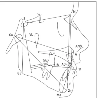
Cephalometric radiographs were obtained at the beginning of the study (T1) and after 12 months of observation (T2). They were scanned and digitized using Radiocef 2.0 Mem-ory Studio computer software (Floresta, Belo Horizonte/MG, Brazil). The customized ceph-alometric analysis used in the study is shown in Figures 2 and 3. A vertical reference line(VL) was constructed perpendicular to sella-nasion through sella to measure anteroposterior posi-tion of the first molars.14
To evaluate the error of the method, 10 pairs of radiographs were randomly selected and digitized by the same operator four times each. An intraclass correlation coefficient (ICC) was calculated to test intra-operator er-ror. Unpaired Student’s t test was used to check for homogeneity between groups for cephalo-metric values at T1.
A A
B
B
C
C
D
FIGURE 1 - Lateral views of a mandibular retrognathism Class II malocclusion subject.
FIGURE 2 - Customized cephalometric analysis: Linear measurements. Co-A: Maxillary length; Co-Gn: Mandibular length; AO’-BO’: Wits appraisal; LAFH: Distance from anterior nasion spine (ANS) to menton (Me), anterior facial height; S-Go: Distance from sella to gonion, posterior facial height; 1/NA (mm): Upper nasion/point A line; 1/NB (mm): Lower incisor-nasion/point B line; U6/VL: Distance from the most anterior point of the upper first molar to vertical line; DU6/VL: Distance from the most posterior point of the upper first molar to vertical line; L6/VL: Distance from the most posterior point of the lower first molar to vertical line.
FIGURE 3 - Customized cephalometric analysis: Angular measure-ments. SNA: Sella-nasion-point A angle; SNB: Sella-nasion-point B angle; SND: Sella-nasion-point D angle; ANB: Point A-nasion-point B angle; NA/APog: Angle formed by nasion-point A and pogonion-point A; GoGn/SN: Angle formed by gonion-gnathion and sella-nasion; SNGn: Sella-nasion-gnation angle; 1/NA (degree): Upper incisor-nasion/point A line; 1/NB (degree): Lower incisor-nasion/point B line; 1/GoGn: Angle formed by lower incisor and gonion-gnation; 1/1: Interincisor angle.
FIGURE 4 - Twin Block appliance used in this study.
S S
VL
Go
Go D6/
/6
6/ AO’-BO’
Me
Gn Gn
Pog B
D B /1 1/
A A
ANS
N N
and were instructed to wear it continuously ex-cept during sports, eating and oral hygiene. The initial construction bite was taken with approxi-mately 4 mm mandible protrusion. A time grid was filled in by the parents, and showed that the appliance had been worn on average for 18 hours and 50 minutes per day.
Descriptive statistics were used to deter-mine the mean and standard deviation of the linear and angular cephalometric values at T1 and T2 for both groups. Wilcoxon test was used to check for statistical differences between T1 and T2 in each group and Mann-Whitney test to evaluate changes resulting from treatment at the end of the observation period (T2) between groups. The confidence level of p<0.05 was used as reference for all statistical tests.
Results
The error of the method demonstrated ex-cellent operator reproducibility, showing ICC values higher than 0.773 for a confidence in-terval of 95%.
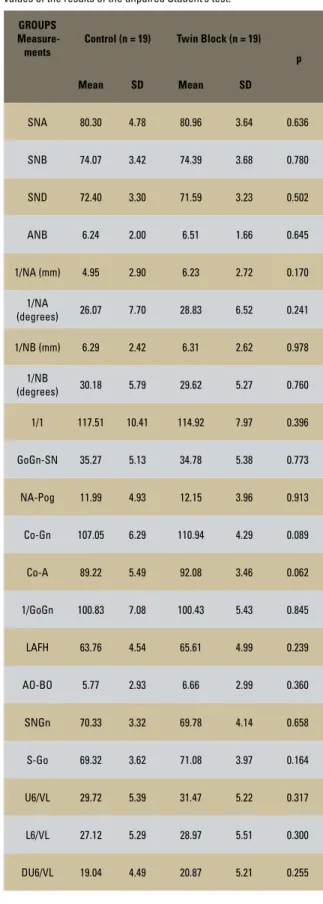
Means, standard deviations and results of the unpaired Student’s t test of the cephalomet-ric measurements at T1 are shown in Table 1. No statistically significant differences between groups were observed, thus confirming the sam-ple homogeneity at T1.
The molar relationship correction was at-tained in 15 of the 19 treated patients (80%), while the CG did not show any modification in Class II relationship.
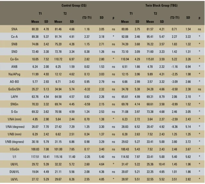
Table 2 shows initial and final cephalometric values of the CG and TBG as well as the results of the Wilcoxon test comparing each group from T1 to T2. Significant increases in mandibu-lar length (Co-Gn= +2.82 mm) and in maxilmandibu-lary length (Co-A= +2.37 mm) occurred in the CG, reflecting the maintenance of the Class II pat-tern with growth, confirmed by the unaltered ANB. The linear measurements for upper inci-sor (1/NA: p<0.30) and for upper and lower
TABLE 1 - Cephalometric measurements of CG and TBG at T1 and the p values of the results of the unpaired Student’s test.
GROUPS
Measure-ments
Control (n = 19) Twin Block (n = 19)
p
Mean SD Mean SD
SNA 80.30 4.78 80.96 3.64 0.636
SNB 74.07 3.42 74.39 3.68 0.780
SND 72.40 3.30 71.59 3.23 0.502
ANB 6.24 2.00 6.51 1.66 0.645
1/NA (mm) 4.95 2.90 6.23 2.72 0.170
1/NA
(degrees) 26.07 7.70 28.83 6.52 0.241
1/NB (mm) 6.29 2.42 6.31 2.62 0.978
1/NB
(degrees) 30.18 5.79 29.62 5.27 0.760
1/1 117.51 10.41 114.92 7.97 0.396
GoGn-SN 35.27 5.13 34.78 5.38 0.773
NA-Pog 11.99 4.93 12.15 3.96 0.913
Co-Gn 107.05 6.29 110.94 4.29 0.089
Co-A 89.22 5.49 92.08 3.46 0.062
1/GoGn 100.83 7.08 100.43 5.43 0.845
LAFH 63.76 4.54 65.61 4.99 0.239
AO-BO 5.77 2.93 6.66 2.99 0.360
SNGn 70.33 3.32 69.78 4.14 0.658
S-Go 69.32 3.62 71.08 3.97 0.164
U6/VL 29.72 5.39 31.47 5.22 0.317
L6/VL 27.12 5.29 28.97 5.51 0.300
first molars (U6/VL and DU6/VL: p<0.014 and L6/VL: p<0.04) also increased statistically. The treatment group showed significant changes in all the cephalometric measurements, except for SNA and GoGn/SN.
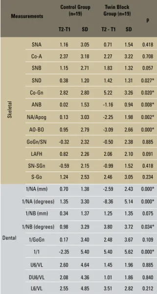
In the comparison between the TBG and CG at T2 (Table 3), the most important skeletal findings in the treated group were a statistically significant increase in mandibular length (Co-Gn) and in the
spatial position of the mandible related to the ante-rior cranial base (SND), and a significant reduction in the measurements related to maxillomandibular sagittal position (ANB, NA/APog = p<0.05, AO-BO = p<0.01). The treated group also showed sta-tistical changes on upper incisors position (1/NA mm and 1/NA degree) and on lower incisors tip-ping (1/NB degree), which resulted in significant reduction of interincisor angle (p<0.01).
TABLE 2 - Cephalometric measurements of the groups at T1 and T2 (n=38) (Wilcoxon test).
Control Group (CG) Twin Block Group (TBG)
T1 T2
(T2-T1) SD p
T1 T2
(T2-T1) SD p
Mean SD Mean SD Mean SD Mean SD
SNA 80.30 4.78 81.46 4.66 1.16 3.05 ns 80.86 3.75 81.57 4.21 0.71 1.54 ns
Co-A 89.38 5.37 91.74 4.91 2.37 3.18 * 92.08 3.46 95.41 5.47 2.27 3.22 *
SNB 74.06 3.42 75.20 4.26 1.15 2.71 ns 74.39 3.68 76.22 3.57 1.83 1.32 *
SND 72.40 3.30 72.78 3.34 0.38 1.26 ns 73.10 3.09 71.60 3.23 1.42 1.31 *
Co-Gn 10.05 7.52 110.72 6.97 2.82 2.80 * 110.94 4.29 115.81 3.59 5.22 3.26 *
ANB 6.24 2.00 6.25 1.59 0.02 1.53 ns 6.51 1.66 4.78 2.32 -1.16 0.94 *
Na/APog 11.99 4.93 12.12 4.62 0.13 3.03 ns 12.15 3.96 9.89 4.31 -2.25 1.98 *
AO-BO 5.77 2.93 6.71 3.43 0.95 2.79 ns 6.66 2.99 3.57 3.32 -3.09 2.66 *
GoGn/SN 35.27 5.13 34.94 5.74 -0.32 2.32 ns 34.78 5.38 34.28 4.66 -0.50 2.38 ns
LAFH 63.76 4.54 64.58 4.57 0.82 2.26 ns 65.61 4.99 69.31 8.79 2.06 2.10 *
SNGn 70.33 3.32 69.74 4.45 -0.59 2.15 ns 69.78 4.14 68.61 3.58 -0.99 1.52 *
S-Go 69.32 3.62 70.56 4.59 1.24 2.53 ns 71.08 3.97 73.38 4.68 2.46 3.05 *
1/NA (mm) 4.95 2.90 5.64 2.44 0.70 1.38 * 6.23 2.72 3.64 2.37 -2.59 2.43 *
1/NA (degrees) 26.07 7.70 27.42 7.29 1.35 3.30 ns 28.83 6.52 20.47 4.92 -8.36 5.14 *
1/NB (mm) 6.29 2.42 6.62 2.51 0.34 1.37 ns 6.28 2.63 7.52 2.43 1.25 1.35 *
1/NB (degrees) 30.18 5.79 31.15 6.86 0.98 3.29 ns 29.62 5.27 33.41 5.00 3.80 3.72 *
1/GoGn 100.83 7.08 101.00 7.65 0.17 3.40 ns 100.43 5.43 7.52 2.43 2.48 3.67 *
1/1 117.51 10.41 115.16 11.40 -2.35 5.40 ns 114.92 7.97 33.41 5.00 5.40 5.62 *
U6/VL 29.72 5.39 32.32 5.72 2.60 4.64 * 31.47 5.22 35.36 10.41 1.45 1.96 *
DU6/VL 19.04 4.49 21.11 5.56 2.08 4.36 ns 20.87 5.21 22.35 4.65 1.01 1.86 *
TABLE 3 - Comparison of CG and TBG mean changes between T1-T2 (Mann Whitney test).
Measurements
Control Group (n=19)
Twin Block Group (n=19)
p
T2-T1 SD T2 - T1 SD
Skeletal
SNA 1.16 3.05 0.71 1.54 0.418
Co-A 2.37 3.18 2.27 3.22 0.708
SNB 1.15 2.71 1.83 1.32 0.057
SND 0.38 1.20 1.42 1.31 0.027*
Co-Gn 2.82 2.80 5.22 3.26 0.020*
ANB 0.02 1.53 -1.16 0.94 0.008*
NA/Apog 0.13 3.03 -2.25 1.98 0.002*
AO-BO 0.95 2.79 -3.09 2.66 0.000*
GoGn/SN -0.32 2.32 -0.50 2.38 0.885
LAFH 0.82 2.26 2.06 2.10 0.091
SN-SGn -0.59 2.15 -0.99 1.52 0.418
S-Go 1.24 2.53 2.46 3.05 0.234
Dental
1/NA (mm) 0.70 1.38 -2.59 2.43 0.000*
1/NA (degrees) 1.35 3.30 -8.36 5.14 0.000*
1/NB (mm) 0.34 1.37 1.25 1.35 0.075
1/NB (degrees) 0.98 3.29 3.80 3.72 0.034*
1/GoGn 0.17 3.40 2.48 3.67 0.109
1/1 -2.35 5.40 5.40 5.62 0.000*
U6/VL 2.60 4.64 1.45 1.96 0.885
DU6/VL 2.08 4.36 1.01 1.86 0.840
L6/VL 2.55 4.85 3.51 2.82 0.212
disCussiOn
Clinically, 80% of the treated group have ben-efited from the correction of the Class II molar relationship, while no improvement was observed in the control group. As the children were treated before the pre-pubertal spurt, the stability of the correction needs to be checked later on.
The TB treatment did not result in inhibition of maxillary forward growth since no statistical
difference was observed in the TBG (Table 3). These results are in agreement with Lund and Sandler,7 while some authors8,9,10 have reported slight maxillary restraint suggesting a headgear effect. Change in the position of point A due to upper incisor retroclination may have hidden the effect upon maxilla in this study, affecting SNA and CO-A changes.
The TB appliance was found to have influ-enced mandibular anterior development when SND was used as reference (p<0.05). In con-trast, SNB angle changes did not reflect the same pattern, probably because point B was influenced by proclination of the lower inci-sors. Previous studies have reported an anterior mandibular displacement related to the anterior cranial base as a result of TB treatment, which means that the mandible was spatially altered in the anteroposterior plane resulting in a correc-tion of Class II malocclusion (Table 3).7,8,10,11,16
A significant increase in mandibular length (Co-Gn=2.4 mm) was observed as well as a sig-nificant reduction in intermaxillary sagittal dis-crepancy (ANB = -1.18°; NA/APog = -2.38°; AO-BO= -4.04 mm), which probably contrib-uted to the Class II correction (Table 3). An increase in total mandibular length was also found by Morris et al6 (3.7 mm in 9 months), Lund and Sandler7 (2.4 mm/year), Mills and McCulloch8 (4.2 mm/year), Toth and McNa-mara10 (3.0 mm in 16 months) and Trenouth11 (3.2 mm/year). These results suggest a response of mandibular growth increments, which may be in part justified by a reported good compli-ance, as the appliance is considered comfort-able and esthetic.
facial height has been attributed to a differ-ent appliance design that allows molar erup-tion.17 Although, molar disocclusion has been mentioned by Clark2 as an advantage of the ap-pliance (Figs 1B and 4C) which could be also observed in this sample at the end of the treat-ment and was considered important in influenc-ing mandibular anterior growth.
The first molars did not show significant linear changes when measured to VL (p>0.05) (Table 3). The upper incisors showed signifi-cant retroclination (1/NA= 3.29 mm, 9.71°); the lower incisors, proclination (1/NB= 2.82°) and the interincisal angle, an increase (1/1= 7.75°) and these results are in accordance with other authors7,8,10,11 who have reported simi-lar findings. The effect on the upper incisors can be mainly attributed to the Hawley arch (Fig 4B) which transmitted a reaction force to the upper incisors. Koroluk et al18 verified that overjet reduction is favorable in early treat-ment, considering the high trauma incidence in preadolescents with a Class II Division 1 mal-occlusion.3 The lower incisor movement may have been due to anchorage loss in response to keeping the mandible in a protrusive posi-tion. This effect may be unfavorable in patients where the incisors are proclined before treat-ment. Therefore, this approach may not be
indicated in such cases or an additional stage of treatment may be required to upright the lower incisors.3
COnClusiOn
The present study assessed changes after a 12-month treatment with the Twin Block appli-ance, compared to a control group. The analysis of the results leads to the following conclusions: 1) Eighty percent of the patients treated with the Twin Block appliance attained Class I relationship.
2) A significant improvement occurred in the total mandibular length and anteroposte-rior relationship.
3) Significant retroclination and proclination was observed in the upper and lower inci-sors, respectively, leading to a decrease in the overjet.
4) No statistically significant change was found in molar position in the treated group. 5) No effect was observed in the position of
the maxilla.
ACKnOWledGeMents
1. Shaw WC. The inluence of children’s dentofacial appearance on their social attractiveness as judged by peers and lay adults. Am J Orthod. 1981;79(4):399-415.
2. Clark WJ. The twin block technique. A functional orthopedic appliance system. Am J Orthod Dentofacial Orthop. 1988;93(1):1-18.
3. Tulloch JF, Phillips C, Profit WR. Beneit of early Class II treatment: progress report of a two-phase randomized clinical trial. Am J Orthod Dentofacial Orthop. 1998;113(1):62-72, quiz 73-4.
4. Dolce C, McGorray SP, Brazeau L, King GJ, Wheeler TT. Timing of Class II treatment: skeletal changes comparing 1-phase and 2-phase treatment. Am J Orthod Dentofacial Orthop. 2007;132(4):481-9.
5. Voudouris JC, Kuftinec MM. Improved clinical use of Twin-block and Herbst as a result of radiating viscoelastic tissue forces on the condyle and fossa in treatment and long-term retention: growth relativity. Am J Orthod Dentofacial Orthop. 2000;117(3):247-66.
6. Morris DO, Illing HM, Lee RT. A prospective evaluation of Bass, Bionator and Twin Block appliances. Part II: The soft tissues. Eur J Orthod. 1998;20(6):663-84.
7. Lund DI, Sandler PJ. The effects of Twin Blocks: a prospective controlled study. Am J Orthod Dentofacial Orthop. 1998;113(1):104-10.
8. Mills CM, McCulloch KJ. Treatment effects of the Twin Block appliance: a cephalometric study. Am J Orthod Dentofacial Orthop. 1998;114(1):15-24.
9. Parkin NA, McKeown HF, Sandler PJ. Comparison of 2 modiications of the Twin-Block appliance in matched Class II samples. Am J Orthod Dentofacial Orthop. 2001;119(6):572-7. RefeRenCes
10. Toth LR, McNamara JA Jr. Treatment effects produced by the Twin-Block appliance and the FR-2 appliance of Fränkel compared with an untreated Class II sample. Am J Orthod Dentofacial Orthop. 1999 Dec;116(6):597-609.
11. Trenouth MJ. Cephalometric evaluation of the Twin-Block appliance in the treatment of Class II division 1 malocclusion with matched normative growth data. Am J Orthod Dentofacial Orthop. 2000;117(1):54-9.
12. Fishman LS Radiographic evaluation of skeletal maturation. A clinically oriented method based on hand-wrist ilms. Angle Orthod. 1982;52(2):88-112.
13. Hassel B, Farman AG. Skeletal maturation evaluation using cervical vertebrae. Am J Orthod Dentofacial Orthop. 1995;107(1):58-66.
14. Brunharo IP, Quintão CA. O aparelho Twin Block: técnica de confecção e aplicação clínica. Rev Bras Odontol. 2001;58(6):373-7.
15. Quintão C, Helena I, Brunharo VP, Menezes RC, Almeida MA. Soft tissue facial proile changes following functional appliance therapy. Eur J Orthod. 2006 Feb;28(1):35-41. Epub 2005 Aug 19.
16. Caldwell S, Cook P. Predicting the outcome of Twin Block functional appliance treatment: a prospective study. Eur J Orthod. 1999;21(5):533-9.
17. Vig PS, Vig KW. Hybrid appliances: a component approach to dentofacial orthopedics. Am J Orthod Dentofacial Orthop. 1986;90(4):273-85.
18. Koroluk LD, Tulloch JF, Phillips C. Incisor trauma and early treatment for Class II division 1 malocclusion. Am J Orthod Dentofacial Orthop. 2003;123(2):117-25; discussion 125-6.
Contact address
Ione Portella Brunharo