Lithograph-Moulded Poly-L-co-D,L Lactide Porous Membranes for Osteoblastic Culture
Andre D. Messiasa,b*, Carolina Lucchesic, Débora C. Coraça-Huberb,d,
Aristides Pavani Filhoe, Eliana A. R. Dueka,b
aDepartment of Material Engineering, Faculty of Mechanical Engineering, State University of Campinas – UNICAMP, Campinas, SP, Brazil bLaboratory of Biomaterials, Faculty of Medical Sciences and Health, Pontifical Catholic University of São Paulo – PUC-SP, Sorocaba, SP, Brazil
cWyss Institute, Harvard Medical School, Boston, MA, USA
dExperimental Orthopedics, Department of Orthopedic Surgery, Medical University Innsbruck, Innsbruck, Austria
eLaboratory of Microsystems Technology, Department of Information Technology, Renato Archer Research Center, CTI, Campinas, SP, Brazil
Received: February 10, 2013; Revised: August 16, 2013
Pore size, shape, wall morphology, porosity, and interconnectivity are important characteristics of the scaffolds. Lithography is a manufacturing technique that allows the production of tridimensional scaffolds with a controllable and reproducible inner architecture. The aim of this study was to use lithography to create a poly-L-co-D,L lactide (PLDLA) scaffold with symmetrical pore size and distribution, and to evaluate its biocompatibility with osteoblasts in vitro. Lithographic moulds were used to produce porous PLDLA membranes by a casting procedure. Osteoblasts were removed from calvarial bones and seeded onto porous and smooth PLDLA membranes after which cell viability and adhesion assays, cytochemical analysis and scanning electron microscopy were used to characterize the cells. Cell viability and adhesion assays, cytochemical analysis, and scanning electron microscopy were carried out. Cell viability was similar on porous and smooth PLDLA membranes but higher than on a polystyrene substrate (positive control). Although osteoblasts adhered to the surface of all the materials tested, cell adhesion to lithographed PLDLA was greater than to smooth PLDLA membranes. In conclusion, osteoblasts interacted well with PLDLA membranes, as shown by the viability and adhesion assays and by the enhanced collagen production.
Keywords: bone tissue engineering, cell adhesion, lithography, osteoblastic cells, poly-L-co-D,L lactide, porosity, viability
1. Introduction
In tissue engineering, cells cultured on biomaterial surfaces provide a simple method for screening the biocompatibility of materials prior to testing in vivo testing1,2. Pore size, shape, porosity and interconnectivity are
important characteristics of scaffolds. These characteristics are important for cell attachment, growth and new tissue formation, diffusion of nutrients and metabolic waste products to and from the implant, and angiogenesis3.
Bone tissue engineering is based on studies of bone cells cultivated on biomaterials in vitro4. Mesenchymal stem
cells seeded on scaffolds can dramatically accelerate bone regeneration when compared to scaffolds without cells5.
Osteoblasts synthesize collagen type I that is involved in bone mineralization and this collagen production is
inluenced by the biomaterial on which these cells are
seeded. Consequently, the compatibility of biomaterials can be partially characterized by the amount of minerals produced as the cells grow on the polymer surface6.
The quality of the pores in scaffolds used for bone tissue engineering is important, with pores >100 µm in diameter being recommended for biomaterials used as bone substitutes. Soluble particles, including salts and carbohydrates, or hydrophobic systems are used to produce pores in a polymeric matrix but allow only partial control of porosity. In addition, porogen waste and solvent residues can be toxic when the scaffold is applied in vitro or implanted in vivo7. An alternative to porogen substances is
the lithography, a controlled manufacturing technique that allows the production of tridimensional (3D) scaffolds with a controllable and reproducible inner architecture8.
Patterned surfaces produced by lithographic techniques show improved adhesion of rat mesenchymal stem cells and greater proliferation on scaffolds containing hyaluronic acid biofunctionalized with peptides9; the proliferation
of osteoblast-like MG-63 cells and rat mesenchymal stem cells on Bioglass®-based glass-ceramic scaffolds is also enhanced10. A micropatterned surface facilitates
the alignment, elongation and colonization of human
osteoblastic-like cells11 and rat mesenchymal stem
cells12in vitro. The use of lithography to modify the
surface morphology of titanium substrates enhances the osteointegration between implants and bone tissue13.
Poly-L-lactide can be used as a scaffold for bone tissue engineering14,15. Poly-L-lactide (PLLA) is a biomaterial
that shows increasing crystallinity during degradation16.
The co-polymer poly-L-co-D,L lactide (PLDLA) has similar mechanical features to poly-L-lactide without the inconvenience of long degradation and high crystallinity. Moreover, PLDLA is a polyester that can be hydrolyzed into lactic acid monomers and eliminated through the tricarboxylic acid cycle17. Finally, PLDLA-based
bioresorbable device are entirely replaced by cells and extracellular matrix18.
The aim of this study was to use lithography to create a PLDLA scaffold with a symmetrical pore size and distribution and evaluate its biocompatibility with osteoblasts in vitro.
2. Material and Methods
2.1.
Mould fabrication using lithography
The mould was designed using the computer-aided design (CAD) software AutoCAD® (Autodesk Inc. San Rafael, CA, USA.) in drawing exchange format (DXF). The design was a bidimensional (2D) hexagonal array of closed polylines with 100 µm between two opposed vertices. The distance between two contiguous pillars was 200 µm. The design was converted to GDSII stream format
(industry standard database binary ile format) to generate a mask. The chrome dark ield glass mask was produced
using electron beam lithography equipment (Electron Beam Microfabricator EBMF 10.5 Leica Lithograph, Cambridge, UK) and a positive tone resist EBR9 (Toray Industries Inc., Tokyo, Japan) process with a resolution of±0.1 µm. A 1.52-mm-thick borosilicate glass plate was used as the mould substrate. The plate was immersed in sulfur acid for 7 min at 60 °C followed by spraying with deionized water (DI). The samples were immersed in a heated ultrasonic detergent
bath for 7 min followed by a DI spray and immersion in an isopropyl alcohol dehydration bath for 7 min, after which they were dried in isopropyl alcohol vapor. The substrate was covered with SU-8 50 resist (Microchem, Newton, MA, USA) by spinning at 1,500 rpm for 40 s to yield a 100 µm
thick ilm and then heated on a hot plate at 65 °C for 5 min
and cured at 95 °C for 15 min. Subsequently, the substrate was exposed to ultraviolet light using a G-line mask copier
(Tamarack model 155, Tamarack Scientiic Co. Inc., Corona,
CA, USA.) at an energy of 350 mJ/cm2 after which the
exposed ilm was immersed in SU-8 developer for 4 min.
The resulting SU-8 structures were hardened by curing on a hot plate at 150 °C for 5 min. The lithographic process yielded an array of pillars 100 µm in height. The resulting
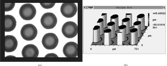
mould was characterized by using an optical proilometer (Zygo NewView 5032, Zygo Corporation, Middleield, CT,
USA; Figure 1).
2.2.
Membrane preparation
Poly-L-co-D,L lactide (PLDLA; Mw=205,000 Da) was prepared by ring-opening polymerization, as previously described by Motta and Duek19 using of L-lactide and
D,L-lactide monomers (70:30, w/w) (Purac Biomaterials, Schiedam, The Netherlands). The membranes were obtained by casting. The co-polymer was dissolved in 5 % (w/v) chloroform (Sigma-Aldrich) for 2 h with mixing and poured into the mould created with the lithograph. After the solvent evaporation, the membrane was removed manually from the plate under sterile conditions. Smooth membranes were used as a control to determine whether the scaffold morphology
inluenced the cellular responses. The membranes were
sterilized in a sterile laminar flow biohazard cabinet (Pachane, Piracicaba, SP, Brazil) with ultraviolet irradiation for 30 min and then placed in 96-well plates for cell culture.
2.3.
Osteoblast isolation and culture
Osteoblasts cells were removed from calvarial bones of 20-day old Wistar rats, as described by Yamamoto et al.20.
The rats were euthanized by cervical displacement followed by decapitation. The calvaria were removed, immersed in
Dulbecco’s Modiied Eagle’s Medium (DMEM) containing
Figure 1. Microscopic view (a) and oblique proilometry plot (b).
gentamicin (150 µg.mL–1) and amphotericin B (15 µg.mL–1)
and subsequently fragmented (about 1 cm2) and subjected to
enzymatic digestion with type I A collagenase (1 mg.mL–1;
Sigma-Aldrich) in DMEM for 2, 4 and 6 h at 37 °C. The cell suspensions were centrifuged three times at 250×g
for 10 min after each interval. Viable cells were quantiied
by Trypan blue dye (Sigma-Aldrich) exclusion in a hemocytometer. The cells (105 cells.mL–1) were plated in
polystyrene tissue culture lasks (Techno Plastic Products)
and grown at 37 °C in a 5% CO2 atmosphere for 4-5 passages
prior to testing. Samples of PLDLA membranes measuring 6 mm in diameter were placed in each well of a 96-well plate (Techno Plastic Products) and DMEM was added. The plates were incubated for 24 h at 37 °C in a 5% CO2 atmosphere
before cell seeding. The osteogenic medium used in the cell experiments consisted of DMEM containing 10% (v/v) fetal bovine serum (FBS; Nutricell – Nutrientes Celulares), L-ascorbic acid (50 µg.ml–1), 100 nM dexamethasone,
10 mM β-glycerophosphate, 0.7 mM calcium chloride, gentamicin (50 µg.mL–1) and amphotericin B (5 µg.mL–1)
(all from Sigma-Aldrich)21,22.
2.4.
Cell viability and adhesion assays
The cell viability and adhesion assays were based on reports by Mosmann23, Lucchesi et al.24 and Uzumaki25.
Polystyrene (the culture plate itself) was used as a positive control in both assays. Phenol (1% solution) was used as a negative control for the cell viability assay while polytetrafluoroethylene (PTFE) dishes were used as a negative control for adhesion26. Six samples of each
membrane type and the controls were tested (n=6). A 100 µL aliquot containing 2×10⁵ cells.mL–1 was seeded
onto membranes and controls in DMEM containing 10% FBS. Osteoblast viability and adhesion were assessed after 24 h and 2 h, respectively. Subsequently, the medium was removed and the samples were washed 3-4 times with phosphate buffered saline (PBS) after which 100 µL of a solution containing 3-(4,5-dimethylthiazol-2-yl)-2,5-diphenyl-tetrazolium bromide (MTT, 0.5 mg.mL–1;
Sigma-Aldrich) in DMEM was added to each well followed by 4 h of incubation (Shel Lab 5212, Cornelius, NC, USA). The MTT solution was subsequently replaced with 200 µL of dimethyl sulfoxide (DMSO; Merck) and 25 µL of glycine/ Sorensen buffer (Nutricell – Nutrientes Celulares). The resulting absorbance was determined at 570 nm using a microplate reader (Elx-800-UV, Bio-Tek Instruments, Winooski, VT, USA).
2.5.
Cytochemical analysis
A 100 µL aliquot of osteoblasts (2×10⁴ cells.mL-1)
was seeded on porous and smooth PLDLA membranes and cultured with osteogenic DMEM. The plates were incubated in a 5% CO2 atmosphere at 37 °C and the culture
medium was replaced every two days. After 6 h and 48 h,
and 7, 14 and 21 days in culture the samples were ixed
with formaldehyde, dehydrated in ethanol and stained with toluidine blue (TB), a dye that bind to basic anionic groups, xylidine ponceau (XP), a dye that binds to cationic groups, von Kossa (VK), which stains mineralization nodules, and picrosirius (PS) which stains collagen type I/III. The
samples were observed with a light microscope (Eclipse E800, Nikon Americas Inc., Melville, NY, USA) and images were captured with an FDX-35 camera (Nikon) attached to the microscope.
2.6.
Scanning electron microscopy (SEM)
The culture conditions (cell number, culture medium and length of culture) were the same as used for the cytochemical analysis. At the end of the culture period, the samples
were ixed for 30 min at room temperature in in ixative
containing 2.5% paraformaldehyde, 2.5% glutaraldehyde, 1% picric acid and 1% tannic acid dissolved in 0.1 M PBS,
pH 7.4. Subsequently, the samples were post-ixed in 1 %
osmium tetroxide for 15 min in the dark, washed in water, dehydrated with ethanol (all reagents from Sigma-Aldrich, St. Louis, MO, USA), critical point dried (Balzers CDT 030, Balzers Inc., Elgin, IL, USA) and coated with gold in a sputter coater (Balzers CDT 050, Balzers Inc., Elgin, IL, USA). The coated specimens were examined with a JEOL JXA-840A scanning electron microscope (JEOL Ltd., Peabody, MA, USA).
2.7.
Statistical analysis
Numerical results are reported as the mean ± standard deviation. Cell viability and adhesion were analyzed statistically with one-way analysis of variance (ANOVA) followed by the Tukey test for post hoc analyses. A value of p<0.05 indicated signiicance. All data analyses were done
using BioEstat version 5.0.
3. Results
3.1.
Cell viability and adhesion
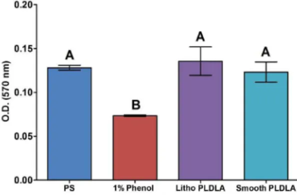
Osteoblast viability was similar in porous and smooth
PLDLA membranes and not signiicantly different from that
seen with polystyrene (Figure 2). In contrast, cell adhesion to lithographed PLDLA membranes was greater than for smooth PLDLA membranes (Figure 3).
3.2.
Cell morphology
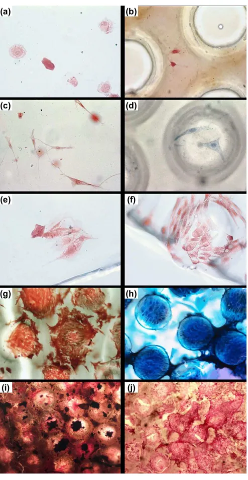
Osteoblasts adhered to the surface of all materials tested. The morphology of these cells on the surfaces of porous and smooth polymeric membranes was examined. On the 14th day and 21st day after seeding, some cells formed multiple layers suggestive of cellular proliferation and synthetic activity. The cell nuclei and cell borders were
dificult to visualize in these layers. Six hours after seeding,
the cells had not yet completed the spreading process because their morphology was spherical and sometimes
slightly lat (Figure 4a and b; Figure 5a). After 48 h the
cells had adhered and showed long, thin protrusions but were still separated from each other (Figure 4c and d). After
7 days, the cells were lat, long and spindle-shaped and
were connected to each other by cellular protrusions such
as ilopodia and lamellipodia (Figure 5b and c); there was
also marked intercellular contact indicative of proliferation (Figure 4e and f). After 14 days, several pores colonized by a cellular monolayer were observed on the lithographed membrane. Cells outside these pores maintained contact with cells inside the pores (Figure 4g and h; Figure 5d). The cell number after 14 days was greater than at previous times. By 21 days post-seeding, the cells formed very tight, multilayered structures that made it difficult to visualize nuclei and the areas inside and outside the pores (Figure 4i and j; Figure 5g and h). The presence of collagen nets showed that the cells were able to synthesize extracellular matrix (Figure 4j) and the occurrence of mineralized nodules indicated that the cells were depositing calcium phosphate crystals (Figure 4i). Some cells stretched from the bottom of the pores to their edge (Figure 5e, f).
4. Discussion
In this study, the interaction between osteoblasts and porous and smooth PLDLA membranes was evaluated. A computer designed metallic mould produced by lithography
was used to obtain porous membranes. The MTT assay was used to assess PLDLA cytotoxicity (based on mitochondrial activity) and to measure cell adhesion. Morphological alterations were assessed by light microscopy after selective staining and SEM was used to examine ultrastructural features.
The measurement of mitochondrial activity is a suitable criterion for assessing cell viability since toxic substances affect not only the molecular structure but also several cellular functions24. PLDLA (70:30) has already been
tested with regards to cytotoxicity, and the MTT, agar and
ilter diffusion assays have shown good cytocompatibility
for this copolymer27. In addition, the mortality of human
osteoblasts in extracts containing PLLA degradation products is ~30%, indicating low cytotoxicity28. These
results support the low cytotoxicity reported here. A number of studies that have investigated the use of PLLA in tissue engineering in vivo have reported satisfactory results for bone regeneration29,30, guided
tissue regeneration31, nerve peripheral regeneration32, and
cartilaginous tissue33. PLDLA copolymer is compatible
with34 and suitable for use as a bone graft substitute35-38,
meniscus replacement39,40, suture cords/threads41 and axon
regeneration42.
Cellular adhesion to biomaterials is extremely important in material sciences. As shown here, more osteoblasts adhered to the surface of porous PLDLA than to smooth PLDLA. Once adhered to the substrate, the cells migrate
and proliferate or show speciic physiological activities,
such as the production of extracellular matrix43,44. Wu et al.45
reported enhanced osteoblast adhesion to scaffolds with larger pores (300-500 µm) compared to those with smaller pores (150-180 µm), and adhesion to the latter was greater than to smooth surfaces. However, other cellular responses such as proliferation and osteogenic function are not
signiicantly inluenced by pore size. Micro- and nano-scale structures on PLLA and polystyrene improve the eficiency
of adhesion when compared to smooth substrates11. Porous
PLDLA membranes (50-70 µm) enhanced mesenchymal cell proliferation, differentiation and activity when compared to smooth membranes46. Nevertheless, the inluence of porosity
and pore size is a controversial. Whiston et al.22 showed
that the surface relief (micro- and nano-scale structures) of PLLA did not enhance the metabolic activities of osteoblasts after two days, as assessed by the MTT test11. Bet et al.47,
reported that ibroblast adhesion to PLLA scaffolds was low
and there was no difference in adhesion among membranes containing pores of different sizes. Pore quality and quantity
do not inluence the proliferation of osteogenic cells from
rat calvaria cultivated on PLGA (75:25) for different periods of time48. Thus, as other studies have demonstrated,
the interaction between osteoblasts and materials with variable porosities and pore sizes does not affect the cellular response49,50. Consequently, different cell types respond
differently to the substrate surface topography11.
The light microscopy and SEM indings described
here indicate that osteoblasts adhered to and spread over smooth and porous membranes, in addition to showing the spindle-shaped and polyhedral cells characteristic of osteoblasts. None of the images showed cells bridging or
covering the pores; rather, they were frequently observed within the pores (lining the bottom or wall). Pores generally increase the surface areas of porous membranes compared to smooth membranes and it was therefore expected that cell adhesion would be greater on porous membranes. Several studies have used SEM to demonstrate the extensive
colonization of biomaterials51-53. Although these studies
have used different cell types, culture conditions and
polymeric substrates, in all cases conluent monolayers with poorly deined cellular limits form a continuous cellular
mat covering the scaffold surface51-53. In contrast to these
indings, as shown here, instead of a conluent layer of cells,
large groups of cells growing separately in various areas of the membranes were observed, especially 14 and 21 days after seeding. Light and scanning electron microscopy revealed a non-homogeneous cell distribution on the membrane surfaces, with osteoblasts always concentrated
in small, condensed groups; this inding agrees with the
observation that cellular aggregation is an important step in
ossiication54. Indeed, membrane pores may favor cellular
aggregation. Substrates with low capability to stimulate adhesion may be able to sustain cellular adhesion55. In
addition, the differentiation and synthesis of extracellular matrix can be stimulated by materials with low adhesion and proliferation rates8,12,13,45. As shown here, cells that adhered
to the membranes were capable of producing collagen. The micropatterned silica films were capable of inducing guided osteoblastic cell adhesion, spreading and propagation56. Isotropic and anisotropic surfaces changes
cell-material and cell-cell interactions57. Therefore, the
surface topography can modulate the way the cells adhere to57 and proliferate on58 the material.
Pelaez-Vargas et al.11, reported that the surface
microtexture modiied cell morphology and spreading,
which could influence important factors such as cell alignment, migration, implant surface colonization, and
function. This suggests that the surface topography and pore uniformity may control cells responses in a different manner than the increased porosity offered by alternative fabrication techniques such as solvent casting59.
5. Conclusion
Based on the results described here, we conclude that osteoblasts interact well with PLDLA membranes. These membranes can sustain adhesion and maintain viable cells, as shown by the ability of cells to produce collagen. PLDLA membranes represent a suitable biomaterial for cultivating osteoblasts and their potential usefulness in vivo deserves further investigation. An increase in the porosity of PLDLA can enhance cellular adhesion.
Acknowledgements
The authors thank the technicians of the Faculty of Mechanical Engineering and of the Electron Microscopy Laboratory of the Institute of Biology, UNICAMP, for help in this investigation, and Dr. Stephen Hyslop for the English review of the manuscript. This work was supported
by FAPESP. The authors have no conlicts of interest with
this work.
8. Lee SJ, Kang HW, Park JK, Rhie JW, Hahn SK and Cho DW. Application of microstereolithography in the development of three-dimensional cartilage regeneration scaffolds. Biomedical
Microdevices. 2008; 10(2):233-241. PMid:17885804. http://
dx.doi.org/10.1007/s10544-007-9129-4
9. Mendes AC, Smith KH, Tejeda-Montes E, Engel E, Reis RL, Azevedo HS et al. Co-assembled and microfabricated bioactive membranes. Advanced Functional Materials. 2013; 23(4):430-438. http://dx.doi.org/10.1002/adfm.201201065
10. Detsch R, Guillon O, Wondraczek L, Boccaccini AR. Initial attachment of rMSC and MG-63 cells on Patterned Bioglass® substrates. Advanced Engineering
Materials. 2012;14(3):B38-B44. http://dx.doi.org/10.1002/
adem.201180068
11. Pelaez-Vargas A, Gallego-Perez D, Carvalho A, Fernandes MH, Hansford DJ and Monteiro FJ. Effects of density of anisotropic microstamped silica thin films on guided bone tissue regeneration – In vitro study. Journal of Biomedical Materials
Research - Part B Applied Biomaterials. 2013; 101(5):762-769.
PMid:23359600. http://dx.doi.org/10.1002/jbm.b.32879
12. Wang P-Y, Li WT, Yu J and Tsai WB. Modulation of osteogenic, adipogenic and myogenic differentiation of mesenchymal stem cells by submicron grooved topography. Journal of Materials
Science: Materials in Medicine. 2012; 23(12):3015-3028.
PMid:22903603. http://dx.doi.org/10.1007/s10856-012-4748-6
13. Prodanov L, Lamers E, Domanski M, Luttge R, Jansen JA and Walboomers XF. The effect of nanometric surface texture on bone contact to titanium implants in rabbit tibia.
Biomaterials. 2013; 34(12):2920-2927. PMid:23380354. http://
dx.doi.org/10.1016/j.biomaterials.2013.01.027
14. Cai Y-Z, Zhang G-R, Wang L-L, Jiang Y-Z, Ouyang H-W and Zou X-H. Novel biodegradable three-dimensional macroporous scaffold using aligned electrospun nanoibrous yarns for bone tissue engineering. Journal of Biomedical Materials Research
Part A. 2012; 100(5):1187-1194. PMid:22345081. http://
dx.doi.org/10.1002/jbm.a.34063
References
1. Beresford JN, Graves SE and Smoothy CA. Formation of mineralized nodules by bone derived cells in vitro: a model of bone formation? American Journal of Medical
Genetics. 1993; 45(2):163-178. PMid:8456798. http://dx.doi.
org/10.1002/ajmg.1320450205
2. Elgendy HM, Norman ME, Keaton AR and Laurencin CT. Osteoblast-like cell (MC3T3-E1) proliferation on b i o e r o d i b l e p o l y m e r s : a n a p p r o a c h t o w a r d s t h e development of a bone-bioerodible polymer composite material. Biomaterials. 1993; 14(4):263-269. http://dx.doi. org/10.1016/0142-9612(93)90116-J
3. Tang ZG and Hunt JA. The effect of PLGA doping of polycaprolactone ilms on the control of osteoblast adhesion and proliferation in vitro. Biomaterials. 2006; 27(25):4409-4418. PMid:16677705. http://dx.doi.org/10.1016/j. biomaterials.2006.04.009
4. S e r v i c e R F . T i s s u e e n g i n e e r s b u i l d n e w b o n e .
Science. 2000; 289(5484):1498-1500. PMid:10991738. http://
dx.doi.org/10.1126/science.289.5484.1498
5. Wang H, Li Y, Zuo Y, Li J, Ma S and Cheng L. Biocompatibility and osteogenesis of biomimetic nano-hydroxyapatite/ polyamide composite scaffolds for bone tissue engineering.
Biomaterials. 2007; 28(22):3338-3348. PMid:17481726. http://
dx.doi.org/10.1016/j.biomaterials.2007.04.014
6. Wan Y, Wang Y, Liu Z, Qu X, Han B, Bei J et al. Adhesion and proliferation of OCT-1 osteoblast-like cells on micro- and nano-scale topography structured poly(L-lactide).
Biomaterials. 2005; 26(21):4453-4459. PMid:15701374. http://
dx.doi.org/10.1016/j.biomaterials.2004.11.016
7. Sarazin P, Roy X and Favis BD. Controlled preparation and properties of porous poly(L-lactide) obtained from a co-continuous blend of two biodegradable polymers.
Biomaterials. 2004; 25(28):5965-5978. PMid:15183611. http://
15. Ciapetti G, Granchi D, Devescovi V, Baglio SR, Leonardi E, Martini D et al. Enhancing osteoconduction of PLLA-based nanocomposite scaffolds for bone regeneration using different biomimetic signals to MSCs. International
Journal of Molecular Sciences. 2012; 13(2):2439-2458.
PMid:22408463 PMCid:PMC3292032. http://dx.doi. org/10.3390/ijms13022439
16. Motta A and Duek E. Síntese, caracterização e degradação in
vitro do poli(L-ácido latico). Polímeros. 2006; 16(1):26-32.
http://dx.doi.org/10.1590/S0104-14282006000100008
17. Agrawal CM and Ray RB. Biodegradable polymeric scaffolds for musculoskeletal tissue engineering. Journal of
Biomedical Materials Research. 2001; 55(2):141-150. http://
dx.doi.org/10.1002/1097-4636(200105)55:2<141::AID-JBM1000>3.0.CO;2-J
18. Peters M and Mooney D. Synthetic extracellular matrices for cell transplantation. In: Liu D and Dixit V. Porous materials for tissue engineering. Materials Science Forum. Einield: Trans Tech Publication; 1997. v. 250, p. 43-52.
19. Motta A and Duek E. Síntese e caracterização do copolímero poli(L-co-D,L-ácido lático). Polímeros. 2007; 17(2):123-129. http://dx.doi.org/10.1590/S0104-14282007000200011
20. Yamamoto N, Furuya K and Hanada K. Progressive development of the osteoblast phenotype during differentiation of osteoprogenitor cells derived from fetal rat calvaria: model
for in vitro bone formation. Biological and Pharmaceutical
Bulletin. 2002; 25(4):509-515. http://dx.doi.org/10.1248/
bpb.25.509
21. Moreira PL, An YH, Santos AR Jr and Genari SC. In vitro
analysis of anionic collagen scaffolds for bone repair.
Journal of Biomedical Materials Research: Part B, Applied
Biomaterials. 2004; 71(2):229-237. PMid:15386402. http:// dx.doi.org/10.1002/jbm.b.30026
22. Whiston SW, Whitson MA, Bowers DE Jr and Falk MC. Factors inluencing synthesis and mineralization of bone matrix from fetal bovine bone cells grown in vitro. Journal of Bone and
Mineral Research. 1992; 7(7):727-741.
23. Mosmann T. Rapid colorimetric assay for cellular growth and survival: application to proliferation and cytotoxicity assays.
Journal of Immunological Methods. 1983; 65(1-2):55-63.
http://dx.doi.org/10.1016/0022-1759(83)90303-4
24. Lucchesi C, Ferreira B, Duek E, Santos A and Joazeiro P. Increased response of Vero cells to PHBV matrices treated by plasma. Journal of Materials Science: Materials in Medicine. 2008; 19(2):635-643. PMid:17619989. http://dx.doi. org/10.1007/s10856-007-0169-3
25. Uzumaki ET, Lambert CS, Santos AR Jr and Zavaglia CAC. Surface properties and cell behaviour of diamond-like carbon coatings produced by plasma immersion. Thin Solid
Films. 2006; 515(1):293-300. http://dx.doi.org/10.1016/j.
tsf.2005.12.081
26. International Organization for Standardization - ISO.
Biological evaluation of medical devices. Part 5: Tests for
cytotoxicity: in vitro methods. ISO; 1992.
27. Ignatius AA and Claes LE. In vitro biocompatibility of bioresorbable polymers: poly(L, DL-lactide) and poly(L-lactide-co-glycolide). Biomaterials. 1996; 17(8):831-839. http://dx.doi.org/10.1016/0142-9612(96)81421-9
28. Marques AP, Cruz HR, Coutinho OP and Reis RL. Effect of starch-based biomaterials on the in vitro proliferation and viability of osteoblast-like cells. Journal of Materials Science: Materials in Medicine. 2005; 16(9):833-842. PMid:16167112. http://dx.doi.org/10.1007/s10856-005-3580-7
29. Coraça DC, Duek EA, Padovani CA and Camilli JA. Osteointegration of poly(L: -lactic acid)PLLA and poly(L: -lactic acid)PLLA/poly(ethylene oxide)PEO implants in rat tibiae. Journal of Materials Science: Materials in Medicine. 2008; 19(7):2699-2704. PMid:18283533. http:// dx.doi.org/10.1007/s10856-008-3397-2
30. Coraça-Huber DC, Duek EA, Etchebehere M, Magna LA and Amstalden EM. The use of vancomycin-loaded poly-l-lactic acid and poly-ethylene oxide microspheres for bone repair: an in vivo study. Clinics. 2012; 67(7):793-798. http://dx.doi. org/10.6061/clinics/2012(07)15
31. Ku Y, Shim IK, Lee JY, Park YJ, Rhee SH, Nam SH et al. Chitosan/poly(L-lactic acid) multilayered membrane for guided tissue regeneration. Journal of Biomedical Materials Research: Part A. 2009; 90(3):766-772. PMid:18615563. http://dx.doi. org/10.1002/jbm.a.31846
32. Pierucci A, De Duek EA and De Oliveira AL. Peripheral nerve regeneration through biodegradable conduits prepared using solvent evaporation. Tissue Engineering: Part A. 2008; 14(5):595-606. PMid:18399734. http://dx.doi. org/10.1089/tea.2007.0271
33. Gong Y, Ma Z, Zhou Q, Li J, Gao C and Shen J. Poly(lactic acid) scaffold fabricated by gelatin particle leaching has good biocompatibility for chondrogenesis. Journal of Biomaterials
Science: Polymer Edition. 2008; 19(2):207-221. PMid:18237493.
http://dx.doi.org/10.1163/156856208783432453
34. Barauna G, Coraça-Huber DC and Duek EAR. In vitro
degradation of Poly-L-co-D, L-lactic acid membranes.
Materials Research. 2013;16(1):221-226. http://dx.doi.
org/10.1590/S1516-14392012005000154
35. Coimbra ME, Elias CN and Coelho PG. In vitro degradation of poly-L-D-lactic acid (PLDLA) pellets and powder used as synthetic alloplasts for bone grafting. Journal of Materials
Science: Materials in Medicine. 2008; 19(10):3227-3234.
PMid:18454304. http://dx.doi.org/10.1007/s10856-008-3425-2
36. Ikavalko M, Skytta ET and Belt EA. One-year results of use of poly-L/D-lactic acid joint scaffolds and bone packing in revision metacarpophalangeal arthroplasty. The Journal
of Hand Surgery, European Volume. 2007; 32(4):427-433.
PMid:17950198. http://dx.doi.org/10.1016/j.jhse.2007.03.006
37. Stares SL, Boehs L, Fredel MC, Aragones A and Duek EAR. Self-reinforced bioresorbable polymer P (L/DL) LA 70:30 for the manufacture of craniofacial implant.
Polímeros. 2012; 22(4):378-383. http://dx.doi.org/10.1590/
S0104-14282012005000056
38. Assaf K, Duek EAR and Oliveira NM. Efficacy of a combination of simvastatin and poly(DL-lactic-co-glycolic acid) in stimulating the regeneration of bone defects. Materials
Research. 2013; 16(1):215-220. http://dx.doi.org/10.1590/
S1516-14392012005000159
39. Pulliainen O, Vasara AI, Hyttinen MM, Tiitu V, Valonen P, Kellomaki M et al. Poly-L-D-lactic acid scaffold in the repair of porcine knee cartilage lesions. Tissue
Engineering. 2007; 13(6):1347-1355. PMid:17518746. http://
dx.doi.org/10.1089/ten.2006.0347
40. Esposito AR, Bonadio AC, Pereira NO, Cardoso TP, Barbo MLP and Duek EAR. The use of PLDLA/PCL-T scaffold to repair osteochondral defects in vivo. Materials
Research. 2013; 16(1):105-115. http://dx.doi.org/10.1590/
S1516-14392012005000155
International Journal of Artificial Organs. 2006; 29(9):893-899. PMid:17033997.
42. Barauna GS, Pierucci A, De Oliveira A, Duarte MAT and Duek E. Estudo da degradação in vivo de poli(L-co-D,L-ácido lático) aplicado como prótese para regeneração nervosa periférica.
Revista Matéria. 2007; 12(2):298-306.
43. L a n g e r R a n d V a c a n t i J P . T i s s u e e n g i n e e r i n g .
Science. 1993; 260(5110):920-926. PMid:8493529. http://
dx.doi.org/10.1126/science.8493529
44. Santos AR Jr, Ferreira BM, Duek EA, Dolder H, Wada RS and Wada ML. Differentiation pattern of Vero cells cultured on poly(L-lactic acid)/poly(hydroxybutyrate-co-hydroxyvalerate) blends. Artificial Organs. 2004; 28(4):381-389. PMid:15084200. http://dx.doi.org/10.1111/j.1525-1594.2004.47199.x
45. Wu YC, Shaw SY, Lin HR, Lee TM and Yang CY. Bone tissue engineering evaluation based on rat calvaria stromal cells cultured on modified PLGA scaffolds.
Biomaterials. 2006; 27(6):896-904. PMid:16125224. http://
dx.doi.org/10.1016/j.biomaterials.2005.07.002
46. Gugala Z and Gogolewski S. Differentiation, growth and activity of rat bone marrow stromal cells on resorbable poly(L/ DL-lactide) membranes. Biomaterials. 2004; 25(12):2299-2307. PMid:14741595. http://dx.doi.org/10.1016/j. biomaterials.2003.09.009
47. Bet MR, Goissis G, Vargas S and Selistre-de-Araujo HS. Cell adhesion and cytotoxicity studies over polyanionic collagen surfaces with variable negative charge and wettability.
Biomaterials. 2003; 24(1):131-137. http://dx.doi.org/10.1016/
S0142-9612(02)00270-3
48. I s h a u g - R i l e y S L , C r a n e - K r u g e r G M , Y a s z e m s k i MJ and Mikos AG. Three-dimensional culture of rat calvarial osteoblasts in porous biodegradable polymers.
Biomaterials. 1998; 19(15):1405-1412. http://dx.doi.
org/10.1016/S0142-9612(98)00021-0
49. Barbanti SH, Santos AR Jr, Zavaglia CA and Duek EA. Porous and dense poly(L-lactic acid) and poly(D,L-lactic acid-co-glycolic acid) scaffolds: in vitro degradation in culture medium and osteoblasts culture. Journal of Materials Science: Materials
in Medicine. 2004; 15(12)1315-1321. PMid:15747184. http://
dx.doi.org/10.1007/s10856-004-5740-6
50. Shi X, Sitharaman B, Pham QP, Liang F, Wu K, Edward Billups W et al. Fabrication of porous ultra-short single-walled carbon nanotube nanocomposite scaffolds for bone tissue engineering. Biomaterials. 2007; 28(28):4078-4090. PMid:17576009 PMCid:PMC3163100. http://dx.doi. org/10.1016/j.biomaterials.2007.05.033
51. Helen W, Merry CL, Blaker JJ and Gough JE. Three-dimensional culture of annulus ibrosus cells within PDLLA/
Bioglass composite foam scaffolds: Assessment of cell attachment, proliferation and extracellular matrix production.
Biomaterials, 2007; 28(11):2010-2020. PMid:17250887. http://
dx.doi.org/10.1016/j.biomaterials.2007.01.011
52. Oliveira AL, Malafaya PB, Costa SA, Sousa RA and Reis RL. Micro-computed tomography (micro-CT) as a potential tool to assess the effect of dynamic coating routes on the formation of biomimetic apatite layers on 3D-plotted biodegradable polymeric scaffolds. Journal of Materials Science: Materials in Medicine. 2007; 18(2):211-223. PMid:17323152. http:// dx.doi.org/10.1007/s10856-006-0683-8
53. Salgado AJ, Figueiredo JE, Coutinho OP and Reis RL. Biological response to pre-mineralized starch based scaffolds for bone tissue engineering. Journal of Materials Science:
Materials in Medicine. 2005; 16(13):267-275. PMid:15744619. http://dx.doi.org/10.1007/s10856-005-6689-9
54. Hall BK and Miyake T. The membranous skeleton: the role of cell condensations in vertebrate skeletogenesis. Anatomy and
Embryology. 1992; 186(2):107-124. PMid:1510240. http://
dx.doi.org/10.1007/BF00174948
55. Lombello CB, Santos AR Jr, Malmonge SM, Barbanti SH, Wada ML and Duek EA. Adhesion and morphology of ibroblastic cells cultured on different polymeric biomaterials. Journal of Materials Science: Materials in Medicine. 2002; 13(9):867-874. PMid:15348552. http://dx.doi.org/10.1023/A:1016552413295
56. Pelaez-Vargas A, Gallego-Perez D, Magallanes-Perdomo M, Fernandes MH, Hansford DJ, De Aza AH et al. Isotropic micropatterned silica coatings on zirconia induce guided cell growth for dental implants. Dental Materials. 2011; 27(6):581-589. P M id:21459429. http://dx.doi.org/10.1016/j. dental.2011.02.014
57. Pelaez-Vargas A, Gallego-Perez D, Ferrell N, Fernandes MH, Hansford D and Monteiro FJ. Early spreading and propagation of human bone marrow stem cells on isotropic and anisotropic topographies of silica thin ilms produced via microstamping.
M i c ro s c o p y a n d M i c ro a n a l y s i s. 2010; 16(6):670-6 7 16(6):670-6 . P M i d : 2 0 9 16(6):670-6 4 8 7 8 . h t t p : / / d x . d o i . o r g / 1 0 . 1 0 1 7 / S1431927610094158
58. Carvalho A, Pelaez-Vargas A, Gallego-Perez D, Grenho L, Fernandes MH, De Aza AH et al. Micropatterned silica thin ilms with nanohydroxyapatite micro-aggregates for guided tissue regeneration. Dental Materials. 2012; 28(12):1250-1260. PMid:23026648. http://dx.doi.org/10.1016/j. dental.2012.09.002
59. Mata A, Kim EJ, Boehm CA, Fleischman AJ, Muschler GF and Roy S. A three-dimensional scaffold with precise micro-architecture and surface micro-textures.
Biomaterials. 2009; 30(27):4610-4617. PMid:19524292