Universidade do Porto
Faculdade de Engenharia da Universidade do Porto
Instituto Ciências Biomédicas Abel Salazar
Instituto de Engenharia Biomédica
Instituto de Patologia e Imunologia Molecular da Universidade do Porto
Establishment of a 3D model of EMT/MET-induction using
molecularly-designed ECM-like hydrogels
Sara Jorge Moreira da Rocha
Masters in Bioengineering – Molecular Biotechnology
Establishment of a 3D model of EMT/MET-induction using
molecularly-designed ECM-like hydrogels
Sara Jorge Moreira da Rocha
FEUP – Faculdade de Engenharia da Universidade do Porto ICBAS-UP – Instituto de Ci ncias Biom dicas de Abel Salazar
Thesis for Masters in Bioengineering
Supervision:
Cristina Barrias, Ph.D.
Assistant Investigator, INEBCo-supervision:
Patrícia Oliveira, Ph.D.
Post-Doctoral Investigator, IPATIMUPSílvia Bidarra, Ph.D.
Post-Doctoral Investigator, INEBINEB – Instituto de Engenharia Biomédica, Universidade do Porto IPATIMUP – Instituto de Patologia e Imunologia Molecular da Universidade do Porto
The most common way people give up their power is by thinking they don’t have any.
i
A
BSTRACT
Epithelial to mesenchymal transition (EMT) is a biologic process that allows a polarized epithelial cell to undergo biochemical changes that enable it to assume a mesenchymal cell phenotype. Therefore, EMT has been associated with a concomitant decrease of epithelial markers (e.g. E-cadherin) and increase of mesenchymal markers (e.g. Vimentin and α-SMA), as well as enhanced migratory and invasion capacity, resistance to apoptosis and increased production of extracellular matrix (ECM) components. EMT-cells may undergo a reverse process, mesenchymal to epithelial transition (MET), through which they are able to recover the epithelial phenotype. In some cases, EMT reversion may not be fully accomplished, giving rise to a metastable phenotype characterized by a simultaneous expression of epithelial and mesenchymal features. EMT has been shown to occur both under physiological settings, namely embryogenesis and wound healing, and in pathological situations, such as fibrosis. Several reports suggested that EMT/MET may also have a role during cancer progression and metastasis establishment. To elucidate this hypothesis, different EMT models have been implemented using traditional two-dimensional (2D) systems. However, such models lack the influence of a proper three-dimensional (3D) ECM-like structure that could better mimic the in vivo microenvironment, a major regulator of the EMT/MET program. Therefore, improved in vitro systems that better mimic the tumour microenvironment are needed in order to closely reproduce the EMT transcriptional program that occurs in vivo.
The central aim of this Master Thesis was the establishment of a 3D in vitro model of TGF-β1-driven EMT/MET induction in EpH4 epithelial cells cultured on an artificial alginate-based matrix with tuneable properties. In addition, we aimed to further characterize the 2D in vitro model of TGF-β1-driven EMT/MET induction previously established in the group, by assessing the stemness features of the EpH4 cells in different stages of the process, as well as their ability to produce matrix metalloproteinases (MMPs).
Our results demonstrated that the 2D EMT-derived cells have enhanced stem-like features (resulting from the enrichment in a CD29+/CD44+ subpopulation) and produce increased levels of MMP9. Regarding the 3D EMT/MET model, our findings showed a TGF-β1-mediated EMT program characterized by E-cadherin impairment and increased mesenchymal markers, both at RNA and protein level (e.g. Vimentin and α-SMA). Moreover, the removal of TGF-β1 stimulus enabled the establishment of a metastable phenotype with concomitant E-cadherin presence at cell membrane and α-SMA expression in the cell cytoplasm.
ii
In conclusion, we created a 3D in vitro TGF-β1-mediated EMT/MET system that makes use of RGD-modified alginate hydrogels that provide a well-defined and tuneable environment. The insights gained with this Master Thesis, may ultimate be useful for the establishment of a high-throughput system for the study of cancer progression and metastasis.
iii
R
ESUMO
A transição epitelial-mesenquimal (EMT) trata-se de um processo biológico através do qual células epiteliais polarizadas sofrem modificações bioquímicas que lhes permitem adquirir um fenótipo mesenquimal. O processo de EMT encontra-se associado a uma concomitante diminuição de marcadores epiteliais (p. ex. Caderina-E) e aumento de marcadores mesenquimais (p. ex. Vimentina e α-SMA), bem como aumento da capacidade de migração e invasão, resistência à apoptose e maior produção de componentes da matriz extracelular (ECM). Células derivadas da transição epitelial-mesenquimal podem sofrer um processo de reversão, transição mesenquimal-epitelial (MET), através do qual recuperam um fenótipo epitelial. Por vezes, esta reversão não é completamente alcançada, originando um fenótipo metastável no qual se verifica uma expressão simultânea de características epiteliais e mesenquimais. A ocorrência da transição EMT tem vindo a ser demonstrada tanto em condições fisiológicas, nomeadamente durante a embriogénese e a cicatrização, como em situações patológicas, tal como a fibrose. Vários estudos sugerem que as transições EMT/MET podem também encontrar-se envolvidas na progressão de cancro e deencontrar-senvolvimento de metástaencontrar-ses. De forma a averiguar tal hipótese, vários modelos de EMT têm vindo a ser implementados usando os clássicos sistemas dimensionais (2D). Todavia, estes modelos não contemplam a influência de uma estrutura tridimensional (3D) semelhante à matriz extracelular, o que permitiria uma melhor mimetização do microambiente in vivo, um importante moderador do programa EMT/MET. Desta forma, é necessário desenvolver sistemas in vitro melhorados, que simulem o microambiente tumoral de forma mais adequada, permitindo assim o estabelecimento de programas de EMT que fidedignamente representam a transição que ocorre in vivo.
O principal objetivo desta Tese de Mestrado foi estabelecer um modelo 3D in vitro de indução de EMT/MET mediada por TGF-β1, em células EpH4 cultivadas em matrizes artificiais de alginato, com propriedades ajustáveis. Paralelamente, tínhamos como objetivo caracterizar o modelo 2D in vitro de indução de EMT/MET também mediada por TGF-β1, previamente estabelecido no grupo, através da caracterização das propriedades estaminais das células EpH4 nos diferentes estadios do processo, bem como da sua capacidade de produção de metaloproteases da matriz (MMPs).
Os nossos resultados demonstraram que as células derivadas do processo de EMT no sistema 2D apresentam aumento das características associadas a células estaminais (resultantes de um enriquecimento da subpopulação CD29+/CD44+) e da produção de MMP9. Relativamente ao
iv
modelo EMT/MET em 3D, as nossas descobertas mostram que o programa de EMT mediado por TGF-β1 é caracterizado por uma deficiente função de Caderina-E e aumento de marcadores mesenquimais tanto ao nível de expressão de ARN como de proteína (p. ex. Vimentina e α-SMA). Adicionalmente, com a remoção do estímulo de TGF-β1 foi alcançado um fenótipo metastável com presença simultânea de Cadherina-E, na membrana celular, e de α-SMA, no citoplasma. Concluindo, foi possível criar um sistema 3D in vitro de EMT/MET mediado por TGF-β1 baseado na utilização de hidrogéis de alginato modificado com RGD, que permitem um ambiente bem definido e regulável. As descobertas alcançadas com esta Tese de Mestrado poderão vir a ser úteis para o estabelecimento de sistemas de alto rendimento para a investigação em progressão de cancro e formação de metástases.
v
A
CKNOWLEDGMENTS
A realização desta Tese de Mestrado só foi possível devido ao empenho de inúmeras pessoas, que incansavelmente me apoiaram, ajudaram e que de alguma forma contribuíram para o meu crescimento como “bioengenheira”, mas mais do que isso, como pessoa. A todos aqueles que não pude mencionar nesta Tese, mas que também me amparam ao longo dos últimos meses, e em alguns casos, ao longo dos últimos cinco anos, o meu mais sincero agradecimento.
À minha orientadora, Dra. Cristina Barrias, por quem desenvolvi um enorme apreço e admiração ao longo dos últimos meses, por me ter dado a oportunidade de participar neste projeto e por toda a simpatia, carinho e preocupação que foram constantes desde o primeiro dia em que reunimos no INEB. Muito obrigada por toda esta experiência!
À Patrícia Oliveira e à Sílvia Bidarra, as minhas co-orientadoras, que foram incansáveis e se mostraram sempre disponíveis para me ajudar. Obrigada por terem apurado o meu espírito crítico e pelas várias discussões científicas que permitiram o desenvolvimento deste trabalho.
À Dra. Carla Oliveira, por também ter permitido que eu entrasse neste projeto e pelo papel fundamental que teve no curso desta Tese. Apesar de não ser minha orientadora ou co-orientadora oficial, agradeço toda a simpatia, apoio e esforços dedicados ao meu trabalho e a forma tão aberta com que fui recebida no grupo Expression Regulation in Cancer.
A todos os elementos dos dois grupos de investigação dos quais fiz parte no INEB e no IPATIMUP (ERiC e BIOCARRIER) que, sem exceção, me acolheram e me fizeram sentir “em casa”. Agradeço toda a vossa ajuda, não só no que diz respeito ao dia-a-dia no laboratório, mas também científica, e a constante preocupação comigo e com o meu trabalho.
Às minhas colegas e amigas Ipatimupianas, Cristina, Filipa, Inês e Joana por terem suportado o meu stress, alguns maus humores e horas de lamúrias, mas também pelos almoços mais divertidos e pelos regulares cafés de pausa, que facilitaram (e muito) o tempo passado no IPATIMUP. Ainda não me esqueci do passeio pelo Porto e do jantar que ficou prometido!
Às minhas colegas de curso, mas mais do que isso, às minhas amigas Joana e Maria, companheiras das muitas horas de desespero, problemas e noites mal dormidas que enfrentamos ao longo dos últimos anos. Nunca irei esquecer o vosso apoio em tempos mais difíceis, nem tão pouco as celebrações dos nossos esforços e da nossa amizade. Mais do que agradecer, desejo que consigamos continuar a alcançar sucessos, juntas!
vi
Ao ex-bioteco/futuro médico Francisco pela amizade e apoio desde o primeiro ano de curso. Obrigada por todas as piadas “à Chico”, por todos os conselhos, paciência e pela companhia diária nas viagens de comboio de regresso a casa.
À Patrícia, a minha amiga dos PCR’s! Nunca pensei que daquele estágio no terceiro ano pudesse surgir uma amizade como a nossa. O teu apoio e confiança em mim foram e continuam a ser uma fonte de entusiasmo para continuar este percurso.
À Sara e à Ana, amigas de longa data, que me mostraram que uma verdadeira amizade supera distâncias físicas e temporais. Depois de tantos anos, é impossível fazer referência a tudo aquilo que já fizeram por mim, e tudo aquilo que já vivemos juntas. Meninas, muito obrigada por estarem sempre disponíveis a apoiar-me!
Aos meus pais, Helena e António, para os quais não tenho (nem nunca conseguirem ter) palavras suficientes para descrever o quão grata e privilegiada me sinto por ser vossa filha. Obrigada por todo o (enorme!) esforço que fizeram para que eu pudesse ter uma boa educação e formação. Obrigada pela dedicação com que me criaram e sempre acompanharam ao longo da vida, e por todas as experiências que me proporcionaram. Sem vocês nada disto seria possível, e tudo o que sou e todos os meus sucessos são, e continuarão a ser também vossos. Espero um dia conseguir retribuir tudo que fizeram por mim! Adoro-vos, são os melhores pais do mundo!
Às minhas maninhas, Sílvia e Sofia, que são sem dúvida as melhores irmãs que eu poderia pedir. Obrigada por partilharem tudo comigo e por me terem apoiado incondicionalmente em todos os capítulos da minha curta história. Agradeço-vos por todo o esforço que fizeram por mim. Sei que já vivemos situações complicadas, mas permanecemos sempre unidas, como aliás iremos continuar! Mais do que irmãs, vocês são as minhas melhores amigas.
Aos meus avós, sempre prontos a mimar-me um pouco mais, pelo constante interesse que demonstraram ao longo do meu percurso académico. Obrigada avó, pelo carinho com que me criaste. Obrigado avô, pelo entusiasmo com que vês a minha contribuição para a ciência.
Finalmente, não poderia deixar de agradecer aos dois meninos dos meus olhos!
Ao João Filipe, que apesar de não perceber nada do que a madrinha faz, é a minha fonte de força para superar as adversidades do dia-a-dia. Tu és a razão de eu acreditar na ciência e naquilo que esta pode fazer por nós. Fizeste-me ver que tudo é possível, basta acreditar!
Ao Pedro, a minha fonte de inspiração! Obrigada por todos os momentos de apoio e motivação. Obrigada por acreditares em mim, e por me teres apoiado quando mais precisei. Contigo aprendi um novo lema de vida, “querer sempre mais, e fazer sempre melhor”, e graças a isso percebi que nenhum sonho é impossível se acreditarmos e trabalharmos para ele. Obrigada por teres vivido todas estas experiências comigo, tanto as boas como as mais difíceis. Um agradecimento nunca irá verdadeiramente refletir tudo aquilo que fizeste e fazes por mim.
vii
T
ABLE OF CONTENTS
Chapter I – Thesis Overview ... 1
1. Thesis Context ... 1
2. Problem Statement ... 1
3. Aims and Objectives of the Thesis ... 2
4. Thesis Outline ... 2
Chapter II – Introduction ... 5
1. General introduction ... 5
2. Epithelial to Mesenchymal Transition ... 6
The biological context of EMT ... 6
Cellular and molecular basis of EMT ... 7
Role of EMT in cancer ... 11
3. In vivo and In vitro Models to Study EMT... 13
In vivo models ... 13
3.2 EMT in vitro models………...14
3.2.1 2D EMT studies ... 14
3.2.2 Advances of 3D in vitro models... 15
3.2.3 EMT studies using 3D in vitro models ... 20
Chapter III – Materials and Methods ... 23
1. 2D culture of EpH4 cells ... 23
2. Alginate hydrogels preparation and formation ... 23
3. 3D culture of EpH4 cells within alginate-RGD hydrogels ... 24
4. Cell viability ... 24
5. Metabolic activity ... 25
6. Transcriptional analyses by real-time PCR ... 25
viii
8. 3D culture characterization ... 27
9. Population molecular characterization by Flow Cytometry ... 27
10. Matrix metalloproteinases secretion by gelatin zymography ... 28
11. Statistical Analysis ... 29
Chapter IV – Results ... 31
1. Complementary studies to characterize the 2D in vitro model of TGF-β1-induced EMT/MET in EpH4 cell line ... 31
2. Establishment of a 3D in vitro model of TGF-β1-induced EMT/MET in EpH4 cell line using ECM-like hydrogels ... 34
2.1 Definition of the 3D in vitro culture conditions ... 34
2.2 Characterization of the EpH4 cell line behaviour in the RGD-alginate hydrogel model 35 2.3 Characterization of EMT induction in EpH4 cells cultured in the RGD-alginate hydrogel model ... 39
2.4 Characterization of MET induction in EpH4 cells cultured in the RGD-alginate hydrogel model ... 44
Chapter V – Discussion ... 53
Chapter VI – Conclusions ... 63
Chapter VII – Future Perspectives ... 65
ix
L
IST OF TABLES
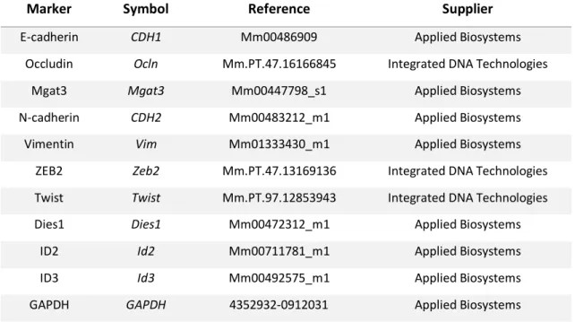
Table 1. Most common 3D cell culture methodologies. ... 18 Table 2. Innovative 3D in vitro models for EMT and cancer research ... 22 Table 3. Fluorescent probes for expression analysis of target genes by real-time PCR ... 26 Table 4. Separating gel and Stacking gel preparation for MMPs analysis via in gel zymography ... 29 Table 5. Summary of the characterization of TGF-β1-induced EMT in the 3D in vitro system ... 59 Table 6. Summary of the characterization of TGF-β1-induced EMT/MET in the 3D in vitro system ... 61
x
L
IST OF FIGURES
Figure 1. Summary of major regulators and signalling pathways involved in EMT progression. .. 9
Figure 2. Post-translational modifications in major EMT regulators SNAIL, bHLH and ZEB ... 10
Figure 3. Two proposed mechanisms of tumour cell dissemination in which EMT and MET are involved ... 13
Figure 4. Balance of main considerations of in vivo and in vitro models ... 15
Figure 5. Adhesive, topographical, mechanical, and soluble cues in 2D and 3D ... 19
Figure 6. Representation of the 2D in vitro TGF-β1-driven EMT/MET induction ... 32
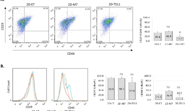
Figure 7. CD29 and CD44 expression during EMT/MET induction in the 2D in vitro model ... 33
Figure 8. Effect of EMT/MET induction on MMP2 and MMP9 secretion ... 33
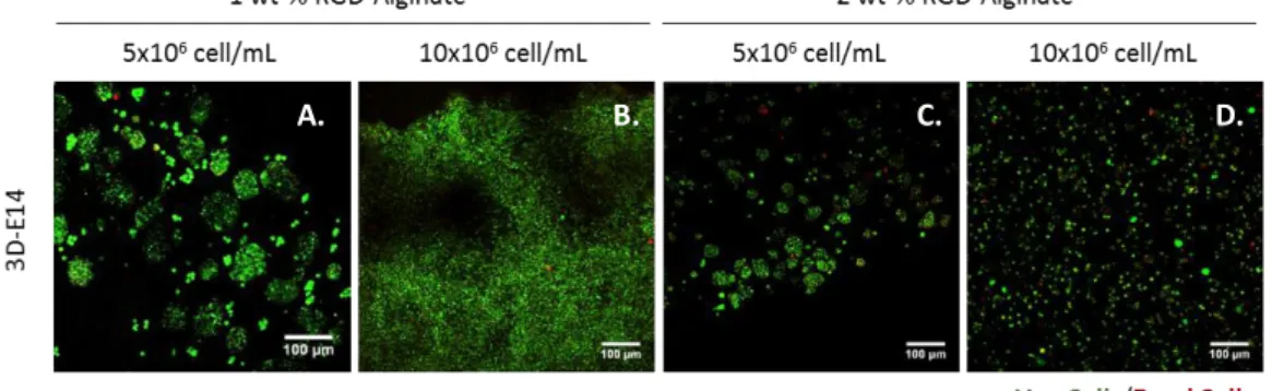
Figure 9. Effect of the 3D in vitro environment on cell viability using the Cytrak OrangeTM/DRAQ7TM live/dead staining ... 35
Figure 10. Morphological and metabolic characterization of EpH4 spheroids in the 1wt-% RGD-alginate hydrogels ... 35
Figure 11. Assessment of the expression of epithelial and mesenchymal markers in 3D epithelial cultures... 37
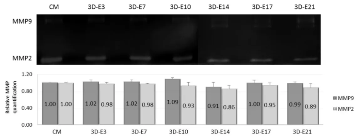
Figure 12. MMP secretion profile of EpH4 cells cultured in the 3D in vitro system ... 38
Figure 13. RNA signature of EpH4 cells cultured in the 3D in vitro system ... 39
Figure 14. Representation of the 3D in vitro TGF-β1-driven EMT ... 39
Figure 15. Metabolic activity during EMT induction ... 40
Figure 16. Assessment of the expression of epithelial and mesenchymal markers in 3D mesenchymal cultures ... 41
Figure 17. MMP secretion profile of EpH4 cells cultured in the 3D in vitro system in normal culture medium supplemented with TGF-β1 ... 42
Figure 18. RNA signature of 3D-M cells and comparison to epithelial RNA profile... 43
Figure 19. Representation of the 3D in vitro TGF-β1-driven EMT/MET induction ... 44
Figure 20. Morphological and metabolic characterization of EMT/MET populations obtained in the 3D in vitro system ... 46
Figure 21. MMP secretion profile during EMT/MET in EpH4 cells cultured in the 3D in vitro system ... 47
Figure 22. Assessment of the expression of epithelial and mesenchymal markers during EMT/MET induction in EpH4 cells cultured in the 3D in vitro system ... 48
xi Figure 23. E-cadherin, CD29 and CD44 expression during EMT/MET induction in the 3D in vitro
system ... 50
Figure 24. RNA signature of cells undergoing EMT/MET in the 3D in vitro system ... 51
Figure 25. RNA signature of cells undergoing EMT/MET in the 3D in vitro system. ... 52
Figure 26. Evaluation of the TGF-β1 potential to induce EMT in the 3D in vitro system ... 56
Figure 27. Architecture of the lumenized spheroids found both in the 3D epithelial and EMT-derived cultures ... 57
Figure 28. MMP2 and MMP9 secretion for 3DE14 and 3DM14 cells. ... 58
Figure 29. Global representation of the 3D in vitro TGF-β1 induced EMT/MET model in RDG-modified hydrogels ... 62
xii
L
IST OF ABBREVIATIONS
2D Two-dimensional
3D Three-dimensional
α-SMA Alpha-smooth muscle actin
bHLH Basic helix-loop-helix
BSA Bovine Serum Albumin
CLSM Confocal laser scanning microscopy
CSC Cancer stem cell ECM Extracellular matrix
EMT Epithelial to mesenchymal transition FBS Fetal Bovine Serum
GDL Glucose delta-lactone GSK Glycogen synthase kinase HIF-1α Hypoxia-inducible factor 1-alpha
HT High-throughput
ILK Integrin linked kinase
MAPK Mitogen-acivated protein kinase
MET Mesenchymal to epithelial transition
miR microRNA
MMP Matrix metalloproteinases
NFκB Nuclear-factor kappa B
PA Polyacrylamide
PBS Phosphate-buffered saline PEG Polyethylene glycol PGA Poly(glycolic acid)
pHEMA Poly(2-hydroxyethyl methacrylate) PI(3)K Phosphoinositide 3-kinase
PLA Poly(lactic acid)
PLG Poly/lactide-co-glycolide) RFU Relative fluorescence units
RGD Arginine-glycine-aspartic acid peptide ROS Reactive oxygen species
R-Smad Receptor Smad
TBS Tris-buffered saline
TGF-β1 Tumour growth factor-beta 1
TGFβRI Tumour growth factor-beta receptor type I TGFβRII Tumour growth factor-beta receptor type II TKR Tyrosine kinase receptor
Chapter I – Thesis Overview |1
C
HAPTER
I
Thesis Overview
1. Thesis Context
Epithelial to mesenchymal transition (EMT) has been described as a phenomenon through which epithelial cells undergo multiple biochemical modifications that enable them to transdifferentiate into cells that evidence a mesenchymal phenotype1. During this biological process, epithelial cells lose their characteristic intercellular adhesion and polarization, and gain mesenchymal features, namely enhanced production and deposition of extracellular matrix (ECM) components, along with enhanced migratory capacity and invasiveness2–4. EMT is a crucial embryonic developmental process that can be recapitulated in adulthood during wound healing and that may be harnessed by cancer cells5. Indeed, epithelial neoplastic cells may undergo EMT and develop the ability to invade through the basement membrane and metastasize, therefore allowing for cancer progression and systemic dissemination of the primary tumour, originating metastasis6. In fact, metastasis are responsible for more than 90% of cancer-related deaths and is currently handled as a dynamic and complex process that holds profound implications for cancer diagnosis, prognosis and treatment7,8. The success of the metastatic process is accomplished when the disseminated cancer cells give rise to a secondary tumour, which entails the reacquisition of an epithelial-like phenotype that can be the outcome of the reverse process of EMT, named mesenchymal to epithelial phenotype (MET)9.
2. Problem Statement
The metastatic process and particularly the contribution of the EMT/MET molecular programs to its success are difficult to study both in vitro and in vivo, due to the lack of proper models. As a matter of fact, the role of EMT in cancer remains dubious since it is difficult to prove and identify a full EMT phenotype in clinical carcinomas and metastases10. Some clinical observations may support the concept of a transient EMT that is only induced in a small number of cancer cells5. During the past 10 years a notable effort was made in order to develop biological models that could contribute to a better understanding of the EMT molecular regulation and signature. Nowadays, EMT studies are driven by the eagerness to discover new molecular targets and therapies, however there is a concerning gap between the results obtained using the in vitro systems and their clinical outcome. Therefore, improved biological systems that better mimic
2 | Chapter I – Thesis Overview
the three-dimensional (3D) microenvironment of the tumour need to be developed in order to closely reproduce the EMT transcriptional program that occurs in vivo. The development of artificial 3D ECM-like matrices may constitute a valuable tool to unravel the molecular mechanisms underlying the metastatic process.
3. Aims and Objectives of the Thesis
The primary aim of this Master Thesis was to establish an advanced 3D culture model of EMT/MET induction that would be useful to complement previous studies performed using traditional two-dimensional (2D) and in vivo models. The model gathers near-normal mouse mammary epithelial cell line EpH4 and alginate hydrogels as synthetic ECM, to create a Tumour Growth Factor (TGF)-β1-induced EMT/MET model with tuneable biochemical and mechanical properties. This model is expected to facilitate the analysis of the cellular behaviour in a well-defined and biologically relevant microenvironment; will allow the identification of several ECM features, to setup an EMT/MET induction within a 3D EpH4 culture; and will provide insight into cell-matrix interactions in this context.
The first objective was to further develop the 2D in vitro model of TGF-β1-driven EMT/MET induction previously established in the group, by characterizing the stemness properties of the cells at different stages of the process, as well as their ability to produce Matrix metalloproteinases (MMPs). The second objective consisted on the establishment of the 3D model that comprised three main tasks, which are identification of the most suitable culture conditions (e.g. stiffness and cell density), determination of TGF-β1 concentration and the time of culture that is necessary in order to achieve each cellular stage of the EMT/MET process, and monitoring of cell behaviour in the 3D artificial environment, in order to evaluate the interaction between epithelial/mesenchymal cells with the surrounding environment, as an integrated system. The final objective was comparison between the 2D and the 3D in vitro models to understand the impact of the third dimension, in the study of EMT/MET processes in vitro.
4. Thesis Outline
Chapter I, Thesis Overview, entails a short context of the Thesis and the problem statement that together represent the motto of this project; the main aims and tasks of the project are also exploited in Chapter I. Chapter II, Introduction, gives a detailed background on the subject that underlies this Thesis, by gathering a review on the extensive knowledge on EMT, particularly related to cancer, and the state-of-the-art of in vivo and in vitro biological models that have been
Chapter I – Thesis Overview |3 developed, in order to study this process. Chapter III, Material and Methods, describes the methods that were implemented during this experimental work, as well as main materials that were used. Chapter IV, Results, reports the observations and findings that resulted from the 2D model study, and from the establishment and validation of the EMT/MET-induction model in the 3D system. Chapter V, Discussion, analyses the obtained results, compares the 2D and 3D EMT/MET systems and correlates them with other models described in literature, offering a critical perspective of the work developed. Chapter VI, Conclusions, reviews the main achievements of this work, their implications, limitations and potential applications. Future research opportunities and perspectives are addressed in the final chapter, Chapter VI.
Chapter II – Introduction |5
C
HAPTER
II
Introduction
1. General introduction
Epithelial cells are defined as cells highly polarized along a basal-apical axis, which normally interact with basement membranes via basal adhesive junctions. These cells are connected by basal and cell-cell adhesive contacts, such as tight and gap junctions, and they are arranged in mono or multilayers giving rise to the epithelium11. The epithelium is a robust tissue that supports the structure of embryos and organs, and acts as an effective barrier against pathogens. It is also recognized by its remarkable dynamic cohesion and adhesion capacity that mainly relies in the establishment of the abovementioned adhesive contacts1,11. Under normal settings, these cells present limited motility due to the fact that they can only migrate laterally along the basal surface12. In certain circumstances, such as embryogenesis, wound healing and cancer, epithelial cells may undergo the EMT transcriptional program that enable them to transdifferentiate into a mesenchymal phenotype1. Mesenchymal cells can be amoeboid or polarized in the anterior-posterior orientation and interact weakly with the ECM components and neighbouring cells leading to an enhanced migratory capacity and invasiveness, two key features of cells that undergo EMT3,4. The EMT process is not an irreversible conversion to a mesenchymal state; cells may suffer a reverse process – MET – by which they are able to partially recover an epithelial phenotype and associated features1,9,13. The microenvironment in which these events occur is a major regulator of the EMT/MET program, thus biomimetic model systems are necessary to study such influence. Over the past years several EMT studies have been performed contributing to the extensive knowledge about this program and related events. The in vitro studies are an essential complement to the in vivo experiments and they have been upgraded accordingly to current technology, evolving from monocultures in monolayer to co-cultures systems, complex 3D environments, microfluidics systems and high-throughput (HT) models. The creation of systems with increasing complexity intends to achieve a more reliable representation of the in vivo microenvironment thereby enabling a consistent and improved study the EMT/MET biological dynamics.
6 | Chapter II – Introduction
2. Epithelial to Mesenchymal Transition
The biological context of EMT
EMT may occur under diversified, although highly specific settings leading to different functional and physiological responses. Accordingly, three main categories of EMT – type 1, 2 and 3 – have been proposed depending on the biological context in which the phenotypic transition is settled1. These three EMT types represent distinctive processes that rely on different biochemical and genetic modification programs that are not yet completely understood. EMT type 1 comprises a well-known multi-step process that plays a central role during embryonic morphogenesis and development. Particularly, EMT type 1 is crucial for embryo implantation and organ development, since epithelial cells are not endowed with the flexibility required for the development of complex body structures as mesenchymal cells are1,11,14,15. Indeed, the differentiation of specialized cells and the complex 3D structure assembly that occurs, for instance, during the internal organs formation, requires sequential rounds of EMT and MET programs, giving rise to the nominations of primary (e.g. gastrulation), secondary (e.g. nephogenesis) and tertiary (e.g. heart formation) EMT13,14.
EMT type 2 occurs as part of the inflammatory response during wound healing, tissue regeneration and organ fibrosis, in order to generate fibroblasts and related cells that enable the reconstruction and remodelling of the damaged tissue1. For instance, during skin wound healing, keratinocytes recapitulate the EMT process through the acquisition of an intermediate phenotype, called “metastable” phenotype, through which cells display simultaneous epithelial and mesenchymal features that allow the movement of these cells without the complete loss of contact16. The wound healing of the ovarian surface epithelium at each menstrual cycle in another example of the importance of EMT type 217. In contrast to EMT type 1, EMT type 2 can be associated to disease, since it enables the conversion of epithelial cells into myofibroblasts that secrete excessive amounts of collagen, which accumulate in the organ creating fibrotic collagen networks that compromise organ function13. This EMT-mediated conversion during fibrosis was already shown in several organs, such as kidneys, liver, lung and heart18–21.
The third type of EMT is also associated to pathophysiological conditions as it has been proposed to occur in neoplastic cells and is thought to be a crucial step for cancer progression. Due to genetic and epigenetic alterations that occur in tumour cells, mechanisms thought to regulate EMT suffer modifications that lead to outcomes far different that those described for EMT type 1 and 21. In the initial steps of carcinogenesis in epithelial tumors, transformed epithelial cells are associated to a gaudy proliferation and angiogenesis, and in the final stages these cells develop the ability to invade through the basement membrane and some of them to metastasize
Chapter II – Introduction |7 leading to a systemic dissemination of the tumour. This malignant phenotypic change has been proposed as being consequence of the activation of an EMT program; furthermore, the eventual dissemination of these cells to originate secondary tumours at distant sites implies the reacquisition of an epithelial phenotype, which may occur via MET6. In some cases, epithelial cells may not be fully transformed into mesenchymal cells, thus accumulating features of both epithelial and mesenchymal phenotypes, suggesting the occurrence of a metastable phenotype22 that could be the result of a partial EMT1,23.
The physiological and pathophysiological conditions in which EMT may be activated, illustrate the biological relevance and highlight the need to study the fundamental mechanisms underlying this process. All three EMT types have been subject of extensive research, however there are many open questions that could break new grounds for improved therapies against cancer, chronic fibrosis and other EMT-associated diseases24. The specific signal or signals that trigger EMT and MET, the molecular stakeholders that orchestrate the response to those signals and the signalling pathways involved are some of the questions that are still not fully understood, particularly in EMT type 2 and 3.
Cellular and molecular basis of EMT
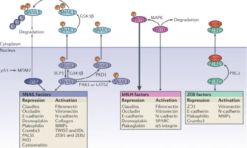
The molecular modifications inherent to the EMT program have been associated to key signalling pathways that may be responsible for the implementation of the cellular response to extracellular signals arising from the cell microenvironment (Figure 1), as it is the case of TGF-β ligand. Indeed, TGF-β signalling pathway is one of the best studied mechanism of EMT induction and its relevance for tumour progression has been intensively investigated and reviewed25–29. Under normal circumstances, TGF-β is a pleiotropic cytokine that is essential to development, differentiation and homeostasis of almost all kinds of mammalian cells and tissues, being a suppressor of uncontrolled proliferation and transformation25,29. However, dysregulation of TGF-β signalling may confer an oncogenic behaviour to this molecule that induces EMT and metastasis. Such functional switch is known as “TGF-β Paradox” and it has been studied in order to further understand the mechanisms underlying EMT induction and eventually to identify novel pharmacological agents that could prevent such mechanisms26. TGF-β superfamily is considered as primary inducer of EMT and it was found to be capable of inducing all three types of EMT9. Although different elements of the family are responsive to different extracellular signals, they share the same transcriptional network and cellular response. To initiate the TGF-β signalling pathway, TGF-TGF-β ligands bind with high affinity to TGF-TGF-β receptor type II (TGFTGF-βRII), a constitutively active cell surface receptor belonging to the serine/threonine kinase family. When the ligand binds to TGFβRII, a conformational change occurs in the receptor allowing the
8 | Chapter II – Introduction
recruitment of an appropriated TGF-β receptor type I (TGFβRI) and therefore, the formation of a tetrameric complex. TGFβRI can directly phosphorylate the receptor Smad (R-Smad) enabling the formation of a complex of two R-Smads and one Smad4 that accumulates in the nucleus and prompts the EMT transcriptional program30. The activation of Smad3 by TGF-β may induce the activation of the Notch pathway that regulates the expression of BMP family members, Notch ligands (e.g. Jagged 1) and targets (e.g. Hey1) and that is associated to E-cadherin repression, enhancement of MMP2/9 and alfa-smooth muscle actin (α-SMA) expression, some of the well-known EMT markers31–33. TGF-β signalling may also induce non-Smad pathways, including Mitogen-activated protein kinases (MAPKs), Phosphoinositide 3-kinase/Akt (PI(3)K/Akt) and Nuclear-factor kappa B (NFκB)13,34,35. All of these pathways converge in the initiation of a transcriptional program that leads to the inhibition of epithelial features and induction of a mesenchymal phenotype. Such outcome results from the activation of key transcription factors such as Snail, Slug, Twist, ZEB1/2, HMGA2, coupled with changes in microRNA (miR) expression, as it is the case of the downregulation of miR-200 family27,36. This specific transcriptional program accounts for characteristic changes in cell-surface, cytoskeletal and ECM protein expression that are understood as EMT markers. The decreased expression of E-cadherin, Cytokeratin and Laminin1, and enhanced expression of N-cadherin, Vimentin and Fibronectin are some of the most common reported changes during EMT at the protein level. TGFβRII may also be triggered in tight junctions where Occludin, Claudin and JAM proteins bind to ZO family proteins, connecting the cell to its neighbour and regulating its own polarity. Once activated, TGFβRII facilitates the interaction between Par6 and Occludin, via Par6 phosphorylation, leading to RhoA inhibition by Smurf1 and consequent polarity loss9,37.
Besides the TGF-β-mediated EMT induction, other signalling pathways have demonstrated an important role in the induction of this event. Wnt signalling can be correlated to the inactivation of glycogen synthase kinase (GSK)-3β, that blocks the activation of the transcription factors Snail and LEF-1, thus inducing the EMT and cell migration via E-cadherin expression decrease and enhanced mesenchymal markers9,38–40. The Tyrosine kinase receptors (TKR) signalling may promote EMT through the induction of Ras-MAPK pathway that eventually activates the transcription factors Snail and Slug13. Met, FGF, IGF, EGF, HGF and PDGF families are some of the TKRs inherently associated with the 3 types of EMT9,17,41. The NFκB pathway can also be seen as an important regulator of type 2 and type 3 EMT, since it leads to the induction of Snail transcription in carcinoma cell lines and mesothelial fibrosis. Indeed, this pathway proved to be fundamental for EMT implementation, since the EMT is blocked when NFκB pathway was inhibited by IκB13,34.
Chapter II – Introduction |9 Figure 1. Summary of major regulators and signalling pathways involved in EMT progression.
Retrieved from Lamouille, Xu and Derynck (2014)42.
Despite of the several EMT induction mechanisms, the outcomes of the transcription program that is implemented always seem to include the loss of functional E-cadherin, which is why this feature is recognized as one of the most important hallmarks of EMT. Indeed, the loss of E-cadherin is widely correlated to all three types of EMT and it has been associated with carcinoma progression and poor prognosis both in human and mouse tumours43. E-cadherin function may be impaired through different mechanisms, which may be developed at the genetic, epigenetic, transcriptional and post-transcriptional levels44. Although rare, mutations in the E-cadherin gene (CDH1) may lead either to the protein absence or truncation, thus impairing E-cadherin mediated cell-cell adhesion. CDH1 inactivating mutations were firstly found in diffuse-type gastric carcinomas45; now, they are known to occur in other types of cancers, such as lobular breast carcinomas46 and synovial sarcomas47. Regarding the epigenetic mechanisms, hypermethylation of the 5’ CpG islands of CDH1 promoter seems to be an important mechanism of E-cadherin silencing and it has been reported in some types of cancers as it is the case of invasive breast ductal carcinoma48, esophageal squamous cells carcinoma49 and gastric cancer50. Indeed, in gastric cancers the hypermethylation of CDH1 was found to be correlated with cancer aggressiveness and metastasis51. The transcriptional repression of CDH1 can be accomplished through the EMT direct activation of zinc-finger transcription factors Snail, Slug and ZEB1/2 that are able to directly bind to E-box repressive elements in the promoter of CDH152,53. Nevertheless, some EMT inducers, such as Twist1, FOXC2 and Goosecoid, have shown the ability to activate the EMT program in epithelial cells without binding directly to CDH1 promoter9. The post-transcriptional modification of E-cadherin commonly occurs through phosphorylation or glycosylation. For instance, adherens junction formation can be negatively regulated by tyrosine kinases, such as Met and Src, which phosphorylate β-catenin and the short cytoplasmic tail of
E-10 | Chapter II – Introduction
cadherin54. On the other hand, the loss of E-cadherin function during EMT may be accomplished by a specific modification of the E-cadherin glycosylation via Mgat3 glycogene expression and GnT-III-mediated glycosylation55. Despite the countless reports that show the loss of E-cadherin function in cells undergoing EMT, it is very important to highlight that when such loss occurs in normal epithelial cells, the result is often cell death rather than EMT, which suggests that E-cadherin loss is not, per se, an EMT inducer9. The loss of cadherin is associated to a cadherin switch in which N-cadherin is up-regulated56 and has an important role in tumour progression. Furthermore, P-cadherin has also been associated with tumour progression and increased migration and invasion57, however its contribution cannot be generalized due to divergent data obtained from independent studies in different types of cancer45.
The mesenchymal differentiation, cell migration and invasion are also recognized hallmarks of EMT and they can be accomplished mainly through post-translational modifications that regulate the activity, subcellular localization and stability of SNAIL, basic helix-loop-helix (bHLH) and ZEB factors (Figure 2), as described by Lamouille, Xu and Derynck42.
Figure 2. Post-translational modifications in major EMT regulators SNAIL, bHLH and ZEB. Adapted from Lamouille, Xu and Derynck (2014)42.
The depicted signalling pathways are very complex and can regulate numerous elements such as transcription factors, effector proteins and microRNAs; additionally, other pathways can be involved in EMT induction. Hence, the precise role of each component and how it affects the EMT program requires further clarification.
Chapter II – Introduction |11
Role of EMT in cancer
The concept of hallmarks of cancer proposed by Hanahan and Weinberg in 2000, and reviewed in 201158, gathered important insights that are seen as crucial pieces in the complex biological puzzle that is cancer. The great knowledge produced worldwide around the EMT mechanisms and its key drivers shows that there is a correlation between EMT implementation and several cancer hallmarks, such as activation of invasion and metastasis, immune system evasion and cell death resistance, which supports the existence of EMT type 3.
Several independent studies have shown that EMT type 3 may be induced mainly by TGF-β and Wnt signalling pathways via microenvironmental stimuli59,60. Hypoxia has also been revealed as a driving force of EMT events. At early stages, hypoxia supports the Snail up-regulation via Reactive oxygen species (ROS)-dependent GSK-3β inactivation and Notch signalling61 and Twist expression may be induced via Hypoxia-inducible factor-1alpha (HIF-1α) activation62. In late stages of the EMT process, invasion, basement membrane degradation and ECM remodelling might be promoted by HIF-1α-mediated Wnt/β-catenin pathway63. Extracellular matrix molecules, such as Collagen type I, may as well contribute to EMT activation64. Collagen type I interacts with α2β1 integrin triggering the increase of Akt, GSK-3β and IκB phosphorylation via integrin linked kinase (ILK) that eventually enables the activation of EMT-related transcription factors, such as NF-κB, Snail and LEF-1, as well as E-cadherin down-regulation38,65. Beyond the multitude of extracellular signals that could be further described in EMT type 3, it is equally important to note the existence of some intracellular stimuli that have been identified as EMT promotes. One of those cases is the intracellular calcium signalling that has been studied in the growth of the primary tumour context and that recently showed some evidences that it may have an important role in cancer cell invasion and migration66,67.
EMT type 3 relies on a transcriptional program that is different from the other two types of EMT, therefore the outcomes of this program will also differ from the ones observed in EMT type 1 and 2. In addition to the general hallmarks of EMT, other cell features were already shown such as cell death evasion, cell cycle and senescence regulation, angiogenesis, immunosuppression induction, resistance to therapy, and also acquisition of stem cell properties, in close resemblance to cancer progression6,9,68. Indeed, the tumour metastasis process is only possible due to a core of tumour cells that retain the self-renewing ability and stem cell features that enable them to initiate new tumours. These cells, named cancer stem cells (CSCs), can be recognized by a CD44high/CD24low phenotype, which allowed the identification of their presence in breast, colon and brain carcinomas69–71 or through a CD44high/CD29high phenotype in squamous cell carcinomas72. Through the induction of EMT in immortalized human mammary epithelial
12 | Chapter II – Introduction
cells, Mani and colleagues showed that cells undergoing EMT also present features of CSC, suggesting that EMT can be involved in the formation of CSC73. The hypothesis of EMT/MET occurrence in these cells could explain how cells that gain motility and invasion capacity are also able to disseminate and initiate another tumour in distant tissues and organs73,74. Tsuji and colleagues suggested that EMT cells might cooperate with non-EMT cells in order to allow a spontaneous metastasis process. In their study, EMT cells induced extracellular matrix degradation enabling invasion and intravasation of both EMT and non-EMT cells75. Unfortunately, none of the studies performed until now demonstrates how these cells can initiate a tumour with similar features in a different microenvironmental niche76. Recent studies showed that the acquisition of an EMT-associated stem cell phenotype can be correlated with decreased expression levels of miR-200c – by p53 loss of function – and miR-203, suggesting that the restoration of the expression levels of this microRNA family may inhibit metastasis77–79. Despite of the lack of irrevocable evidences of EMT in human cancer tissues, the increasing number of studies reporting the potential role of EMT in promoting CSC features and chemoresistance8,80, lead some authors to suggest the development of therapies based on the use of CSC or EMT markers to target CSCs and consequently reduce the tumour growth, metastasis and patient chemoresistance81. Others, propose the use of drugs to induce cancer cells to differentiate into benign stromal fibroblasts82.
The strong evidences of the EMT importance for tumour development and dissemination, and the hypothetical role of MET in the establishment of metastases (Figure 3)83 suggest that EMT/MET and associated signalling pathways could be useful to improve cancer treatment and find new cancer therapies. Moreover, the identification of clinically validated EMT/MET markers would lead to an improved and perhaps earlier diagnosis, which could be translated into better prognosis. Such expectation has been nourishing several studies that aim to magnify the knowledge about EMT and its potential role in cancer therapy.
Chapter II – Introduction |13 Figure 3. Two proposed mechanisms of tumour cell dissemination in which EMT and MET are involved. A. EMT as a complete or partial process. Primary epithelial tumour cells (blue cells) undergo EMT either partially (turquoise) or completely (green), and then undergo MET at a distant site to form the metastatic lesion. B. EMT and cell cooperation. Mesenchymal cells (green) invade the vasculature, enabling epithelial (blue) cells to pass through as passengers. Adapted from Krebs et al. (2014)84.
3. In vivo and In vitro Models to Study EMT
In vivo models
There is no doubt about the outstanding contribution of the scientific studies performed in vivo for cancer research purposes and for biomedical research in general. In vivo studies are currently the ultimate barrier to the implementation of new therapies and treatments in all therapeutic classes, and for that reason these models are extensively used for screening purposes. These models are also largely employed to study the complex biological environment and dynamics, however it is important to highlight the high cost and ethical issues intrinsically associated with their implementation. Furthermore, these models are very time consuming and in some cases they fail to reflect the human condition, which may lead to a failure in the translation of the research outcomes85–87. In the particular case of cancer research, the most common models arise from the surgical implantation of tumour cells in animals, which are frequently immunodeficient, or the generation of genetically-engineered models that accomplish a spontaneous tumour formation88. A major lack in this field is the development of animal models that recreate deadly cancers89. Additionally, classical animal models and in vivo studies are associated to a higher degree of unpredictability of the model behaviour and ethical issues. These limitations suggest the need for better animal models that would enable to follow the spatial and temporal modifications in tumour cells. Until now, in vitro 2D systems have been used as an additional and complementary tool in order to overcome the considerable restrictions of in vivo models. Regarding the particular case of the EMT study, the in vivo study is even more complex due to the lack of proper biomarkers that would allow the identification of the EMT associated cells90. Indeed, despite the large number of biomarkers identified through 2D standard in vitro cultures, the success rate of their implementation in clinics is extremely low which translates the need for more reliable and representative models91.
14 | Chapter II – Introduction
EMT in vitro models
3.2.1 2D EMT studiesThe conventional 2D studies accomplished in flat culture plastic have given rise to major advances in the understanding of EMT. These systems are particularly interesting to study the effect of individual variables and factors, or to achieve constant and stringent conditions of analysis92. In fact, these models provide almost total control over the biochemical and topographical properties of cells and they can be easily manipulated and integrated with a wide range of techniques93. For all these reasons, 2D models have an important role in understanding the influence of the individual EMT intervening factors in the global biological process and to relate them with the observed outcomes.
Although 2D studies represent an easy, flexible and accurate way to analyse EMT related effects, they fail in reconstructing the 3D complex environment in which the EMT takes place. One of the major criticisms concerning studies in 2D is that when cells are removed from their biological environment they tend to show a non-natural behaviour that may induce modifications in the global process. Once cultured in monolayer, cells become polarized with only part of their surface anchored in the substrate while the remaining surface is exposed to the culture media, hence cells are limited to their planar and spread morphology which can lead to the loss of their tissue-specific functions93,94. When tissue derived cells or cell lines are cultured in 3D scaffolds they tend to develop complex networks of cell-cell and cell-matrix interactions that affect the penetration of nutrients, biological factors (hormones and growth factors) and drugs, thus influencing cell growth, differentiation and death95. Other restrictions of 2D models are the lack of stromal components and reduced cell-matrix interaction, which are essential stimuli found in the in vivo environment95. In order to overcome some of these limitations, several studies attempt to incorporate ECM-like components on the 2D model by coating the flat dishes with various proteins, such as Collagen, Fibronectin and Laminin, creating a natural basal adhesion. The most complex 2D models also include co-cultures that intend to recreate the natural intercellular communications. In any case, cells remain in a monolayered arrangement that impairs the generation of a multidimensional structure.
Chapter II – Introduction |15 3.2.2 Advances of 3D in vitro models
The limitations of both 2D cell models and in vivo animal models provided the opportunity to the establishment of 3D models that aim to bridge the outcomes derived from the traditional models, and simultaneously to connect cell culture and human biology, both in physiological and pathological conditions. Such models attempt to recapitulate the tumour biology at all levels, which includes tumour gene signatures, cellular morphology, proliferation and differentiation, phenotypic heterogeneity of the population and the tumour microenvironment, through a fine balance between 2D in vitro simplicity and in vivo complexity (Figure 4).
Figure 4. Balance of main considerations of in vivo and in vitro models. 3D cultures that recreate the complex cellular microenvironment more precisely than traditional 2D cultures, due to the incorporation of multiple physical, mechanical and chemical cues that arise from ECM–cell and cell–cell interactions. Animals do not capture important facets of human behavior and they are not feasible for HT screening applications. Adapted from Alemany and Semino (2014).
The differences between 2D and 3D systems were noticed for the first time in the beginning of the 20th century, when Ross Harrison cultured embryonic frog cells in different conditions and observed a different cell behaviour in 2D and 3D hanging drop cultures96. Another type of 3D cell culture had its beginning in 1957 when McLimans and its colleagues reported a “disturbing feature” of HeLa cells grown in shake culture that was the formation of “large matrices of cells”97. This was the first time that a multicellular spheroid derived from mammalian cells was described. Three years later, in 1960, Wichterle and Lim proposed the use of poly(2-hydroxyethyl methacrylate), also known as pHEMA, in contact lenses giving rise to the concept of hydrogels98. It took twenty years to expand this concept to cell culture that came in the first place through cell encapsulation99. The 3D culture concept was brought to the cancer field by Miller and its colleagues that showed that tumour cells exhibit greater drug resistance when grown as multicellular spheroids in a collagen gel than when cultured in monolayer100. Since then, many important achievement have been accomplished as it is the case of the pioneer work
16 | Chapter II – Introduction
of Mina Bissell that used 3D culture systems to model the molecular mechanisms underlying breast cancer cell invasion and revealed that when cells from tumour of epithelial origin are cultured in Matrigel, their shape is changed and resembles what is seen in in vivo tumour progression101. These findings sparked the interest of the scientific community, which led to the improvement of the 3D cell culture systems. Important advances were achieved in the field of biomaterials, giving rise to the concept of biologically active materials. The bioactivity of synthetic polymers could be accomplished by the incorporation of cell-recognition sites, such as adhesion factors and enzymatic cleavage moieties102. One example of those bioactive biomaterials are the polyethylene glycol (PEG) hydrogels designed by West and Hubbell (1999) that incorporated peptide domains susceptible to cellular proteases, which allowed the hydrogel degradation by cells103. A few years later, the work of Mooney’s group in adapting scaffolds initially developed for tissue engineering purposes for cancer research, allowed for the first time the combination of these scaffolds with human carcinoma cells for in vitro and in vivo experiments104. Since then, many other groups have expanded the applications of the 3D in vitro models in cancer, for instance, Hutmacher and Clements lab developed 3D in vitro models for breast and prostate cancer bone metastasis90 and currently there are several groups trying to develop HT systems to produce and analyse 3D cultures, which can be very important to improve the screening of anticancer drugs and to develop personalized therapies.
According to the tumour microenvironment and its characteristics, or a particular process/feature that is aimed to mimic, different 3D models can be used, differing not only in their composition but also in terms of cell culture methodology and analysis (Table 1). Regarding the composition of these systems, they can be constituted by purely natural to purely synthetic materials105. Models derived from natural materials encompass as main advantages their cytocompatibility, ease usage, the ability to create a porous matrix or fibrous structure, and the possibility of modulating the stiffness simply through modulation of polymer composition and gelling conditions. Additionally, ECM-derived materials are provided with bioactivity due to the presence of cell adhesion sites in their composition. Despite of the broadly usage of these materials, they present relevant limitations, such as batch-to-batch variability and their complex or sometimes unknown molecular composition that challenges the reproducibility of the experiments. While studying EMT, it is important to understand that the unknown and variable composition on growth factors, namely TGF-β, may strongly compromise the replicability of the outcomes. Some examples of naturally derived materials are Matrigel® (derived from the basement membrane of murine sarcoma), ECM components from animal tissues such as Collagen type I, Laminin and Fibrin, and components derived from other biological sources as it is the case of Alginate106 and Chitosan107. Both Matrigel and Collagen have been largely used in
Chapter II – Introduction |17 cancer research, and EMT studies are not an exception, however they present weak mechanical strength and the modification of the matrix mechanics often alters the matrix biochemistry. Alginate is a natural polymer derived from polysaccharides that has been used in tissue engineering, for instance to transplant cells108. Nevertheless, 3D alginate scaffolds, such as AlgiMatrixTM, have already proved their value as tumour models, namely for anticancer drug screening109. The typical slow biodegradation of the alginate hydrogels93,110 can be adjusted through their partial oxidation, combination of polymer chains with different molecular weight111 or even the incorporation of MMP-sensitive peptides112.
Synthetic materials can also be used to engineer 3D models for cancer research, being their main advantage the great control over the experimental conditions. Additionally, they provide batch-to-batch uniformity and allow a selective tuning of physical and biochemical composition. The major lack of these models is the absence of intrinsic bioactivity since their ultimate goal is the recreation of a physiological microenvironment that mimics as closely as possible the in vivo scenario. Therefore, hydrogels and scaffolds based in synthetic materials can be modified giving rise to hybrid artificial matrices. The incorporation of cell adhesive peptides (e.g. Arginine-Glycine-Aspartic acid (RGD) and Tyrosine-Isoleucine-Glycine-Serine-Arginine (YIGSR)) and proteins (growth factors); the sensibility to cellular matrix-proteases (e.g. MMPs); and the modulation of the scaffold stiffness and surface topography are some of examples of matrices modifications93,113,114. Among the most commonly used synthetic materials are PEG115, poly(lactide-co-glycolide) (PLG)116 or polyacrylamide (PA)117. PEG hydrogels are biocompatible and resistant to protein adsorption, since they have high hydrophilicity, neutrality and mobility. PEG bioactivity can be easily tuneable and controlled, and it has been used as an hydrogel to culture different types of cells118,119. PLG is a synthetic and biocompatible polymer that allows for an easy reproduction and is convenient to use, which enables its utilization in a large-scale, for instance for cancer progression research and antitumor drug screening104. PA models are ideal to study mechanoregulatory mechanisms in the tumour; indeed, these models enabled researchers to understand that matrix stiffness improves tumorigenesis and that increased cellular traction stresses, as a response to increased stiffness, may be considered as an index of enhanced malignant potential120. Other synthetic materials can be used depending the microenvironment that is aimed to mimic, thus enabling the study of different metastatic sites. For instance, poly(lactic acid) (PLA), poly(glycolic acid) (PGA) or the combination of these two polymers provide a very high stiffness that can be used to biomimic the bone ECM121.
18 | Chapter II – Introduction
Table 1. Most common 3D cell culture methodologies.
Cell culture Methods Fundamentals Multicellular
Spheroids
Cells undergo self-assembly when in the absence of an attachment surface or scaffold, leading to the formation of cellular aggregates122
Spinner culture
Constant stirring cell collision, thus avoiding single cells in suspension to settle and promoting the spheroid formation.122,123 This technique allows the
formation of heterotypic spheroids and the control over spheroid characteristics via medium changes, stirring time and agitation speed. Mass diffusion along the spheroid can be promoted by fluid movement, avoiding the gradient of soluble components. This process can be easily scaled up, however it cannot be used for culturing low cohesive, shear sensitive or adherent cells. Additionally, cell visualization during aggregation is not possible due to the constant mixing.122,124
Liquid overlay
Cells are seeded in flat tissue culture dishes made of low-adhesive surface (e.g. coated with agar, agarose and pHEMA) and spheroid formation is achieved by rocking the plates, together with a small amount of shaking. This technique is easy to perform, yet spheroids present a heterogeneous size and shape.122,124
Rotating wall vessel
Cell suspension is slowly rotated about an x-axis, creating a microgravity environment.95 As cells start to aggregate, the rotation speed is increased to
avoid its settlement. Through this method high yield of spheroid formation can be achieved, using low levels of shearing forces. Moreover, the culture medium can be easily changed by perfusion allowing long-term studies or changing conditions during the process.125 Nevertheless, the size of the
spheroids is highly variable and it is difficult to track spheroid assembly.
Hanging drop
A small volume of a cell suspension is pipetted in the inside part of the lid of a tissue culture plate, and when the lid is inverted, the drop will stay attached due to surface tension and spheroid formation occurs through gravity-enforced cell self-assembly.126,127 This method allows for a high-throughput
spheroid formation, with a fine spheroid size and cellular composition regulation.128 In this system, it is difficult to track spheroid assembly and to
change or add cell culture medium/drugs.
Concave plate
Cells are isolated in microwells of non-adhesive substrates allowing them to cluster and grow into compact cell spheroids through cell-cell adhesion. This method enables the creation of a high-throughput system of spheroid production, as well as easy track of spheroid formation and medium change.
External force
External forces, such as ultrasound, electric and magnetic fields induce an increase in cell concentration into a high density that facilitates spheroid formation. Physiological cell changes induced through this method are still not well characterized.122
Chapter II – Introduction |19
Scaffolds Cell-cell and cell-matrix interactions induce cell spheroid mimicking the ECM behaviour95,105
Microporous scaffolds
Cells interact with the substrate being lodge within the porous. Due to the large size of those porous (≈100 µm), cells tend to adhere and form few cell-cell interaction which leads to a 2D-like culture rather than a 3D culture.105
Nanofibrous scaffolds
Cells interact with a fibrous substrate that provides a 3D architecture for cell culture. Yet, these substrates tend to be structurally weak to handle the stress needed for mechanotransduction.105
Hydrogels
Cells interact with crosslinked networks that are characterized by high porosity and water content that enable free diffusion of nutrients, oxygen, growth factors, cell metabolites and other soluble components.93,105 These
matrices closely mimic the nature of most soft tissues and allow the study of cell behaviour in different conditions, since the medium can be easily changed. Retrieving the cells from the 3D culture seems to be the main difficulty associated to this method.124
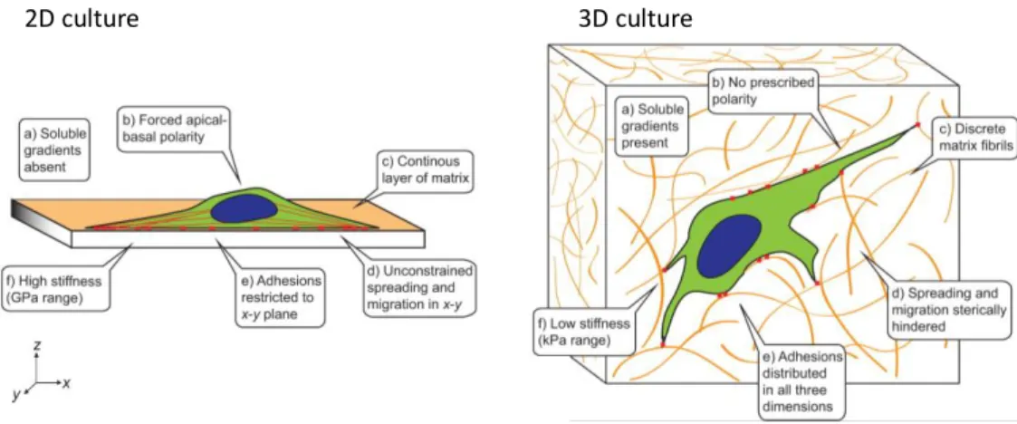
Regardless the cell culture methodology or composition of the 3D model, epithelial cells tend to form cellular spheroids that provide a more physiological arrangement that the 2D cultures (Figure 5), that can be generally described by additional mechanical inputs, dissimilarity in morphology, distinct cell adhesion and contraction, as well as modified intracellular signalling, which may ultimately change the functional behaviour of these cells129,130.
Figure 5. Adhesive, topographical, mechanical, and soluble cues in 2D and 3D. The cues encountered by a cell are strikingly different between an ECM-coated glass or plastic surface (2D) and a typical 3D ECM. Adapted from Baker and Chen (2012)130.
2D culture 3D culture