Elucidating the role
of macrophages
and CD4
+
CD25
+
regulatory T cells
on colorectal cancer
cell invasion
Maria Helena Cristiano Brigas
Master in Cellular and Molecular BiologyDepartment of Biology
2014/2015
Supervisor
Maria José Cardoso Oliveira, Assistant Researcher, I3S/INEB
Co-Supervisor
Todas as correções determinadas pelo júri, e só essas, foram efetuadas. O Presidente do Júri,
Acknowledgments
A primeira pessoa a quem tenho de agradecer é à minha orientadora Maria José Oliveira. És das melhores pessoas que conheço. Muito obrigada por teres acreditado em mim e por me teres deixado participar nesta grande aventura. Foi um grande privilégio teres-me confiado um projeto de raiz. Obrigada por todo o conhecimento científico e orientação que me deste na construção e realização deste projeto. Tenho imensa admiração por ti e pelo teu trabalho.
Quero agradecer à minha co-orientadora, Marta Oliveira, que desenhou este projeto e me deu todas as condições para o realizar. Obrigada pelo otimismo, estímulo, compreensão e orientação assim como por todas as contribuições preciosas que deste para este trabalho.
Marta, foste o meu anjinho. Nunca te agradecerei o suficiente por toda ajuda e apoio que me deste. Obrigada por teres estado comigo em toda esta caminhada com toda a tua paciência, disponibilidade e carinho. Ajudaste-me sempre a ver as coisas com mais clareza. Obrigada por todo o conhecimento cientifico e laboratorial que me deste.
Ana, muito obrigada por teres ajudado no planeamento de experiências e teres discutido comigo os resultados. Estiveste sempre disponível para me ajudar quando eu precisava.
Flávia, muito obrigada pelo conhecimento que partilhaste comigo e por todas as discussões científicas que tiveste comigo. Agradeço teres sido sempre sincera e crítica relativamente ao meu trabalho.
Hugo, obrigada pela tua amizade. Obrigada pelo apoio que me deste e por teres tornado os momentos menos fáceis em momentos cheios de alegria (e de comida e de café). A tua paixão pela ciência é contagiante, obrigada.
Mãe, obrigada por seres sempre carinhosa, mas ao mesmo tempo exigente. Deste-me a oportunidade de chegar onde cheguei. Pai, muito obrigada por todo o apoio e preocupação que sempre demonstraste. Obrigada, por acreditares em mim e por me estimulares a dar sempre o melhor de mim. Agradeço ao meu irmão e à minha irmã por acreditarem mais em mim do que qualquer outra pessoa. Obrigado por me terem apoiado em todos os momentos stressantes e por estarem sempre prontos para me ajudar.
Gustavo, tenho de te agradecer por todas os conselhos que me deste e todo amor, compreensão e paciência que tiveste comigo durante este ano. Muito obrigada.
Abstract
Solid tumours became malignant when cancer cells invade the surrounding tissues, becoming prone to intravasate blood and lymph vessels and to metastasize. In solid tumours, tumour-associated macrophages (TAMs) and regulatory T cells (Tregs) are reported to favour tumour progression. However, in colorectal cancer (CRC) their intratumour infiltration often correlates with good prognosis. Depending on the tumour microenvironment, TAMs may polarize into a pro- or an anti-inflammatory profile, modulating cancer cell activities and participating in tumour progression. On their turn, Tregs may steer macrophage differentiation, modulating macrophage inflammatory profile and functions within the tumour microenvironment.
This study aims to elucidate how TAM-CD4+CD25+ Treg crosstalk affects macrophage phenotype, clarifying the role of Treg-educated macrophages on cancer cell invasion, as well as dissecting the underlying invasion-related pathways. Therefore, macrophage precursors and CD4+CD25+ T cells were co-cultured at 1:1 and 2:1 ratios, for 3 or 7 days. The monocyte-macrophage profile was then assessed through the analysis of specific cell surface receptor expression and cytokine secretion. The impact of CD4+CD25+ T cells-educated macrophages on RKO and SW620 CRC cell invasion was evaluated through 3D-matrigel-invasion-assays and through analysis of the matrix metalloproteinases (MMPs) proteolytic profile. Phosphorylation of epidermal growth factor receptor (EGFR) and of its downstream regulators was also evaluated as part of the invasion-related pathway. Furthermore, FoxP3-positive cell infiltration on human CRC specimens was characterized by immunohistochemistry.
Our results evidenced that CD4+CD25+ Treg-educated macrophages lost CD80 maintaining the expression of CD163 and displaying a mixed pro- and anti-inflammatory cytokine secretion profile. Only RKO cell invasion was significantly promoted by soluble factors produced by macrophages and macrophage/CD4+CD25+ Treg co-cultures, but not by Tregs alone. At 2:1 ratio co-cultures, macrophage pro-invasive properties were enhanced. This effect was concomitant with increased pro-MMP9 activity and with the activation of EGFR (at the residue Y1086). In addition, using histopathological sections derived from human CRC specimens, we observed that FoxP3-positive cells infiltrate preferentially at the tumour nest and at the invasive front.
In conclusion, our in vitro results suggest that CD4+CD25+ T cells modulate macrophages toward a more anti-inflammatory profile and that, at 2:1 ratio, they promoted
macrophage pro-invasive activities. The higher FoxP3 infiltration found at the tumour nest and at the invasive front of CRC patient surgical resections are supported by previous reports. In the future, we intent to cross CRC FoxP3+ subpopulation characterization and TAMS differentiation profile with patients’ clinicopathological information. This research will elucidate the modulatory role of Tregs on macrophage pro-tumour properties, contributing to the design of novel therapeutic strategies targeting macrophages and/or Tregs.
Keywords: Tumour microenvironment; M1-like pro-inflammatory macrophages; M2-like anti-inflammatory macrophages; Regulatory T cells; Colorectal cancer cell invasion; Invasion-related EGFR pathway; Matrix metalloproteinases
Resumo
Os tumores sólidos tornam-se malignos quando as células tumorais ultrapassam os limites dos tecidos, tornando-se capazes de entrar nos vasos sanguíneos e linfáticos e metastisar. Nos tumores sólidos, os macrófagos associados ao tumor (TAMs) e os linfócitos T reguladores (Tregs) estão descritos como promotores da progressão tumoral. No entanto, no cancro colorretal (CRC) a sua elevada infiltração intratumoral está geralmente associada a bom prognóstico. De acordo com o microambiente tumoral, os macrófagos podem diferenciar-se em pró- ou anti-inflamatórios, modelando a atividade das células tumorais e participando na progressão tumoral. Por sua vez, os Tregs podem modelar a diferenciação dos macrófagos, modelando o seu perfil inflamatório e as suas funções dentro do microambiente tumoral.
Com este projeto pretende-se elucidar como é que a interação TAM-CD4+CD25+ Treg afeta o fenótipo dos macrófagos, regulando a sua capacidade de estimular a invasão das células tumorais, bem como identificar as vias de sinalização envolvidas nessa atividade. Para tal, os precursores dos macrófagos (monócitos) e as células T CD4+CD25+ foram co-cultivados nos rácios 1:1 e 2:1, durante 3 ou 7 dias. O perfil de diferenciação monócito-macrófago foi assim caraterizado através da análise da expressão de recetores celulares de membrana específicos e da produção de citocinas. O impacto dos macrófagos, diferenciados na presença de células T CD4+CD25+, na invasão das linhas colorectais RKO e SW620 foi avaliado por ensaios de invasão 3D em Matrigel e por análise do perfil proteolítico de metaloproteases da matriz (MMPs). A fosforilação do recetor do fator de crescimento epidérmico (EGFR) e dos seus reguladores a jusante foram avaliadas, como parte de uma via de sinalização associada à invasão. Por último, a infiltração de células FoxP3 positivas nos tumores de doentes com cancro colorectal foi caraterizada por imunohistoquímica.
Os nossos resultados demonstram que os macrófagos diferenciados na presença de células T reguladoras CD4+CD25+ perderam a expressão de CD80, mantendo a expressão de CD163 e apresentando um perfil inflamatório misto com secreção de citocinas pró- e anti-inflamatórias. Os macrófagos e as co-culturas de macrófagos/CD4+CD25+ Tregs, mas não as Tregs sozinha, produziram fatores solúveis que estimularam apenas a invasão das células RKO. No entanto, a capacidade pró-invasiva dos macrófagos foi maior no rácio 2:1. Este efeito foi concomitante com o aumento da atividade da pró-MMP9 e da ativação do EGFR (no resíduo Y1086). Recorrendo a cortes histopatológicos provenientes de
amostras de pacientes com cancro colorrectal, observámos que as células FoxP3-positivas estão preferencialmente infiltradas na região intratumoral e na frente invasiva.
Concluindo, os nossos resultados in vitro sugerem que as células T CD4+CD25+ modelam os macrófagos para um perfil mais anti-inflamatório e que, no rácio 2:1, promovem as propriedades pró-invasivas dos mesmos. O nível elevado de células FoxP3 positivas, encontradas no centro do tumor e na frente invasiva de recidivas cirúrgicas de pacientes com CRC, está de acordo com a literatura. No futuro, pretendemos cruzar a caracterização das populações FoxP3+ com o perfil de diferenciação dos macrófagos nas amostras de CRC e com a informação clinico-patológica dos pacientes. Esta investigação permitirá elucidar o papel Tregs na modelação das propriedades pró-tumorais dos macrófagos, contribuindo para o planeamento de novas estratégias terapêuticas que tenham como alvo os macrófagos e/ou os Tregs.
Palavras-chave: Microambiente tumoral; Macrófagos pró-inflamatórios tipo-M1; Macrófagos anti-inflamatórios tipo-M2; Linfócitos T reguladores; Invasão das células de cancro colorretal; Invasão mediada pela via do EGFR; Metaloproteases da matriz.
Contents
List of figures and tables ... 11
List of Abbreviations ... 12 Introduction ... 15 1.Cancer ... 15 1.1. Colorectal Cancer ... 15 2.Tumour microenvironment ... 17 3.Macrophages ... 17
3.1. Macrophages and cancer ... 19
3.2. Macrophages in colorectal cancer ... 23
4.Regulatory T cells ... 24
4.1. Tregs in cancer ... 26
5.Macrophage-Treg crosstalk ... 29
Aim ... 30
Materials and methods ... 32
Cell line culture ... 32
Immune cells isolation ... 32
CD14+ monocyte isolation ... 32
Human CD4+CD25high T cell isolation ... 33
Mono-culture of human CD14+ monocytes or of CD4+CD25+ T cells ... 34
Flow cytometry ... 35
Co-cultures of human CD14+ monocytes and CD4+CD25+ Tregs ... 36
Cell viability and metabolic assays ... 37
Immunocytochemistry ... 37
Enzyme-linked immunosorbent assay (ELISA) ... 38
Zymography ... 39
Western blot ... 39
Immunohistochemistry ... 40
Measurement of FoxP3 density ... 41
Statistical Analysis ... 41
Results ... 42
Isolation of CD4+CD25+ T cells with StemCell isolation kit allows an autologous setting between CD14+ monocytes and CD4+CD25+ T cells ... 42
Isolated T cells modify their morphology and FoxP3 expression profile after 3 and 7 days of culture ... 45
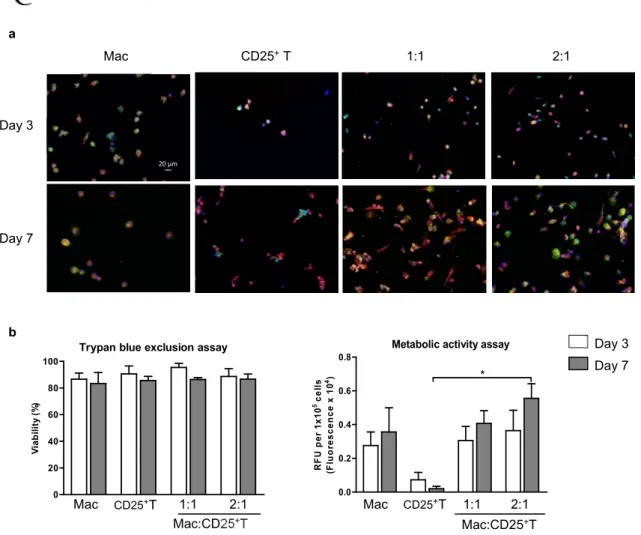
Macrophages and CD4+CD25+ T cell-educated macrophages are viable and metabolically active while CD4+CD25+ T cells are viable but less active ... 47
Co-cultures with isolated CD4+CD25+ T cells express CD163, but not CD80 ... 49
Macrophages and CD4+CD25+ T cell-educated macrophages significantly induce RKO but not SW620 cancer cell invasion ... 51
CD4+CD25+ T cell-educated macrophages in indirect contact with RKO cells express CD163, but not CD80, and display a “mixed” cytokine secretion profile ... 52
Pro-MMP9 activity is higher in CM of macrophage and macrophage co-cultures ... 54
Conditioned media from macrophages and from CD4+CD25+ T cell-educated macrophages induce phosphorylation of EGFR in RKO cancer cells ... 56
FoxP3-positive cells infiltrate preferentially at the tumour nest and invasive front on human CRC specimens ... 58
Discussion ... 60
Conclusions and future perspectives ... 67
Supplementary Data ... 69
List of figures and tables
Figure 1. The tumour microenvironment………18 Figure 2. TAMs facilitate tumour cell survival and growth, migration and invasion,
angiogenesis, metastasis and immune suppression………..21
Figure 3. Model proposal for the macrophage-mediated invasion pathway………..23 Figure 4. Mechanisms of Treg-mediated suppression….……….27 Figure 5. Macrophage display higher levels of adhesion and differentiation on 24-well plates
with glass coverslips ... 43
Figure 6. Isolation of Tregs with StemCell isolation kit allows an autologous setting between
CD14+ monocytes and CD4+CD25+ T cells ... 44
Figure 7. Isolated T cells modify their aspect ratio and FoxP3 expression profile after 3 and
7 days of culture ... 46
Figure 8. Macrophage and CD4+CD25+ T-educated macrophages are viable and metabolically active while CD4+CD25+ T cells are viable but less active ... 48
Figure 9. Co-cultures with isolated CD4+CD25+ T cells express CD163, but not CD80 .. 50 Figure 10. Macrophages and CD4+CD25+ T-educated macrophages significantly induce RKO but not SW620 cancer cell invasion ... 51
Figure 11. CD4+CD25+ T-educated macrophages in indirect contact with RKO cells express CD163, but not CD80 ... 53
Figure 12. CD4+CD25+T-educated macrophages in indirect contact with RKO cells display a “mixed” cytokine secretion profile... 54
Figure 13. Pro-MMP9 activity is higher in CM of macrophage and macrophage co-cultures
... 55
Figure 14. Conditioned media from macrophages and CD4+CD25+ T cell-educated macrophages induce phosphorylation of EGFR in RKO cancer cells ... 57
Figure 15. FoxP3–positive cells infiltrate preferentially the tumour nest and invasive front on
human CRC specimens ... 59
Figure S1. In vitro experimental set-up performed to dissect Treg-educated macrophages
profile and pro-invasive properties... ... 69
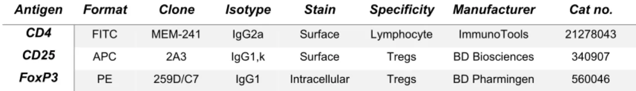
Table 1. Antibodies used for monocytes and CD4+CD25+T cell phenotyping by FACS….36
Table 2. Mean number of FoxP3 in normal colonic mucosa (NCM), tumour nest (TN) and
List of Abbreviations
ACF – Aberrant crypt focus
Akt – v-AKT murine thymoma viral
oncogene homologue
APC – Antigen-presenting cells APC – Allophycocyanin
ATCC – American type culture collection aTreg – Activated regulatory T cell Avidin-HRP D – Horseradish peroxidase
avidin D
CAC – Colitis-associated colorectal
cancer
CaCl2 – Calcium chloride
cAMP – Cyclic adenosine monophosphate
CCL – Chemokine ligand CD – Cluster of differentiation
Cdc42 – Cell division cycle protein 42 CM – Conditioned media
CO2 – Carbon dioxide
CpG – Cytosine-phosphate-Guanine CRC – Colorectal cancer
CSF-1 – Colony stimulating factor-1 c-Src – Cellular Homologue of Rous
sarcoma virus protein
CTL – Cytotoxic T lymphocyte
CTLA-4 – Cytotoxic T-lymphocyte antigen
4
CXCL – Chemokine (C-X-C motif) ligand DAPI – 6-diamidino-2-phenylindole DC – Dendritic cell
DNA – Deoxyribonucleic acid
ECM – Extracellular matrix
EDTA – Ethylenediaminetetraacetate EGF – Epidermal growth factor
EGFR – Epidermal growth factor receptor ELISA – Enzyme-linked immunosorbent
assay
EMT – Epithelial-mesenchymal transition ERK1/2 – Extracellular regulated MAP
kinase1/2
FAP – Familial adenomatous polyposis FACS – Fluorescence-activated cell
sorting
FBS – Fetal bovine serum
FITC – Fluorescein isothiocyanate FSC – Forward scatter
FoxP3 – Forkhead box protein 3
Gab1 – GRB2-associated binding
protein-1
GM-CSF – Granulocyte-macrophage
colony-stimulating factor
GTP – Guanosine-5'-triphosphate
GITR – Glucocorticoid induced TNFR
family-related protein
H2O2 – Hydrogen peroxide
HA – Hyaluronic acid HCl – Hydrochloric acid
HLA-DR – Human leukocyte antigen-D
related
HNPCC – Hereditary nonpolyposis
colorectal cancer
IBD – Inflammatory bowel disease
ICC – Immunocytochemistry
IDO – Indoleamine 2,3-dioxygenase IF – Tumour invasive front
IFN – Interferon Ig – Immunoglobulin IL – Interleukin
IL – 2Rα Interleukin-2 receptor α-chain iNOS2 – Inducible nitric oxide synthetase
tyoe 2
iTreg – Induced regulatory T cell
K-RAS – Kirsten rat sarcoma viral
oncogene homologue LPS – Lipopolysaccharide M1 – M1-like pro-inflammatory macrophage M2 – M2-like anti-inflammatory macrophage
mAB – Monoclonal antibody Mac – Macrophage
MCP-1 – Monocyte chemoattractant
protein-1
MHC – Major histocompatibility complex MIP- 1β – Macrophage inflammatory
protein-1β
MIP-1α – Macrophage inflammatory
protein-1α
MMP – Matrix metalloproteinase MMR – Mismatch repair
MSI – Microsatellite instability NaF – Sodium fluoride
NCM – Normal colonic mucosa NK – Natural killer
nTreg – Natural regulatory T cell
ON – Overnight
P53 – Tumour protein-53 PB – Peripheral blood
PBMC – Peripheral blood mononuclear
cell
PBS – Phosphate buffered saline PDGF – Platelet derived growth factor PDL1 – Programmed cell death protein 1 PE – Phycoerythrin
PFA – Paraformaldehyde PGE2 – Prostaglandin E2
PI1-K – Phosphatidylinositol 3-kinase PIGF – Placental growth factor PLC-γ – Phospholipase C-gamma PMF – Phenylmethylsulfonyl fluoride RAF/MAPK – Raf-mitogen-activated
protein Kinase
RANTES – Regulated on activation,
normal T cell expressed and secreted RA
RIPA – Radioimmunoprecipitation assay RFU – Relative fluorescence units
RhoA – Ras homolog gene family,
member A
RT – Room temperature
rTreg – Resting regulatory T cell SC – Stem cell
SD – Standard deviation
SDF-1 – Stromal-derived factor 1 SDS – Sodium dodecyl sulfate
SMAD – Contraction of Sma and Mad
(Mothers against decapentaplegic)
STAT – Signal transducer and activator of
transcription
TAM – Tumour-associated macrophage Tconv – Conventional T cell
TCR – T cell receptor
TGFBRII – Type II TGFβ receptor
TGF-β – Transforming growth factor-beta Th – T helper cell
TILs – Tumour-infiltrating lymphocytes TLR – Toll-like receptor
TMB – 3,3',5,5'-tetramethylbenzidine TME – Tumour microenvironment
TN – Tumour nest
TNF-α – Tumour necrosis factor-alpha Tr3 – Type-3 regulatory T cells Tr1 – Type-1 regulatory T cells Tregs – Regulatory T cells
TSDR – Treg-specific demethylated
region
tTGF-β – Total transforming growth
factor-beta
VEGF – Vascular endothelial-growth
Introduction
1. Cancer
Cancer is a progressive disease that occurs by a multistep regulated process. Typically, it arises from a series of mutations and epigenetic alterations that encompass gain of function of oncogenes and loss of function of tumour suppressor genes. These series of genetic changes confer growth advantages to proliferating cells and drive normal cells to transform into a neoplasic state. The progression of solid malignancies is reflected in the genetic heterogeneity of tumour cell subpopulations that share a common genetic background within a tumour1,2. In fact, only 10% of cancers are linked to germline mutations while the vast majority are caused by somatic mutations and environmental factors3.
1.1. Colorectal Cancer
1.1.1. Epidemiology
Colorectal cancer (CRC) is the third cause of cancer-related death worldwide4 and one of the most frequent and deadliest cancer in Portugal (Globocan, 2012), being a major health concern in industrialized and developed countries. Moreover, its incidence is increasing in developing countries, while remaining low in less-developed countries5. Environmental risk factors such as an inadequate dietary pattern, obesity, tobacco-smoking, excessive alcohol intake and sedentarism are considered to increase CRC risk6. These environmental factors have been associated with intestinal microbiota alterations and modifications on crucial molecular pathways related to CRC tumourigenesis5. Besides dietary and lifestyle factors, inherited and somatic mutations, epigenetic changes, as well as chronic intestinal inflammation have also prominent roles in CRC occurrence5.
Despite advances in CRC diagnosis, knowledge of novel biomarkers and therapies, many patients with advanced or metastatic disease, or with local recurrence following primary tumour resection, will still die from this disease7. This stands out the need for early detection, diagnosis and more effective non-invasive treatments.
1.1.2. Pathogenesis
The molecular basis underlying the progression of CRC is still subject of intense research. Typically, CRC is driven by the gradual accumulation of genetic mutations and epigenetic modifications. These modifications induce the activation of oncogenes or loss of function of
tumour suppressor genes in proliferating cells, driving the neoplastic process9. Histological changes which result in the transition from normal colon mucosa to adenomas and from adenomas to invasive carcinomas may them occur8.
The bulk of CRC cases are sporadic (80%) and result from somatic mutations arising during patients’ lifetime. The other 20% have a hereditary component, frequently associated with the hereditary nonpolyposis colorectal cancer syndrome (HNPCC or Lynch syndrome) and the familial adenomatous polyposis (FAP), and arise from subtle germ-line mutations10. In general, the earliest and most common mutation in CRC is the inactivation of
adenomatous polyposis coli (APC), a tumour-suppressor gene. Consequently, there is an
overactivation of the wingless-type(Wnt)/β-catenin signalling pathway, favouring transcription of tumour-promoting genes and cell growth. This mutation underlies the FAP cases, as well as 70–80% of all sporadic CRCs, being frequently found in the aberrant crypt focus (ACF), the first identifiable lesion in CRC formation10.
Subsequent mutations in other genes, most prominently K-RAS, P53 and TGFBRII, are related to colon carcinogenesis. The proto-oncogene Kirsten rat sarcoma (K-RAS) encodes highly conserved proteins involved in signal transduction. By overactivation of the Raf-mitogen-activated protein Kinase (RAF/MAPK), JNK and phosphatidylinositol 3-kinase (PI3K) pathways11, K-RAS mutations favour carcinoma growth. This is an early mutational event that underlies the transition from early to intermediate adenoma and occurs in 37-41% of CRCs11. Tumour protein-53 (P53) is a tumour suppressor transcription factor which regulates cell-cycle arrest, genomic stability and apoptosis12. When mutated, P53 may foster the malignant transition from intermediate adenoma to carcinoma. Progression of CRC can also involve alteration of the TGF-β signalling pathways through mutations in type II TGFβ
receptor (TGFβRII) gene, that occur in 30% of CRCs13, or in members of these pathways
namely in SMAD4/2. Genetic mutations in TGF-β signalling are frequently associated with microsatellite instability (MSI). MSI, characterized by insertion and deletion in repetitive DNA sequences due to the failure of DNA mismatch repair (MMR) mechanisms, gives rise to 15% of sporadic CRC and is highly associated with the HNPCC syndrome.
Epigenetic mechanisms, such as DNA methylation of CpG islands and pos-translational modifications of histones, have also a prominent role in the alteration of chromatin conformation and consequently may favour the transcription of genes that mediate CRC development14. Furthermore, specific bacterial pathogens as well as gut microflora and its metabolites can induce DNA damage, cell apoptosis and pro-inflammatory
conditions, thus promoting colon carcinogenesis15. In fact, individuals with inflammatory bowel disease (IBD) display an increased incidence of CRC16.
All of the above mentioned factors can act in concert to promote CRC development and malignant progression, ultimately leading to cancer cell spreading and dissemination to distant organs, primarily to the liver17.
2. The tumour microenvironment
Typically, tumour formation comprises collaborative interactions between neoplastic cells and the surrounding host microenvironment. This intercellular communication occurs through a complex and dynamic network of cytokines, chemokines and matrix remodelling enzymes that will dictate the tumour fate18. In general, when solid tumours emerge, the immune system initiates an anti-tumour response that actively eliminates antigen-expressing tumour cells, characteristic of a T helper (Th)1-like inflammatory response. However, over time, tumour cells elicit several changes in the surrounding stroma and escape the host response, shifting the immune environment from a Th1- towards a Th2-type19. In this way, malignant cells can instruct non-malignant stroma elements to support and promote tumour growth, survival, spread and even to affect response to treatment20,21. Therefore, together with cancer cells, immune cells, fibroblasts, pericytes, extracellular matrix (ECM) components, blood and lymphatics vessels constitute the tumour microenvironment (TME) (Figure 1)21. The immune infiltrate found at the TME can include multiple cell types. These populations can display both pro- and anti-tumour functions and can vary in their activation status, density and localization within the tumour22. Macrophages and regulatory T cells (Tregs) are key mediators between tumour cells and stroma components and are highly represented in most neoplastic tissues, including CRC23,24. The complete understanding of the tumour biology encompasses the study of cellular and molecular interactions in the TME that will open novel avenues for enhanced therapeutics and better clinical outcomes21.
3. Macrophages
Mononuclear phagocytes play an essential role in innate immunity and regulate adaptive responses25. Tissue macrophages derive from embryonic haematopoietic progenitors that persist and maintain the macrophage pool into adulthood26. However, in tissues that are exposed to microbiota (e.g. skin and gut) or under extensive inflammatory conditions, blood
monocytes can differentiate into tissue macrophages 27. Macrophages are highly diverse cells capable of undergoing phenotypic switches depending on environmental cues and on the milieu found at the tissue site28. Macrophages may undergo a classical M1-like activation (stimulated by IFN (interferon), IFN/LPS (lipopolysaccharide) and Toll-like receptor (TLR) ligands or an alternative M2-like activation (induced by IL-10, IL-4/IL-13, IL-33, IL-21)29-31. These states mirror the Th1/Th2 dichotomy of T cells and represent two extremes of a continuum spectrum of activation. M1-like macrophages express high levels of co-stimulatory receptors for lymphocytes (CD86/CD80, CD40), antigen-presenting molecules (MHC-class II such as human leukocytes antigen-D related protein [HLA-DR]) and produce high levels of pro-inflammatory cytokines (e.g. interleukin (IL)-6, tumour necrosis factor-alpha (TNF-α), IL-12, IL-23 and IL-1β). They target infectious agents or damaged cells, promoting cytotoxic activities28,29. Alternatively, M2-like macrophages display high phagocytic activity, high expression of the hemoglobin-scavenger receptor (CD163) and of the mannose receptor (CD206) and secrete anti-inflammatory cytokines (e.g. IL-10 and transforming growth factor-beta (TGF-β))29,32. This macrophage subtype contributes to
Figure 1. The tumour microenvironment. Tumours are highly complex niches composed by tumour cells, host cells such as
pericytes, immune cells and fibroblasts, vascular network (blood and lymph vessels) and extracellular matrix components. The molecular crosstalk established between tumour cells and the surrounding microenvironment dictates tumour fate. Adapted from Junttila et al. (2013) Nature Reviews.
parasite clearance33, tissue remodelling34, tumourigenesis28,32 and tumour progression35. The distinct functional profile of M1/M2 subsets is further reflected in the selective recruitment of other immune cells, such as T cells. While M1-like macrophages induce the recruitment of helper (Th1 and Th17) and cytotoxic T lymphocytes (CTL), by producing CXCL9 and CXCL10, M2-like macrophages recruit Th2 and regulatory T cells (Tregs), by releasing CCL17, CCL22 and CCL2436,37. In established tumours, the presence of pro-inflammatory Th1, Th17 cells and CTL within the tumour is associated with an anti-tumour immune response, whereas the presence of Th2 and Tregs is frequently linked to anti-tumour immune escape38.
3.1. Macrophages and cancer
In the majority of the solid tumours, macrophages dominantly infiltrate the tumour microenvironment and are frequently associated with tumour initiation, progression and metastasis (Figure 2)3,39-41. Macrophage-mediated tumourigenesis is tightly related with an inflammatory microenvironment, which promotes mutagenesis and growth of tumour cells3. In a developing primary tumour, macrophages hamper the adaptive immune system, particularly T cells’ response, and promote angiogenesis, tumour cell migration, invasion and intravasation39,42. At the metastatic site, macrophages induce tumour cell extravasation, survival and growth39. Different subpopulations of macrophages underlie each of these pro-tumour activities, highlighting these cells as attractive targets for anticancer therapies39.
Typically, blood monocytes are chemoattracted from the periphery to the tumour site by growth factors (colony stimulating factor-1 (CSF-1), vascular endothelial-growth factor (VEGF), placental growth factor (PIGF)) and/or by potent chemoattractant chemokines (macrophage inflammatory protein-1α [MIP-1α] (CCL3), MIP-1β (CCL4), regulated on
activation, normal T cell expressed and secreted [RANTES] (CCL5), macrophage-derived
chemokine (CCL22))40,43,44. Once within the tissues, monocytes differentiate into tumour-associated macrophages (TAMs) that share functional features with M2-like (anti-inflammatory) macrophages, reflecting the Th2-like anti-inflammatory response found at the tumour site25. M2-like macrophage polarization can be induced by IL-4, released from tumour cells or CD4+ T cells42, as well as by tumour cell-derived growth factors, like tumour granulocyte-macrophage colony-stimulating factor (GM-CSF) and colony-stimulating factor type 1 (CSF1)45. During tumour progression, TAMs levels are maintained by the proliferation of recruited monocytes or of resident macrophages and by their retention at the tumour site46.
3.1.1. Tumour cell survival and growth
In general, TAMs and lymphocytes produce IL-6, which is described as promoting tumourigenesis, tumour cell survival and growth47. The excess of this pro-inflammatory cytokine activates the transcription factor signal transducer and activator of transcription 3 (STAT3)48, one of the major components of cancer-related inflammation47. The loss of the anti-inflammatory cytokine IL-10, that acts through STAT3, also enhanced carcinogen-induced tumourigenesis in the intestine15. Increased activation of STAT3 is also linked to
enhanced tumour cell growth and survival, invasion, metastasis, and modulation of tumour-promoting T-cell subtypes49. At high concentrations tumour necrosis factor-alpha (TNF-α),
one of the major mediators of inflammation, is also able to induce tumourigenesis50.
3.1.2. Tumour cell invasion
The ability of cancer cells to cross tissue boundaries and invade neighbouring areas is what confers them malignancy. In breast cancer, macrophages were described as obligate partners in cancer cell migration, invasion and metastasis35. Cancer cell-derived CSF1 was described to induce macrophage secretion of epidermal growth factor (EGF), which in turn activates cancer cell EGF receptor (EGFR), stimulating tumour cell migration and invasion 51-54. Moreover, it was described that macrophages are able to induce an angiogenic switch55 and that tumour cell intravasation occurred in association with perivascular macrophages52. The relevance of this CSF1-EGF loop was further confirmed when ablation of macrophages, through depletion of CSF1, decreased circulating tumour cells and metastasis52. Further, the use of CSF1R antagonists inhibited the migration of both tumour cells and macrophages56. In line with these results, our team described that soluble factors produced by human macrophages promoted gastric and colorectal cancer cell invasion, motility, migration and proteolysis57. These results were described to be dependent on matrix metalloproteinase (MMP)-9 and 2 activities and on the activation of EGFR (at the residue Y1086), PLC-γ (phospholipase C-gamma), Gab1 (GRB2-associated binding protein-1), cSrc (Cellular Homologue of Rous sarcoma virus protein) and ERK1/2 (Extracellular regulated MAP kinase1/2). When EGF was depleted from the medium, macrophage-mediated cancer cell invasion and motility was impaired, suggesting EGF as one of the major pro-motile and pro-invasive factors produced by macrophages. Further, macrophages also induced phosphorylation of Akt (v-AKT murine thymoma viral oncogene homologue), and increased RhoA (Ras homolog gene family, member A) and Cdc42 (Cell division cycle protein 42)
activity. Interestingly, Akt phosphorylation seemed to be induced independently of EGFR activation (Figure 3)57.
Macrophage are major contributors to tissue and matrix remodelling in tumours, being great producers of MMPs57-59 and of other proteolytic enzymes (e.g. osteonectin (SPARC)60, cathepsin proteases61). Secreted MMPs degrade the ECM releasing trapped pro-invasive, pro-motile and pro-angiogenic factors and opening paths for cell migration62. This favours local cancer cell invasion, intravasation into blood/lymph vessels and spreading63
. Our group demonstrated that IL10-stimulated macrophages are more efficient in stimulating gastric and colorectal cancer cell migration, invasion and angiogenesis than their LPS-stimulated counterparts. Interestingly, these differences seemed to be related to the enhanced MMP-2 and MMP-9 activities of IL-10-stimulated macrophages and not to differences in the activation of the EGFR-signalling pathway, which is identical in both
Figure 2. TAMs facilitate tumour cell survival and growth, migration and invasion, angiogenesis, metastasis and immune suppression. TAMs produce cytokines and growth factors (IL-6, low concentrations of TNF-α and EGF) that support tumour growth and survival. TAMs produce high levels of MMPs, favouring degradation of extracellular matrix and facilitating local tumour cell invasion, blood vessels intravasation and metastatic dissemination. Tumour invasion is tightly linked to the CSF1-EGF loop that occurs between macrophages and tumour cells. In addition, TAMs induce neoangiogene through the secretion of pro-angiogenic factors, such as VEGF and CXCL8. Additionally, macrophage-mediated immune suppression is correlated to their ability to counteract cytotoxic T cell function, through the release of IL-10 and TGF-β, and to recruit regulatory T cells (Tregs) through the release of chemokines such as CCL20 and CCL22. Adapted from Marelli et al. (2015) Cancer Research Frontiers.
macrophage populations58. The process of invasion may also involve macrophage-derived TGF-β, that promotes epithelial to mesenchymal transition (EMT)64, a mechanism that promotes the invasive and metastatic behaviour of epithelial cancer cells65.
Yet, the molecular mechanisms that drive cancer cell invasion and metastasis as well as the contribution of the cellular and non-cellular components of the tumour microenvironment remain poorly understood and constitute still major challenges in the cancer research field.
3.1.3. Angiogenesis
Macrophages increase the density of blood vessels at the tumour site, providing oxygenation and nutrients to tumour cells, in a process referred as “angiogenic switch”66. TAMs favour neoangiogenesis through the production of angiogenic factors such as VEGF-A, CXCL8, PIGF and prokineticin67. The genetic depletion of macrophage VEGF attenuated angiogenesis68, while depletion of macrophages impaired tumour angiogenesis which was only restored via ectopic overexpression of VEGF69. The combination of increased migration and angiogenesis induced by macrophages facilitates cancer cell intravasation, facilitating metastasis70.
3.1.4. Immunosuppression
TAMs suppress the anti-tumour adaptive immune responses. In fact, TAMs can directly inhibit the role of effector cells (e.g. natural killer cells [NK], conventional CD4+ T cells [Tconvs] and CTL) of the adaptive immune response by expressing the programmed cell death protein 1 (PDL1) and the co-stimulatory molecules CD80/CD86, which engage the receptor cytotoxic T-lymphocyte antigen 4 (CTLA-4)71 and/or by releasing IL-10 and TGF-β72. Additionally, TAMs are able to shape the type of leukocyte infiltrate at the tumour site through the release of chemokines (e.g., CCL17, CCL18, and CCL22) that will recruit immune suppressive cells, such as Tregs42, inhibiting the proliferation and function of effector cells and favouring tumour immune escape70. Additionally, M2-like macrophages have been implicated in de novo generation and clonal expansion of Tregs, as well as in the inhibition of IL-17-producing CD4+ T cells (Th17), further assisting the formation of a high anti-inflammatory microenvironment.
3.2. Macrophages in colorectal cancer
In line with the findings mentioned above, TAMs infiltration is frequently associated with poor prognosis73. However, in CRC the prognostic value of such infiltration is still contradictory74. While some studies associate massive macrophage infiltration with poor prognosis75,76 others correlate high density of TAMs with improved outcome73,77,78. However, some of these studies lack TAMs phenotypic characterization77, solely using a lineage-macrophage marker (e.g. CD68), without discriminating macrophage subpopulations. Due to the plasticity of these cells, one critical aspect in macrophage characterization is the discrimination between
Figure 3. Model proposal for the macrophage-mediated invasion pathway. Soluble factors produced by macrophages,
namely EGF, lead to EGFR phosphorylation in cancer cells. By siRNA experiments, EFGR-interacting partners such as Gab1 and PLC-y were demonstrated to be required for macrophage-mediated cancer cell invasion. Other molecules such as cSrc, ERK, Akt, and the small GTPAses Cdc42 and RhoA were demonstrated to be phosphorylated/activated. Silencing of EGFR demonstrated that macrophage-mediated cancer cell c-Src and ERK but not Akt occur downstream of EGFR signalling. Dashed arrows indicate putative interactions described in the literature. Closed circles indicate inactivation by pharmacological inhibitors. Unshaded rectangles indicate transient knocked-out expression by siRNAs. Black circles indicate increased protein phosphorylation. Elipses indicate increase in the GTP-bound form of the protein. Adapted from Cardoso et al (2013)
M1 and M2 subpopulations79. Several studies use inadequate M1- or M2-like markers78,80. For instance, inducible nitric oxide synthetase 2 (iNOS2), used as a M1-like marker78, was described to be a specific marker for these macrophages in mice, but not in humans. Further, the use of HLA-DR, frequently used as a M1-like marker, was also questioned80. Nevertheless, TAMs location at different tumour regions appears to be crucial for prognostic prediction, since accumulation of TAMs at the tumour invasive front, a region less affected by cancer cells, was associated with increased patients’ disease-free survival73,77. Our group is assessing macrophage infiltration in CRC specimens and has observed interesting results: stronger infiltration of M1-like macrophages (positive for CD80) at normal colonic mucosa in contrast to M2-like (positive for CD163) macrophage accumulation at normal colonic mucosa, tumour nest and invasive front (MLPinto, unpublished data). Future work includes correlation of these results with the clinicopathologic data of patients, to evaluate possible clinical associations.
4. Regulatory T cells
Regulatory T cells (Tregs) are known to develop and maintain peripheral self-tolerance (recognition and unresponsiveness to self-constituents), and control immune responses to non-self-antigens81,82.
Tregs derive from the thymus83 and from thymic emigrants that were induced into Tregs in the periphery84 and include CD4+, CD8+ T cells and CD4-CD8- double negative subtypes85. The majority of Tregs comprise a small subset of CD4+ T lymphocytes in mice and human (5-10%)86 and are characterized by high cell surface expression of the interleukin-2 receptor α-chain (IL-2Rα) (CD25)86, required for Treg survival and IL-2 absorption87 and by the expression of Forkhead box protein 3 (Foxp3)88. FoxP3 is an essential Treg lineage-specifying transcription factor required for Treg development and suppressive competence89. Among other factors, demethylation of the Treg-specific demethylated region (TSDR), a FoxP3 locus, was shown to be crucial to maintain stable and high the expression of FoxP390. Tregs molecular signature requires additional nuclear factors and is dependent on several molecules and signals such as lL-2, TGF-β, and co-stimulatory molecules such as CD28, triggered by its ligands CD80/CD86, expressed in APCs91. Treg cells display a distinct T cell receptor (TCR) repertoire, with increased affinity with self-antigen compared with that of Tconvs92.
Currently, FoxP3 is one of the selected markers to identify Tregs, but its nuclear localization requires permeabilization of cellular and nuclear membranes, inhibiting the
isolation of viable Tregs93. Although the majority of FoxP3-expressing CD4+ T cells are considered to be suppressive Tregs in mice, CD4+FoxP3+ T cells in humans display a heterogeneous phenotype and function92. For instance, CD4+CD25- Tconvs can transiently upregulate the expression FoxP3 without acquiring suppressive competence94. Furthermore, not all CD4+CD25+FoxP3+ T cells in PBMCs possess suppressive competence95. It is imperative to identify distinct human FOXP3+ subpopulations to better understand Treg subpopulations in normal and disease states and to specifically target them to control pathological immune responses. To delineate FoxP3+ T cell populations several cell surface molecules were investigated: IL-7 receptor α-chain (CD127), glucocorticoid induced TNFR family-related protein (GITR), CTLA-4, CD45RA (or CD45RO) and sialyl Lewis x (CD15s)92,96-98. These markers are expressed at different levels and reflect differential functional stages of the heterogeneous Treg population. However, these markers still carry some drawbacks and have to be used in concert. For instance, CD25 expression forms a continuum being difficult to accurately define a CD25high population and can be upregulated by Tconvs92.
Miyara et al.95, described that CD4+CD25+FoxP3+ T cells can be divided into three phenotypically and functionally distinct subpopulations: 1) naïve or resting Tregs as CD25+CD45RA+FOXP3low (rTreg), 2) effector or activated Tregs as CD25highCD45RA -FOXP3high (aTreg), and 3) non-suppressive CD4+ T cells as CD25+CD45RA-FOXP3low (FOXP3+ non-Treg). In vitro and in vivo studies demonstrated that rTregs proliferate upon TCR stimulation and convert into terminally differentiated aTregs, which are hyporesponsive and highly apoptotic after activation and suppression. On the other hand, non-Tregs abundantly secrete pro-inflammatory cytokines, such as IL-17, are less active in FoxP3 expression and do not suppress Tconvs proliferation in vitro95. In addition, in vitro and in vivo studies demonstrated that previously suppressive FoxP3+ Tregs lost their suppressive ability under Th1 and Th17-polarizing environments and acquire the ability to produce pro-inflammatory cytokines99. Altogether these data show the heterogeneity but also the plasticity of Treg populations in different microenvironments.
Additionally, Tregs can also be induced (iTregs) from Tconvs/naïve via TCR stimulation in the presence of IL-10 and TGF-β, generating Type-1 regulatory T cells (Tr1) and Th3, respectively. Tr1 express high levels of IL-10 in the absence of FoxP3 expression100; Th3, are a TGF-β-secreting Treg subset characterized by low IL-10 and intermediate levels of FoxP3 expression101.
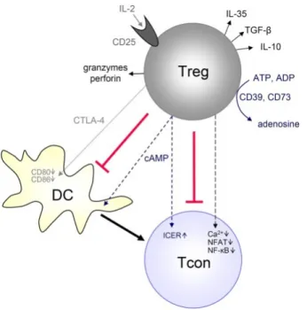
Tregs are able to suppress activation, proliferation and function of distinct immune cells, including CD4+ and CD8+ T lymphocytes, NK and NKT cells, and antigen-presenting cells (APCs) (Dendritic cells [DCs] and macrophages) and B cells70,102. Treg-mediated suppression can occur by cell-to-cell contact or in a cytokine dependent manner, via multiple mechanisms. The activated mechanisms may depend on the local microenvironment, the suppressed target cell or on the state of activation of Tregs (Figure 4)102. Tregs inhibit T cells through release of inhibitory cytokines, such as IL-35, IL-10 or TGF-1β102,103, but a direct cell contact between Tregs and Tconvs is required118,119. In turn, these inhibitory cytokines induce other types of Tregs from CD4+ T cells104. The high levels of IL-2R chain at Tregs surface can lead to competitive consumption of IL-2, scavenging it from other T cells and consequently inhibiting their proliferation; Tregs can generate and secrete adenosine, degrade tryptophan and transfer cAMPs to other T cells, leading to their metabolic disruption105. Via perforin and granzyme-dependent pathways Tregs can directly elicit effector T cell apoptosis. Finally, Tregs can indirectly affect Tconvs by hampering APCs function. CTLA-4, highly expressed at the surface of Tregs, binds to co-stimulatory molecules, such as CD80/CD86 from the surface of APC, leading to their removal106. The direct interaction established between Tregs and APC prevents the stable contacts between APC and the co-stimulatory molecule CD28 of CD4+ T cells, inhibiting T cell activation106.
4.1. Tregs in cancer
Tregs are mediators of peripheral tolerance to self-antigens and innocuous environmental antigens81. Tumour-associated antigens are modified or aberrantly expressed self-antigens. It is, therefore, expected that Tregs are the major precursors of tumour immune evasion107. In fact, Tregs were described to recognize a broad range of tumour-associated self-antigens and to suppress the response of tumour-antigen-specific effector T cells108. Additionally, Tregs have more self-reactive TCR repertoires and thus present higher ability to recognize self-tumour antigens than other CD4+ T cells92.
Previous studies have reported that during tumour initiation Tregs are enriched in the tumour tissue and in advance-stage disease their number increase systemically, in comparison to healthy individuals109. Tregs accumulate at the tumour microenvironment by recruitment, expansion and de novo generation. Tumour cells and stroma components produce and secrete several factors that are chemoattractants for Tregs, recruiting them to
the tumour site. For instance, in colorectal cancer, CCL20 secreted by TAMs induces the recruitment of CCR6+ nTreg cells110 and, in ovarian cancer, TAMs are described to secrete CCL22 that mobilizes CCR4+ nTreg cells111. After recruitment, Tregs are activated and expanded in the tumour. The high levels of TGF-β and IL-10 produced by tumour cells and TAMs favour pre-existing Tregs clonal expansion101. Furthermore, effector T cells were proposed to secret IL-2 that could induce the proliferation of Tregs112. The combined presence of TGF-β and IL-10 with DC or TAMs promotes the conversion of naïve or conventional T cells into suppressive Tregs, Th3 and Tr1113,114. Prostaglandin E2 (PGE2), indoleamine 2,3-dioxygenase (IDO), and TGF-β derived from the tumour have also been correlated with the conversion of iTregs from Tconvs/naïve115,116.
The immunosuppressive role of Tregs was also indirectly correlated with metastatic advantages117,118. In breast cancer models, recruitment of Tregs to the primary tumour, increased CTL and NK cells’ apoptosis and metastasis. Additionally, in the same model, the suppression of lung metastasis was accompanied by decrease of CD4+CD25+ T cells in the
Figure 4. Mechanisms of Treg-mediated suppression. Tregs suppress directly or indirectly conventional T cells (Tcon) via
different mechanisms, depending on the local immune environment, in the target cell or even according to Tregs status. Tregs secrete immunosuppressive adenosine or transfer cAMP to Tconvs, produce immunosuppressive cytokines (10, TGF-β, IL-35), suppress Tconvs by IL-2 consumption or induce effector cell death via granzyme and perforin. Furthermore, Tregs can suppress Tconvs indirectly by downregulating costimulatory molecules on APCs (such as DCs) via CTLA-4. Adapted from Schmidt et al. (2012), Frontiers in immunology.
primary tumour121. Additionally, systemic Treg depletion in patients induced regression of melanoma metastases120.
4.1.1. Prognostic value of Tregs in colorectal cancer
The infiltration of Tregs into tumours has been reported to be associated with an unfavourable outcome in several human cancers, such as ovarian119, breast, gastrointestinal, lung, liver, pancreatic cancer and melanoma120. Contrarily, in CRC the results are controversial with high densities of FoxP3+ Tregs mostly correlating with favourable outcomes121-125 Tregs infiltrate preferentially at the tumour nest (TN)122,125. In a cohort of 967 CRC specimens, high density of FoxP3+ T cells at the TN was associated with better prognosis, whereas their accumulation at the surrounding healthy tissues was associated with worse prognosis122. Similar results were found by Frey et al. in a cohort of 1420 patients, where high FoxP3 infiltration at tumour site was an independent positive predictor of 5-years disease free survival in proficient patients, but not in MMR-deficient patients121. Even in a metastatic setting, higher levels of FoxP3+ T cells, within the tumour of patients subjected to chemotherapy, was associated with better prognosis126. Ling and colleagues demonstrated that high levels of FoxP3+ T cells correlated with a good prognosis in patients with low infiltration of CD8+T cells125. Moreover, they described that accumulation of FoxP3 at the invasive front and TN alone are correlated with better prognosis, bearing strong prognostic information125. Additionally, higher intratumoural infiltration of Tregs than IL-17+ T cells were associated with suppressed MMPs activities and decreased metastases score in CRC cancer upon resection127. Nevertheless, Treg-mediated suppression also involves interactions with APCs, and the prognostic role of the balance between macrophages and/or DCs and Tregs has not yet been performed in a CRC context.
The role of FoxP3+ tumour-infiltrating lymphocytes in CRC may depend on the immune response found at the tumour. If the tumour microenvironment is infiltrated with immune cells that promote tumourigenesis and/or tumour progression, the immunosuppressive role of Tregs may be beneficial. However, if a Th1-like response, characteristic of a tumour antigen-specific immune response is dominant, the suppression by Tregs can be deleterious promoting tumour immune escape and progression128. The controversial results of FoxP3 infiltration can be further explained by the phenotypic and functional heterogeneity of FOXP3+ T cell subpopulations129. The contribution of each Treg subpopulation (rTregs, aTregs and non-Tregs) at CRC microenvironment is still unknown.
A higher infiltration of non-Treg, capable of producing pro-inflammatory cytokines, could contribute to a better prognosis of FoxP3+ T cells in CRC130. The controversial role of Tregs in cancer may also rely on the low inter-study comparability due to the use of different techniques, among others. The role and the prognostic significance of FoxP3+ infiltration and FoxP3+ subpopulation is still scarce. However, it seems that Tregs exert distinct influences at different inflammatory microenvironments, which may be reflected on different FoxP3+ subpopulations128. The impact of Tregs in tumour progression may be determined by the type and location at the tumours as well as by the balance between Tregs and other immune populations within the tumour mass22. It is then imperative to clarify the role of FoxP3+ T cells in the tumour microenvironment, so as to adopt adequate different strategies to manipulate Tregs, in order to improve patient survival.
5. Macrophage-Treg crosstalk
By cell-to-cell contact and in a cytokine-dependent way, Tregs exert a suppressive effect on monocyte/macrophage activation and effector function131,132. In humans, the inhibitory functions of Tregs on monocyte-derived macrophages were provided by in vitro autologous co-cultures between CD4+CD25+ T cells and monocytes131. These monocytes displayed a resting phenotype, producing lower levels of IFN-, TNF-α and IL-10 than when co-cultured with CD4+CD25- T cells. When further challenged with LPS, Treg-educated macrophages were inhibited ofproducing IFN-, TNF-α and IL-6 and exhibited downregulation of CD86 when comparing with control monocytes. The different macrophage activation was reflected in the hampered APC function of Treg-educated macrophages131. Later, Tiemessen and colleagues132, showed that Tregs steered macrophage differentiation towards an immunosuppressive phenotype132, characterized by the upregulation of CD206 and CD163 and downregulation of HLA-DR and CD86. In addition, Treg-educated monocytes increased secretion of CCL18, enhanced phagocytic capacity and displayed a diminished capacity to respond to LPS, producing low levels of pro-inflammatory cytokines (IL1-β, IL-6, IL-8 TNF-α). The authors described that the upregulation of CD163 was dependent on IL-10. Contrarily, the upregulation of CD206 was cytokine-independent suggesting the need of cell-to-cell contact, which was further demonstrated using transwell assays132. Kryczek et al. reported a similar suppressive profile on human macrophages cultured with Tregs, but further described that the higher levels of IL-10 produced by Treg-educated macrophages induced the upregulation of the Tconvs cell inhibitory molecule B7-H4 on monocytes133. In
line with these reports, the presence of Tregs after myocardial infarction induced macrophages toward an anti-inflammatory profile, contributing to inflammation resolution and wound healing135, in a mouse model. In the physiologic intestinal microenvironment, commensal microbiota promotes colonic Tregs differentiation, which in turn contribute to the immune homeostasis in the colon136. Tregs differentiation is also favoured by colonic DCs which repress the expression of LPS response genes, promoting microbiota protection, as well as, inhibiting macrophage activation due to hyporesponsiveness to TLR stimulation136. Nevertheless, the modulatory role of Tregs on macrophages phenotype and functions in a cancer context is still scarce.
Conversely, macrophages induce Tregs differentiation113,114 and recruitment137, extending their population within tumours and maintaining an immunosuppressive microenvironment that may favour tumour progression138. Along with cancer cells, TAMs are major source of TGF-β and directly induce Tregs by cell-to-cell interaction via membrane bound TGF-β137. In a colitis mouse model, macrophage-derived IL-10 was described to maintain FoxP3 expression in Tregs139. Further, in an immunocompetent mouse model with renal injury, IL-10/TGF-β–modified macrophages attenuated renal inflammation140. The suppression of CD4+ T cells by IL-10/TGF-β–modified macrophages induced proliferation regulatory T cells from CD4+CD25- T cells and increase Tregs in renal draining lymph nodes140. These results highlight the role of anti-inflammatory macrophages in de novo generation and recruitment of Tregs. Nevertheless, under pro-inflammatory conditions (e.g. rheumatoid arthritis), monocyte-derived IL-6, IFN- and IL-1β was described to induce aTregs to produce both pro-inflammatory (IL-17, IFN-, TNF-α) and anti-inflammatory (IL-10) cytokines, without losing their suppressive functions141. These data indicate that different
macrophage subsets display distinct roles in the modulation of Tregs’ properties, however the exact mechanisms underlying these modulation is still not completely understood. Furthermore, it is also likely that different Treg subpopulations could distinctly modulate macrophage inflammatory profiles.
Aim
Tumours are considered malignant when cancer cells invade the surrounding tissues, becoming prone to intravasate vessels and to metastasize. TAMs are considered obligate partners in the malignant transformation. Additionally, Tregs, which display immunosuppressive properties facilitate tumour immune escape, favouring cancer cell invasion. Given the modulatory role of Tregs on macrophage inflammatory phenotype, it is likely that Treg-educated macrophages may have enhanced pro-tumour activities. However, in CRC the role of TAMs and Tregs is controversial since their intratumoural infiltration is often correlated with good prognosis. Also, the influence of Tregs on monocyte/macrophage pro-invasive properties is presently unknown. Similarly, the phenotypic and functional profile of these immune cell populations and respective subpopulations in a CRC context remains to be elucidated.
Therefore, the main goal of this project is to better understand the role of Tregs on macrophages behaviour, uncovering their impact on CRC cell invasion. Specific objectives include:
1) Evaluate the impact of Tregs on monocyte-derived macrophage differentiation; 2) Assess the role of Tregs-educated macrophages on colorectal cancer cell
invasion;
3) Investigate the mechanisms underlying Tregs modulation of macrophage-mediated cancer cell invasion, such as on MMPs proteolytic activity and the activation of the EGFR-invasion related signalling pathway;
Materials and methods
Cell line culture
The RKO (CRL-2577) and SW620 (CCL-227) stablished cancer cell lines, derived from human colon carcinomas, were obtained from the American Type Culture Collection (ATCC, Manassas, VA, USA) in 2012. RKO and SW620 cells were maintained at 37ºC, under a 5% CO2 humidified-atmosphere, in complete RPMI1640-GLUTAMAX medium (Invitrogen, Merelbeke, Belgium) or in DMEM+F12 medium (Invitrogen), respectively. Media were supplemented with 10% fetal bovine serum (FBS) (Lonza, Basel, Switzerland), 100 U/ml penicillin and 100 mg/ml streptomycin (Invitrogen).
Immune cells isolation
Human monocytes and regulatory T cells were isolated from healthy blood donors’ buffy coats, provided by the Immunohemotherapy Department, Hospital São João, Porto, Portugal. For an autologous setting, the same blood donor was used to isolate CD14+ monocytes and CD4+CD25+ T cells. To combine monocyte with BD regulatory T cell isolation (BD Biosciences, Madrid, Spain), buffy coats were initially divided. Alternatively, to combine monocyte isolation with the StemCell regulatory T cell isolation kit (StemCell Technologies, Grenoble, France), peripheral blood mononuclear cells (PBMCs) collected from buffy coats were divided.
CD14
+monocyte isolation
Monocytes were isolated using the Human Monocyte Enrichment Cocktail (StemCell Technologies), which consists in a negative selection of CD14+ cells. From our previous work using this isolation system, over 80% of isolated monocytes were found to be CD14-positive57. Briefly, buffy coats were centrifuged at 1200 xg, without brake, for 20 minutes at room temperature (RT). The whitish layer containing PBMCs was then collected and used to isolate CD14+ monocytes. PBMCs were incubated for 20 minutes under rotation with the RosetteSep™ Human Monocyte Enrichment Cocktail (StemCell Technologies), according to manufacturer’s instructions. The enriched mixture was diluted (1:1 ratio) in phosphate buffered saline (PBS) supplemented with 2% FBS (Lonza), gently layered over equal volume of Histopaque®-1077 (Sigma-Aldrich, Madrid, Spain) and centrifuged at 1200 xg, without break, for 20 minutes at RT. The interface layer, enriched in human CD14+ monocytes, was
collected, washed with PBS and centrifuged at 98 xg, with brake, for 17 minutes at 4ºC. The supernatant was discarded and the pellet with the isolated cells were resuspended with complete RMPI1640-GLUTAMAX medium supplemented with 10% FBS, 100 U/ml penicillin and 100 mg/ml streptomycin. For the in vitro studies, monocytes were seeded in the same culture medium, according to the experimental set up.
Human CD4
+CD25
highT cell isolation
The Human Regulatory T Lymphocyte Separation Set (BD Biosciences) or the Regulatory T cell Isolation Kit (StemCell Technologies) were used to isolate the broadest phenotypic variety of Tregs. Isolations consisted in a negative (CD4+) followed by a positive (CD25+) selection of cells.
1. Human Regulatory T Lymphocyte Separation Set (BD Biosciences)
To isolate PBMCs, blood from buffy coats were gently layered over Histopaque®-1077 and centrifuged at 1200 xg, without brake, for 20 minutes at RT. The whitish layer containing PBMCs was then collected, washed with PBS, centrifuged at 18 xg for 10 minutes and the supernatant discarded. Then, PBMCs were resuspended in the Human Regulatory T Lymphocyte Separation Cocktail for 15 minutes at RT. The labelled cells were washed in 1x BD IMag™ buffer (1:10 ratio), centrifuged at 300 xg for 8 minutes, and the supernatant discarded. For CD4+ T lymphocytes enrichment, cell pellet was resuspended in BD IMag™ Streptavidin Particles Plus – DM and incubated for additional 30 minutes at RT. The labelled cell suspension was brought to 60x106 cells/mL with 1x BD IMag™ buffer, transferred to 5 mL round-bottom polystyrene tube (BD Falcon) and placed on the EasySep® magnet for 7 minutes. The supernatant (enriched fraction) was transferred to a new sterile tube. The enrichment step was repeated twice. The tube containing the combined enriched fraction was placed on the magnet for 7 minutes and the supernatant was removed to a new tube for a twice-enriched fraction containing CD4+ T lymphocytes. For the positive selection of CD4+CD25+ T cells, the twice-enriched fraction was centrifuged, as previously, and the cell pellet was resuspended in the BD IMag™ Anti-APC Particles – DM and incubated for 30 minutes at RT. The labelled cell suspension was brought to 60x106 cells/mL with 1X BD IMag™ buffer, transferred to 5 mL round-bottom tube and placed on the magnet for 8 minutes. With the tube on the magnet, supernatant was discarded. After two wash steps, the tube was removed from the magnet and isolated CD4+CD25+ T cells were resuspended in complete RMPI1640-GLUTAMAX medium supplemented with 10% FBS, 100 U/mL penicillin, 100 mg/mL streptomycin and 50 ng/mL anti-CD3 (OKT3, Abcam, UK).
2. Regulatory T cell Isolation Kit (StemCell Technologies)
Buffy coats were centrifuge at 1200 xg, without brake, for 20 minutes at RT. The whitish layer containing PBMCs was then collected and used to isolate CD4+CD25+ T cells. PBMCs were incubated for 20 minutes under rotation with RosetteSep® Human CD4+ T Cell Enrichment Cocktail, following manufacturer’s instructions. The enriched mixture was diluted (1:2 ratio) in PBS + 2% FBS, gently layered over Histopaque®-1077 and centrifuged, as previously. The interface layer, enriched in CD4+ cells, was collected, washed with PBS + 2% FBS, centrifuged at 1200 xg, with brake, for 17 minutes at 4ºC and the supernatant was discarded. The enriched sample was transferred to a 5 mL round-bottom polystyrene tube and incubated under rotation with EasySep® Positive Selection Cocktail, for 15 minutes. Then, EasySep® Magnetic Nanoparticles were added to the suspension, for 10 minutes, following manufacturer’s instruction. The cell suspension was brought to a volume of 2.5 mL with PBS + 2% FBS, resuspended and place into the magnet for 5 minutes. The supernatant fraction was then discarded while the magnetically labelled cells remained inside the tube. The positively selected cells were resuspended with complete RMPI1640-GLUTAMAX medium supplemented with 10% FBS, 100 U/mL penicillin, 100 mg/ml streptomycin and 50 ng/mL anti-CD3 (Abcam). For the in vitro studies Tregs were seeded in the same culture medium. A fraction of CD4+CD25+ T cells was analysed by flow cytometry to evaluate the efficiency and purity of the cell separation procedure. Isolation resulted in 77% CD4+CD25+ T cells with 94% expressing FoxP3.
Mono-culture of human CD14
+monocytes or of CD4
+CD25
+T cells
Isolated human CD14+ monocytes from two different healthy blood donors were seeded in 58 cm2 tissue-culture petri dishes (Greiner, Frickenhausen, Germany) at a density of 1x10⁷ monocytes per plate and maintained at 37ºC, under a 5% CO2 humidified-atmosphere for 24 hours. Then, monocytes were transferred to three different substrates: 6-well tissue culture-treated plates (Falcon, Durham, USA) at a density of 1,2x10⁶ monocytes per well (0,13 x10⁶ monocytes/cm2); 6-well tissue culture-nontreated plates at a density of 1,2x10⁶ monocytes per well (0,13 x10⁶ monocytes/cm2) (Falcon); and 24-well tissue culture-treated plates (Falcon) with coverslips (Deckglasser, Lauda-Konigshofen, Germany) at a density of 5x10⁵ monocytes per well (0,26 x10⁶ monocytes/cm2). Non-treated plates are hydrophobic polystyrene surfaces whereas treated plates are hydrophobic polystyrene surfaces that where chemically treated to become hydrophilic, usually by increasing their negative charge,