Recebido para publicação: Março de 2010 • Aceite para publicação: Abril de 2010
1245
Enfarte agudo do miocárdio associado
a origem anómala das artérias coronárias
(um caso raro de origem da artéria
coronária esquerda no ostio da artéria
coronária direita)
[83]
ALEXANDRARAMOS, LUÍSBRIZIDA, RAMIROCARVALHO, FERNANDOMATIAS, LUÍSMOURÃO
Serviço de Cardiologia, Hospital Curry Cabral, Lisboa, Portugal
Rev Port Cardiol 2010; 29 (07-08): 1245-1251
RESUMO
A origem da artéria coronária esquerda a partir do ostio ou do segmento proximal da artéria coronária direita é uma anomalia congénita rara (0,03 a 0,4%), e um achado angiográfico pouco comum (0,6 a 1,3%) (artéria coronária única). A anomalia congénita coronária mais frequente é a origem separada da artéria descendente anterior e da artéria circunflexa no seio coronário esquerdo. A segunda anomalia mais comum é a origem da artéria circunflexa no seio coronário direito. Descrevemos um caso de enfarte agudo do miocárdio com localização infero lateral, que evoluiu com choque cardiogénico e disfunção ventricular esquerda graves, após angioplastia primária de oclusão proximal da artéria coronária direita, e em que não foi possível cateterizar a artéria coronária esquerda por inexistência de ostio no seio coronário esquerdo. Destaca-se o papel importante das novas técnicas de imagem, nomeadamente da angio-TC das coronárias, na identificação das
anomalias congénitas das artérias coronárias.
Palavras-chave:
Enfarte do miocárdio; Anomalias congénitas das artérias coronárias;
Tomografia computorizada
Acute myocardial infarction associated with anomalous origin of coronary arteries (a rare case of origin of the left coronary artery from the ostium of the right coronary artery)
ABSTRACT
Origin of the left coronary artery from the ostium or proximal segment of the right coro-nary artery (single corocoro-nary artery) is a rare congenital anomaly (0.03 to 0.4%) and an unusual angiographic finding (0.6 to 1.3%). The most common congenital anomaly is separate origin of the left anterior descending artery and circumflex artery from the left coronary sinus.
The second most common anomaly is the origin of the circumflex artery from the right coronary sinus.
We present a patient with acute inferior myo-cardial infarction and cardiogenic shock, in whom it was not possible to perform catheteri-zation of the left coronary artery because of the absence of the ostium of the left coronary sinus. Multislice computed tomography of the coro-nary arteries was the tool that enabled us to visualize the origin and course of the left coronary artery.
Key words
Myocardial infarction; Congenital coronary anomalies; Multislice computed angiography
CASO CLÍNICO
T
rata-se de uma doente do sexo feminino, de 72 anos, que tem, como factores de ris-co para doença das ris-coronárias, hipertensão arterial e diabetes não insulino tratada.Foi internada no nosso serviço por enfarte agudo do miocárdio com supra desnivelamento do segmento ST de localização inferolateral.
Realizou-se Coronariografia de urgência, com 4h 30 min de evolução, que revelou oclu-são da artéria coronária direita no seu segmen-to proximal Não foi visualizada e não se conse-guiu cateterizar a artéria coronária esquerda.
Foi feita angioplastia coronária, com colo-cação de stent metálico Driver 3,5x 9,00 mm na artéria coronária direita proximal e de Stent metálico Driver 3,00x18,oo mm na artéria co-ronária direita distal.
Foi necessária a colocação de pacemaker provisório por ritmo idioventricular muito len-to e hipotensão
Na Unidade Coronária instalou-se quadro de choque cardiogénico, com necessidade de ventilação invasiva e de terapêutica com ami-nas simpaticomiméticas e vasopressores.
O ecocardiograma revelou ventrículo es-querdo com função sistólica muito diminuída (fracção de ejecção 30%) e ventrículo direito não dilatado e com cinética conservada.
Verificou-se melhoria progressiva hemodi-nâmica, com extubação ao quinto dia.
O ECG evoluiu com onda Q em D2, D3, AVF, V4, V5 e V6 e QS em V1, V2 e V3 (en-farte combinado inferior e anterior).
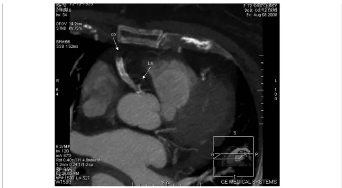
Como não tinha sido possível cateterizar a artéria coronária esquerda, e suspeitando-se de origem anómala, foi decidido realizar angio-TC das artérias coronárias que mostrou (Figura 1, Figura 2, Figura 3 e Figura 4): arté-ria descendente anterior e artéarté-ria circunflexa com origem anómala, nascendo ambas do ostio da artéria coronária direita; artéria des-cendente anterior com percurso anterior e com placa mole significativa; artéria Circunflexa com percurso posterior e é um vaso doente antes da primeira obtusa marginal.
Tendo a doente a função ventricular es-querda diminuída, havendo a suspeita de
en-CASE REPORT
A
72-year-old female patient with hyper-tension and treated type 2 diabetes as risk factors for coronary disease was admitted to our department with inferolateral ST-seg-ment elevation acute myocardial infarction.Urgent coronary angiography carried out at 4 hours 30 min of evolution revealed occlu-sion of the proximal segment of the right coro-nary artery; the left corocoro-nary artery could not be visualized or catheterized.
Coronary angioplasty was performed, with a 3.5 x 9.00 mm Driver bare-metal stent being implanted in the proximal right coronary artery and a 3.0 x 8.00 mm Driver bare-metal stent implanted in the distal right coronary artery.
A provisional pacemaker was implanted due to very slow idioventricular rhythm and hy-potension.
In the coronary care unit, a setting of cardio-genic shock began, the patient requiring inva-sive ventilation and sympathomimetic amines and vasopressors.
The echocardiogram revealed very poor left ventricular (LV) systolic function (ejection fraction 30%) and a non-dilated right ventri-cle with preserved kinetics.
Progressive hemodynamic improvement was seen and she was extubated on the 5th day.
The ECG showed Q waves in D2, D3, aVF, V4, V5 and V6, and QS complex in V1, V2 and V3 (combined inferior and anterior infarc-tion).
Since the left coronary artery could not be catheterized, and an anomalous origin was suspected, it was decided to perform CT angiography of the coronary arteries, which showed (Figures 1-4) anomalous origin of the anterior descending and circumflex arteries, both arising from the ostium of the right coro-nary artery; anterior descending artery with an anterior course and a significant soft plaque; circumflex artery with a posterior course; and a diseased vessel before the first obtuse mar-ginal.
Given the patient’s diminished ventricular
1246
1247
Recebido para publicação: ????????????? • Aceite para publicação: ?????????
Figura 1. AngioTC: origem da artéria descendente anterior no ostio da artéria coronária direita com estenose proximal
Figure 1. TC angiography: origin of the anterior descending artery in the ostium of the right coronary artery, with proximal stenosis.
Figura 2. Angio TC: origem anómala da artéria descendente anterior no ostio da artéria coronária direita
Figure 2. TC angiography: anomalous origin of the anterior descending artery in the ostium of the right coronary artery.
farte combinado (inferior e anterior) e verifi-cando-se a existência de placa na artéria des-cendente anterior, decidiu-se a realização de cintigrafia de perfusão miocárdica, com a fi-nalidade de avaliar a presença de isquemia no território desta artéria.
function, the suspicion of combined infarction (inferior and anterior) and the existence of atherosclerotic plaque in the left anterior descending artery, it was decided to perform myocardial perfusion scintigraphy in order to assess the presence of ischemia in the
territo-Este exame revelou: defeito de captação grave de toda a parede inferolateral e porção adjacente da parede inferior com reversibili-dade ligeira; defeito de captação grave do apex, segmentos médio e apical da parede anterior, porções adjacentes do segmento septoapical e da restante parede anteroseptal e segmento lateroapical com reversibilidade, persistindo pequena área de hipocaptação anteroseptal; fracção de ejecção do ventrículo esquerdo 43% com volumes normais.
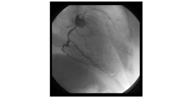
Tendo-se confirmado a ocorrência de en-farte inferolateral e anteroseptal com extensas áreas de isquemia reversível da parede ante-rior, bem como a existência de placa signi-ficativa na artéria descendente anterior, rea-lizou-se nova coronariografia para catete-rização da artéria coronária esquerda (Figura 5 e Figura 6).
Este exame revelou a origem da artéria coronária esquerda logo após o ostio da artéria coronária direita; artéria descendente anterior com estenose proximal de 95%; artéria circun-flexa atrófica; artéria coronária direita domi-nante e sem reestenose dos stents previamente colocados; artéria postero-lateral com lesão de 60-70% na origem; insuficiência mitral ligeira
ry of this artery. This revealed a severe uptake defect throughout the inferolateral wall and in the adjacent part of the inferior wall, slightly reversible; a severe uptake defect in the apex, mid and apical segments of the anterior wall, parts adjacent to the septoapical segment and the remainder of the anteroseptal and lat-eroapical segment, reversible, with a small persistent area of reduced anteroseptal uptake; and LV ejection fraction of 43%, with normal volumes.
These findings confirmed inferolateral and anteroseptal infarction with extensive areas of reversible ischemia in the anterior wall, as well as the existence of significant plaque in the anterior descending artery. Coronary angiography of the left coronary artery was repeated (Figures 5 and 6), which revealed the origin of the left coronary artery immediately after the ostium of the right coronary artery; 95% proximal stenosis of the anterior descending artery; atrophied circumflex artery; dominant right coronary artery, without restenosis of the previously implanted stents; posterolateral artery with 60-70% stenosis at the origin; mild mitral regurgitation; and reduced left ventricular function, with
ejec-1248
Recebido para publicação: ????????????? • Aceite para publicação: ?????????
Figura 3. AngioTC: origem anómala das artérias descendente anterior e circunflexa no ostio da artéria coronária direita e lesão
proximal da artéria descendente anterior
Figure 3. TC angiography: anomalous origin of the anterior descending and circumflex arteries from the ostium of the right coronary
e função do ventrículo esquerdo diminuída com fracção de ejecção calculada em 35%.
O ecocardiograma realizado revelou: aurí-cula esquerda ligeiramente dilatada; ven-trículo esquerdo não dilatado, com fracção de ejecção 39%, com hipocinésia do segmento médio e apical da parede inferior, do septo e segmento apical da parede anterior; insufi-ciência mitral ligeira (área do orifício regurgi-tante 0,14 cm2 e volume regurgiregurgi-tante 21 ml).
1249
Recebido para publicação: ????????????? • Aceite para publicação: ?????????
Figura 4. AngioTC: origem anómala das artérias descendente anterior e circunflexa no ostio da artéria coronária direita e lesão
proximal da artéria descendente anterior
Figure 4. TC angiography: anomalous origin of the anterior descending and circumflex arteries from the ostium of the right coronary
artery, with proximal stenosis of the anterior descending artery.
tion fraction estimated at 35%.
The echocardiogram revealed a slightly dilated left atrium; non-dilated left ventricle, ejection fraction of 39%, with hypokinesia of the mid and apical segments of the inferior wall and septum and the apical segment of the anterior wall; and mild mitral regurgitation (regurgitant orifice area 0.14 cm2and
regurgi-tant volume 21 ml).
The patient was referred for myocardial
Figura 5. Coronariografia: origem anómala das artérias descendente anterior e circunflexa no ostio da artéria coronária direita e lesão
proximal da artéria descendente anterior
Figure 5. Coronary angiography: anomalous origin of the anterior descending and circumflex arteries from the ostium of the right
Foi proposta a cirurgia de revascularização miocárdica com a realização de pontagem de artéria mamária esquerda para a artéria descendente anterior e de veia safena para a artéria postero-lateral.
Após a cirurgia a doente encontra-se sem angor e em classe funcional I-II da NYHA.
O ecocardiograma realizado dois meses após a cirurgia revela aurícula esquerda ligei-ramente dilatada; ventrículo esquerdo com hi-pocinésia inferomediana, inferoapical e ante-roapical e fracção de ejecção de 40% e insufi-ciência mitral ligeira.
DISCUSSÃO
Esta anomalia coronária é das mais raras mencionadas na literatura(1-7). O facto de a
revascularization surgery with left mammary-left anterior descending and saphenous vein-posterolateral artery grafts.
Following surgery she is without pain and in NYHA functional class I-II.
Echocardiographic study two months after surgery showed a slightly dilated left atrium, inferomedial, inferoapical and anteroapical hypokinesis of the left ventricle, ejection frac-tion of 40% and mild mitral regurgitafrac-tion.
DISCUSSION
The coronary anomaly reported here is one of the rarest in the literature(1-7). The fact that
the patient presented with a clinical setting of ST-segment elevation myocardial infarction and hemodynamic instability, a situation in
1250
Recebido para publicação: ????????????? • Aceite para publicação: ?????????
Figura 6. Coronariografia: origem anómala das artérias descendente anterior e circunflexa no ostio da artéria coronária direita e lesão
proximal da artéria descendente anterior
Figure 6. Coronary angiography: anomalous origin of the anterior descending and circumflex arteries from the ostium of the right
coro-nary artery, with proximal stenosis of the anterior descending artery.
doente se apresentar com uma quadro clínico de enfarte com supradesnivelamento do seg-mento ST e com instabilidade hemodinâmica, em que “tempo é músculo”, e ainda a presen-ça de oclusão proximal da artéria coronária direita, condicionou a abordagem terapêutica (angioplastia) e o facto de não se fazerem mais
which “time is muscle”, as well as the pres-ence of occlusion of the proximal right coro-nary artery, determined the therapeutic approach (angioplasty) and meant no further attempts were made to catheterize the left coronary artery.
tentativas de cateterização da artéria coroná-ria esquerda.
O quadro de choque cardiogénico na Uni-dade Coronária, as alterações da cinética segmentar do VE, correspondentes ao ter-ritório da artéria descendente anterior, e a evolução electrocardiográfica, levaram-nos ao diagnóstico de enfarte combinado inferior e anterior.
A angio-TC das artérias coronárias pareceu--nos o exame indicado para a visualização da origem da artéria coronária esquerda(8,9).
Pensamos que o período de hipotensão e bradicárdia, durante a realização de interven-ção coronária, aliado à existência de lesão crítica no segmento proximal da artéria des-cendente anterior, foram os responsáveis pela evolução para enfarte combinado.
coronary care unit, the LV regional wall motion alterations corresponding to the terri-tory of the left anterior descending artery, and the electrocardiographic evolution, led us to diagnose combined inferior and anterior infarction.
We considered TC angiography of the coro-nary arteries to be the exam of choice to visu-alize the origin of the left coronary artery(8, 9).
We believe that the period of hypotension and bradycardia during the percutaneous coronary intervention, together with the exis-tence of a critical lesion in the proximal seg-ment of the left anterior descending artery, were responsible for the evolution to com-bined infarction.
Pedido de Separatas Adress for Reprints Alexandra Ramos Serviço de Cardiologia Hospital Curry Cabral Rua da Beneficência, 8 1069-166 LISBOA
1251
Recebido para publicação: ????????????? • Aceite para publicação: ?????????
1. Turkay C, Golbasi I, Bayezid O. A single coronary artery from the right sinus of Valsalva associated with atherosclero-sis. Acta Cardiol.2002 Oct;57(5):377-9
2. Makaryus AN, Orlando J, Katz S. Anomalous origin of the left coronary artery from the right coronary artery: a rare case of a single coronary artery originating from the right sinus of Valsalva in a man with suspected coronary artery disease: Invasive Cardiol. 2005 Jan;17(1):56-8
3.Kang WC, Ahn TH, Shin EK. Successful percutaneous nary intervention for severe stenosis of an anomalous left coro-nary artery originating from the proximal right corocoro-nary artery. Invasive Cardiol. 2006 May; 18(%):E154-6
4.Yokoyama HA, Pessoa CM, Carvalho FC, Campos RF, Franco RJ, Bregagnollo EA. Percutaneous coronary interven-tion in single coronary artery in a patient with high risk unsta-ble angina. Arq Bras Cardiol 2007 Mar; 88(3):e53-5 5.Latsios G, Tsioufis K, Tousoulis D, Kallikazaros I, Stefanadis C. Common origin of both right and left coronary arteries from the right sinus of Valsalva. Int J Cardiol. 2008 Aug 18; 128(2): e60
BIBLIOGRAFIA / REFERENCES
6. Vincelj J, Todorovic N, Marusic P, Puksic S. Anomalous ori-gin of the left coronary artery from the right sinus of Valsalva in a 62-year-old woman with unstable angina pectoris: A case report. Int J Cardiol, 2009 Jan 12.
7. Raddino R, Pedrinazzi C, Zanini G, Leonzi O, Robba D, Chieppa F, Portera C, Dei Cas L. Percutaneous coronary angio-plasty in a patient with anomalous single coronary artery aris-ing from the right sinus of Valsalva. Int J Cardiol. 2006 Oct 10; 112(3):e60-2
8. El-Menyar AA, Das KM, Al-Suwaidi J. Anomalous origin of the three coronary arteries from the right aortic sinus of Valsalva: role of MDCT coronary angiography. Int J Cardiovasc Imaging. 2006 Oct; 22(5):723-9
9. Komatsu S, Sato Y, Ichikawa M, Kunimasa T, Ito S, Takagi T, Lee T, Matsumoto N, Takayama T, Hiryama A, Mishima M, Saito S, Kodama K. Anomalous coronary arteries in adults detected by multislice computed tomography: presentation of cases from multicenter registry and review of the literature. Heart Vessels. 2008 Jan; 23(1): 26-34