Universidade de Aveiro 2015
Departamento de Biologia
Patrícia Alexandra
Sousa Jegundo
Polimorfismos de genes com atividade antioxidante
e desintoxicante na predisposição para o Cancro
Colo-retal
Antioxidant and detoxify genes polymorphisms in
colorectal cancer predisposition
DECLARAÇÃO
Declaro que este relatório é integralmente da minha autoria, estando devidamente referenciadas as fontes e obras consultadas, bem como identificadas de modo claro as citações dessas obras. Não contém, por isso, qualquer tipo de plágio quer de textos publicados, qualquer que seja o meio dessa publicação, incluindo meios eletrónicos, quer de trabalhos académicos.
Universidade de Aveiro 2015
Departamento de Biologia
Patrícia Alexandra
Sousa Jegundo
Polimorfismos de genes com atividade antioxidante
e desintoxicante na predisposição para o Cancro
Colo-retal
Antioxidant and detoxify genes polymorphisms in
colorectal cancer predisposition
Dissertação apresentada à Universidade de Aveiro para cumprimento dos requisitos necessários à obtenção do grau de Mestre em Biologia Molecular e Celular, realizada sob a orientação científica da Professora Doutora Lina Carvalho, Professora Associada com Agregação do Instituto de Anatomia Patológica da Faculdade de Medicina da Universidade de Coimbra e da Mestre Sandra Balseiro, Professora Assistente da Escola Superior e Saúde Dr. Lopes Dias, Instituto Politécnico Castelo Branco e da Professora Doutora Maria de Lourdes Pereira, Professora Associada com Agregação do Departamento de Biologia da Universidade de Aveiro.
Este trabalho contou com a colaboração do Instituto de Anatomia e Patologia Molecular da Faculdade de Medicina da Universidade de Coimbra e foi financiado pelos projetos da CIMAGO (projeto nº19/2009) e do GAI (projeto LCarvalho.GAI2015) da FMUC.
Dedico este trabalho aos profissionais de saúde, pacientes e famílias que se deparam com a luta contra o cancro no seu dia-a-dia, bem como, aos que de algum modo deram o seu contributo para esta batalha.
“You may have to fight a battle more than once to win it.” Margaret Thatcher
O júri
Presidente Prof. Doutor António Carlos Matias Correia,
Professor Catedrático, Departamento de Biologia, Universidade de Aveiro
Prof. Doutora Lina Carvalho
Professora Associada com Agregação, Faculdade de Medicina, Universidade de Coimbra
Prof. Doutor Fernando de Jesus Regateiro
Agradecimentos Em primeiro lugar agradeço às minhas orientadoras. À Professora Doutora Lina Carvalho pela oportunidade que me deu para desenvolver a minha tese no serviço de Anatomia Patológica da FMUC, pela sua disponibilidade e orientação científica. À Mestre Sandra Balseiro também pela oportunidade e disponibilidade, bem como, pela transmissão do seu conhecimento, por todo o ensinamento, paciência e ajuda no contorno dos obstáculos, pelo apoio e pela amizade durante todos estes anos que me leccionou e orientou. E à minha co-orientadora Professora Doutora Maria de Lourdes Pereira pela sua orientação interna na Universidade de Aveiro.
Agradeço também aos meus colegas de trabalho Raquel Pandeirada, Sofia Fernandes, Tifanny e José Pedro Mendes pela cooperação dentro e fora do laboratório, pelo bom ambiente partilhado e pelas suas amizades.
Não menos importante, agradeço à equipa do serviço do IAP-FMUC, nomeadamente, à Maria Silva, à Maria João e à Natália pela disponibilidade que sempre demonstraram e pelo bom ambiente que proporcionaram.
Agradeço ainda à Doutora Adriana Brito pela flexibilidade de trabalho que me deu, permitindo conciliar trabalho e estudos.
Por fim e em especial, agradeço à minha família, namorado e amigos pelo incentivo, pelo apoio incondicional, pela motivação e por acreditarem sempre nas minhas capacidades e no meu trabalho, nos bons e maus momentos. Cada um da sua maneira contribuiu para a realização de mais este meu projeto, por isso um grande bem-haja a todos.
Palavras-chave Adenocarcinamo colo-retal esporádico; Superóxido Dismutases; Glutationas S-Transferases; Polimorfismos comuns; PCR.
Resumo O cancro colo-retal (CCR) resulta de um conjunto gradual de alterações histológicas e genéticas, que se traduz numa proliferação celular descontrolada. Embora existam causas genéticas para a origem deste carcinoma, a maioria dos autores assume causas multifactoriais para a génese do CCR. Das causas não genéticas, a baixa atividade física, uma dieta rica em gorduras e pobre em fibras, bem como, os hábitos tabágicos parecem ter um papel preponderante no desenvolvimento desta patologia. Do mesmo modo, tem vindo a ser descrito que diferentes níveis de stresse oxidativo podem influenciar o desenvolvimento deste tipo de cancro. Desta forma, a manutenção celular do estado oxidação-redução parece ser crucial para a conservação da função dos tecidos e prevenção da carcinogénese. Variações genotípicas nos genes envolvidos neste processo, tais como, MNSOD, SOD3, GSTP1, GSTT1 e GSTM1, podem ser importantes biomarcadores para o CCR.
Neste trabalho pretendeu-se determinar a distribuição da frequência dos polimorfismos mais comuns dos genes envolvidos na regulação do stresse oxidativo (MNSOD, SOD3, GSTP1, GSTT1 e GSTM1) em indivíduos com adenocarcinoma colo-retal esporádico (ACE) e em controlos saudáveis, avaliando assim a sua possível correlação com o risco para o desenvolvimento do ACE. A análise dos polimorfismos dos genes com atividade desintoxicante e desintoxicante (MNSOD T175C, SOD3 R213G, GSTP1 A105G, GSTP1 C114T, GSTT1del e GSTM1del) foi feita através da técnica de PCR-SSP. Neste estudo encontrámos uma prevalência dos genes mutados nos pacientes com ACE, comparativamente com o grupo controlo: MNSOD 175CC (55% vs 2%; p<0,0001; OR: 58,5; CI 13,3 a 256,7), SOD3 213GG (31% vs 2%; p<0,0001; OR: 21,89; CI 4,93 a 97,29), GSTP1 105GG (46% vs 12%; p<0,0001; OR: 6,14; CI 2,85 a 13,26), GSTP1 114TT 38% vs 0%; p<0,0001; OR: Infinito) e GSTT1 del (75% vs 28%; p<0,0001; OR: 7,71; CI 3,83 a 15,56). Além disto, observámos também que os genótipos mutados GSTP1 114TT
(52% vs 27%; p=0,003; OR: 2,88; CI: 1,41 a 5,89) e GSTT1 del (87% vs 65%; p=0.003; OR: 3.66; CI 1.51 a 8.84) estavam associados com o colon.
Deste modo, os nossos resultados sugerem uma associação positiva entre os polimorfismos dos genes estudados e a prevalência do ACE. Assim sendo, a desregulação dos genes MNSOD, SOD3, GSTP1, GSTT1 e GSTM1 pode ser associada com um aumento de ROS no tecido do colon-retal. Além disto, o stresse oxidativo nas células do tecido colon-retal pode também induzir uma desregulação da via da p53. Este estudo evidência assim que os polimorfismos MNSOD 175C, SOD3 213G, GSTP1 105G, GSTP1 114T e
GSTT1 del poderão estar envolvidos no risco para o ACE, permitindo clarificar esta patologia multifactorial.
Keywords Sporadic coloretal adenocarcinoma; Superoxide dismutases; Glutatione S-transferases; common Polymorfisms; PCR.
Abstract Colorectal cancer (CRC) results from histologic and gene alterations can lead to a massive cellular proliferation. Most of the authors assume multifactorial causes to CRC genesis. Low physical activity, a fat diet poor in fibers and smoking habits seems to have an important role in CRC. However, there are also genetic causes associated with CRC risk. It has been described that oxidative stress levels could influence CRC development. Thus, cellular balance reactive species and defense enzymes involved in oxidative stress are crucial to maintain a good tissue function and avoid neoplasic process. Therefore, genome variations on these defense enzymes, such as MNSOD, SOD3, GSTP1, GSTT1 and GSTM1, could be important biomarkers to colorectal adenocarcinomas.
We intend to determine frequencies distribution of most common polymorphisms involved on oxidative stress regulation (MNSOD, SOD3, GSTP1, GSTT1 and GSTM1) in patients with sporadic colorectal adenocarcinoma (SCA) and in healthy controls, evaluation their possible correlation with SCA risk. Samples common polymorphisms of antioxidant and detoxify genes (MNSOD T175C, SOD3 R213G, GSTP1 A105G, GSTP1 C114T, GSTT1del and GSTM1del) analysis was done by PCR-SSP techniques.
In this study we found a higher prevalence of MNSOD 175CC (55% vs 2%; p<0.0001; OR: 58.5; CI 13.3 to 256.7), SOD3 213GG (31% vs 2%; p<0.0001; OR: 21.89; CI 4.93 to 97.29), GSTP1 105GG (46% vs 12%; p<0.0001; OR: 6.14; CI 2.85 to 13.26), GSTP1 114TT (38% vs 0%; p<0.0001; OR: Infinity) and GSTT1 null (75% vs 28%; p<0.0001; OR: 7.71; CI 3.83 to 15.56) mutated genotypes among SCA patients, while the normal genotypes were associated with SCA absence. Furthermore, we found GSTP1 114TT mutated genotype (52% vs 27%; p=0.003; OR: 2.88; CI: 1.41 to 5.89) and GSTT1 null genotype (87% vs 65%; p=0.003; OR: 3.66; CI 1.51 to 8.84) associated with colon samples.
These findings suggest a positive association between most of common polymorphisms involved on oxidative stress regulation and SCA prevalence. Dysregulation of MNSOD, SOD3, GSTP1, GSTT1 and GSTM1 genes could be associated with an increase of ROS in colon and rectum tissue and p53 pathway deregulation, induced by oxidative stress on colonic and rectal cells. The present study also provides preliminary evidence that MNSOD 175C, SOD3 213G, GSTP1 105G, GSTP1 114T and GSTT1 null polymorphisms, may be involved in SCA risk and could be useful to clarify this multifactorial disorder.
Index
I. Introduction ... 1
1.1. Colorectal Cancer... 1
1.2. Types of Colorectal Cancer ... 3
1.3. Environmental factors: antioxidant nutrients ... 4
1.4. Oxidative stress ... 6
1.5. Genetic Factors: Oxidative stress regulators ... 9
1.5.1. Antioxidant regulators - Superoxide Dismutases ... 10
1.5.2. Detoxify regulators: Glutathione S-transferases ... 14
1.6. Antioxidant and Detoxify regulators in colorectal cancer ... 18
II. Material and methods ... 21
2.1. Material ... 21
2.2. Methods ... 24
2.2.1. DNA extraction... 24
2.2.2. Analysis of concentration and quality of the extracted DNA ... 24
2.2.3. Genotyping ... 24
2.2.4. Electrophoresis in agarose gel... 25
2.2.5. Statistical analysis ... 26
III. Results ... 27
3.1. Clinical pathology data ... 27
3.2. Analysis of purity and concentration of DNA ... 27
3.3. Molecular analysis of antioxidant and detoxify genes... 28
3.3.1. Polymorphisms in SCA and controls ... 28
3.4. Patient’s polymorphisms stratified by: ... 32
3.4.2. Tumor localization ... 34
IV. Discussion ... 37
4.1. SCA subjects and controls ... 38
4.1.1. Antioxidant genes ... 38
4.1.2. Detoxify genes ... 40
4.2. Gender ... 43
4.3. Distinction between colonic and rectal Adenocarcinomas ... 43
V. Conclusion ... 47
List of abbreviations Ala – Alanine Arg – Arginine CAT – Catalase CI – Confidence Interval CRC – Colorectal cancer ECM – Extracellular matrix
EC-SOD – Extracellular superoxide dismutase FAP – Familial adenomatous polyposis Gly – Glycine GSH – Glutathione GSH-Px – Glutathione peroxidase GSSRG-R – Glutathione reductase GST – Glutathione S-transferase H2O2 – hydrogen peroxide
HNPCC - Hereditary nonpolyposis colon cancer HO- – hydroxyl radical
Ile – Isoleucine
MAP – MYH-associated polyposis
MnSOD – Manganese dependent superoxide dismutase NADPH – Nicotinamide adenine dinucleotide phosphate O2 – Molecular oxygen
O2- – Superoxide radical/anion,
OD – Optical Density OR – Odd Ratio
PCR-SSP – Polymerase Chain Reaction - Single Specific Primers RNS – reactive nitrogen species
ROS – reactive oxygen species RR – Relative Risk
SCA – Sporadic Colorectal Adenocarcinoma SNP – Small nucleotide polymorphism SOD – Superoxide dismutase
Val – Valine
Antioxidant and detoxify genes polymorphisms in colorectal cancer predisposition Patrícia Alexandra Sousa Jegundo, 2015
1
I. Introduction
1.1. Colorectal Cancer
Cancer is characterized by uncontrolled growth and spread of mutated cells (Figure 1). External factors (such as alcohol and radiation), as well, internal factors (such as hormones and mutations) may trigger initiation and promotion of carcinogenesis.1-5 When tumor progression is uncontrolled, it can result in fatality. However, prevention can be achieved in certain types of cancer by inhibiting the effects of these factors. Research shows that environmental factors, such as diet, influence gene expression control mechanisms (for example, epigenetic processes) that can eventually lead to the development of malignant disease.1-5 Studying the impact of nutrients on genes, their encoded proteins and the influence of genetic factors on diet is essential for the development of strategies of prevention.
Colorectal cancer (CRC) is the second major cause of death on the developed countries and the second more incident in both genders (Figure 2).6 The incidence of CRC patients and their overall survival change between countries, ethnic groups and lifestyles.1-4 The interaction between diet and other environmental factors with genetic factors can explain the difference in incidence between geographic regions.1-4 Mortality by CRC has increased approximately
Figure 1: Carcinogenesis process.
Antioxidant and detoxify genes polymorphisms in colorectal cancer predisposition Patrícia Alexandra Sousa Jegundo, 2015 2
80% during the last two decades of the 20th century.5 Based on statistics from data of 2012, CRC is responsible for 13% of death by in Europe and its 16% of the total oncologic mortality rate in Portugal. In Portugal CRC have the 2nd higher mortality after lung in males and breast cancer in females (Figure 3).6
Figure 2: Cancer incidence in Portugal by World Health Organization (2014). Adapted from: http://www.who.int/nmh/countries/prt_en.pdf?ua=1.
Figure 3: CRC incidence in males and females in Portugal by World Health Organization (2014). Adapted from: http://www.who.int/nmh/countries/prt_en.pdf
Antioxidant and detoxify genes polymorphisms in colorectal cancer predisposition Patrícia Alexandra Sousa Jegundo, 2015
3 1.2. Types of Colorectal Cancer
There are three known types of this disease: sporadic forms, inherited syndromes and family forms.7,8 Approximately 10%-30% of all CRC cases occur in the context of a family history (such as Lynch Syndrome) but the predisposing genetic factors are still unknown. Familial adenomatous polyposis (FAP), MYH-associated polyposis (MAP), and hereditary nonpolyposis colon cancer (HNPCC), which are highly penetrant and inherited CRC syndromes, are less common examples, accounting for up to 5% of CRC cases. 9-11 These cases result from mutations highly associated to specific genes that lead to the development of two distinct syndromes: familial adenomatous polyposis (germinal mutation in APC gene) and non-associated polyposis colon inherited carcinoma (Syndrome of Lynch - germinal mutations in genes involved in DNA repair).12-14 The accumulation of these mutations results from an inactivated repair system, and this is defined as the mutant pathway of carcinogenesis.15 When there is a medical history of CRC in a relative of first and second degree the risk increases to 20%, and in individuals who present one of the syndromes referred before, the risk reaches 80-100%.16 In spite of the familial cases, approximately 70% of CRC cases occur occasionally, indicating that health behaviors are strongly correlated to disease development. 17,18
Sporadic form of CRC (Sporadic Colorectal Carcinoma - SCA) is characterized by accumulation of mutations, loss of function of several tumor suppressor genes and activation of oncogenes. 19 These oncogenes are involved in regulation of cell proliferation and tumor suppressor genes. Gain-of function of oncogenes and loss-of-function mutations in tumor suppressor lead to altered cell proliferation.20,21 In SCA, random mutations during carcinogenesis follow a
well-Figure 4: Multistep model of Colorectal carcinogenesis. Adapted from: http://csls-text2.c.u-tokyo.ac.jp/large_fig/fig07_04.html
Antioxidant and detoxify genes polymorphisms in colorectal cancer predisposition Patrícia Alexandra Sousa Jegundo, 2015 4
established sequence (Figure 4). The inactivation of APC gene located in chromosome 5 is the first step to dysplasia in an adenoma. Additional mutations accumulate in oncogenes (RAS family) and tumor suppressor genes in chromosomes 18q (DCC, SMAD2, SMAD4) and 17q (p53), leading to an accentuated dysplasia and subsequently to carcinoma.22
It is also know that colitis associated to CRC has a contribution to carcinogenesis. The mutation and transformation process of a normal into a cancer cell can be triggered by accumulation of free radicals at the early stages and result in cancer progression. Generally is a slow process and often takes decades from tumor initiation to diagnosis.23
1.3. Environmental factors: antioxidant nutrients
Diet and environment have been recognized as main factors to CRC development risk. It is known that low physical activity, a rich diet in fats and sugars and poor in food fibers, as well smoking habits were associated with a greater CRC risk. On the other hand, high consumption of vegetables and an active life have been associated with a low risk. 24-27 Various nutrients with either pro-inflammatory or anti-inflammatory activities may work together to influence CRC risk. High consumption of processed and red meats, refined grains, soda, and sweets pattern has been associated with increased CRC risk. In contrast, high intake of fruits, vegetables, fish, poultry, and whole-grain products has been associated with lower risk. Glutathione, carotenoids (β-carotene, vitamin A and lycopene), vitamin C, vitamin E and flavonoids have potent oxidative and anti-inflammatory properties, which confers protective effects against oxidative stress and tumorigenesis. Prospective studies have shown that antioxidant nutrient intake is associated inversely with CRC risk and can reduce adenoma recurrence. However, antioxidant intake may be essential for overall health, but is unlikely to prevent CRC. 27-32
Antioxidant and detoxify genes polymorphisms in colorectal cancer predisposition Patrícia Alexandra Sousa Jegundo, 2015
5 Among CRC patients, higher intake of a Western dietary pattern after
diagnosis may increase the risk of cancer recurrence and mortality, while a Mediterranean diet has been associated with lower cancer mortality and lower CRC risk.27 Despite of the consistent associations between CRC and some food behaviors (diet, smoking habits, physical activity), there are several inconsistencies which suggest that genes variants (polymorphisms) are involved on body individual responses to their behavior. These inconsistencies can reflect the complex and multifactorial genesis of this pathology (Figure 5). Furthermore, polymorphisms can interfere with bioactive process, absorption and elimination. Molecular targets and their distribution can be altered, as well. In this view, at first, it is essential understand the interaction between those factors and their role in CRC development. 33-36
Figure 5: Main sources of ROS production.
Adapted from: http://transformationalwellnessproject.blogspot.pt/2014/03/topic-2-reduction-oxidation-free.html
Antioxidant and detoxify genes polymorphisms in colorectal cancer predisposition Patrícia Alexandra Sousa Jegundo, 2015 6
Inflammation, fat metabolism, meat consumption, alcohol and tobacco smoking are the main factors in CRC genesis by reactive oxygen species (ROS) release (Figure 6). Activated macrophages during inflammation, oxidation of polyunsaturated fatty acids, intake of heme present in red meat release large amounts of ROS and reactive nitrogen species (RNS) witch oxidize DNA/RNA, proteins and lipids and induce DNA damage and cell proliferation of colonic epithelial cells. High levels of insulin and glucose, in combination with oxidative stress and chronic inflammation, can increase the risk of developing cancer amongst patients with diabetes.28 Although controversial, epidemiologic studies of tobacco smoking have demonstrated high levels of oxidative damaged in the leukocytes of lung cancer patients. However the molecular mechanism of development of CRC remains unclear.29
1.4. Oxidative stress
There are several metabolisms involved in cancer beginning. From those, the redox metabolism has been one of the most studied on cancer research. Redox homeostasis is maintained by regulated production of redox active molecules. Imbalance on this metabolism leads to an overproduction of ROS, increasing the rates of cellular oxidative stress.28-30, 37-42 ROS are molecules or ions formed by the incomplete one-electron reduction of oxygen. Mitochondria are essential for the production of energy inside the cell. This is the most important
Figure 6: Risk factors to CRC carcinogenesis. Adapted from: http://www.who.int/nmh/countries/prt_en.pdf
Antioxidant and detoxify genes polymorphisms in colorectal cancer predisposition Patrícia Alexandra Sousa Jegundo, 2015
7 source of ROS. They are unstable metabolites of molecular oxygen (O2) that have
high reactivity, such as, superoxide radical (O2-), hydroxyl radical (HO-) and hydrogen peroxide (H2O2). These species are generated as byproduct of normal aerobic metabolism. In balance conditions, they contribute to microbicide activity of phagocytes, regulation of signal transduction and gene expression. 32,43,44
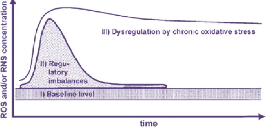
Oxidative stress is defined as overproduction of reactive oxygen species combined with failure in protective mechanisms of redox defense. The concentrations (threshold), pulse duration (flux) and sub-cellular localization are responsible for their responses (Figure 7).32,44,45 Low or moderate concentrations, reactive oxygen species act as mediators/second messengers of specific physiological processes and signaling pathways regulating numerous cellular processes, including proliferation and apoptosis. However, ROS and RNS high concentrations can be responsible for: damage on DNA, RNA, proteins and chromosome degradation; polyunsaturated fatty acids and amino acids oxidation; enzymatic inactivation, abnormal inflammatory reactions and interfere on cell-signaling molecule, such as apoptotic regulators and antioxidant enzymes. Those alterations seem to be associated with the initiation or progression of human cancers by the disruption of cell, tissue or organ functions (Figure 8). 28, 29, 41, 42, 44, 46-51
Figure 7: Reactive species concentration and their responses. Adapted from: Graves DB. The emerging role of reactive oxygen and nitrogen
species in redox biology and some implications for plasma applications to medicine and biology. J Phys D Appl Phys. 2015; 45: 263001
Antioxidant and detoxify genes polymorphisms in colorectal cancer predisposition Patrícia Alexandra Sousa Jegundo, 2015 8
Free radicals are molecules with high instability and reactivity due to the presence of an odd number of electrons in the outermost orbit of their atoms. Cells can generate ROS from exogenous sources as well as endogenously. Endogenously ROS production results mainly from the mitochondrial electron transport chain but also from proteins metabolism (metabolism of fatty acid, xanthine oxidases, cytochrome P450 reductase, nitric oxide synthase, nicotinamide adenine dinucleotide phosphate (NADPH) oxidases, peroxisomes and myeloperoxidase.23, 41, 44, 51, 52 Molecular oxygen is the last electron acceptor during oxidative phosphorylation. Incomplete reduction of O2 in the mitochondrial electron transport chain can lead to accumulation of O2−, OH- and H2O2, which as highly reactive species.30,43
Figure 8: ROS-mediated mechanisms in carcinogenesis.
Adapted from: Trachootham D. Alexandre J. Huang P. Targeting cancer cells by
ROS-mediated mechanisms: a radical therapeutic approach? Nature Reviews Drug
Antioxidant and detoxify genes polymorphisms in colorectal cancer predisposition Patrícia Alexandra Sousa Jegundo, 2015
9 1.5. Genetic Factors: Oxidative stress regulators
Nowadays, we know that human gene sequence is shared for 99,9% of individuals and the slight variation is essentially in just one nucleotide (SNPs - small nucleotide polymorphisms). 87 Polymorphism is variation in a gene or in DNA that occurs with a relative high frequency on population. The recent medicine attempts to relate those polymorphisms with the individual response to diet behavior, tobacco and alcohol consumption with disease predisposition. Noting that individual genetic constitution alters individual response to bioactive components.38, 39, 53-59 On this view, in last years it has been reported several variations, directly involved or not, involved in metabolic pathways with direct risk to CRC development.25-27, 33-36, 60-63
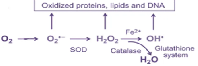
Eukaryotic cells have developed defense mechanisms that eliminate ROS. The human’s antioxidant endogenous defense system consists in a range of extracellular and intracellular antioxidants that are able to protect tissues from ROS and RNS. This antioxidant defense consists in non-enzymatic and enzymatic system. Non-enzymatic antioxidants are nutrient compounds, which includes: fenol, glutathione, vitamins C and E, β-carotene, α-tocopherol, and cytochrome c. Enzymatic process includes antioxidant enzymes, such as superoxide dismutase (SOD), glutathione reductase (GSSRG-R), glutathione peroxidase (GSH-Px), glutathione S-transferase (GST), and catalase (CAT). SOD catalyzes the dismutation of superoxide anion (O2-) into hydrogen peroxide (H2O2) plus O2. GSH-Px and catalase reduce H2O2 to O2 and H2O (Figure 9). GSH-Px uses glutathione as a reducing agent (electron donor). 23, 32, 41, 42, 45, 47, 48, 51, 64-68, 69, 70
Figure 9: ROS elimination process.
Adapted from: Bigarella CL. Liang R. Ghaffari S.. Stem cells and the impact of ROS signaling. Development. 2014; 141(22): 4206–4218.
Antioxidant and detoxify genes polymorphisms in colorectal cancer predisposition Patrícia Alexandra Sousa Jegundo, 2015 10
Some genes have polymorphic variants, single nucleotide polymorphisms (SNP) that are responsible for the individual’s genetic constitution/response. Between the genes with antioxidant and detoxifying activity, superoxide dismutases (SOD) and glutathione-S-transferases (GST) genes are highly polymorphic and often contain numerous mutations, increasing the risk of the cancer development on esophagus, stomach, large intestine, breasts, lungs and lymph nodes.39, 48, 53-56 Thus, we can suppose that some polymorphisms, involved in oxidative stress regulation, may predispose to disease and particularly on CRC. The strength of the biological impact will also depend on heterozygosity or homozygosity of the variant allele.71
1.5.1. Antioxidant regulators - Superoxide Dismutases
It is assumed that carcinogenesis is a result of cellular proliferation and differentiation control loss. Superoxide dismutase genes seem to have an important role on oxidative metabolism balance, particularly in cellular detoxify. SODs are a family of enzymes, responsible for the first line of antioxidant defense against ROS.67,72 In humans there are three main isoforms of these genes: SOD1,
SOD2 and SOD3 (Figure 10). They encode proteins that require copper, zinc or
manganese on their active center in order to their enzymatic function, respectively SOD1, SOD3 (Cu/Zn- SOD) and SOD2. O2- dismutation mechanism into H2O2 by SOD, which involves alternate reduction and re-oxidation of these metals.73 SOD1 is located in the cytosol of liver, kidney, erythrocytes, and central nervous system cells – mutations on this enzyme are associated with neural diseases, such as amyotrophic lateral sclerosis. SOD2 is found in mitochondria and it’s associated with aging, cancer, asthma and transplant rejection as well. SOD3 is detected as extracellular enzyme, plays an important role in regulating blood pressure, vascular contraction and also in neurologic, pulmonary and arthritic diseases. SODs catalyze the inactivation (dismutation) of superoxide ion by convert in oxygen peroxide, a less toxic molecule. 23, 38, 39, 43, 56, 64, 74, 75
Antioxidant and detoxify genes polymorphisms in colorectal cancer predisposition Patrícia Alexandra Sousa Jegundo, 2015
11 There is evidence that decreased of intracellular SOD levels are related to
malignant transformation, nevertheless, this data was not reported for SOD1.67, 72, 76
In the past, it was supposed that induction higher levels of SOD causes a return to the non-malignant phenotype.51, 65 Nowadays, it was found that manganese-dependent superoxide dismutase (MnSOD) or SOD2 overexpression appears to enhance invasiveness and migration of malignant cells.38,39 Moreover, several studies have reported that SOD2 is upregulated during tumor progression in prostate, colon and lung.47 On the other hand, other studies report that SOD2 overexpression can suppress tumor incidence and proliferation in other cancers such as in breast and skin cancers. Innumerous polymorphic variants of these genes confer different expression and active proprieties on antioxidant cellular defense.38, 39, 53-56 In colon cancer mutations, adenomatous polyposis, could begin a neoplasia process even when SOD2 levels are often low. The SODs activities are often lowered during early cancer development. However, SOD2 mRNA and protein levels increase during early and intermediate stages in lung and colorectal cancers.42, 47, 67, 69, 76, 77
Figure 10: Role of SODs in oxidative metabolism balance. Adapted from: http://cdn.intechopen.com/pdfs-wm/38459.pdf
Antioxidant and detoxify genes polymorphisms in colorectal cancer predisposition Patrícia Alexandra Sousa Jegundo, 2015 12
SOD2 gene
The role of SOD2 is still not exactly understood. SOD2, also named as MnSOD, regulates cellular redox homeostasis that is known to regulate proliferative and quiescent growth states. SOD2 expression is induced by a wide variety of factors such as hyperoxia, irradiation, cytokines (IL-1, TNF-α), oxidized LDL and the cellular redox state. Disturbances in the functioning of SOD isoforms lead to numerous pathological changes in the human organism, including tumor disease. Therefore, MnSOD and ROS rates are believed to be critical regulators.41, 43, 50, 65-67, 76
The SOD2 gene located in chromosome 6q25, it’s the most well-known (Figure 11).27, 43, 64, 76 The Val16Ala (47 T>C; rs4880) polymorphism has a single peptide mutation, a substitution of a valine acid for an alanine amino acid on its 16 codon. This mutation leads to a conformational change in the helical structure of the protein.23,31 It is predicted that the Ala variant encodes proteins with a higher MnSOD activity than the Val variant, suggesting that Ala/Ala homozygous subjects may have higher SOD2 activity. Therefore, the defense response to oxidative stress will be altered as well.23, 27, 31, 64, 76 It is hypothesized that the higher activity variant (Ala) suppresses carcinogenesis; however, this overproduction of SOD2 increases the levels of H2O2, as well. Epidemiologic studies associated the Ala variant with increased risk of carcinogenesis, particularly among people with lower intakes of exogenous antioxidants on their diet.43, 76, 78
There are several different views on how MnSOD expression can contribute to cancer development. Several authors reported that a loss of MnSOD activity results in aberrant proliferation, and was also related with poor 5-year overall survival.31, 41, 65, 67, 68 One explanation for such discrepancies is the different clinical stages of the tumors studied.66 CRC have been characterized
Figure 11: Genomic view of MnSOD gene. Adapted from: http://cdn.genecards.org/images/v4/genomic-location/SOD2-gene.png
Antioxidant and detoxify genes polymorphisms in colorectal cancer predisposition Patrícia Alexandra Sousa Jegundo, 2015
13 immunohistochemically by a decreased of MnSOD and total SOD activity when
compared with adjacent normal mucosa, suggesting that MnSOD acts a tumor suppressor.50 There are authors that showed a decreased or unchanged expression and activity of SOD isoenzymes.43, 65, 74 While others observed increased expression and activity of SOD isoenzymes in various types of tumors, implying MnSOD in tumors progression, aggressiveness and metastatic potential.66, 67, 72 MnSOD overexpression has shown to slow down cancer cell growth, but it also has a metastasis-promoting activity by the upregulation of matrix-degrading metalloproteases and blocking apoptosis.44, 68, 78
SOD3 gene
Extracellular superoxide dismutase (EC-SOD; SOD3) has Cu and Zn in the catalytic center and a heparin binding domain (HBD). SOD3 is highly expressed in plasma, blood vessels, heart, lungs, kidney, placenta and extracellular fluids. This enzyme is produced by resting macrophages and it’s associated with the cell surface through extracellular matrix (ECM)-binding region.76 SOD3 is a protective molecule which catalyzes the conversion of superoxide anions into hydrogen peroxide and oxygen, thus protecting from oxidative fragmentation of matrix components. Inherited change in SOD3 expression or function could affect organ matrix homeostasis and influence its normal function.23, 31, 73, 76, 79, 80 SOD3 binds matrix components and inhibits their fragmentation in response to oxidative stress. Their fragmentation stimulate inflammatory cell migration, so SOD3 could play a central role in tissue defenses against oxidative stress.23,79 It was observed that non-neoplastic tissue has more SOD3 expression than neoplastic tissue which supports the previously predicted role of SOD3 in tumorigenesis. Thus, dysregulation of extracellular oxidant-moderating proteins, such as SOD3, is significant in cancer. However, there is not much knowledge about EC-SOD in human tumors so further studies are needed in order to characterize its potential role.81, 82, 83
The most studied single nucleotide polymorphism (SNP) within SOD3 is Arg231Gly (rs1799895; R213G) on chromosome 4 (Figure 12). About 2-3% of the population carries this polymorphism. The substitution of arginine to glycine in its
Antioxidant and detoxify genes polymorphisms in colorectal cancer predisposition Patrícia Alexandra Sousa Jegundo, 2015 14
heparin binding domain at amino acid 213 (R213G) was first identified in patients with heart failure.31, 73, 81, 82 This variant is known to reduce the binding capacity of SOD3 and may thus have an impact on the cellular distribution.68, 76 Transcript levels of SOD3 significantly differed between tumor and non-neoplastic tissues. A low expression level of SOD3 induces primary cell proliferation and immortalization, whereas, high expression levels induce growth arrest, senescence, and apoptosis though signaling pathway activation (p53 and p21).84, 85
Despite the enzymatic activity of SOD3 should not be affect by this mutation, it was observed in lung adenocarcinoma that SOD3 mRNA and protein expression were significant decreased.75, 85
While SOD2 was upregulated in the tumor tissue, this SOD3 polymorphism leads to a higher concentration of SOD3 in plasma and lower in tissues, once it’s anchoring/binding to the extracellular matrix is compromised. Thus SOD3 has strong downregulation in tumor tissue samples.31, 78, 82, 86 Evidence of downregulation in SOD3 expression in lung and oral tumors, previously reported, have suggested that SO3 can have potential effects on extracellular regulation of multiple factors that regulate angiogenesis and invasion, increasing relative risk for this disease.73, 77-80, 82-84, 87 It was reported that SOD3 overexpression, delays the onset of increased breathing frequency and significantly reduces breathing rates in wild-types mice exposed to a source of oxidative stress (ionizing radiation).78 SOD3 overexpression also inhibits the invasive capacity of human prostate cancer cells.82 78 Therefore, arginine 213 is critical for maintaining proper organ function through moderating the normal innate immune response, suggesting the potential to suppress aggressive tumor behavior.78, 80, 82, 86
1.5.2. Detoxify regulators: Glutathione S-transferases
Figure 12: Genomic view of SOD3 gene. Adapted from: http://cdn.genecards.org/images/v4/genomic-location/SOD3-gene.png
Antioxidant and detoxify genes polymorphisms in colorectal cancer predisposition Patrícia Alexandra Sousa Jegundo, 2015
15 Glutathione S-transferases (GSTs) super family is a family of
Phase II detoxification enzymes that are involved in the detoxification of xenobiotic compounds. They catalyze the conjugation of glutathione (GSH) to a wide variety of endogenous and exogenous electrophilic compounds (Figure 13) formed during oxidative stress including those result from lipid peroxidation. 88-103
This process usually inactivates the electrophiles and facilitates their excretion into urine or bile.88, 89 On the other hand, this detoxification ability plays an important role in cellular protection from environmental and oxidative stress, yet is also implicated in cellular resistance to drugs acquired by an accelerated detoxification of drug substrates.90 The GSTs are found on liver and in the epithelium of the human gastrointestinal tract, thus providing a protective role for the cells in the gut.88, 104, 105 GST activities may be reduced in colonic mucosa affected by chronic inflammatory conditions.105 GSTs have also been shown to play critical roles in kinase signaling. However, this pathway is not fully understood. The lack of function of these enzymes has been correlated with a higher risk of cancer. Studies have linked aberrant expression of GST isozymes with the development and expression of resistance to a variety of chemicals. Therefore, differences in the human GST isozyme expression patterns influence cancer susceptibility, prognosis and treatment.38, 53, 54, 88, 91-96, 98, 99, 100, 102, 106-108
Glutathione S-transferases are high polymorphic among ethnicities. Human GSTs are divided into three main families with have similar ability to catalyze the conjugation of GSH: cytosolic, mitochondrial and membrane-bound microsomal. Based on identity sharing greater than 60% within a class and focuses mainly on the highly conserved N-terminal, the mammalian cytosolic family of GSTs is further
Figure 13: Graphic representation of GSTs detoxify role.
Adapted from: Gorrini C. Harris IS. Mak TW. Modulation of oxidative stress
as an anticancer strategy. Nature Reviews Drug Discovery 2013;12: 931–947
Antioxidant and detoxify genes polymorphisms in colorectal cancer predisposition Patrícia Alexandra Sousa Jegundo, 2015 16
divided into seven classes: alpha, mu, omega, pi, sigma, theta and zeta.90, 94, 96-98, 101, 104, 108-110
Recently, was described plus one class – lambda (K).103 From these classes above, alpha, mu, theta and pi have been identified in the human colon; however, the pi class predominates.105, 110, 111 SNPs in those isoforms cause a steric change at the substrate-binding site of the enzyme that changes its catalytic activity which seems to denote impaired ability to detoxify carcinogens, conferring an increased cancer risk.90, 110, 112
GSTP gene
The GST Pi class is encoded by a single gene located on chromosome 11 (Figure 14). Four functionally different polymorphisms have been identified (GSTP1*A–D). For GSTP1, two genetic polymorphisms are known for this gene: in exon 5, a substitution A>G at codon 105 of GSTP*B gene (Ile105Val; rs1695), turns amino acid isoleucine to valine; in exon 6, transition of C>T at codon 114 turns in a substitution of amino acid alanine to valine (Ala114Val; rs1138272).27, 90, 94-97, 106-108,110, 111
These SNPs cause a steric change at the substrate-binding site of the enzyme that changes its catalytic activity without affecting the binding affinity to glutathione.90, 110
The GSTP1 genotype has been associated with differences in chemotherapeutic response and cancer susceptibility and is overexpressed in a wide variety of tumors including ovarian, bladder, testicular, lung, breast, colon, pancreas, larynx and lymphoma.94, 95, 97, 105, 110 GST expression and/or activity of
Figure 14: Genomic view of GSTP gene. Adapted from:
Antioxidant and detoxify genes polymorphisms in colorectal cancer predisposition Patrícia Alexandra Sousa Jegundo, 2015
17 specific isoforms are lost in some individuals with allelic variation and are
proposed to occur during pathogenesis of the disease. Lack of GSTP1 expression results on enzyme activity reduction, followed by an increase of cellular oxidative stress which allows the human colon cancer cell survival and proliferation.94, 102, 110 The Ile105Val results in a catalytic activity reduction and thus a diminished detoxification capacity in individuals V105 when compared with I105.97,112 It was observed a slightly better survival in GSTP1 Ile/Ile patients compared with patients with the Ile/Val or Val/Val genotypes.98,112 However, the association between the
Glutathione S transferase-P1 (GSTP1) Ile105Val polymorphism and colorectal
cancer (CRC) susceptibility are still inconsistent between studies.102, 106, 110 GSTP1
Val114 polymorphism apparently contributes to esophageal and colorectal cancer
susceptibility.102, 110
GSTT gene
The Theta class of GSTs consists of two different subfamilies: GSTT1 and GSTT2. Genes encoding both proteins are localized on chromosome 22 (Figure 15). Polymorphisms exist within both genes. GSTT1 has a functional and a non-functional allele. Homozygosity for the nonnon-functional allele of GSTT1 is the null phenotype and exhibits an absence of catalytic activity. Individuals homozygous for this deletion are thought to be at increased risk for malignancies (such as cancer, cardiovascular and respiratory diseases) as a consequence of a decreased capacity to detoxify possible carcinogens.95, 94, 99, 101-104, 113, 114 It was demonstrated that 20% of Caucasians carries this genotype.114 The GSTT1 polymorphism was significant in breast cancer and for CRC risk in Caucasians.95, 101, 115
However, this association is not consistent between all studies reported.
Figure 15: Genomic view of GSTT gene.
Adapted from: http://www.ncbi.nlm.nih.gov/gene/2952
Antioxidant and detoxify genes polymorphisms in colorectal cancer predisposition Patrícia Alexandra Sousa Jegundo, 2015 18
GSTM gene
Five GSTM isoforms belonging to the mu class (GSTM1- 5) have been described. A gene cluster located on chromosome 1 encodes for GSTM1-5, four different alleles allowing for several M1 class polymorphisms (Figure 16). The presence of the GSTM1 allele has been associated with a decreased risk of bladder cancer.91, 94, 104 Loss of GSTM enzyme function is described to a homozygous deletion of this gene resulting in the GSTM1 null allele. It was showed that 20-50% of Caucasians carries GSTM1 null genotype.114, 115 The
GSTM null phenotypes are associated with an increased risk of the lung, head,
colon and bladder cancer and were also been associated with response rates to some chemotherapy. Data have suggested that GSTM1 polymorphism is associated with an increased risk of CRC, especially in the Caucasian population.88, 89, 94-96, 99, 103-105,108, 114
1.6. Antioxidant and Detoxify regulators in colorectal cancer
The gene variants can affect the risk of sporadic colorectal cancer associated with various environmental and dietary factors. Diet and social behavior are a fundamental risk factor for development of CRC, and its influence appears to be stronger during post-initiation phases of carcinogenesis. Accordingly, antioxidant and detoxify molecular mechanisms should be considered among potential risk modulators for this neoplasia. Several gene polymorphisms involved in regulation detox defense. However, there still inconsistencies on CRC ethology.
Figure 16: Genomic view of GSTM gene. Adapted from: http://cdn.genecards.org/images/v4/genomic-location/GSTM1-gene.png
Antioxidant and detoxify genes polymorphisms in colorectal cancer predisposition Patrícia Alexandra Sousa Jegundo, 2015
19 Both antioxidant and detoxify genes are crucial to redox hemostasis.
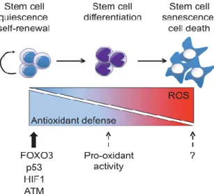
Imbalance on these defense mechanisms leads to ROS accumulation and predispose DNA cell to damage. This damage can change protein activity and function. ROS accumulation on colorectal environment predisposes to risk of CRC. The balance between antioxidants and ROS is lost. Therefore, colonic and rectal cells will be prone to DNA damage. This damage induces changes on enzymatic function and activity. The continuous exposure of colonic and rectal cells to ROS damage will allow to neoplasic development (Figure 17). The
MNSOD, SOD3, GSTP1, GSTT1 and GSTM1 genes have an important role in
antioxidant and detoxify metabolisms management. Numerous studies reported that their polymorphic variants have an huge impact on their functions. However, some studies about association and influence on CRC didn’t agree with majority.
The knowledge about detox genes polymorphisms involved on CRC genesis and development will be essential for new approaches on cancer research. However, Sporadic Colorectal Adenocarcinoma (SCA) origin hasn’t been studied until now. Therefore, we wanted verify if MNSOD, SOD3, GSTP1,
GSTT1 and GSTM1 gene polymorphisms influence the risk of SCA occurrence.
Moreover, we want to know if those polymorphisms are associated to gender and colonic and rectal localization among SCA subjects.
Figure 17: Colonic and rectal cells neoplastic development induced by continuous ROS exposure. Adapted from: Bigarella CL. Liang R. Ghaffari S.. Stem cells and the
impact of ROS signaling. Development. 2014; 141(22): 4206–4218.
Antioxidant and detoxify genes polymorphisms in colorectal cancer predisposition Patrícia Alexandra Sousa Jegundo, 2015 20
Antioxidant and detoxify genes polymorphisms in colorectal cancer predisposition Patrícia Alexandra Sousa Jegundo, 2015
21
II. Material and methods
2.1. Material
A global of 68 samples of SCA (mean age of 67 +/- 18 years; 75% men and 25% women) was used in the research of the defined polymorphisms (Table 1). A total of 31 colon and 37 rectal biopsies suffered the typical process of fixation in formaldehyde and inclusion in paraffin and were selected from the archives of Anatomical Pathology Institute of Faculty de Medicine of University of Coimbra (Figure 18). Samples were collected between 2009 and 2011, tumor diagnosis and differentiation grade of the tumor were established by histological evaluation of tumor fragments using criteria according to World Health Organization (Table 2).
A total of 100 healthy subjects biopsies (with normal colic mucosa) were used as control group (mean age of 73 +/- 9 years; 79% men and 21% women) (Table 1). This study was supported and approved by local ethics committee (CIMAGO - Faculty of Medicine of the University of Coimbra, Coimbra, Portugal).
Table 1: Clinical data of SCA and control samples.
Tumor location
Distribution Age Colon Rectum Total
% Mean Stand. Dev. n % n % Gender Patients Male 75 67 18 25 81 26 70 51 Female 25 6 19 11 30 17 Total 100 31 100 37 100 68 Controls Male 79 73 9 Female 21 Total 200
Antioxidant and detoxify genes polymorphisms in colorectal cancer predisposition Patrícia Alexandra Sousa Jegundo, 2015 22
Figure 18: (A) and (B) Histologic picture of normal colonic mucosa: 40x HE, 200x HE; (C) and (D) – Histological picture of well differentiated sporadic adenocarcinoma. 200x HE (hematoxylin-eosine); 100x HE. Images from IAP-FMUC.
A B
Antioxidant and detoxify genes polymorphisms in colorectal cancer predisposition Patrícia Alexandra Sousa Jegundo, 2015
23 Histological classification according to WHO
Epithelial tumours Non-epithelial tumours
Adenoma Lipoma
Tubular Leiomyoma
Villous Gastrointestinal stromal tumour
Tubulovillous Leiomyo sarcoma
Serrated Angiosarcoma
Kaposi sarcoma
Intraepithelial neoplasia (dysplasia) associated with chronic inflammatory diseases Malignant Melanoma Others Low-grade glandular intraepithelial neoplasia High-grade glandular intraepithelial neoplasia Malignant lymphomas Carcinoma
Marginal zone B-cell lymphoma of MALT Type
Adenocarcinoma Mantle cell lymphoma
Mucinous adenocarcinoma
Diffuse large B-cell lymphoma
Signet-ring cell carcinoma Burkitt lymphoma
Small cell carcinoma
like /atypical Burkitt-lymphoma
Squamous cell carcinoma Others
Adenosquamous carcinoma Medullary carcinoma Undifferentiated carcinoma Secondary tumours Polyps
Carcinoid (well differentiated endocrine
neoplasm) EC-cell, serotonin-producing neoplasm Hyperplastic (metaplastic)
L-cell, glucagon-like peptide
and PP/PYY producing tumour Peutz-Jeghers
Juvenile
Mixed
carcinoid-adenocarcinoma
Others
Antioxidant and detoxify genes polymorphisms in colorectal cancer predisposition Patrícia Alexandra Sousa Jegundo, 2015 24
2.2. Methods
2.2.1. DNA extraction
DNA genomic isolation from biopsies were made according to the extraction protocol from NZY Tissue gDNA Isolation Kit (NZYTech, Lisbon, Portugal), after microdissection of the normal colic tissue (5 to 10 dissections of 10µm of thickness to each sample). Samples were prepared by adding 1 ml of xylene to each tube to paraffin removal. After centrifugation at 11,000xg for 3 min, supernatant was discarded and samples were washed with 1ml of ethanol (96%-100%), repeating the centrifugation step. Lysis of the cell wall by Proteinase K and NZY was then performed overnight and purification of DNA obtained through columns and according to the procedure of the referred kit. The DNA was further stored at – 20ºC.
2.2.2. Analysis of concentration and quality of the extracted DNA
DNA samples were quantified in a spectrophotometer GeneQuant pro (Biochrom, Cambridge, England). RNAse-free water was applied as reference and 7 μl of DNA sample were inserted in the ultra-microvolume cuvette in order to perform the quantification and measurement of concentration and purity of the sample, by reading adequate optical densities (230 nm, 260 nm and 280 nm). The existence of nucleotides and proteins was detected at a wavelength of 280 nm, while at the 260 nm wavelength there is detection of nucleotides only. At a wavelength of 230 nm, the presence of contaminants is assessed.
2.2.3. Genotyping
The genotyping of polymorphisms was carried through commercial kits “Nutri Box Kit” (Genebox, Cantanhede - Portugal) using PCR-SSP technique. These kits included internal, negative and positive controls for each sample.
MNSOD T175C, SOD3 R213G, GSTP1 A105G, GSTP1 C114T, GSTT1 del, GSTM1 del mutations detection was performed using manufacturer protocol
Antioxidant and detoxify genes polymorphisms in colorectal cancer predisposition Patrícia Alexandra Sousa Jegundo, 2015
25 SYBR Safe (Molecular Probes, Oregon – USA) in 2% agarose gel and visualized
in a ultra-violet (UV) transilluminator (UVi Tech, Cambridge, United Kingdom).
Step Temperature Time Number of cycles
Initial denature 95º C 1 Min 1
Denature Annealing Extension 95º C 70º C 72º C 25 Sec 45 Sec 30 Sec 5 Denature Annealing Extension 95º C 65º C 72º C 25 Sec 45 Sec 30 Sec 21 Denature Annealing Extension 95º C 55º C 72º C 25 Sec 1 Min 2 Min 4
Final extension 72º C 10 Min 1
Table 3: Protocol of amplification by PCR-SSP (Polymerase Chain Reaction – Single Specific Primers), from Genebox, Cantanhede – Portugal.
2.2.4. Electrophoresis in agarose gel
PCR reactions, after amplification, were submitted to electrophoresis by 2% agarose gel in order to identify the amplified products. Agarose Routine Grade (NZYTech, Lisbon, Portugal) was dissolved in 1x TAE (Tris-acetate-EDTA) (NZYTech, Lisbon, Portugal) and distillated water and agitated for 15 seconds. Then the solution was transferred to the microwaves to complete dissolution of agarose (approximately 2 min at 900w). Afterwards, the solution was cool down and 1x10⁻⁵ SYBR Safe (Molecular Probes, Oregon – USA) a dye that allows the visualization of DNA under the UV light incidence, were added and the solution was agitated for 15 seconds for homogenization. Solution was after putted into a cradle until it solidifies under the environment temperature. After solidification, the solidified gel was inserted in the plastic gel box, previously filled with 1x TAE
Antioxidant and detoxify genes polymorphisms in colorectal cancer predisposition Patrícia Alexandra Sousa Jegundo, 2015 26
(NZYTech, Lisbon, Portugal). PCR samples were inserted into the gel wells and they were left running for 10 minutes under the 300 volts of the Power Pac Basic device (Bio-Rad, California, USA). Finally, PCR products were visualized under UV light by a transilluminator (UVi Tech, Cambridge, United Kingdom). Results were further registered by a digital camera (NIKON DMX1200F).
2.2.5. Statistical analysis
2.2.5.1. Analysis of purity and concentration of DNA
DNAs purity and concentration study consisted on the calculation of the means, standard-deviations and confidence levels relatively to their concentrations and contamination quantity. This statistical analysis aimed to verify if the conditions of the DNAs were acceptable for the validation of the results in the study.
2.2.5.2. Frequencies of allele and genotype polymorphisms
Allele and genotype frequencies were calculated from obtained percentages for each studied polymorphism. In order to assess if the mutations were in equilibrium, chi-square test was performed using the Hardy-Weinberg equilibrium as reference. MnSOD T175C, SOD3 R213G, GSTP1 A105G, GSTP1 C114T,
GSTT1 del, GSTM1 del frequencies were compared between different groups
(SCA versus control and among tumor localization) using STATISTICA 14 (StatSoft, Inc., 2013) based on chi-square (2x2) test and Exact Fisher test. The significance level was set at p<0.05, odds ratio (OR) and 95% confidence intervals (CI) for relative risks (RR) were also calculated for each variation.
Antioxidant and detoxify genes polymorphisms in colorectal cancer predisposition Patrícia Alexandra Sousa Jegundo, 2015
27
III. Results
3.1. Clinical pathology data
The individual distributions of the SCA subjects included in the study, according to the clinical pathologic and biological features of biological samples are presented in the Table 1. The distribution of SCA subjects by gender shows the predominance of the disease in male subjects comparing with female individuals (75% versus 25%) (Table 1). In terms of tumors localization, it was observed a small difference between gender distribution among colon and rectum groups, however, no significant differences were found (Table 4). Moreover, 46% of the SCA biopsies were located in colon and 54% of the SCA biopsies were located in rectum (Table 4). However, there were no significant differences between mean age in both groups (patient samples: mean age of 67 +/- 18 years; controls: mean age of 73 +/- 9 years) (Table 1).
Colon Rectum n % N % p OR RR Male 25 81 26 70 NS Female 6 19 11 30 Total 31 46 37 54
Table 4: Gender distribution among colon and rectum groups.
3.2. Analysis of purity and concentration of DNA
Although the concentration of DNA is not uniform (+/-29.3g/ml) and lower than the standard value (100 g/ml) (Table 5), the amplification by PCR-SSP occurred without problems, as the protocol of amplification was adapted to the concentration of DNA of samples. Although the mean values of purity of DNAs range between acceptable limits, 1.6-1.8 to O.D.260nm/O.D.280nm and 0.4-0.6 to O.D.230nm/O.D.260nm, some DNAs show high quantities of contaminants and proteins with a confidence interval being above certain acceptable limits.
Antioxidant and detoxify genes polymorphisms in colorectal cancer predisposition Patrícia Alexandra Sousa Jegundo, 2015 28
Furthermore, coefficients of variation show the existence of samples that deviate from acceptable patterns, whether in concentration of DNA and whether in quantity of contaminants. These DNAs can affect some results; however the majority of samples show a level of purity highly acceptable (Table 5). In general, PCR-SSP didn’t have major amplification problems.
Concentratio n O.D.260nm/ O.D.280nm O.D.230nm/ O.D.260nm Mean 42.4 1.83 0.54 Standard Deviation 29.3 0.09 0.15 Variation Coefficient 69% 5% 28% Confidence Interval (95%) 1.7-1.9 0.4-0.7
Table 5: Means and standard-deviations of DNA samples purity and concentration values.
3.3. Molecular analysis of antioxidant and detoxify genes 3.3.1. Polymorphisms in SCA and controls
When compared SCA with controls, we notice that MNSOD 175C (p<0.0001; 69% vs 32%; OR:4.76; CI: 2.97 to 7.61), SOD3 213G (p<0.0001; 52% vs 25%; OR:3.37; CI: 2.11 to 5.36), GSTP1 105G (p<0.0001; 59% vs 30%; OR:3.33; CI: 2.11 to 5.26) and GSTP1 114T (p<0.0001; 53% vs 12%; OR:8.66; CI: 5.00 to 15.00) mutant alleles were more frequent among SCA subjects (Table 6-7; Graphic 1-2).
Patients and controls
Alleles Patients Controls p OR RR
n % n % MNSOD T175 C (rs4880) T 42 31 136 68 <0.0001 0.21 (0.13-0.34) 0.40 (0.30-0.53) C 94 69 64 32 4.76 (2.97-7.61) 2.52 (1.90-3.38) Total 136 100 200 100 SOD3 R213G (rs1799895) C 65 48 151 75 <0.0001 0.30 (0.20-0.47) 0.51 (0.40-0.65) G 71 52 49 25 3.37 (2.11-5.36) 1.97 (1.53-2.53) Total 136 100 200 100
Antioxidant and detoxify genes polymorphisms in colorectal cancer predisposition Patrícia Alexandra Sousa Jegundo, 2015
29
Alleles Patients and controls
Patients Controls p OR RR n % n % GSTP1 A105G (rs1695) A 56 41 140 70 <0.0001 0.30 (0.19-0.47) 0.50 (0.38-0.65) G 80 59 60 30 3.33 (2.11-5.26) 2 (1.54-2.60) Total 136 100 200 100 GSTP1 C114T (rs1138272) C 64 47 177 88 <0.0001 0.12 (0.07-0.20) 0,35 (0.28-0.45) T 72 53 23 12 8.66 (5.00-15.00) 2.85 (2.25-3.62) Total 136 100 200 100
Table 7. Allelic frequency of detoxify genes in SCA and control group. 0 10 20 30 40 50 60 70 80 T C C G MNSOD T175C SOD3 R213G
Antioxidant frequency alleles
patients controls
Graphic 1: Allelic frequency of antioxidant genes in SCA and control group (p<0.0001).
0 20 40 60 80 100 A G C T GSTP1 A105G GSTP1 C114T
Detoxify frequency alelles
patients controls
Antioxidant and detoxify genes polymorphisms in colorectal cancer predisposition Patrícia Alexandra Sousa Jegundo, 2015 30
Graphic 3: Genotype frequency of antioxidant genes in SCA and control group.
We also found a higher prevalence of MNSOD 175CC (55% vs 2%; p<0.0001; OR: 58.5; CI 13.3 to 256.7), SOD3 213GG (31% vs 2%; p<0.0001; OR: 21.89; CI 4.93 to 97.29), GSTP1 105GG (46% vs 12%; p<0.0001; OR: 6.14; CI 2.85 to 13.26), GSTP1 114TT (38% vs 0%; p<0.0001; OR: Infinity) and GSTT1 null (75% vs 28%; p<0.0001; OR: 7.71; CI 3.83 to 15.56) mutated genotypes between SCA patients. GSTM1 del mutated genotype was not statistical significance between SCA patients and control group (Table 8-9; Graphic 3-4).
Patients and controls
Genotypes Patients Controls p value OR RR
n % n % MNSOD T175 C (rs4880) TT 11 16 38 38 0.002 0.32 (0.15-0.67) 0.47 (0.27-0.82) TC 20 29 60 60 <0.0001 0.30 (0.20-0.47) 0.51 (0.40-0.65) CC 37 55 2 2 59 (13.3-256.7) 3.95 (2.88-5.41) Total 68 100 100 100 SOD3 R213G (rs1799895) CC 18 26 53 53 0.0006 0.32 (0.16-0.62) 0.49 (0.32-0.77) CG 29 42 45 45 NS GG 21 32 2 2 <0.0001 21.9 (4.93-97.29) 2.82 (2.16-3.68) Total 68 100 100 100
Table 8: Genotype frequency of antioxidant genes in SCA and control group.
0 10 20 30 40 50 60 TT CT CC CC CG GG MNSOD T175C SOD3 R213G
Antioxidant frequency genotypes
Patients Controls p<0.0001 p<0.0001 p=0.0006 p=0.002 p<0.0001
Antioxidant and detoxify genes polymorphisms in colorectal cancer predisposition Patrícia Alexandra Sousa Jegundo, 2015
31
Patients and controls
Genotypes Patients Controls
p value OR RR n % n % GSTP1 A105G (rs1695) AA 19 28 52 52 0.002 0.36 (0.19-0.69) 0.53 (0.34-0.82) AG 18 26 36 36 NS GG 31 46 12 12 <0,0001 6.14 (2.85-13.26) 2.44 (1.75-3.38) Total 68 100 100 100 GSTP1 C114T (rs1138272) CC 22 36 77 77 <0,0001 0.14 (0.07-0.29) 0.33 (0.23-0.50) CT 20 30 23 23 NS TT 26 38 0 0 <0,0001 Infinity 3.4 (2.62-4.36) Total 68 100 100 100 GSTT1del NOR 17 25 72 72 <0.0001 0.13 (0.06-0.26) 0.30 (0.19-0.47) NULL 51 75 28 28 7.71 (3.83-15.56) 3.38 (2.14-5.34) Total 68 100 100 100 GSTM1del NOR 65 96 99 99 NS NULL 3 4 1 1 Total 68 100 100 100
Table 9: Genotype frequency of detoxify genes in SCA and control group.
0 10 20 30 40 50 60 70 80 90 100
AA AG GG CC CT TT NOR NULL NOR DEL
GSTP1 A105G GSTP1 C114T GSTT1 del GSTM1
Detoxify frequency genotypes
Patients Controls p=0.002 p<0.0001 p<0.0001 p<0.0001 p<0.0001 p<0.0001
Antioxidant and detoxify genes polymorphisms in colorectal cancer predisposition Patrícia Alexandra Sousa Jegundo, 2015 32
3.4. Patient’s polymorphisms stratified by: 3.4.1. Gender
The distribution of SCA subjects by gender shows no significant differences between men and women (Tables 10-13).
Alleles Gender Male Female p OR RR n % n % MNSOD T175 C (rs4880) T 32 31 10 29 NS C 70 69 24 71 Total 102 100 34 100 SOD3 R213G (rs1799895) C 45 44 20 59 NS G 57 56 14 41 Total 102 100 34 100
Table 10: Allelic frequency of antioxidant genes by gender.
Alleles Gender Male Female p OR RR n % n % GSTP1 A105G (rs1695) A 38 37 18 53 NS G 64 63 16 47 Total 102 100 34 100 GSTP1 C114T (rs1138272) C 48 47 16 47 NS T 54 53 18 53 Total 102 100 34 100
Antioxidant and detoxify genes polymorphisms in colorectal cancer predisposition Patrícia Alexandra Sousa Jegundo, 2015
33
Gender
Genotypes Male Female
p OR RR n % n % MNSOD T175 C (rs4880) TT 9 18 2 12 NS CT 14 27 6 35 CC 28 55 9 53 Total 51 100 17 100 SOD3 R213G (rs1799895) CC 12 24 6 35 NS CG 21 41 8 47 GG 18 35 3 18 Total 51 100 17 100
Table 12: Genotype frequency of antioxidant genes by gender.
Gender
Genotypes Male Female
p OR RR n % n % GSTP1 A105G (rs1695) AA 12 24 7 41 NS AG 14 27 4 24 GG 25 49 6 35 Total 51 100 17 100 GSTP1 C114T (rs1138272) CC 16 31 6 35 NS CT 16 31 4 24 TT 19 38 7 41 Total 51 100 17 100 GSTT1del NOR 12 24 5 29 NS NULL 39 76 12 71 Total 51 100 17 100 GSTM1del NOR 46 90 17 100 NS NULL 5 10 0 0 Total 51 100 17 100 GSTM del NOR 7 14 2 12 NS NULL 44 86 15 88 Total 51 100 17 100