UNIVERSIDADE DE LISBOA
Faculdade de Medicina Veterinária
PRECISION EVALUATION OF DIFFERENT FLOTATION SOLUTIONS IN THE
RESULTS OF FOUR COPROLOGICAL TECHNIQUES IN UNGULATES: IMPACT
ON DETECTING ANTHELMINTIC RESISTANCE
PEDRO AFONSO DAS NEVES PEREIRA DA SILVA
CONSTITUIÇÃO DO JÚRI
ORIENTADORA
Doutora Maria Isabel Neto da Cunha Fonseca
Dra. Martine van Zijll Langhout
Doutor Luís Manuel Madeira de Carvalho
Doutor David Wilson Russo Ramilo
CO-ORIENTADOR
Doutor Luís Manuel Madeira de
Carvalho
2019
UNIVERSIDADE DE LISBOA
Faculdade de Medicina Veterinária
PRECISION EVALUATION OF DIFFERENT FLOTATION SOLUTIONS IN THE
RESULTS OF FOUR COPROLOGICAL TECHNIQUES IN UNGULATES: IMPACT
ON DETECTING ANTHELMINTIC RESISTANCE
PEDRO AFONSO DAS NEVES PEREIRA DA SILVA
Dissertação de Mestrado Integrado em Medicina Veterinária
CONSTITUIÇÃO DO JÚRI
ORIENTADORA
Doutora Maria Isabel Neto da Cunha Fonseca
Dra. Martine van Zijll Langhout
Doutor Luís Manuel Madeira de Carvalho
Doutor David Wilson Russo Ramilo
CO-ORIENTADOR
Doutor Luís Manuel Madeira de
Carvalho
2019
ii
“I know of no pleasure deeper than that which comes from contemplating the natural world and trying to understand it” David Attenborough, in Life on Air: Memoirs of a Broadcaster, 2002
i
Acknowledgments
To Dr. Martine van Zijll Langhout, thank you for the opportunity to train and learn from all your expertise and for the chance to experience the truth about the “dream job”! I appreciate all the advices and inputs you’ve shared with me. It wouldn’t have been the same without all our deep conversations about work or any other worldly topic.
To Dr. Marno Wolters, a special thanks for letting me accompany him in his clinical rounds at ARTIS, allowing me to deepen my knowledge in the field. I’m wishing the next time we meet, we’ll meet as colleagues! To Daphne Valk, thank you for all your help with the material and I’m sorry for all the mess.
To Prof. Doctor Luís Madeira de Carvalho, your guidance and assurances throughout my insecure moments were crucial to the success of this phase. Thank you for all your hard work and dedication, not only during the writing of this thesis, but since I started being a student of yours. Your passion for Parasitology was what led me to gain interest in the subject and in the end led the “eterno presidente” writing a thesis about it.
To Dr. Lídia Gomes, thank you for all the help, knowledge, music and friendship shared in the laboratory. It would have been too dull without your company.
To Prof. Doctor Isabel Neto, I am very grateful for all your help with the statistical analysis and review.
To the Laboratory of Parasitology and Parasitlogical Diseases of the Center for Interdisciplinary Research in Animal Health of the Faculty of Veterinary Medicine, thank you for supporting me with the material needed to proceed and finish the final part of this study.
A special thank you to my Parents that since day one supported me and fought for the best education they could find me. Without them, I would most certainly not be where I am today and I’m very grateful to them for helping me in all my academic achievements. To my little sister, thank you for always bugging me while we were children and strengthening my most stubborn side. Because of that training, I’ll be able to deal with far more annoying things in my professional life! Love you sis!
To Jacky and Carsten, thank you so much for your company and friendship during my internship and afterwards. Thank you for your hospitality and showing me the A’dam life. You’ve made all the difference in the world!
ii
To Gonçalo, I have to thank you very much for putting up with all my frustrations and anxieties through this past 3 years. Your support led me forward and helped me evolve to the person I am today. I hope to be there by your side when your big moment comes as well.
To Annie, the girl I met on the bus and would end up being one of my rocks during this last 6 years of college. Even separated mid-way, your importance in my personal and academic life wasn’t diminished. Thank you for every laugh and tear, every confession and advice.
To my Zenda and all AEFMV’s boards I was involved in, thank you for all the tenacity and determination you instilled in me. Because of those 3 years of associative movement, I grew confident in my work and became more demanding of it.
To a special group of friends, I have to thank for all the endless calls and uncountable messages that made me feel that I was never alone during my internship. Carolina, for all the laughter you brought to my life every time we spoke. Catarina, for the companionship throughout our wild adventure abroad and thesis-related warnings every step of the way. Luísa, for every hour spent on the phone unwinding from work and making sure I was fine. Tatiana, for visiting me, bringing me a piece of home when I most needed it. Minês, for discovering a bit more of Holland with me and all the memories that came from it.
To Babs, for all your faith, support and inspiration. You were and always will be a role model.
iii
Resumo
A resistência a anti-helmínticos (RAH) de parasitas gastrointestinais de ruminantes e cavalos tem sido largamente descrita em todo o mundo nas últimas duas décadas. De modo a atrasar, ou até prevenir, o seu desenvolvimento, uma nova abordagem à desparasitação destes animais deve ser feita, considerando as ferramentas de diagnóstico disponíveis atualmente. Este estudo tem como objetivo evidenciar os desempenhos diferenciais de quatro métodos coprológicos comummente empregados, usando diferentes soluções de flutuação, e fazer algumas recomendações práticas sobre qual a técnica que parece ser mais adequada para enfrentar este problema emergente no maneio de equinos.
Esta tese divide-se em duas partes. Na primeira, três espécies de ungulados selvagens foram analisadas no ARTIS Amsterdam Royal Zoo para determinar a repetibilidade de quatro métodos coprológicos testados - flutuação simples (SF), flutuação com centrifugação (CF), McMaster (McM - sensibilidade de 50 ovos por grama OPG) e Mini-FLOTAC (MF - sensibilidade de 5 OPG) - em associação com três soluções de flutuação de diferente gravidade específica (GE): de sal (GE = 1,20), de sulfato de magnésio (MgSO4) (GE = 1,24) e de açucar (GE = 1,28) onde cada amostra foi analisada 10 vezes. Na segunda parte, foram colhidas 17 amostras fecais de equinos da raça Sorraia, em Abril de 2019 (12 fêmeas na pastagem e cinco machos estabulados), e analisadas com os mesmos métodos, mas apenas com as soluções de sal e açúcar. Cada amostra foi analisada em triplicado para SF e CF e em duplicado para McM e MF. Como parâmetro semi-quantitativo, o número de ovos foi contado em 10 campos de cada lâmina microscópica e a média foi obtida para cada réplica.
Na primeira parte, a CF foi capaz de evidenciar maiores contagens totais com menores coeficientes de variação (CV), principalmente com as soluções densas. MF apresentou muito boa precisão em todas as espécies, com diferentes soluções aparentemente melhores para cada uma. Na segunda parte, o SFsal obteve os melhores resultados para a deteção de
Triodontophorus spp., embora não significativos (p> 0,05). No entanto, teve um CV menor do
que as de CF, o que a torna a técnica qualitativa mais precisa. Foram observadas altas cargas parasitárias (média de 1825 OPG para todas as técnicas) e MFaçúcar foi a técnica mais consistente pela sua baixa variabilidade. Através de coproculturas confirmou-se a presença e prevalência de Cyathostomum s.l. tipo D (100%) e S. vulgaris (5,9%).
Os resultados obtidos aqui suportam o uso de CF e MF em parques zoológicos como ferramentas de diagnóstico válidas. Em suma, MF mostrou-se mais preciso em todo o estudo, exigindo a revisão das diretrizes atuais para o diagnóstico de RAH, nomeadamente em equídeos. Surpreendentemente, uma a solução de sal parece ser mais adequada para métodos qualitativos e a solução de açúcar mais adequada para os quantitativos.
Palavras-chave: Ungulados, zoo, cavalos de Sorraia, métodos coprológicos,
iv
Abstract
Anthelmintic resistance (AHR) in gastrointestinal parasites of ruminants and horses has been continuously described all over the world in the last two decades. In order to delay, or even prevent, the further development of resistance to anthelmintics, a new deworming approach must be taken, considering the diagnostic tools available nowadays. This study aims to evidence the differential performances of four commonly employed coprological methods, using different flotation solutions, and make some practical recommendations on which technique seems better-suited to face this emerging problem in horse management.
This thesis contains two parts. In the first one, three wild ungulate species were analyzed at ARTIS Amsterdam Royal Zoo to determine the repeatability of four common coprological methods – simple flotation (SF), centrifugal flotation (CF), McMaster (McM - sensitivity of 50 eggs per gram EPG) and Mini-FLOTAC (MF – sensitivity of 5 EPG) – in association with three flotation solutions with different specific gravities: salt (SG=1.20), magnesium sulphate (MgSO4) (SG=1.24) and sugar solution (SG=1.28); where each sample was analyzed 10 times. In the second part 17 fecal samples of Sorraia horses were collected in April 2019 (12 females on the pasture and five stabled males) and analyzed with the same methods but only with the salt and sugar solutions; each sample was analyzed in triplicates for SF and CF and in duplicates for McM and MF. As a semi-quantitative parameter, the number of eggs were counted in 10 fields of each slide and the mean was obtained for each replicate. In the first part of the study, CF was able to evidence a higher amount of total egg counts, especially with the denser solutions, with lower coefficients of variation (CV). MF performed with very good precision across the species, with different solutions apparently better for each one of them. In the second part of the study, SFsalt obtained the best results for detecting Triodontophorus spp. eggs, although not significantly (p>0.05). Yet, it showed lower CV than CF techniques, which makes it a more precise and reliable technique. High parasitic burdens (mean of 1825 EPG for all techniques) were registered and MFsugar performed more consistently as seen by its low variability. Through coprocultures, the prevalence of
Cyathostomum s.l. type D (100%) and S. vulgaris (5.9%) were also detected.
The results obtained here support the use of CF and MF methods in zoos as valid diagnostic tools. In general, MF was shown to be more precise and reliable across the study, urging the need to review current guidelines for the diagnosis of AHR, namely in horses. Unexpectedly, a clear association was evidenced, as salt solutions seem to be better suited for qualitative methods, whereas the sugar solutions better suited for quantitative ones instead.
Keywords: Ungulates, zoos, Sorraia horses, coprological methods, strongyle,
v
Introductory note
The present research was carried out in ARTIS Amsterdam Royal Zoo and in the Laboratory of Parasitology and Parasitic Diseases of the Centre for Interdisciplinary Research in Animal Health, Faculty of Veterinary Medicine, University of Lisbon (CIISA-FMV-UL) as part of the author’s training period during the 6th year of the Integrated Master in Veterinary Medicine. This work has already resulted in:
- A poster communication in the Proceedings of the 18th International Conference “Life Sciences for Sustainable Development”, 26th to 28th of September, 2019, Cluj-Napoca, Romania (see Appendix A)
vi Table of Contents Acknowledgments ... i Resumo ... iii Abstract ... iv Introductory note ... v List of Figures ... ix List of Graphs ... xi List of Tables ... xi
List of Abbreviations and Symbols ... xiii
1. Description of Internship Activities ... 1
2. Introduction ... 3
3. Bibliography review ... 5
3.1. Parasitism and coprology ... 5
3.1.1. Collection and preservation of fecal samples ... 5
3.1.2. Coprological Methods ... 6
3.2. ARTIS Amsterdam Royal Zoo... 9
3.2.1. History and context ... 9
3.2.2. Animals used as experimental models ... 10
3.3. Horse as the subject host ... 12
3.3.1. Sorraia breed ... 12
3.3.2. Parasitism in Sorraia horses ... 13
3.4. Parasites used as experimental models ... 14
3.4.1. Family Ascarididae... 14
3.4.2. Family Molineidae ... 17
3.4.3. Family Strongylidae ... 18
3.5. Anthelmintic resistance ... 23
3.5.1. Resistance in equine nematodes ... 26
3.5.2. Delaying or preventing advancements in resistance ... 28
3.5.3. Current situation – farm practices and legislation ... 32
3.5.4. Portugal’s panorama ... 34
vii
5. Materials and Methods ... 34
5.1. Repeatability Study of Coprological Methods in Zoo Animals ... 34
5.1.1. Weather characterization ... 34
5.1.2. Sample collection and preservation ... 35
5.1.3. Coprological methods ... 36
5.1.4. Microscopic reading ... 38
5.1.5. Data analysis and statistics ... 39
5.1.6. Fecal Egg Count Reduction Test ... 39
5.2. Performance Comparison of Coprological Methods – CIISA-FMV-ULisboa ... 39
5.2.1. Horse farm Alter do Chão ... 39
5.2.2. Sample collection and preservation ... 40
5.2.3. Coprological methods ... 41
5.2.4. Coprocultures ... 41
5.2.5. Microscope reading ... 41
5.2.6. Data analysis and statistics ... 42
6. Results ... 43
6.1. Repeatability Study of Coprological Methods – ARTIS ... 43
6.1.1. Red river hogs ... 43
6.1.2. Greater kudu ... 43
6.1.3. Grevy’s Zebra ... 44
6.1.4. Fecal Egg Count Reduction Test ... 44
6.2. Performance Comparison of Coprological Methods – CIISA-FMV-ULisboa ... 46
6.2.1. Qualitative techniques’ assessment and sensitivity ... 47
6.2.2. Qualitative techniques’ agreement ... 48
6.2.3. Data distribution ... 48 6.2.4. Descriptive statistics ... 49 6.2.5. Correlations ... 51 6.2.6. Association Triodontophorus-EPG ... 54 6.2.7. Coprocultures ... 54 7. Discussion ... 55
viii
7.1.1. Red river hogs ... 55
7.1.2. Greater kudus ... 57
7.1.3. Grevy’s zebra ... 58
7.1.4. General considerations ... 60
7.2. Performance Comparison of Coprological Methods – CIISA-FMV-ULisboa ... 62
7.2.1. Triodontophorus spp. prevalence ... 62
7.2.2. Qualitative techniques’ agreement ... 63
7.2.3. Techniques comparison ... 64
7.2.4. Correlations ... 68
7.2.5. Association Triodontophorus-EPG ... 69
7.2.6. Coprocultures ... 69
7.2.7. Parasitic burden ... 70
7.2.8. Influence of the used technique in the classification of shedders ... 71
8. Conclusions... 75
8.1. Repeatability of Coprological Methods – ARTIS ... 75
8.2. Performance Comparison of Coprological Methods – CIISA-FMV ... 75
9. Future Perspectives and Recommendations ... 77
9.1. Limitations of the study ... 78
10. Bibliography ... 79
APPENDIX A – Poster communication for the Proceedings of the 18th International Conference “Life Sciences for Sustainable Development”, 26th to 28th of September, 2019, Cluj-Napoca, Romania ... 94
APPENDIX B – Parasitology Logbook of ARTIS’ Veterinary Department from September to December 2018 ... 95
APPENDIX C – Calculation of k value of agreement... 110
... 110
APPENDIX D – Significance values ... 111
ix
List of Figures
Figure 1 – Examples of internship activities. Left to right, top row: cardiopulmonary
auscultation during hoof trimming of a red river hog (Potamochoerus porcus); collection of blood sample from a jaguar (Panthera onca); an X-ray of a bladder stone in a common chuckwalla (Sauromalus ater) prior to cystoliththotomy; corneal edema in a californian sea lion (Zalophus californianus). Bottom row: anesthesia reversal in an african wild dog (Lycaon
pictus) after contraceptive injection; anesthesia of a yellow-throated marten (Martes flavigula)
for a health check-up; gill probing of an ocellate river stingray (Potamotrygon motoro) (Originals). ... 2
Figure 2 – McMaster slide scheme, evidencing each chamber's volume and dimensions
(source: Taylor, Coop and Wall, 2016) ... 8
Figure 3 – Fill-FLOTAC with its sampling kit, filter and container and the reading disk,
Mini-FLOTAC (Original) ... 9
Figure 4 - Map of ARTIS, evidencing the Grevy's zebra and greater kudus’ enclosure (1), the
laboratory of the veterinary department (2) and the red river hogs’ enclosure (3) (source: ARTIS, 2019). ... 10
Figure 5 – Greater kudu in ARTIS with its spiral horns (Original). ... 10
Figure 6 – Grevy's zebras grazing in ARTIS (Original). ... 11
Figure 7 – Red river hog evidencing its hair tuffs pending from its ears (source:
www.arkive.org). ... 12
Figure 8 – Herd of Sorraia horses with their white-fringed hair (source:
www.autoctones.ruralbit.com). ... 13
Figure 9 – Ascaris suum evidencing the characteristic three lips surrounding the stoma of
ascarids (source: Bowman, 2014). ... 14
Figure 10 – Parascaris equorum and Ascaris suum egg showing its multi-layered appearance
and thick walls. Adapted from Taylor, Coop and Wall, 2016... 14
Figure 11 – Parascaris equorum life cycle: (A) Hatching of third stage larvae (L3) in the
stomach and small intestine, penetration of intestinal veins; (B) larvae reach liver via portal vein, migration through liver tissue and penetration of liver veins; (C) Larvae reach lung via vena cava and right heart, penetration into lung alveoles and migraton via trachea and pharynx to small intestine (moulting to L4 and L5 prior to development into adults). Adapted from ESCCAP, 2019. ... 16
Figure 12 – Nematodirus sp. egg, one of the largest nematode eggs of ruminants. Adapted
from Taylor, Coop and Wall, 2016... 17
Figure 13 – A cyathostomin egg, typically strongylid. Adapted from Taylor, Coop and Wall,
x
Figure 14 – Life cycle of small strongyles: (A) egg shedding; (B) oral uptake of third stage
larvae (L3) with grass; (C) exsheathment through gastric fluids; (D) passage of exsheathed L3 through small intestine; (E) invasion of mucosa/submucosa of colon and caecum, moult to fourth stage, return to intestinal lumen and final moult before development to adult stage.
Adapted from ESCCAP, 2019. ... 19
Figure 15 – A Strongyus spp. Egg with its characteristic morula inside. Adapted from Taylor, Coop and Wall, 2016. ... 20
Figure 16 – Anterior end of Strongylus spp. evidencing each species buccal capsule, leaf crown and presence or absence (in the case of S. edentatus) of tooth-like structures at the base of the buccal capsule. Adapted from ESCCAP, 2019. ... 21
Figure 17 – Life cycle of Strongylus vulgaris: (A) free-living stages; (B) arterial and intestinal migration of larvae. Adapted from ESCCAP, 2019. ... 21
Figure 18 – One of the studied greater kudus, a giraffe and the studied zebra in their mixed enclosure at ARTIS (Original). ... 35
Figure 19 - Flotation solutions used in ARTIS with their specific gravity measured with a hydrometer. From left to right: salt, MgSO4 and sugar solutions (Original). ... 36
Figure 20 – Hettich Universal 32 centrifuge used in ARTIS’ laboratory, with a rotating motor of 7 cm of radii (Original). ... 37
Figure 21 – Mini-FLOTAC full and locked ready to be read under the microscope (Original). ... 38
Figure 22 – Fill-FLOTAC with its container, sampling kit and built in filter and the reading disk, Mini-FLOTAC, on the right (Original). ... 38
Figure 23 – Centromix II-BL centrifuge used in CIISA's laboratory, with a rotating motor of 7 cm of radii (Original). ... 41
Figure 24 – Helminth eggs found in zoo animals at ARTIS; A – Ascaris suum egg from feces of red river hogs; B – Nematodirus spp. from feces of greater kudus; C – Parascaris spp. from feces of Grevy’s zebra (Originals). ... 44
Figure 25 – Typical strongylid eggs found (Original).C ... 44
Figure 25 – Typical strongylid eggs found (Original). ... 47
Figure 26 – Triodontophorus spp. egg (Original). ... 47
Figure 27 – Histograms of every technique’s distribution, not considering sexes, evidencing the non-normal distribution of CFsugar, McMsalt and MFsalt. ... 49
Figure 28 – Strongylus vulgaris larva from coprocultures of Sorraia horses (Original). ... 54
Figure 29 – Cyathostomin type D from larval cultures of Sorraia horses (Original). ... 54
Figure 30 – Feces’ morphology of different ungulate species from ARTIS. From left to right:
xi
feces, as individualized acorn-sized pellets, coated with mucous; and Grevy’s zebra’s fecal bolus with obvious fiber content and easily broken apart (Originals). ... 57
List of Graphs
Graph 1 – Temperature (C) and precipitation (mm) in Amsterdam during the year of 2018,
including the internship period (Weather underground WU, 2019). ... 34
Graph 2 - Temperature (ºC) and precipitation (mm) in Portalegre, as recorded in the Badajoz
airport weather station (WU, 2019). ... 40
Graph 3 – Simple flotation (SF) results obtained with both salt and sugar solutions for Sorraia
horses at CIISA-FMV. ... 65
Graph 4 – Centrifugal flotation (CF) results obtained with both salt and sugar solutions for
Sorraia horses at CIISA-FMV ... 65
Graph 5 – McMaster (McM) results obtained with both salt and sugar solutions for Sorraia
horses at CIISA-FMV ... 65
Graph 6 – Mini-FOTAC (MF) results obtained with both salt and sugar solutions for Sorraia
horses at CIISA-FMV ... 65
List of Tables
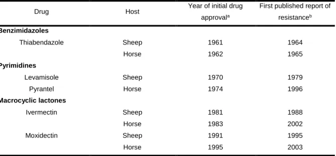
Table 1 – Year of approval of broad-spectrum anthelmintic drugs in sheep and horses
comparing to the first published report of its resistance. Adapted from Kaplan, 2004. ... 24
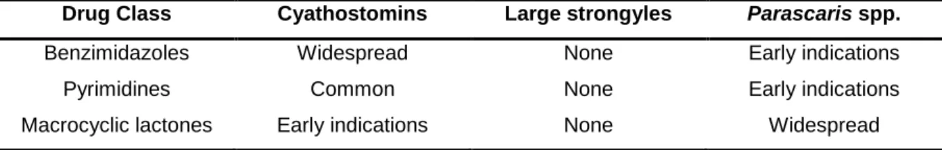
Table 2 – Summary of reported resistance worldwide of the main equine nematodes to
broad-spectrum anthelmintics. Adapted from Nielsen et al, 2019. ... 26
Table 3 – Thresholds of fecal egg count reduction test results to determine the presence of
anthelmintic resistance to equine broad-spectrum anthelmintics. Adapted from Nielsen et al, 2019. ... 27
Table 4 – Egg reappearance periods of equine broad-spectrum anthelmintics when the drug
is fully effective on cyathostomins. Adapted from Nielsen et al, 2019; ... 27
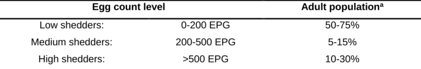
Table 5 – Classification of horses according to their fecal egg count and their respective
proportion in the population. Adapted from Nielsen et al, 2019; ... 29
Table 6 – Adulticidal and larvicidal action of broad-spectrum anthelmintics in main equine
nematodes. Adapted from Decision Tree Horse, accessed in June 2019. ... 30
Table 7 – Assessment risk of infection from pasture considering various factors and practices
xii
Table 8 – Pre-treatment (D0) and post-treamtent (D15) FECs of the studied species at ARTIS
... 44
Table 9 - Descriptive statistics for the values obtained with the flotation techniques – ARTIS. ... 45
Table 10 – Mean values obtained with the flotation techniques performed with faecal samples from Sorraia Horses– CIISA-FMV. ... 46
Table 11 – Prevalence of Triodontophorus spp., according to the qualitative technique and solution used. ... 47
Table 12 – Presence of Triodontophorus spp. eggs in qualitative methods with salt and sugar solutions ... 47
Table 13 – Agreement statistics for associations between all methods and solutions – CIISA-FMV; ... 48
Table 14 – Shapiro-Wilk test of normality results for all techniques – CIISA-FMV ... 49
Table 15 – Spearman correlations of the techniques performed at CIISA-FMV (n=17) ... 51
Table 16 – Descriptive statistics of all techniques performed at CIISA-FMV. For the separate sexes’ statistics, the median was presented as it is a more conservative central tendency statistic for normal and non-normal distributed variables. ... 53
Table 17 – FECRT (%) values for the FECs performed at ARTIS, 15 days after treatment. . 62
Table 18 – Classification of shedders according to different techniques ... 72
Table 19 – Percentage of high, medium and low level of parasitism of the tested samples, globally and sex-dependently, according to Madeira de Carvalho (2006) – CIISA-FMV. ... 73
Table 20 – Mean differences (%) between the obtained results for quantitative techniques performed with Sorraia breed horse samples – CIISA-FMV. ... 74
Table 21 – Contingency table for the association CFsalt-CFsugar; ... 110
Table 22 – Contingency table for the association SFsalt-SFsugar; ... 110
Table 23 - Contingency table for the association SFsugar-CFsugar; ... 110
Table 24 - Contingency table for the association SFsalt-CFsalt; ... 110
Table 25 - Contingency table for the association SFsalt-CFsugar; ... 110
Table 26 - Contingency table for the association CFsalt-SFsugar; ... 110
Table 27 - Contingency table for the association the salt and sugar solutions; ... 110
Table 28 – Summary of the significance values (p) for the tested associations of techniques performed. ... 111
Table 29 – Summary of the comparison between all the techniques performed with the three species at ARTIS. ... 114
xiii
List of Abbreviations and Symbols
cm – centimeter mm – millimeter m – micrometer AH – anthelmintic
AHR – anthelmintic resistance BZ – benzimidazoles
CF – centrifugal flotation EPG – eggs per gram FEC – fecal egg count
FECRT – fecal egg count reduction test IVM – ivermectin LEV – levamisole McM – McMaster MF – Mini-FLOTAC ML – macrocyclic lactones MOR – morantel MOX – moxidectin PYR – pyrantel
SAT – selective anthelmintic treatment SF – simple flotation
1
1. Description of Internship Activities
The author’s curricular internship took place at ARTIS, the Amsterdam Royal Zoo, between the 3rd of September and the 21st of December, consisting of a total of 640 hours. A description of the institution is given in the chapter 3.2 - ARTIS, Amsterdam Royal Zoo.
During his training period, the author was responsible for the diagnostic parasitology, analyzing over 150 fecal samples (Appendix B), while still accompanying the attaining vet in the routine rounds and check-ups. The parasitological work consisted of coprological analysis of animals of different classes, with two routine methods: a direct fecal smear to evaluate the presence of pathological protozoans; and a simple flotation method with a commercial magnesium sulphate solution to evidence any parasite eggs present. If any parasite infections were detected, the decision to deworm the animals was made by the veterinary team.
The clinical work at the zoo included: animals’ chemical or physical immobilization and handling; primate vaccination and identification with microchip; ungulates, canids and primates contraceptive administration; wound cleaning and treatment of various mammals (tamarins, beavers and tapirs); and necropsies, among other routine activities. The student also participated in non-common procedures of zoological medicine, such as: health check-ups for transportation of African wild dogs (Lycaon pictus) and jaguars (Panthera onca); induction and monitorization of anesthesia of a chuckwalla (Sauromatus ater); clinical emergencies, including fractures in birds and primates and standing sedations in Asian elephants (Elephas
maximus).
Besides these specific situations, there were some clinical cases that accompanied the author for the majority of its internship. Two clinical cases important to highlight were a Californian sea lion (Zalophus californianus) with corneal edema refractory to treatment and continuous pain management of a spinal hernia and a yellow-throated marten (Martes
flavigula) with chronic hemorrhagic diarrhea refractory to treatment. Cases discussions with
the chief veterinarian, Dr. Martine van Zijll Langhout, about these cases were common as changes in treatment of both cases were frequent. Euthanasia was also a topic of discussion in which the student was involved, when the well-being of the animals could not be sustained.
The author also had the chance to collaborate in a nature conservation project concerning the captive breeding and reintroduction of Polynesian tree snails (Partula affinis, P. hyalina and P. nodosa). In this project, he was involved in performing regular health check-ups of snails from the ARTIS collection and from St. Louis Zoo. These check-ups intended to screen the snails for Cryptosporidium spp. and heavy burdens of parasites to prevent the introduction of these pathogens in the wild. The importance of this preventive measure is that the
2
dissemination of these pathological agents could decimate other gastropods populations and lead to their extinction, as most of them are insular species.
In addition to following the daily work operations of the vet and zookeepers teams in ARTIS, the author was also able to go to other Dutch institutions, such as Wildlands Emmen Zoo and Gaia Zoo, to perceive other zoological realities. The opportunity to perform a necropsy on a Californian sea lion (Zalophus californianus) at the Faculty of Veterinary Medicine of Utrecht University, and the day spent at the primates rescue center Stichting AAP, must be highlighted, as they were pivotal moments for the student to contact with other areas of veterinary medicine, such as pathology and rehabilitation medicine of wild animals.
Figure 1 – Examples of internship activities. Left to right, top row: cardiopulmonary auscultation during hoof trimming of a red river hog (Potamochoerus porcus); collection of blood sample from a jaguar (Panthera onca); an X-ray of a bladder stone in a common chuckwalla (Sauromalus ater) prior to cystoliththotomy; corneal edema in a californian sea lion (Zalophus californianus). Bottom row: anesthesia reversal in an african wild dog (Lycaon pictus) after contraceptive injection; anesthesia of a yellow-throated marten (Martes flavigula) for a health check-up; gill probing of an ocellate river stingray (Potamotrygon motoro) (Originals).
3
2. Introduction
As part of veterinary medicine, diagnosing disease is probably the most important step to assure animal health and welfare, as in most cases, without it, no sort of treatment or action plan is implemented (Constable, Hinchcliff, Done and Grünberg, 2017). This is especially true when considering animals that are not as medically approachable as domestic and companion animals, such as livestock, due to their large individual numbers (Hoffman et al, 2012), or wild fauna, because of their habit to hide clinical signs of disease (Kouba and Willard, 2005), for example. Constant research on new diagnostic methods and evaluation of well-established methods, is then of great importance to guarantee the most accurate diagnosis.
In the past few years, an increase in the development of molecular-based methods, such as polymerase chain reaction (PCR) tests (Ryan, Paparini and Oskam, 2017) or
enzyme-linked immunosorbent assays
(ELISA) (Andersen, Howe, Olsen and Nielsen, 2013), to confirm the existence of pathogens and diseases is clearly noticeable. However, this type of techniques is off reach for a great part of the veterinary community, whether for practical, geographical or economic reasons, among others (Gonçalves et al, 2014). Thus, diagnostic methods that can be applied at the field level still have value and their development should not be discouraged, as they tend to be non-invasive, easier and cheaper to perform and their use more widespread as a consequence. Parasitology is probably one of the veterinary fields that most relies on these low technological methods, in order to easily study the presence of parasitic infections in animal populations worldwide and to rapidly make on-the-spot treatment-based decisions.A perfect example of the reliance of veterinarians on diagnostic parasitology methods are the zoological institutions. Although zoos are perceived as prime places for the investigation of wild animals, the actual veterinary-related knowledge they produce is normally scarce and of difficult access. Among other reasons for this, the fact that zoos harbor large collections of endangered species threatened in their natural habitats invokes caution when handling them, limiting the collection of valuable data on behalf of the well-being of the animals. As a consequence, the greater part of the veterinary data collected from wild animals for diagnostic or research purposes comes from non-invasive methods, such as feces or urine collection or post mortem procedures like necropsies (Panayotova-Penecheva, 2013). Because of this, parasitology and pathology are two of the most routinely applied veterinary sciences in zoological institutions, as confirmed during the author’s internship at ARTIS. However, despite necropsy procedures being fairly similar and well established worldwide, the parasitological methods applied lack that same consistency across institutions.
4
There is an immense variety of parasite types and stages that can be detected through parasitological methods (e.g. as blood smears for hemoparasites, muscle enzymatic digestion for the detection of Trichinella spp., urianalysis for Stephanurus edentatus in pigs, lymph node biopsy for evidencing amastigote forms of Leishmania spp., or necropsy for collection of adult parasitic forms from the pulmonary or digestive tract) (Foreyt, 2001; Bowman, 2014; Taylor, Coop and Wall, 2016). However, gastrointestinal parasites are more commonly diagnosticated, particularly in livestock, because of the morbility and production losses associated with them and probably due to the easy access to samples and user-friendly diagnostic techniques. Thus, analyzing feces by means of coprologic methods comprises the great majority of diagnostic logistics in parasitology, especially through flotation methods (simple floatation or McMaster) (Hendrix and Robinson, 2012; Ballweber, Beugnet, Marchiondo and Payne, 2014).
Furthermore, when considering the use of flotation methods to evidence the presence of eggs in the feces, for example, various factors become important to determine and evaluate, such as density of the flotation used, the density of the eggs, the straining mesh used and the time allowed for flotation (Vidyashankar, Hanlon and Kaplan, 2012). This has led to a great variability in the available proceedings of parasitological diagnostic methods, making it difficult to implement one as a gold standard. In the absence of this, it is then crucial that the performance of different processing steps and parasitological methods must be under continuous research, rendering a satisfactory assessment on the accuracy of each method’s variation. Besides accuracy, the repeatability of each method, its precision, is also an important characteristic of a diagnostic test to determine its reliability (Gonçalves et al, 2014).
With recent reports of resistance to anthelmintics in all livestock species, this type of analysis on established parasitological methods is of extreme importance, taking into consideration that they are the basis of treatment-based decisions (Kaplan and Nielsen, 2010). The accuracy and reliability of these methods must be evaluated, in order to determine which methods should be used as trusted diagnostic and decision tools. Only then it will be possible to correctly tackle this emerging problem in animal management and prevent its further development.
5
3. Bibliography review
3.1. Parasitism and coprology
With the increasing concern for welfare and well-being of animals worldwide, the importance of applying non-invasive techniques of diagnostic and managing animal’s health is also increasing. One of the veterinary sciences that uses this type of techniques routinely is parasitology, in particular coprology applied to it. The study of feces to evidence parasitism in animals has long been a reality in veterinary medicine. In the following bibliographical review, some of the methods and factors influencing coprological examinations will be addressed. For the purposes of this study, the concept of parasitism will be defined, according to Hendrix and Robinson (2012), as the relation established between two individuals of two different species, where one (parasite) lives on or within the other (host), for just a phase or its whole life. During this time, the parasite is metabolically dependent of the host and thus interferes with the metabolism of the latter, either positively or negatively. The latter defines parasitosis.
3.1.1. Collection and preservation of fecal samples
In order to perform an accurate coprological examination with any of the methods mentioned afterwards, a proper collection protocol must be carried out. Feces ought to be fresh for reliable results, either collected directly from the animal’s rectum or from the field, preferably after witnessing the animal defecating, as long as they are not more than twelve hours old (Foreyt, 2001; Nielsen et al, 2010a). Samples should be collected individually, at least five grams each, with a minimum of 10 samples per herd or flock and put in a wide mouth plastic container duly identified with the animal’s identification and sampling date (Taylor et al, 2016). Unless examined in the same day of the collection, samples must be preserved according to the techniques to which they are intended. Cooling is the method of choice, being the easiest and allowing for later application of fixatives. Temperatures below 6ºC prevent development and hatching of the eggs, as does an anaerobic environment (Nielsen et al, 2010a). In fact, in 1986 Foreyt demonstrated that conserving samples at 4ºC allows for the recovery of up to 80% of the eggs, even after 50 days, setting the storage temperature traditionally used (Foreyt, 2001). On the other hand, temperatures below zero should be discouraged as they tend to result in egg rupture due to crystallization and, consequently, the inability to float.
Other preservation methods include the use of chemicals, depending on the diagnostic technique in view. Formalin is a very versatile and commonly used fixative considering it is readily available and preserves not only the helminth larvae and eggs but also protozoan cysts.
6
It consists of a diluted solution of formaldehyde at 37%1 and thus it has carcinogenic potential, which implies some care while handling. The recommended concentrations to fixate the mentioned parasitic forms are 10% and 5%, respectively, with lower concentrations resulting in continued development of the eggs (Garcia, 2002). Despite that, a 2,5% concentration has showed higher egg recovery rates than 10 or 5% for at least one hundred days (Foreyt, 2001), making it a better choice for long-term studies. Unfortunately, fixation by formalin has been proved to alter eggs’ density and, inevitably, altering those structures’ floating properties (Smith, Wiles, Malone and Monahan, 2007).
3.1.2. Coprological Methods
A brief historical review of the coprological methods used in this study during author’s internship is given below. Other methods are used in parasitology, such as the Baermann technique, which was not used and, therefore, not described in this thesis.
3.1.2.1. Direct Fecal Smear
As with many of the veterinary medical subjects, coprology, as the examination of stools for the study of parasitic infections, began gaining interest with human medicine developments. In this case, in 1878 when Grassi, Parona and Parona proved that parasite eggs could be evidenced in a direct fecal smear (Ballweber et al, 2014). Direct fecal smear is in fact the most basic technique for qualitatively assessing the presence of various
gastrointestinal
parasiticelements in an infected individual. The procedure comprises of
mixing a very small amount of feces with a drop of saline solution on the slide, in a thin layer through which newspaper can be read, and observe with a coverslip afterwards (Hendrix and Robinson, 2012). The use of saline instead of water is to prevent any osmotic shock that may lead to distortion of trophozoites and amoebas, making them more difficult to identify (Bowman, 2014). Despite having a very low sensitivity due to the little amount of sample analysed (Zajac and Conboy, 2012), this technique is still in use nowadays because it is a very economic andtime inexpensive procedure.
In spite of being crude, this method is valid to determine the main parasitic species in heavily infected animals. However, the main purpose of direct smears today is to examine the presence of protozoans like flagellates, ciliates or amoebas based on their motility, since the technique does not kill them (Bowman, 2014). The high amount of debris turns it difficult to identify helminth eggs, but it makes the protozoan movements easier to inspect, making this method a good routine procedure for the detection of protozoal infections. This takes particular
7
importance when considering that this type of parasites ranks second in intestinal parasitosis’ frequency reports in zoos (Panayotova-Pencheva, 2013).
However, this technique implies that the sample is analysed very shortly after being collected while it is still fresh. Ideally, samples should be no more than 5 minutes old (Broussard, 2003), although, according to the author’s internship experience, positive results for motile protozoans can be found even after two to four hours, at room temperature. If distorted protozoans or loss of motility are suspected, a range of stains can be applied only to evidence their morphology, since all of them kill protozoans, with the most common in use being the Lugol’s solution (Zajac and Conboy, 2012).
3.1.2.2. Flotation
In 1906, the first scientific publication on a new coprologic method involving flotation of parasitic eggs appeared where Bass described a very simple flotation method using a solution of sodium chloride to evidence uncinariasis in humans (Faust et al, 1939). This new technique was based in the different densities of fecal debris and eggs of parasites causing them to have floating properties that allowed them to be separated in a liquid heavier than water. Because most fecal matter has a specific gravity (SG) of 1.3 g/mL or higher and some of the most studied helminthic eggs have a SG between 1.05-1.24 g/mL, using a flotation solution which has a higher density than the eggs, but lower than the fecal debris, can bring up a great amount of eggs present in the sample, while achieving clear slides to examine them on (David and Lindquist, 1982; Hendrix & Robinson, 2012). According to the solution used, the results may vary, as different parasites have different egg densities (David and Lindquist, 1982; Norris et al, 2018).
The principle of flotation can also be applied when using a centrifuge to concentrate eggs at the top of a denser solution. This technique was first described by Lane in 1924 (Lane, 1924 as cited by Ballweber et al, 2014) and has since then suffered many modifications to increase its analytical sensitivity (Stoll, 1930; Egwang and Slocombe, 1982). However, factors such as centrifugation time, rotations per minute (RPM), rotor radii and, consequently, relative centrifugal force (RCF) influence results and are important in the comparison of different studies (Ballweber et al 2014).
3.1.2.3. Egg-counting Techniques
The amount of eggs in the feces can also be measured by techniques that use the principle of flotation of eggs in denser solutions. Fecal egg counts (FEC) are obtained from
8
these methods and they are usually described as the amount of eggs per gram (EPG) in the feces. A brief description of the two egg-counting techniques used in this study is given next.
3.1.2.3.1. McMaster
The most used method to determine EPG of infected animals is the McMaster technique, created in the McMaster Animal Health Laboratory in Sydney (Gordon and Whitlock, 1939), which gives it the name. This technique uses a reading slide with two chambers, each with a counting grid in the undersurface of the top glass. When fully filled, each chamber has the volume of 0.5 cm3, but only of 0.15 cm3 under the counting grid, as seen in fig. 2. This method is usually performed with a detection limit of 50 EPG (Taylor et al, 2016). Several modifications have also been described concerning the flotation solution used, the ratio of feces to fluid, amount of reading chambers and, consequently, its analytical sensitivity, as reviewed by Ballweber et al (2014). The main advantages of this method are: the waiting time to read the slide is very short, due to the low height of the chambers (0.15 cm); and that the slides can be washed and reused, making them ideal for field assessments.
3.1.2.3.2. Mini-FLOTAC
Over the years, many variations of the McMaster technique have been presented. The most recent one is the Mini-FLOTAC method, which uses a mixing chamber, the Fill-FLOTAC, and a reading disk, Mini-FLOTAC (Cringoli et al, 2013). The Fill-FLOTAC is a unique apparatus in the sense that it facilitates the sample collection, weighting, homogenization and filtration all in one equipment. It includes a sampling kit, a mixer and a 250 m filter built in (fig. 3). This is one of the main advantages of this method, as it can be applied virtually anywhere, without the need of any other laboratory materials. The other main advantage is its detection limit, being able to go as low as 5 EPG. This is mainly due to the high volume of fecal suspension analysed with the reading disk (fig. 3), as each of the two gridded chambers have a reading volume of 1 mL. Because of these reasons, this technique is becoming of increasingly common use in human and veterinary medicine (Cringoli et al, 2013). The recommended floating solution to be used with this method by its developers is saturated saline, but recent studies achieved good results with other sensitive solutions for different human, domestic and wild animals’
Figure 2 – McMaster slide scheme, evidencing each chamber's volume and dimensions (source: Taylor, Coop and Wall, 2016)
9
parasite eggs (Barda et al, 2014; Maurelli et al, 2014; Noel, Scare, Bellaw and Nielsen, 2017; Alvarado-Villalobos et al, 2017; Bortoluzzi et al, 2018; Dias de Castro et al, 2017).
3.1.2.4. Fecal Cultures
Nematode eggs recovered from fecal samples, namely strongyle type eggs, can be hatched in the laboratory using a hot air oven or incubator. This method is particularly useful when trying to determine the exact genus/species of parasite infecting animals, by obtaining their infective stage larvae. These are normally the third stage larvae, in the case of strongylids, and they are mainly distinguished by the number of intestinal cells present in their bodies, among other characteristics, such as body length and oesophagus type (Bowman, 2014; Taylor et al, 2016). Various identification keys have been proposed over the years, concerning all types of parasites, focusing on these characteristics (Madeira de Carvalho, 2001; Santos, Madeira de Carvalho and Molento, 2018).
3.2. ARTIS Amsterdam Royal Zoo 3.2.1. History and context
ARTIS is the oldest zoo in the Netherlands and it is located in Amsterdam. Its full name,
Natura Artis Magistra, means “Nature is the teacher of the Arts”, motto of its original owners,
Messrs Westerman, Werleman and Wijsmuller. They intended to create a publicly accessible zoo, unlike most zoological institutions at that time. Due to risk of bankruptcy in 1939, ARTIS was transferred to the City of Amsterdam, making it publicly owned until today (ARTIS, 2017).
Even confined in the city centre, the zoo extends for 14 hectares in the Plantage neighbourhood and comprises, among other enclosures, an aquarium, a pheasentry and a planetarium (fig. 4). Moreover, ARTIS also includes Micropia, the only microbe museum in the world. With almost 800 species and over 8000 specimens (World Association of Zoos and Aquariums, 2016), the zoo is listed as a full member of the European Association for Zoos and Aquariums (EAZA) and participates in various conservation projects such as reintroduction of
Figure 3 – Fill-FLOTAC with its sampling kit, filter and container and the reading disk, Mini-FLOTAC (Original)
10
jaguars in Argentina, vultures in Sardinia and snails in Polynesia. It receives over a million visitors every year and is considered the 14th best zoo in Europe (Sheridan, 2016).
3.2.2. Animals used as experimental models
A description of the animals used in ARTIS as experimental models is given below along with a brief clinical history of their parasitology. All the information was obtained from the Zoological Information Management Software (ZIMS) from Species360.
3.2.2.1. Greater Kudu
Greater kudus (Tragelaphus strepsiceros) are one of the largest antelopes in Africa and span over most of southern Africa and, in lesser numbers, in the eastern side of the continent. Its most distinguishable characteristic is their spiral horns with an average length of 120 cm (fig 5.), making them the biggest of the bushbuck family (Estes, 1991). They inhabit mostly shrublands and are currently considered as a species of least concern in the Red List by the International Union for
Conservation of Nature and Natural Resources (IUCN) because of their stable population
Figure 4 - Map of ARTIS, evidencing the Grevy's zebra and greater kudus’ enclosure (1), the laboratory of the veterinary department (2) and the red river hogs’ enclosure (3) (source: ARTIS, 2019).
Figure 5 – Greater kudu in ARTIS with its spiral horns (Original).
1
2
11
numbers and their only threats being competition for natural resources with humans (IUCN, 2016).
The three male greater kudus studied were all born around September 2014 and arrived at ARTIS in May 2015. As the group shared the same interior enclosure, individual sample collection was not possible, thus the animals’ general clinical history was considered the same for the three of them. At arrival, a Trichuris spp. and a Cryptosporidium spp. infections were detected and treated with febantel (Rintal) and toltrazuril (Baycox), respectively. The febantel treatment was repeated three more times with an interval of three months. After that, a deworming program with ivermectin (IVM) (Eraquell and Equimectin) was started with an interval of 6 months. In November 2018, a Nematodirus spp. infection was detected and treated with IVM. In December 2018, strongylid eggs were found in fecal samples and a dose of IVM and praziquantel (Iverpraz) was given.
1. H 2. N 3. 3. 1. J 3. 2. K 3. 2. 1. J 3. 2. 2. K 3.2.3. d 3.2.3.1. j 3.2.3.2. Grevy’s Zebra
The Grevy’s zebra (Equus grevyi) is the largest of the three species of zebra and is distinguishable from the other two by its white belly, stripeless area around the base of the tail and narrow stripes (fig. 6). Their habitat consists of semi-arid grasslands or scrublands with very little pluviosity, making them the zebra species less dependent on water, not having the need to drink every day (Rubenstein, 2010). Having been extinct in former territories, nowadays the Grevy zebra’s range is confined to Kenya and a small area in Ethiopia (Rubenstein et al, 2016). The significant decrease observed throughout the years for these populations is not only due to anthropomorphic actions, such as trophy hunting and competition with livestock and local communities for water sources, but natural causes have also been important, as it is shown by the last anthrax outbreak in Kenya (Muoria et al, 2007).
The female zebra studied was born in ARTIS in September 2017. In April 2018, it had a Parascaris spp. infection. The treatment for it consisted in dosages of IVM in April and July (Ivomec), August and December (Eraquell) and a pyrantel (PYR) treatment (Strongid) in September. As of the ending of the traineeship in December 2018, the zebra had negative results in coprology. However, this infection was present throughout the whole year and even during the author’s curricular internship.
Figure 6 – Grevy's zebras grazing in ARTIS (Original).
12
3.2.3.3. Red River Hog
Red river hogs (Potamochoerus porcus) are an African swine species distributed across west and central sub-Saharan Africa inhabiting its forests. What differentiates this species from other swine species is its reddish colour, dorsal white strip and characteristic hair tuffs pending from its ears (fig. 7). Both male and female present big tusks as in other wild pigs (Nowak, 1999). Although these hogs are considered as a least concern species by IUCN, its population numbers are decreasing due to hunting and trapping of terrestrial animals in those areas (Reyna, Jori, Querouil and Leus, 2016).
Three adult males, two born in 2011 and one in 2013 comprised the group of studied red river hogs. As of August 2014, all three specimens were at ARTIS and were dewormed with IVM (Eraquell). In December 2014, treatments with febendazole (Pigfen) and levamisole were given. In 2016, after adult worms were found in feces, three treatments with rotational anthelmintics (AH)
were performed: one in January with praziquantel; in June with febantel; and in December with IVM (Eraquell). In 2017, three doses of febendazole were given, with a 6-month interval. In addition, in November 2017, a treatment with praziquantel and pyrantel were given. Also, in this month, two treatments with febendazole, with an interval of two weeks, were performed. In 2018, five treatments with febendazole were given, with an average interval of 2 months. Three treatments with IVM (Eraquell) were also given, in February, November and December, after Ascaris spp. eggs were found in fecal samples each time. A treatment with flubendazole (Flubenol) was also made in May. As of January 2019, the hogs were negative for worm ova in coprology.
3.3. Horse as the subject host 3.3.1. Sorraia breed
The Sorraia breed is one of the four Portuguese horse breeds and is considered one of the most threatened of extinction. Due to its low national breeders and population numbers, it is currently considered as a particularly rare breed in Portugal (Carolino, Afonso and Calção, 2013) and as a critical-maintained breed by the Food and Agriculture Organization (FAO) (FAO, 2000). As of the last survey in 2016, according to Sociedade Portuguesa de Recursos
Genéticos Animais (SPREGA), 152 females and 149 males were registered in Portugal, with
Figure 7 – Red river hog evidencing its hair tuffs pending from its ears (source: www.arkive.org).
13
15 national breeders in regions of Ribatejo and Alentejo (SPREGA, n.d.). Moreover, since this breed was recovered from only 12 specimens (7 females and 5 males), the inbreeding coefficients in the existing horses are concerningly high, being higher than 25% with a rate of inbreeding of 5.2% per generation (Pinheiro, Kjöllerström and Oom, 2013). This fact only underlines even more the risk of extinction that this breed is under and the need to take concrete steps in the conservation of this genetical resource.
With an average height of 1.44m for females and 1.48m for males, this breed is considered a small sized horse and is commonly used as a working or riding breed (Pinheiro, 2008). Sorraia horses are described as having a subconvex body shape and a long convex profile head with a dark muzzled area. However, its most distinguishable characteristics are its dun or grullo color, darken extremities (ears, hooves and muzzle), black dorsal stripe, sometimes zebra stripes on the legs and
two-colored hair in the mane and tail, mainly dark and fringed with light-two-colored hair (Pinheiro et al, 2013), as depicted in fig. 8. All these characteristics point to the primitive origin of this breed, as they look very similar to the Paleolithic representations of pre-historic horses. Further evidence can be found on genetic studies, suggesting that the Sorraia breed is the most likely ancestor of all the Southern-Iberian horse breeds (Luís, Bastos-Silveira, Cothran and Oom, 2006).
3.3.2. Parasitism in Sorraia horses
Very few studies concerning the gastrointestinal parasites of Sorraia horses have been performed. To the author’s knowledge only three were developed, one in the horse stud farm of Alter do Chão, in Alentejo, and two in the Escola Superior Agrária de Santarém, in Ribatejo. In the first one, Cyathostomum spp. and S. vulgaris were detected (Osório, 2011), whereas the other two detected a larger variety of gastrointestinal nematodes. Cyathostomum spp. was present in all analysed horses, followed by Triodontophorus spp., Strongylus spp.,
Trichostrongylus axei, and Oesophagostomum spp., in decreasing order of prevalence
(Crespo, Rosa e Ferreirinha, 2003; Crespo, Tagarroso, Rosa, Vicente e Borges, 2004). The FEC for these two studies were initially of about 740 EPG and 500 EPG, respectively.
Figure 8 – Herd of Sorraia horses with their white-fringed hair (source: www.autoctones.ruralbit.com).
14
3.4. Parasites used as experimental models
In this study, six genera of parasites belonging to three families were found in the analysed fecal samples. Their morphology, epidemiology and ecology are briefly addressed below.
3.4.1. Family Ascarididae
Ascarids are some of the largest nematodes found in animals and have a very distinct pathogenesis from other nematodes. The two species mentioned in this study share most morphological traits and pathogenicity so a joined description of Ascaris suum and Parascaris spp. will follow. After that, an individualized short review of their epidemiology, ecology and control will be given.
As a main morphological characteristic, ascarids share three lips surrounding their mouth, making them very distinct from other nematodes (fig. 9). The reason for these lies in their form of feeding, as they use the serrated teeth in the lips to cut the intestine mucosa and obtain blood. Their adult forms are rigid parasites and are also very recognizable by their size. Ascaris
suum is the largest intestinal nematode of swine species worldwide and Parascaris spp. is one
of the biggest in horses, with females from both species reaching lengths of about 40 cm (Bowman, 2014).
Their life cycle is direct and does not differ in-between these species and is presented in section 3.4.1.2.. Their eggs are also similar with the ones from Ascaris suum being ovoid (50-75 x 40-55 µm) and the ones from
Parascaris spp. being spherical (85-100 x 80-90 µm),
depicted in fig. 10 (Taylor et al, 2016). They are easily identifiable under the microscope as they are of medium size with a very thick and multilayered shell, with an outer layer that is irregularly mammilated (fig.10).
Once ingested, the infective eggs hatch in the stomach and the third-stage larvae (L3) enters the portal vein via
the large intestine walls, causing some mild hemorrhage. In the liver, the migration of the larvae through the parenchyma will lead to fibrosis and the formation of “milk spots”, discovered in slaughterhouses. However, these lesions will self-cure after a few weeks and, when present, only indicates recent infection by these ascarids. After the liver, the larvae migrate to the lungs
Figure 9 – Ascaris suum evidencing the characteristic three lips surrounding the stoma of ascarids (source: Bowman, 2014).
Figure 10 – Parascaris equorum and Ascaris suum egg showing its multi-layered appearance and thick walls. Adapted from Taylor, Coop and Wall, 2016.
15
via the vena cava and the pulmonary artery, where they will penetrate the alveoli and continue to the trachea. This may be accompanied with coughing and nasal discharges. It’s in this phase that L3 moults to a fourth-stage larva (L4) and is swallowed to enter in the digestive system again, maturing into a fifth-stage larva (L5) in the small intestine and completing the liver-lung-tracheal-enteric migration, characteristic of these parasites (Cooper and Figueiredo, 2013; Bowman, 2014; Reinemeyer and Nielsen, 2018). The prepatent periods of these parasites are also similar, being 7-9 weeks for A. suum and 10 weeks for Parascaris spp. (Taylor et al, 2016).
The effect of these migrations through various organs is massive, but the most important pathogenic effect of ascarids is the interference with normal nutrition and growth and the risk of peritonitis associated with small intestine obliteration and rupture, which emphasizes the economical need to prevent this type of parasitosis, either in pigs or horses.
3.4.1.1. Ascaris suum
Being one of the most important helminths in swine, A. suum is the most prevalent intestinal species worldwide in younger pigs. In developed countries, prevalence ranging between 15-35% has been registered, depending on the production phase and age. These prevalence rates are also greatly influenced by the production systems in place, as in intensive systems, prevalence as low as 11% has been detected and in extensive/organic systems, as high as 73% (Thamsborg, Nejsum and Mejer, 2013). Nonetheless, age must be one of the predominant factors in A. suum infection, since natural resistance to this parasite has been showed to increase with multiple infections (Cooper and Figueiredo, 2013).
Because of their thick shell, the infective eggs are very resistant to desiccation and disinfectants and can survive for long periods in the environment (up to 4-8 weeks), depending on sun exposure (Murrell, 1986; Gaasenbeek and Borgsteede, 1998), making this the biggest problem to prevent the transmission of Ascaris suum. Furthermore, due to the foraging behavior of pigs and their natural resting position laying on the ground, the highest prevalence of this parasite is detected in early age piglets that suckle in the mammary skin of sows highly contaminated with A. suum eggs (Taylor et al, 2016). Hygiene of the environment is thus the most important factor in preventing its transmission (Murrell, 1986). As seen from the prevalence of this parasite in intensive production systems, hygiene does not exclude it from pig farms. However, the great majority of AHs used in pigs are effective against A. suum (fenbendazole, levamisole, ivermectin, among others), with only pyrantel tartrate being effective against infective L3 in the intestines (Bowman, 2014). Resistance of this parasite to AH drugs may be rising as some reports suggest it, in particular to levamisole (Zhao, 2017).
16
3.4.1.2. Parascaris spp.
It is commonly accepted that Parascaris equorum is the model for its genus. However, recent studies have suggested that the majority of Parascaris spp. eggs found nowadays are in fact from P. univalens. Nonetheless, P. equorum is used as the model species of these parasites in the following characterization. The distinction between P. equorum and P.
univalens can only be made through molecular methods, such as PCR (Nielsen et al, 2014a;
Reinemeyer and Nielsen, 2018). Since in the present study none of these techniques were perfomed, the ascarid eggs found in horses were registered as being Parascaris spp., in order to be more scientifically correct.
In similarity to A. suum, P. equorum is also a widespread parasite in all equids and the most pathogenic to foals of under one year old. The larval migration through the intestinal wall and liver, evidenced in fig. 11, lead to a state of malabsorption and hypoalbuminemia, compromising the normal growth and leading to an oedematous appearance (Taylor et al, 2016). In spite of this, hosts infected with this parasite demonstrate the same immunological behaviour, as the ones with A. suum, since infected foals tend to develop immune resistance to the parasite larvae after one year of age (Reinemeyer, 2012; Bowman, 2014).
Due to differences in livestock management of pigs and horses, hygiene is not so easily obtained with foals as with suckling pigs and prevention relies only on eliminating the contact of foals with fecal matter. The widespread of this parasitosis reflects the frequency of these contacts and so deworming is often used as a prevention tool. Nowadays, piperazine compouds, fenbendazole, pyrantel, ivermectin and moxidectin, among other equine AHs, are effective against P. equorum (Bowman, 2014). However, lower rates of efficacy have been reported in the past few years, as will be discussed further. One thing to consider is that, because of the rigid nature and size of this species, deworming infected animals with adult
Figure 11 – Parascaris equorum life cycle: (A) Hatching of third stage larvae (L3) in the stomach and small intestine, penetration of intestinal veins; (B) larvae reach liver via portal vein, migration through liver tissue and penetration of liver veins; (C) Larvae reach lung via vena cava and right heart, penetration into lung alveoles and migraton via trachea and pharynx to small intestine (moulting to L4 and L5 prior to development into adults). Adapted from ESCCAP, 2019.
17
parasites may pose an increased risk of impaction and complete bowel obstruction, as dead parasite plugs may form in the intestinal lumen (Owen and Slocombe, 1985; Bowman, 2014).
3.4.2. Family Molineidae
3.4.2.1. Nematodirus spp.
Being a part of the Trichostrongyloidea superfamily, this genus differs considerably from the rest of the parasites discussed in this thesis. These parasites are identifiable by a dorsal triangular tooth in the stoma, anterior longitudinal cuticular ridges and a vesicle in the cephalic region. They tend to be of small size, with females reaching 25 mm and a spine in the end of the tail, and males of the different species of Nematodirus spp. are differentiated by the morphology of their two spiculae, which tips are fused together (Bowman, 2014; Taylor et al, 2016). Another thing that separates this genus from the other trichostrongylids, is their large-sized eggs, as seen in fig. 12, which can be up to twice the size of the normal trichonstrongylid egg, becoming instantly recognizable under the microscope (Taylor et al, 2016).
These parasites are typically found in domestic and wild ruminants, such as cattle, small ruminants, camelids and deer, with one species specific of rabbits. From the eight recognized species of Nematodirus spp., N. battus and N. filicollis are the most important and prevalent in domestic ruminants in temperate climates (Michel, 1969; Taylor et al, 2016). Nonetheless, all species have direct life cycles and they are very similar in-between species but greatly different from other trichostrongylids. Whereas in eggs from Trichostrongylus spp. or Ostertagia spp. a first-stage larva (L1) is hatched, the eggs of Nematodirus spp. hatch an already infective L3, depending on extrinsic stimuli (Bowman, 2014). The level of dependence on these external factors, especially temperature thresholds, is particularly high for N. battus, since it needs a prolonged period of chill (11C) followed by a warmer climate (17-20C) to hatch. This justifies the occurrence of clinical disease in spring in the northern hemisphere and, normally, with only one generation of parasites per year. It may also be the reason for the presence of N. battus to be geographically limited to temperate areas, when compared to other more widespread and less demanding Nematodirus spp., such as N. filicollis, N. spathiger and
N. helvetianus (Michel, 1969; Van Dijk and Morgan, 2009). These species also have different
prepatent periods, being around 2 weeks for N. battus and 3 weeks for N. filicollis and N.
helvetianus (Taylor et al, 2016).
Figure 12 – Nematodirus sp. egg, one of the largest nematode eggs of ruminants. Adapted from Taylor, Coop and Wall, 2016.