UNIVERSIDADE DE LISBOA
FACULDADE DE FARMÁCIA
p53, A NOVEL MOLECULAR TARGET OF
URSODEOXYCHOLIC ACID IN MODULATING
HEPATOCYTE APOPTOSIS
Joana São José Dias Amaral
DOUTORAMENTO EM FARMÁCIA
BIOQUÍMICA
UNIVERSIDADE DE LISBOA
FACULDADE DE FARMÁCIA
p53, A NOVEL MOLECULAR TARGET OF
URSODEOXYCHOLIC ACID IN MODULATING
HEPATOCYTE APOPTOSIS
Joana São José Dias Amaral
Research advisor:
Cecília M. P. Rodrigues, Ph.D.
DOUTORAMENTO EM FARMÁCIA BIOQUÍMICA
p53, A NOVEL MOLECULAR TARGET OF
URSODEOXYCHOLIC ACID IN MODULATING
HEPATOCYTE APOPTOSIS
p53, UM NOVO ALVO MOLECULAR DO ÁCIDO
URSODESOXICÓLICO NA MODULAÇÃO DA
APOPTOSE EM HEPATÓCITOS
Dissertação apresentada à Faculdade de Farmácia da Universidade de
Lisboa para obtenção do grau de Doutor em Farmácia (Bioquímica)
Joana São José Dias Amaral
2008
The studies presented in this thesis were performed at the Centro de Patogénese Molecular, Faculdade de Farmácia da Universidade de Lisboa under the supervision of Professor Cecília M. P. Rodrigues.
Joana São José Dias Amaral was the recipient of a Ph.D. fellowship (SFRH/BD/17799/2004) from Fundação para a Ciência e a Tecnologia (FCT), Lisbon, Portugal. This work was supported by grants POCTI/BCI/44929/2002, POCI/SAU-FCF/62479/2004, POCI/SAU-MMO/57936/2004, and PTDC/SAU-FCF/67912/2006) (to C.M.P.R.) from FCT, Portugal.
De acordo com o disposto no ponto 1 do artigo nº 41 do Regulamento de Estudos Pós-Graduados da Universidade de Lisboa, deliberação nº 93/2006, publicada em Diário da República – II Série nº 153 – 5 de Julho de 2003, a Autora desta dissertação declara que participou na concepção e execução do trabalho experimental, interpretação dos resultados obtidos e redacção dos manuscritos.
Aos meus avós “lá em cima”
Aos meus pais e irmã
e a mim...
vii
Contents
Preface ix Summary xv Sumário xvii Abbreviations xxiiiChapter 1: General Introduction 3
Objectives 75
Chapter 2: No evidence of direct binding between ursodeoxycholic acid and p53 DNA-binding domain 79
Chapter 3: p53 is a key molecular target of ursodeoxycholic acid in modulating apoptosis 97
Chapter 4: Ursodeoxycholic acid modulates the ubiquitin-proteasome degradation pathway of p53 131
Chapter 5: Concluding Remarks 159
Acknowledgments
ix
Preface
“He flew into the water, and swam towards the beautiful swans. The moment they espied the stranger, they rushed to meet him with outstretched wings.Kill me!, cried the poor animal, hanging his head down to the surface of the water,
awaiting death. But what did he see in the clear stream below? He saw his own image, no longer an ugly, dark gray bird, but a graceful and beautiful swan. There is no harm in being born in a farmyard when you hatch from a swan’s egg.” Hans
Christian Andersen, The Ugly Duckling.
Similar to “The ugly duckling” of our childhood, was the history of p53, a chaotic voyage from the obscure world of oncogenes to the rewarding world of tumor suppressors. In 1979, p53 was discovered independently by Lane and Levine as a cellular protein in complex with the large T antigen (LT) of Simian virus 40. Consistent with its association with LT, p53 was found to cooperate with other oncogene products in in vitro transformation assays. This, combined with several observations that p53 was overexpressed in human cancer cell lines, suggested that p53 acted as an oncogene. At that time, who would have imagined that in a few years p53 would achieve the title of “molecule of the year” attributed by Science, in 1993. The popularity of the p53 protein emerged more than a decade after its discovery. A litany of studies demonstrated that normal, wild type p53 could inhibit transformation of cells in culture and that the p53 gene was mutated in a large percentage of human tumors. In fact, p53 appeared to be the
most frequently mutated gene in cancer cells, and the first results of in vitro transformation assays resulted from artifactual problems related to involuntary cloning of mutant p53 instead of wild-type. Subsequent work during the 1990’s showed that once activated, p53 acted as a sequence-specific transcription factor regulating the expression of several genes involved in a wide variety of cellular responses to stress. By now, it is clearly demonstrated that p53 can specifically modulate cell cycle progression, senescence, and apoptosis. In fact, the apoptotic activity of p53 is considered by most cancer researchers to be signature feature of the tumor suppressor, as well as a key target for cancer therapy. However, the net consequence of widespread p53 activity, and subsequent apoptosis may also be harmful to healthy cells other than tumors, and lead to degeneration of non-renewable cell types such as neurons.
Apoptosis was indeed my motivation. It was in 1972 that the pathologist John Kerr identified within cells structural changes different from the prevailing concepts of cell death. He observed single liver cells, shrinking to half their original volume, losing contact to neighbors, showing aggregated chromatin, and emerging as condensed, membrane-bounded bodies. Shortly after, he named this process, apoptosis from the Greek word , whose prefix “apo” ( generally means “separation”, and the suffix “ptosis” ( ) the “act of falling off”. The complete word can be translated as the falling of leaves from trees in the autumn and refers to the fragmentation of dying cells into characteristic small bodies. The beauty of the apoptosis process is that death is the basis for establishing life. In fact, apoptosis is an integral part of normal embryonic development and tissue homeostasis during adulthood. Sculpture of fingers and toes is a classic example of apoptosis, where cells between the digits dye to separate them. Nevertheless, deregulation of apoptosis can also account for several pathological conditions, ranging from cancer to neurodegenerative disorders. Importantly, in 1998, Rodrigues and co-workers demonstrated that the endogenous
xi
hydrophilic bile acid, ursodeoxycholic acid (UDCA), widely used in the treatment of several liver diseases, was a potent inhibitor of mitochondrial membrane perturbations during the apoptosis process in hepatocytes. In addition, the studies showed that the antiapoptotic function of UDCA was not tissue-dependent, thus highlighting UDCA as a potential therapeutic agent for several diseases associated with high levels of apoptosis. Curiously, UDCA is a major constituent of black bear bile, which has been used in traditional Chinese medicine for the treatment of a litany of diseases for thousands of years. Unfortunately, and despite all efforts from animal rights movements worldwide to avoid it, it has been reported that a few traditional Chinese medicinal solutions still use bile extracted from endangered bears, when there are pharmaceutical-grade alternatives already available.
However, although sometimes strange to occidental eyes, one of the concepts that provide the intellectual framework of Chinese scientific thinking, especially in fields like biology and medicine, suits some of the contents of this thesis. The principle of Yin and Yang describes two opposing and, at the same time, complementary aspects of any one phenomenon. For example, illness is seen as a disturbance in the balance of Yin and Yang and therapy thus depends on accurate diagnosis of the source of the imbalance. In this regard, the p53 apoptotic activity, and apoptosis, in general, may be seen as dualistic mechanisms that when imbalanced may result in disease. Interestingly, “Guardian of the genome”, “Death Star”, and “Good and bad cop” are just a few of the opposing names that have been attributed to p53 over recent years.
When I started my Ph.D. in the laboratory of Professor Cecília M. P. Rodrigues, my main scientific ambition was to elucidate the precise molecular mechanisms underlying the antiapoptotic activity of UDCA. I still remember that, in addition to their outstanding scientific contributions, I found their studies on antiapoptotic UDCA to be a promising and appealing area of therapeutic research.
In addition, as a molecule already in use as first-line treatment in several liver diseases, and with promising clinical use in other apoptosis-related disorders, it becomes imperative to characterize the precise mode of action of UDCA. Although aware that many of the initial questions remain to be answered, the advances achieved with this work are the tasteful rewards of my challenging four-year journey. From the beginning, the purpose of my Ph.D. work, now presented and discussed in this thesis, was to further explore the precise molecular mechanism(s) by which UDCA inhibited apoptosis triggered by p53. In Chapter 1, we begin by giving a general, up-to-date review on the process of apoptosis. In addition, we provide an overview on the activities of p53, specifically as an inducer of apoptotic cell death. We also focus on describing the role of bile acids as modulators of apoptosis. In Chapter 2, we show that modulation of p53 apoptosis pathway by UDCA does not involve direct binding between the bile acid and the DNA binding domain of the tumor suppressor. Possible interactions with other p53 structural domains are also discussed. Chapter 3 demonstrates that UDCA inhibits different levels of p53 activity. Further, Mdm-2 emerges as a crucial player of the antiapoptotic role of the bile acid. The involvement of Mdm-2-dependent ubiquitination and proteasomal degradation of p53 in UDCA-mediated response is discussed in detail as part of Chapter 4. Finally, Chapter 5 integrates all the data and provides a critic and detailed discussion of our overall findings.
The involvement of p53 and apoptosis in the modern world’s most common non-infectious diseases, including cancer, neurodegeneration, ischemia or atherosclerosis underscores the importance of understanding this molecule and how it works. As an example, after the paradigm shift of p53 function, more than 45 000 papers have been published on this topic. With this thesis, we hope to contribute to this mass of knowledge on the regulation of apoptosis, further dissecting the beneficial role of UDCA as a strong modulator of intracellular
xiii
signaling pathways. Ultimately, these new findings on the molecular mode of action of UDCA may open the door to a wider clinical applicability of this molecule.
xv
Summary
Ursodeoxycholic acid (UDCA) is an endogenous bile acid in clinical use for the treatment of certain liver diseases. There is now strong evidence that the cytoprotective effects of this molecule result, in part, from its ability to reduce the apoptotic threshold in several cell types through modulation of classical mitochondrial pathways. In addition, UDCA modulates upstream molecular targets involved in apoptosis signaling. In these studies, we hypothesized that p53 may represent an important target for UDCA to reduce apoptosis and investigated the molecular mechanisms underlying UDCA protection against p53 triggered hepatocyte apoptosis. Our results using circular dichroism spectroscopy showed that UDCA does not interact directly with the central region of p53, containing the DNA binding domain. Nevertheless, subsequent functional studies revealed that UDCA reduced both transcriptional and DNA binding activity of p53 tumor suppressor, while promoting its nuclear export in primary rat hepatocytes. More importantly, these effects resulted in abrogation of all apoptotic hallmarks induced by p53 overexpression, such as Bax mitochondrial translocation, cytochrome c release and caspase-3 activation. Further, we evaluated whether UDCA inhibited p53 via its major repressor, the Mdm-2 protein. Indeed, increased association between p53 and Mdm-2 was detected in hepatocytes preincubated with UDCA. We suggested that by inducing Mdm-2/p53 complex formation, UDCA reduced p53 activity simultaneously blocking its transactivation domain and enhancing itsxvi
export to the cytosol. Posttranscriptional silencing of the mdm-2 gene confirmed the crucial role of this protein in the antiapoptotic function of UDCA. Finally, the fact that proteasomal degradation has been described as the main mechanism by which Mdm-2 inhibits p53 prompted us to investigate the role of UDCA in this pathway. Our data indicated that the bile acid stimulated Mdm-2-dependent ubiquitination of p53, and further increased proteasome activity triggered by wild-type p53. After proteasomal inhibition, UDCA pre-treatment resulted in accumulation of Mdm-2-dependent ubiquitinated p53. It is important to note that the protective function of UDCA was abolished by inhibiting proteasome activity.
In conclusion, the work presented here provides additional insight into the molecular mechanisms underlying the antiapoptotic function of UDCA. This bile acid protects cells from p53-induced apoptosis, by enhancing complex formation between p53 and its inhibitor Mdm-2. Furthermore, by acting as a chaperone-like molecule, UDCA is capable of modulating specific and diverse regulatory events, such as transcription, subcellular localization, and degradation of precise apoptosis-related molecular targets.
xvii
Sumário
O ácido ursodesoxicólico (UDCA) é um ácido biliar endógeno largamente utilizado no tratamento de diversas doenças hepáticas, como a cirrose biliar primária. Actualmente, sabe-se que o efeito citoprotector do UDCA se deve, em parte, à sua capacidade de inibir uma forma específica de morte programada da célula, designada por apoptose. A modulação da apoptose por parte do UDCA ocorre essencialmente ao nível da mitocôndria. Na verdade, estudos anteriores demonstraram que o UDCA estabiliza a membrana mitocondrial, prevenindo a libertação de factores apoptogénicos para o citoplasma e a consequente activação de caspases, responsáveis últimos pela execução do processo apoptótico. Porém, os mecanismos de sinalização celular iniciados por este ácido biliar são ainda pouco conhecidos. O facto de estar já bem estabelecido que os vários produtos do metabolismo lipídico, incluindo os ácidos biliares, possuem propriedades sinalizadoras, conduziu à hipótese de que a acção anti-apoptótica exercida pelo UDCA poderia envolver a modulação da transcrição génica, ou até mesmo de mecanismos pós-transcricionais, como sejam a activação de vias de sobrevivência e de transdução de sinal. De facto, estudos recentes demonstraram que o UDCA tem a capacidade de modular a expressão de alvos moleculares situados a montante da mitocôndria e que estão envolvidos na sinalização da apoptose, nomeadamente a via apoptótica E2F-1/Mdm-2/p53 induzida pela citocina pró-fibrogénica TGF-β1. Foi ainda demonstrado que, dada a sua semelhança estrutural com o colesterol, esteácido biliar interage com uma região específica do domínio de ligação dos receptores nucleares de esteróides, nomeadamente com os receptores glucocorticóide e mineralocorticóide, o que lhe permite migrar para o núcleo dos hepatócitos e modular a expressão génica.
No presente estudo, colocou-se a hipótese de que a proteína supressora de tumores, p53, sendo um factor de transcrição que desempenha um papel fundamental na regulação da expressão de vários genes envolvidos no controlo do ciclo celular e da apoptose, poderia representar um alvo importante do UDCA na sua função moduladora da apoptose. Os mecanismos moleculares subjacentes ao papel citoprotector deste ácido biliar contra a apoptose dependente de p53 foram investigados, recorrendo a diversas abordagens metodológicas, tais como sobre- -expressão e silenciamento pós-transcricional de genes de interesse, ensaios de imunoprecipitação e imunofluorescência e, ainda, ensaios espectrométricos.
Na primeira parte do trabalho, optou-se por utilizar duas abordagens biofísicas distintas para determinar, in vitro, se o UDCA se liga directamente ao domínio da proteína p53 responsável pela ligação ao DNA. A escolha desta região, em detrimento dos outros domínios estruturais da proteína p53, prendeu-se com o facto de ser através desta zona que ocorre a ligação específica entre o factor de transcrição e os seus genes alvo. Utilizou-se a espectrometria por dicroísmo circular, uma técnica amplamente utilizada em estudos de interacção, dada a sua elevada sensibilidade para detectar alterações na estrutura secundária das proteínas, e a anisotropia de fluorescência, outro método espectrométrico altamente sensível às alterações da mobilidade rotacional de um fluoróforo. A formação de estruturas de ligação entre duas moléculas em solução resulta numa restrição do seu movimento rotacional que, por sua vez, se traduz num aumento da anisotropia. Os resultados obtidos por ambas as técnicas não demonstraram a existência de qualquer ligação entre o UDCA e o domínio de ligação da proteína p53 ao DNA. Tanto o espectro de dicroísmo circular, como as experiências de titulação pela
xix
mesma técnica, não revelaram alterações significativas da estrutura secundária da proteína p53 na presença do ácido biliar. Do mesmo modo, a análise da anisotropia não detectou qualquer alteração na mobilidade de p53, ou de UDCA, quando em conjunto. Não se pode excluir, no entanto, a existência de interacção entre o UDCA e outra região da proteína p53. O domínio de transactivação localizado na região N-terminal da proteína, por exemplo, é um local de ligação de inúmeros reguladores de p53, tais como o co-activador p300 e o repressor Mdm-2, constituindo uma região alternativa para a ligação do UDCA.
Numa fase seguinte do trabalho, realizaram-se estudos funcionais em hepatócitos primários de rato, que permitiram concluir que o UDCA reduz a actividade transcricional de p53 e a sua capacidade de ligação ao DNA, ao mesmo tempo que induz a migração do factor de transcrição, do núcleo para o citoplasma. É de salientar que estes efeitos resultaram na inibição de todas as alterações apoptóticas induzidas pela sobre-expressão da forma wild-type de p53, nomeadamente a translocação da proteína Bax para a mitocôndria, a libertação de citocromo c e a activação de caspase-3. De seguida, pretendeu-se averiguar se a inibição de p53 pelo UDCA era mediada pela proteína Mdm-2. Esta proteína é o principal repressor de p53 que, ao ligar-se à região N-terminal do factor de transcrição, bloqueia o seu domínio de transactivação e ao mesmo tempo promove o seu transporte para o citoplasma. Curiosamente, ensaios de imunoprecipitação revelaram que a presença do UDCA estimula a ligação entre p53 e Mdm-2. Por outro lado, o silenciamento pós-transcricional do gene mdm-2 provocou uma redução significativa da função protectora do UDCA contra a apoptose dependente de p53, bem como uma acumulação de p53 no núcleo, mesmo na presença do
ácido biliar, o que confirma o envolvimento desta proteína na função anti- -apoptótica do UDCA.
Por fim, o facto da degradação proteossomal de p53 ser o mecanismo último e principal da sua inibição pelo repressor Mdm-2, serviu de fio condutor para a
parte final deste trabalho. A actividade de ubiquitina ligase característica da proteína Mdm-2 confere-lhe a capacidade de “marcar” a proteína p53 para a via de degradação pelo proteossoma, o que levou ao estudo do envolvimento do UDCA nesta via. A actividade do proteossoma foi medida em extractos proteicos de hepatócitos primários de rato transfectados com um plasmídeo de sobre-expressão de p53 wild-type, ou com um plasmídeo vazio para controlo, na presença ou ausência de UDCA. Os resultados revelaram que a actividade do proteossoma nas células que sobre-expressam p53 está aumentada em relação aos respectivos controlos que não sobre-expressam qualquer proteína. Porém, a pré-incubação das células com UDCA resulta numa ainda maior activação do proteossoma no caso dos hepatócitos que sobre-expressam p53, não apresentando, no entanto, qualquer efeito nos hepatócitos transfectados com o plasmídeo controlo. Além do mais, verificou-se que o UDCA diminui o tempo de semi-vida de p53 em células HeLa. De seguida, avaliou-se o papel do UDCA na ubiquitinação da proteína p53. Ensaios de imunoprecipitação demonstraram que a presença do ácido biliar estimula a ubiquitinação de p53 em hepatócitos. Além do mais, a inibição química do proteossoma conduz a uma acumulação significativa de p53 ubiquitinado, o que indica que a degradação proteossomal está, de facto, activa. Resultados obtidos após silenciamento do repressor Mdm-2 permitiram, ainda, concluir que o aumento de p53 ubiquitinado, induzido pelo UDCA, é dependente de Mdm-2. Por fim, verificou-se que um proteossoma activo é fundamental para a acção citoprotectora do UDCA em hepatócitos com níveis aumentados de p53.
Em suma, o presente trabalho revela novos mecanismos moleculares que suportam e melhor caracterizam o papel anti-apoptótico do UDCA. A sobre- -expressão de p53 em hepatócitos primários de rato resulta em níveis desregulados de apoptose que, por sua vez, são contrabalançados pela acção protectora do UDCA. Estes estudos demonstram que o ácido biliar tem a capacidade de modular diversos fenómenos de regulação celular, tais como a transcrição, a localização
xxi
subcelular e a degradação de alvos moleculares específicos e associados ao processo apoptótico. O recente envolvimento da apoptose dependente de p53 em vários contextos patológicos, como por exemplo nas doenças neurodegenerativas, na aterosclerose e, acima de tudo, nos efeitos secundários graves das actuais terapias contra o cancro, assim como o modo de acção generalizado do UDCA, consolidam o papel deste ácido biliar como possível opção terapêutica, ou coadjuvante, na regulação de níveis desajustados de morte celular, não só no fígado, como em outros órgãos.
Palavras-chave: Ácidos Biliares – Apoptose – Mdm-2 – p53 – Proteossoma – Ubiquitina
xxiii
Abbreviations
A amyloid peptide
AIF apoptosis-inducing factor
Apaf-1 apoptosis protease-activating factor 1
ATP adenosine triphosphate
BH Bcl-2 homology domain
CAD caspase activated deoxyribonuclease
CAT chloramphenicol acetyltransferase
CD circular dichroism
CREB cAMP-response element-binding protein
ΔΨm mitochondrial transmembrane potential
DBD DNA binding domain
DCA deoxycholic acid
DED death effector domain
DIABLO direct IAP binding protein with low pI
ER endoplasmic reticulum
ERK extracellular signal-regulated kinase
FADD Fas-associated death domain
FasL Fas ligand
FITC fluorescein isothiocyanate
GC glucocorticoid
GR glucocorticoid receptor
IAP inhibitor of apoptosis protein
IκB NF-κB inhibitory protein
IM mitochondrial inner membrane
JNK c-Jun N-terminal kinase
LBD ligand binding domain
MAPK mitogen-activated protein kinase
Mdm-2 mouse double minute 2
MMP mitochondrial membrane permeabilization
MPT mitochondrial permeability transition
MR mineralocorticoid receptor
NES nuclear export signal
NF-κB nuclear factor κB
NSR nuclear steroid receptor
OM mitochondrial outer membrane
PBS phosphate-buffered saline
PI3K phosphatidylinositide 3’-OH kinase
pRb retinoblastoma protein
PXR pregnane X receptor
RING really interesting new gene
ROS reactive oxygen species
RSK ribosomal S6 kinase
siRNA short interference RNA
Smac mitochondria-derived activator of caspases
TAD transactivation domain
tBid truncated Bid
xxv
TNF tumor necrosis factor
TRAIL TNF-related apoptosis inducing ligand
TUDCA tauroursodeoxycholic acid
UDCA ursodeoxycholic acid
UPP ubiquitin-proteasome pathway
General Introduction
1
Part of this chapter is included in Expert Reviews of Endocrinology and
Metabolism, Vol. 2, Joana D. Amaral, Susana Solá, Clifford J. Steer, and Cecília
M. P. Rodrigues, Function of nuclear steroid receptors in apoptosis: role of ursodeoxycholic acid, 497-501, Copyright 2007. Permission to reproduce the material in this thesis has been granted by the publishers. All rights reserved.
General Introduction
_________________________________________________________________________
3
1.
Apoptotic Cell Death
Early evidence for the existence of two morphological distinct types of cell death came from the pioneering work of Kerr on the effects of hepatic ischemia in hepatocytes (Kerr 1965). Both necrotic and a novel type of cell death were observed. In the latter, cells exhibited histological properties that were very different from necrosis. They included the absence of inflammation, intact lysosomes, and the conversion of scattered individual hepatocytes into small round cytoplasmic masses, some of which contained pyknotic nuclei. In order to highlight its kinetic significance, Kerr named the novel death process “apoptosis”, from the Greek word for “falling of the leaves” (Kerr et al. 1972).
Electron microscopy examination has further identified the morphological changes during apoptosis to include chromatin condensation, cytoplasmic shrinkage, membrane blebbing, and the generation of small apoptotic vesicles containing intact cytoplasmic organelles as well as nuclear remnants (Kerr et al. 1972). These are rapidly eliminated by resident phagocytic and neighboring cells, preventing the release of cellular components into the extracellular space, and consequent inflammatory response. Besides the morphological changes, apoptotic cells undergo a number of distinct biochemical events involving the loss of mitochondrial membrane potential, DNA fragmentation, and protein cleavage (Reed 2002).
Apoptosis has been documented as a prominent player in normal embryonic and postembryonic development, morphogenesis, immune regulation, tissue homeostasis, as well as in pathological and therapeutic settings. Surprisingly, per day, the human body destroys ~ 60 x 109 cells through an apoptotic process, in response to physiological, pathogenic, or cytotoxic stimuli, underscoring the
Chapter 1
_________________________________________________________________________
4
relevance of this orchestrated form of cellular suicide (Reed 2002). Apoptotic cell death is a highly regulated mechanism, which can be viewed as a process to eliminate superfluous, damaged, or mutated cells according to the rule “better death than wrong”. However, defects in physiological pathways of apoptosis may occur, contributing to the development of numerous medical illnesses for which adequate therapy or prevention is lacking. In fact, excessive apoptosis can lead to T cell depletion, neurodegenerative diseases, or hepatocellular degeneration, while impaired apoptosis contributes to oncogenesis, autoimmune diseases, and persistent infections (Daniel 2000). Thus, it is not surprising that apoptosis has become a topic of intensive research to identify molecular targets and propose effective therapies in the management of apoptosis-associated disorders.
Apoptosis can be divided into at least three distinct phases: initiation, integration/decision, and execution/degradation (Kroemer et al. 1997). The initiation phase is highly heterogeneous and depends on the nature of the death-inducing signal. The integration/decision phase involves the activation of a complex network of apoptotic pathways. During this phase, the “decision to die” is taken and the “point of no return” is trespassed. Finally, the execution/degradation phase culminates in cell death, and is common to distinct types of apoptosis (Kroemer et al. 2007). Moreover, the apoptotic process may occur by several molecular pathways. The best characterized and most prominent, however, is the intrinsic pathway, involving the mitochondria (Shi 2002), and the extrinsic pathway, activated by death receptors located at the cellular membrane (Ashkenazi 2002) (Fig. 1). Although apparently independent, these two apoptotic pathways may interact through a delicate coordination and cross-talk involving key proteins that are common to both pathways.
General Introduction
_________________________________________________________________________
5
Fig. 1. Schematic overview of extrinsic and intrinsic apoptotic pathways. In the death
receptor pathway, after interacting with their ligands, the death receptors recruit adaptor proteins such as FADD and activate caspase-8 and -10. These initiator caspases then cleave effector caspase-3, -6, and -7, which activate key downstream targets and execute the apoptotic process. In the mitochondrial pathway, death stimuli target mitochondria either directly or through transduction by proapoptotic Bax and Bak. Mitochondria then release cytochrome c, Smac/DIABLO, and other apoptogenic factors. Cytochrome c induces oligomerization of Apaf-1 that recruits and activates procaspase-9. Caspase-9 then
Death Receptor APOPTOSIS Bax Smac/DIABLO IAPs Bid tBid Procaspase-8/-10 Caspase-8/-10 Apoptosome Procaspase-9 Procaspase-3/-6/-7 Caspase-3/-6/-7 oligomerization Apaf-1 unfolding Cytochrome c Mitochondrial pathway Apaf-1 DED
DED DED DED
Bcl-2 Ligand
Death receptor pathway
Bcl-xL Cytochrome c DED DED DED DED DED DED Bak Bax
Chapter 1
_________________________________________________________________________
6
activates effector caspases. The crosstalk between both pathways is mediated by Bid, which is truncated and activated by caspase-8. See text for more complete description. DED, death effector domain.
1.1.
Mitochondrial pathway
During the second half of the 20th century, mitochondria were exclusively considered the cell powerhouse; organelles whose particular architecture and biochemical composition would serve the major purpose of maximizing energy production by oxidative phosphorylation. Around 1995, it became clear that mitochondria have a second crucial function, the control of cell death. At first, the idea of mitochondrial cell death control was controversial. On one hand, specialists in bioenergetics wondered how it was possible that the cell’s vital forces concentrated in mitochondria could be perverted to serve a lethal purpose. On the other hand, cell death experts were reluctant to recognize that an organelle that does not suffer major alterations in its ultrastructure during apoptosis, compared with the nucleus, can control cell fate (Kroemer et al. 2007).
Today, the controversy has been overcome and mitochondria are seen as critical players in both physiological and pathological cell death. Following an apoptotic stimulus, such as oxidative stress, DNA damage, or protein misfolding, levels of calcium are increased, the mitochondrial membrane is rapidly permeabilized, releasing apoptogenic factors from the intermembrane space to the cytoplasm and disrupting the mitochondrial membrane potential, which culminates in cell death (Ricci et al. 2003).
General Introduction
_________________________________________________________________________
7
1.1.1. Mitochondrial membrane permeabilization
Under physiological conditions, mitochondrial inner membrane (IM) is nearly impermeable to all ions including protons. The charge imbalance that results from the generation of an electrochemical gradient across the IM forms the basis of the inner mitochondrial transmembrane potential (ΔΨm) (Mitchell & Moyle 1965). Although a transient loss of the ΔΨm may occur in physiological circumstances, a long-lasting or permanent ΔΨm dissipation is often associated with cell death (Zamzami et al. 2005). The permeability of the mitochondrial outer membrane (OM) is also well regulated. It is generally accepted that the OM is freely permeable to small metabolites and solutes, such as ATP, ADP, and respiratory substrates, mainly by the presence of voltage-dependent anion channels (VDAC) (De Pinto & Palmieri 1992).
During apoptosis, the process of mitochondrial membrane permeabilization appears to represent a point of no return for many cell types. Systematic analysis revealed that mitochondria release all soluble proteins, some of them with important proapoptotic functions, from the intermembrane space into the cytoplasm, through the permeabilized OM. Mitochondrial membrane permeabilization (MMP) may occur by the opening of the mitochondrial permeability transition (MPT) pore, or through the formation of specific release channels in the OM, promoted by proapoptotic members of the Bcl-2 family.
The exact molecular composition of the MPT pore is still a matter of debate, although there is growing consensus that it is a multicomponent protein complex that spans the IM and the OM, and not a single protein. The adenine nucleotide translocator (ANT), located in the IM (Brustovetsky & Klingenberg 1996), the VDAC (De Pinto & Palmieri 1992), and cyclophilin D, from the matrix (Crompton
Chapter 1
_________________________________________________________________________
8
proapoptotic proteins from the Bcl-2 family, namely Bax and Bak, can also engage in a close molecular cooperation with some components of the MPT complex, such as the VDAC and/or the ANT (Tsujimoto & Shimizu 2002), or form themselves pores, further enhancing mitochondrial permeabilization (Wolter et al. 1997). MPT pore complex may exhibit several distinct opening states. Ca2+, reactive oxygen species (ROS), proapoptotic Bcl-2 family members, and some chemotherapeutic agents are only a few of the many agents that induce the opening of MPT pore leading to massive mitochondrial swelling.
1.1.2. Release of cell death effectors from mitochondria
The opening of the MPT pore and the loss of the ΔΨm results in the release of two main groups of normally sequestered proapoptotic proteins from the intermembrane space into the cytosol. The first group consists of cytochrome c, second mitochondria-derived activator of caspaes/direct IAP binding protein with low pI (Smac/DIABLO), and the serine protease HtrA2/Omi. These proteins activate a family of death-inducing cysteine proteases, termed caspases, which cleave a wide array of cellular substrates, ultimately resulting in cell demise. The second group of proteins includes apoptosis-inducing factor (AIF) and endonuclease G (EndoG), whose function is independent of caspases, and finally DNA fragmentation factor 40 (DFF40) or caspase activated DNase (CAD) (Elmore 2007).
Once in the cytosol, cytochrome c oligomerizes with the apoptotic protease-activating factor-1 (Apaf-1), in the presence of dATP, recruiting procaspase-9 to form the apoptosome. This complex formation results in cleavage and activation of caspase-9, which in turn cleaves and activates other caspases that function as downstream effectors of the cell death program finalizing the apoptotic process
General Introduction
_________________________________________________________________________
9
(Zou et al. 1999). Smac/DIABLO (Du et al. 2000) and Htr2/Omi (Martins 2002) are both proteolytically processed within the intermembrane space to yield mature polypeptides, which in turn are reported to promote apoptosis by inhibiting members of the inhibitor of apoptosis protein (IAP) family.
The link between MMP and nuclear degradation has been established by the Kroemer group through the activity of the second group of proapoptotic proteins released from mitochondria (Susin et al. 1999). In healthy cells, AIF is required for optimal detoxification of ROS, and assembly or maintenance of the respiratory chain complex I. However, during apoptosis, the mitochondrial localization sequence of AIF is cleaved giving rise to a mature form of the protein that is translocated from the mitochondria to the cytosol, and subsequently to the nucleus. Here, it promotes chromatin condensation and large-scale DNA fragmentation. A similar role is played by EndoG, a mitochondria-specific endonuclease that translocates to the nucleus during apoptosis to cleave single and double stranded DNA and RNA (Li et al. 2001). CAD is the last protein being released and it translocates to the nucleus after cleavage by caspase-3, leading to DNA nucleosomal fragmentation and a more pronounced and advanced chromatin condensation (Enari et al. 1998).
1.2.
Death receptor signaling
The cell fate is deeply influenced by components of the extracellular matrix, which in turn are closely associated with changes in expression and/or functioning of death receptors and their ligands (Fig. 1). Death receptors are type 1 transmembrane proteins with three domains, belonging to the tumor necrosis factor (TNF) receptor superfamily (Locksley et al. 2001), including TNF receptor
Chapter 1
_________________________________________________________________________
10
(TNFR)1, Fas (CD95/Apo-1), TNF-related apoptosis inducing ligand (TRAIL) receptors -1 and -2, death receptor (DR) 3 and DR6. They are usually sequestered in intracellular vesicles associated with the Golgi complex, and are transported to the cytoplasmic membrane after specific stimuli. The members of TNF receptor family share similar cysteine-rich extracellular domains (N-terminal) and have a cytoplasmic domain of about 80 amino acids called the “death domain” (C-terminal) (Ashkenazi & Dixit 1998). This death domain plays a critical role in transmitting the death signal from the cell surface to intracellular signaling pathways. In fact, the triggering point in death receptor signaling is the engagement of transmembrane proteins by their cognate ligands, followed by recruitment of intracellular adaptor molecules, such as the Fas-associated death domain (FADD), forming the death-inducing signaling complex (DISC) (Kischkel
et al. 1995). Once activated, death receptors induce the cleavage and activation of
procaspase-8, and -10 via dimerization of the death effector domain (DED). In type I cells, the apoptotic cascade continues as caspase-8, and -10 directly activate downstream caspases, such as caspase-3 or -7, perpetuating the apoptotic process (Peter & Krammer 2003). In type II signaling cells, such as hepatocytes, the progression of the apoptotic cascade depends entirely on its amplification by mitochondria (Li et al. 2002). In this case, caspase-8 cleaves inactive cytoplasmic Bid, exposing an active truncated fragment (tBid) (Scaffidi et al. 1998). Once activated, tBid induces conformational changes in proapoptotic Bax and Bak and subsequent translocation to mitochondria (Eskes et al. 2000). Moreover, tBid can also inhibit antiapoptotic proteins, such as Bcl-2, (Kim et al. 2000), or even directly permeabilize the mitochondrial OM (Goonesinghe et al. 2005), inducing the release of cytochrome c, which ultimately results in caspase-3 activation and perpetuates the apoptotic process (Fig. 1).
General Introduction
_________________________________________________________________________
11
Tumor necrosis α (TNF-α) overlaps with Fas signaling in many aspects. In fact, TNF-R1 activation is followed by recruitment of the adaptor TNF-R-associated protein with DED, which in turn may also recruit FADD as well as other adaptor proteins. Signaling by both receptors plays a pivotal role in liver diseases including alcoholic hepatitis and fulminant hepatic failure (Bird et al. 1990, Streetz et al. 2000). However, TNF-R1 is unique in its ability to trigger survival signals, including the activation of nuclear factor B (NF- B) (Barnhart & Peter 2003), underscoring the relevance and complexity of death receptors.
Interestingly, the activation of caspase-8 by the death receptor pathway, and subsequent activation of Bid, is key to activating the mitochondrial apoptosis machinery, as an amplifying loop of weaker apoptotic signals. This coordinated cross-talk between the extrinsic and intrinsic apoptotic pathways reinforces the critical role of the mitochondrial organelle during programmed cell death.
1.3.
Other molecular players in the apoptotic process
The control, regulation and execution of apoptosis involve many proteins, enzymes, and different factors within the cell. Two families of proteins play essential roles in the apoptotic process, the Bcl-2 and caspase families. In fact, to integrate death signals and perform the multiple reactions that culminate in cell death, several components, interactions, and biochemical processes are necessary, in a complex organization that defines the efficiency of apoptosis.
1.3.1. Bcl-2 family
Many studies have indicated that the multifunctional capabilities of Bcl-2 proteins enable them to serve as powerful regulators of the apoptotic cell death program
Chapter 1
_________________________________________________________________________
12
(Reed 1997). This family gives its name to the gene for B cell leukaemia/lymphoma 2 (bcl-2) originally identified as the proto-oncogene involved in the human follicular B cell lymphoma. Curiously, Bcl-2 was found to inhibit cell death, rather than promote proliferation, and it was identified as a mammalian homologue to the apoptosis repressor Ced-9 in Caenorhabditis elegans (C.
elegans).
Bcl-2 members can be categorized into three groups, according to their structure and function. The antiapoptotic multidomain proteins (prototypes: Bcl-2, Bcl-XL) share four conserved Bcl-2 homology (BH) domains (BH1-4); proapoptotic multidomain proteins (prototypes: Bax, Bak), which contain three BH domains (BH1-3); and proapoptotic BH3-only proteins (prototypes: Bid, Bad) (Cory et al. 2003). Interestingly, BH domains have been shown to mediate protein interactions forming either homo- or hetero-complexes.
The mechanism(s) by which Bcl-2 family members regulate membrane permeability remains controversial. The three most popular theories postulate that they either form de novo protein channels in membranes, interact with and regulate pre-existing mitochondria membrane pores, or alter membrane lipid order to produce pores (Sharpe et al. 2004). In healthy cells, Bak is associated with the mitochondrial OM, whereas Bax resides in the cytosol. However, upon induction of apoptosis, Bax undergoes conformational changes, translocates to mitochondria, oligomerizes, and inserts into the OM (Kirsch et al. 1999). Here, Bax is thought to form giant protein-permeable pores, alone or in association with Bak or truncated Bid allowing the release of cytochrome c (Kuwana et al. 2002). Further, Bax and Bak can enhance the loading of the endoplasmic reticulum (ER) calcium store, boosting the calcium load to mitochondria (Scorrano & Korsmeyer 2003).
General Introduction
_________________________________________________________________________
13
Curiously, antiapoptotic Bcl-2 resides in the cytoplasmic face of mitochondrial OM, ER, and nuclear envelope, promoting the integrity of membranes, and thereby enhancing cell survival (Yang et al. 1997). Both Bcl-2 and its homologue Bcl-XL heterodimerize with proapoptotic proteins through BH1 and BH2 domains, preventing pore formation and release of apoptogenic factors (Antonsson et al. 1997). In addition, Bcl-XL physically interacts with Apaf-1 to prevent the activation of the caspase cascade and subsequent apoptosis (Hu et al. 1998). Finally, some authors suggested that Bax and Bcl-2 also modulate the MPT pore (Vieira et al. 2000) while others have demonstrated that Bax does not have a major role in regulating the MPT in isolated mitochondria (De Marchi et al. 2004).
BH3-only proteins link the death receptor and the mitochondrial pathways of apoptosis (Luo et al. 1998) (Fig. 1). These proapoptotic proteins exert their action by two different mechanisms. They can interact with antiapoptotic proteins, dissociating them from other BH3-only or from BH1-3 proteins, and promoting MMP (e.g. Bad), or they can directly activate BH1-3 proteins to initiate MMP, either by stimulating the translocation of Bax to mitochondrial OM or by local effects on Bak (e.g. tBid). Puma and Noxa are two BH3-only proteins that are also involved in the apoptotic process. Together with Bax, they play an important role in apoptosis induced by the tumor suppressor protein p53 (Miyashita & Reed 1995, Michalak et al. 2008). In fact, p53 has been shown to be critical in the regulation of Bcl-2 family proteins, antagonizing the antiapoptotic function of Bcl-XL, Bak, or Bcl-2 (Mihara et al. 2003, Sot et al. 2007), through both transcription-dependent and -independent mechanisms.
Chapter 1
_________________________________________________________________________
14
1.3.2. Caspases
Most morphological changes of apoptotic cells are caused by a set of cysteine proteases that are activated specifically during apoptosis. They cleave a restricted set of target substrates after an aspartate residue, and are termed cysteinyl aspartate-specific proteases or caspases (Thornberry & Lazebnik 1998). These death proteases are part of a large family, which can be thought as the central executioners of the apoptotic pathway. Members of the caspase family are highly conserved through evolution. They are found from humans all the way down to insects, nematodes and hydra (Budihardjo et al. 1999, Cikala et al. 1999). In 1991, studies in C. elegans identified two genes, ced-3 and ced-4, required for apoptosis in the worm: if either gene was inactivated, cell death of the 131 cells during development failed to occur (Ellis et al. 1991). The great similarities between CED-3 and caspase-3 established for the first time a connection between caspases and apoptosis (Yuan et al. 1993).
To date, at least 14 members of this family have been identified in mammals although not all of them function during apoptosis. Caspases are synthesized as inactive zymogens or precursor forms, and must undergo a process of activation during apoptosis. These zymogens are composed of an N-terminal prodomain and two small caspase subunits, p20 and p10. Usually, caspase activation involves proteolytic cleavage between the p20 and p10 domains, and also between the prodomain and the p20 domain (Wolf & Green 1999). The mature and active enzyme consists of a heterotetramer containing two p20/p10 heterodimers and two active sites (Earnshaw et al. 1999). According to their function, caspases are subdivided in upstream proteases, termed initiator caspases (caspase2, 8, 9 and -10), and their downstream targets known as effector or executioner caspases (caspase-3, -6 and -7) (Thornberry & Lazebnik 1998).
General Introduction
_________________________________________________________________________
15
The initiator caspases contain larger prodomains with protein-protein interaction modules, which allow them to bind and associate with their upstream adaptors and regulators. Caspase-2 and -9 contain a caspase activation and recruitment domain (CARD), whereas caspase-8 and -10 contain a death effector domain (DED) (Hofmann et al. 1997, Ashkenazi & Dixit 1998). Through these domains initiator caspases interact with adaptor proteins that recruit them to specific “death complexes”, which are large multiprotein complexes mediating caspase activation. In mammals, death complexes include the apoptosome, in the mitochondrial pathway, where procaspase-9 is recruited through CARD-CARD interactions to its adaptor Apaf-1, and rapidly processed into active caspase-9 (Thornberry & Lazebnik 1998), and/or the DISC, in the death receptor pathway, where caspase-8 is recruited via binding to its adaptor FADD (Varfolomeev et al. 1998). Caspase-10 recruitment during TRAIL and Fas-mediated apoptosis also requires FADD.
Finally, caspase-2 was one of the first caspases discovered, but its physiological function and activation remain obscure. Recently, it was found that activation of caspase-2 involves a large protein complex, the PIDDosome, formed by the p53-induced protein with a death-domain (PIDD) and the receptor interacting protein (RIP)-associated ICH-1/Ced-3-homologous protein with death domain (RAIDD) adaptor protein (Tinel & Tschopp 2004). It is often localized in the cytosol, nucleus, and Golgi, although its protein targets remain largely unclear. Nevertheless, it was shown that caspase-2 is responsible for the mitochondrial OM permeabilization in response to DNA damage in some cells (Zhivotovsky & Orrenius 2005). Further, caspase-2 is involved in response to ER stress- (Upton et
al. 2008), oxidative stress- (Tamm et al. 2008), and p53-mediated apoptosis
Chapter 1
_________________________________________________________________________
16
independently of its protease activity, such as by activation of mitogen-activated protein kinase (MAPK) and NF- B signaling pathways (Lamkanfi et al. 2005).
The extrinsic and intrinsic apoptotic pathways end with the execution phase, considered the final step of apoptosis. Effector caspases are responsible for cleaving specific cellular substrates involved in the apoptotic process, modifying their function. Caspase-3 is the main downstream effector caspase, which can be activated via the death receptor pathway, following activation by caspase-8, and through the mitochondrial pathway, by caspase-9 (Porter & Janicke 1999). In fact, caspase-3 is activated by any of the initiator caspases. It is responsible for cleavage of many substrates, including nuclear lamins and cytoskeletal proteins associated with morphological changes in apoptotic cells (Kothakota et al. 1997). In addition, caspase-3 cleaves the inhibitor of caspase-activated DNase (ICAD), promoting the activation of the endonuclease CAD, which induces the characteristic nucleosomal DNA fragmentation (Sakahira et al. 1998). Caspase-3 is also responsible for the cleavage of poly(ADP-ribose) polymerase (PARP), inhibiting its capacity to repair DNA (Rosen & Casciola-Rosen 1997).
Caspase-7 is highly homologous to caspase-3, with similar substrate specificity and redundant functions in the majority of general apoptotic events. It can be activated by caspase-8 and caspase-9, and has also a specific role in the ER-stress response pathway (Rao et al. 2001). A novel role for caspase-7 was also described recently during cell proliferation at mitosis (Hashimoto et al. 2008). Caspase-6, although structurally similar to caspase-3 and -7 has different substrate specificities. Its function and activation are still not entirely understood. However, some caspase-6 substrates have been already described and include lamin A (Takahashi et al. 1996). Curiously, caspase-6 proteolytic activity has been recently
General Introduction
_________________________________________________________________________
17
implicated in Alzheimer’s and Huntington’s diseases (Klaiman et al. 2008, Warby
et al. 2008).
Even with apparently similar functions, effector caspase-3, -6 and -7 have different relevance to the apoptotic process. In fact, depletion of caspase-3 in a cell-free apoptotic system inhibited most of the downstream events, including DNA fragmentation and chromatin condensation, while elimination of caspase-6 and -7 did not produce the same effects (Slee et al. 2001). Nevertheless, if caspase-3 is missing or not functioning, other effector caspases can compensate the catalytic mechanisms, creating alternative and novel networks.
Of note, certain caspases may also play an essential role in inflammation (Cornelis et al. 2007). Activation of inflammatory caspases such as caspase-1 and -5 occurs following the assembly of an intracellular complex designated inflammasome. Caspase activation then leads to activation of proinflammatory cytokines. Finally, caspase-12 also appears to have a distinct role in the ER stress-mediated pathway, which is correlated with disruption of calcium homeostasis (Lamkanfi et al. 2004). The full-length human caspase-12 appears to be enzymatically inactive; thus as an alternative to caspase-12 in human, caspase-4 is involvedin ER stress-induced cell death pathway. Both murine caspase-12 and human caspase-4 are localized to the ER and cleaved specifically by ER stress (Hitomi et al. 2004).
Overall, caspases are prominent players in the apoptosis process due to their role in abrogating survival pathways and activating downstream events that are responsible for cell dismantling and death. Giving their importance and ability to destroy cells, caspases are tightly regulated in normal cell function. The inhibitor of apoptosis proteins (IAPs) are the primary inhibitors of caspase activation, whose homologues have been subsequently described in all eukaryotes, from yeast to
Chapter 1
_________________________________________________________________________
18
humans (Crook et al. 1993). The p53 transcription factor (Erster et al. 2004) and CmrA (a cytokine response modifier gene) (Ray et al. 1992) can also regulate the activation of caspases. In the last years, several peptide and non-peptide caspase inhibitors have been developed, providing novel therapeutic tools in prevention of apoptosis associated with pathogenic situations, such as neurodegenerative and infectious diseases, and ischemia-reperfusion disorders. In fact, a pancaspase inhibitor was shown to reduce the severity of myocardial reperfusion injury in rat and mouse models of myocardial infarction (Mocanu et al. 2000). Further, caspase-1 specific inhibitors have been developed to treat rheumatoid arthritis and other inflammatory conditions (Le & Abbenante 2005). However, other studies did not result in reduced cell death, since the use of caspase inhibitors often sensitizes cells to necrosis and/or autophagy. Moreover, cells can also undergo apoptosis through caspase-independent pathways, involving several other proteases such as cathepsins, calpains, and granzymes. Future advances will doubtlessly lead to the discovery and development of better therapeutic solutions to pathological apoptosis.
1.4.
Cell survival signaling
Impaired apoptotic responses resulting in extensive or faulty cell death contribute to a variety of human diseases. However, several molecular pathways protect cells from pathologic apoptosis. The so-called survival signaling pathways include NF-κB, phosphatidylinositol 3-kinase (PI3K), and MAPK pathways.
A wide range of signals trigger cell survival pathways, including cytokines from the TNF superfamily (Gaur & Aggarwal 2003) and bile acids (Schoemaker et
General Introduction
_________________________________________________________________________
19
al. 2004), among others. Survival stimuli generally trigger intracellular signaling
through activation of transmembrane receptors.
Despite its involvement in apoptosis, TNF-α can also stimulate survival pathways via NF- B-dependent gene expression (Barnhart & Peter 2003). NF- B is a ubiquitous transcription factor that mediates a variety of proinflammatory responses (Ghosh & Karin 2002). In unstimulated cells, NF- B is tightly bound to inhibitory protein IκB in the cytoplasm, which prevents its translocation to the nucleus. Following cellular stresses, I B is phosphorylated by specific kinases, leading to its proteasomal degradation and subsequent dissociation from NF- B. In the nucleus, NF- B binds to specific response elements in the promoter of target genes, including proinflammatory cytokines but also antiapoptotic Bcl-2 elements. Thus, activation of NF- B promotes either cell injury or protection.
The PI3K family of enzymes has also been implicated in controlling cell survival in response to trophic factors and bile acids (Rajesh et al. 2005, Ueno et
al. 2007). In fact, activation of transmembrane receptors results in the recruitment
of PI3K isoforms to the inner surface of the plasma membrane as a result of ligand-regulated protein-protein interactions (Toker & Cantley 1997). Downstream effectors of PI3K signaling include a key component in cell survival, the serine/threonine kinase Akt. In turn, Akt targets several proteins to keep cells alive, including apoptosis regulators and transcription factors (Franke et al. 2003). It is thought that activation of Akt induces phosphorylation of Bad, which will no longer associate with and inhibit antiapoptotic Bcl-XL. In addition, Akt phosphorylates and inactivates caspase-9, suppresses translocation of proapoptotic Bax to mitochondria, and activates three transcription factor families, including forkhead, cAMP-response element-binding protein (CREB), and NF- B, all of which are involved in regulating cell survival. Whereas the phosphorylation of
Chapter 1
_________________________________________________________________________
20
forkhead family members by Akt negatively regulates death promoting signals, phosphorylation of CREB and I B kinase (IKK) stimulates survival pathways. Interestingly, IKK phosphorylation by Akt links the PI3K signaling cascade with the NF- B pathway.
Finally, the MAPK pathway is another important survival pathway. MAPKs are a superfamily of homologous enzymes that integrate and transduce intracellular and extracellular signals into the nucleus. This pathway is activated by the small GTP-binding protein, Ras, and the downstream cascade includes the sequential phosphorylation and activation of kinases Raf, extracellular signal-regulated kinase (ERK), and MAPK/ERK kinase (MEK) (Chong et al. 2003). The effect of the MAPK pathway on survival is mediated, at least partly, by activation of the pp90 ribosomal S6 kinase (RSK) family members. Like Akt, RSKs phosphorylate Bad, and both kinases might act synergistically at inhibiting Bad proapoptotic activity. RSKs are also potent activators of CREB, which is known to activate transcription of bcl-2, thereby stimulating cell survival directly (Bonni et al. 1999). Thus, although there are several survival pathways triggered by different survival signaling, they all converge on the same set of proteins to inhibit the apoptosis program.
Another mechanism through which cells are believed to acquire resistance to apoptosis is by overexpression of IAP proteins. The IAP gene family is highly conserved in a wide range of organisms from insects to humans. The first IAP was discovered in baculovirus, where it was shown to suppress death of viral-infected host cells (Clem et al. 1991). To date, the eight known human IAPs are cellular IAP1 (c-IAP1), c-IAP2, neuronal AIP (NAIP), survivin, livin, X chromosome-linked IAP (XIAP), testis-specific IAP (TsIAP) and baculovirus IAP repeat (BIR)-containing ubiquitin conjugating enzyme (BRUCE) (reviewed in Vaux & Silke
General Introduction
_________________________________________________________________________
21
2005). In addition to BIR repeats, most IAP proteins contain carboxy-terminal RING (really interesting new gene) domains that function as ubiquitin ligases to promote proteasomal degradation. Unlike the Bcl-2 family of proteins, which exert their regulatory effects through mitochondria, the antiapoptotic activity of IAPs was originally attributed to their ability to bind and inhibit caspases. However, recent studies have demonstrated that under physiological conditions such activity is only inherent to XIAP, while others exhibit weak binding to and inhibition of caspases (Eckelman et al. 2006). XIAP was shown to inhibit caspase-3, -7, and -9 (Deveraux et al. 1997); however, it was also shown to promote prosurvival signaling by inducing NF- B and Jun NH2-terminal kinase (JNK) pathways (Lu et al. 2007, Sanna et al. 2002).
Importantly, inhibitory activity of IAPs is also subjected to negative control. This control is carried out by Smac/DIABLO. The serine protease HtrA2/Omi also binds and antagonizes a number of IAP proteins. Finally, recent studies have described other functions for IAPs. Survivin and Bruce seem to modulate apoptotic signaling through the control of cell cycle and proliferation, as well as p53 destabilization (Li et al. 1999, Ren et al. 2005), while c-IAP1 and c-IAP2 are recruited to the type-2 TNF-receptor complex, where they modulate caspase-8 activity (Rothe et al. 1995).
2.
The p53 Tumor Suppressor Protein: Role in Apoptosis
Throughout their life cycle, organisms constantly face severe DNA damage. Depending on multiple factors, including the source and extent of an insult, DNA damage leads either to cell cycle arrest, in which the cell is given the opportunity to
Chapter 1
_________________________________________________________________________
22
repair damaged DNA, or to the complete disposal of the cell by apoptosis. One of the main players deciding the fate of a cell following DNA damage is the tumor suppressor protein p53, famously dubbed “The Guardian of the Genome” (Lane 1992).
The p53 protein was first described in 1979 as a cellular protein in complex with the large T antigen of SV40 (Lane & Crawford 1979, Linzer & Levine 1979). The description of this protein and its gene has changed from a virus-associated tumor antigen to an oncogene, and finally to a tumor suppressor gene (Lane & Benchimol 1990). Loss or mutation of p53 is strongly associated with increased susceptibility to cancer, and most functions of p53 have been considered in the light of how p53 might protect from malignant progression (Vogelstein et al. 2000). However, it is now becoming clear that this protein also influences other aspects of health and disease apart from cancer development. Indeed, p53 is at the center of an intricate regulatory protein network exhibiting diverse and global functions, in which cell cycle arrest, senescence, and apoptosis are the most characterized (Fig. 2).
It has been reported that p53 dysfunction induces pathological conditions beyond tumorigenesis, specifically associated with deregulated levels of apoptosis (Vousden & Lane 2007). In addition, numerous recent studies have linked p53 to a number of different and sometimes unexpected responses, such as regulation of glycolysis (Matoba et al. 2006), autophagy (Tasdemir et al. 2008), angiogenesis (Teodoro et al. 2006), and differentiation (Murray-Zmijewski et al. 2006). p53 functions primarily as a transcription factor with complex functional domains, that both activate or repress the expression of different subsets of genes to accomplish its many biological functions.
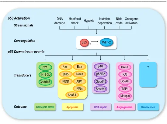
General Introduction _________________________________________________________________________ 23 p53 Activation p53 Downstream events Stress signals Core regulation Transducers Outcome Mdm-2 p53 DNA damage Hypoxia Nitric oxide Nutrition deprivation Heat/cold shock Oncogene activation
Cell cycle arrest
p48 p53R2 Gadd45 Sestrins BAI-1 KAI Gd-AIF TSP1 Maspin
DNA repair Angiogenesis
Apoptosis p21 14-3-3σ Gadd45 Fas DR5 PIDD Bax Noxa PIGs AIP1 Apaf-1 Senescence ?
Fig. 2. Schematic diagram of p53 activation and response. Several types of stress signals
are detected by the cell and communicated to the p53 protein and its direct regulators. Once activated, p53 functions as a transcriptional modulator of p53-regulated genes. This results in three major outcomes of cell cycle arrest, cellular senescence, or apoptosis. Other p53 target genes are involved in different cellular responses, such as inhibition of angiogenesis or DNA repair. The role of p53 is likely to depend on the context in which the activation occurs.
2.1.
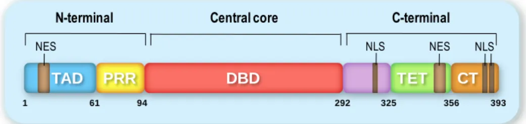
Structure and function of p53 domains
The functional complexity of p53 is mirrored in its chemical structure (reviewed in (Joerger & Fersht 2008). Human and murine p53 exist predominantly as tetramers of unusual shape, and to a smaller extent as higher oligomers in solution (Friedman