Évora 2019
1
Universidade de E´vora - Escola de Ciˆencias e Tecnologia
Mestrado Integrado em Medicina Veterin´aria
Disserta¸c˜ao
Exploring the impact of canine idiopathic epilepsy on
hemogram parameters
Daniela Filipa da Silva Pinto
Orientador(es) | Joana da Costa Reis
Évora 2019
Universidade de E
´vora - Escola de Ciˆencias e Tecnologia
Mestrado Integrado em Medicina Veterin´aria
Disserta¸c˜ao
Exploring the impact of canine idiopathic epilepsy on
hemogram parameters
Daniela Filipa da Silva Pinto
Orientador(es) | Joana da Costa Reis
A disserta¸c˜ao foi objeto de aprecia¸c˜ao e discuss˜ao pu´blica pelo seguinte ju´ri nomeado
pelo Diretor da Escola de Ciˆencias e Tecnologia:
• Presidente | Rita Payan Carreira (Universidade de E´vora)
• Vogal | David Orlando Alves Ferreira (Universidade de E´vora)
• Vogal-orientador | Joana da Costa Reis (Universidade de E´vora)
I
ACKNOWLEDGMENTS
Being surrounded by people who inspire you and want to see you at your greatest is one of the best things we can experience from life. And there are many people I would like to express my gratitude for making my dream come true.
To the woman of my life, my mum. Your strength, kindness and resilience are some of your qualities I admire the most and you are undoubtedly my biggest inspiration and role model. All this would not be possible without your guidance and sacrifice. Thank you for making me a better person and for supporting me in all my decisions.
To my imperfectly perfect family for supporting me throughout this journey. To my grandparents, for all the never-ending love and for all the cookies and sweets, which in your opinion were very important to maintain proper concentration and reasoning. To my aunt (also called I) for all the endless support and affection. To my godmother for being such a caring and trustful person and for making me believe in my capabilities. To Victor and Magda for bringing joy to my life.
To my supervisor Joana Reis for all the friendship and for the prompt availability (especially on Facebook) to help me with all my doubts and thoughts. Thank you for believing in my capabilities since the beginning.
To my externship supervisor Juliana Moreira for accepting and supporting me on this study, making this all possible and for providing your guidance and knowledge to me.
To all the staff from the Hospital Veterinário Breed Paredes, all the veterinarians, nurses, veterinary assistants and trainees. I have learned so much with you all and I could not be more pleased and thankful for all your support and guidance throughout this externship.
To Professor Alfredo, who promptly offered to help me with all the data and statistical work. To my dearest group of friends, also known as VetDamas. Each of you have a special place in my heart and I truly could not be more thankful to the universe to put people like you in my way. You all turned a tough and stressful journey into one of the most amazing phases of my life. Within this amazing group, I would like to leave a big shout out for two people in particular: Bárbara and Ana.
To Bárbara, my crazy cat lady (as am I, hence the chemistry), for never leaving my back at any circumstance. Even in one of my hardest moments you were always available for me, always kind and supportive. Thank you for all the deep conversations, music-sharing, study sessions,
II
homemade marmalades, veterinary congresses, vegetarian food, walking moments and for making your house my second home in Portugal (literally).
To Ana, the sweetest and kindest girl I ever met. Thank you for all your advices about all the imaginable topics, your sweet good-morning messages, little runs over the town, study sessions and, most importantly, for all your endless patience with me (and with all my swear words, not offensive though).
To my university friends, especially those from my classroom, with whom I shared a lot of amazing moments.
To my residence friends, with whom I shared wonderful moments, from the motorway journeys to the study sessions, I will keep all those moments for the rest of my life.
To Daniela for being such a crazy and special friend, for helping me with my anxiety, for creating the most incredible memes and for making sure I would never have toilet rolls or chocolate missing (sharing is caring).
To Carolina for being the most unpredictable but best company during our 6-month journey in Vila Real. Those pastries will not be forgotten, neither will you.
To my room colleague Inês, for all the amazing moments shared in our room 124. I strangely miss that annoying phone alarm ringing that would awaken everyone but you.
To João and his family for believing in me and my capabilities and for all the unconditional support through those four years of university.
To my goddaughter, who was the biggest surprise in my academic life. I could not be more grateful for being chosen as your godmother and I am forever thankful for all your friendship.
To Matt, for all the lovely moments shared, for supporting me and for being totally available for reviewing my dissertation beforehand.
To my hometown friends, Joana, Mafalda, Maria José and Mariana for being the craziest friends and for looking out for me for the past 9 years.
Last but not least, I would like to thank to Professor Hélder and Andrew, you made my other externship equally amazing and for making me see the beauty in laboratory routine. Also, a big mention goes to Cristina for accompanying me in this unforgettable experience.
III
ABSTRACT
Epilepsy is one of the most common diseases that affect both dogs and humans and may be potentially threatening when not treated. The cause of idiopathic epilepsy remains unclear, but many factors have been identified as potential triggers for developing epilepsy. Nowadays many drugs are available for the treatment of epilepsy, but the cases of total remission are rare and there is no cure. Moreover, most of anti-epileptic drugs (AEDs) induce side effects.
This dissertation includes a literature review outlining the topic of idiopathic epilepsy, together with a preliminary, exploratory study with the objective of investigating possible alterations on blood parameters that might be induced by repeated seizures. Neuroinflammation may be a consequence of seizures, which possibly induces alterations on blood parameters.
The results suggest that a status of inflammation is likely following seizures, however, further studies regarding the topic are necessary to achieve more clear and precise information.
IV
RESUMO
O IMPACTO DA EPILEPSIA IDIOPÁTICA CANINA NOS PARÂMETROS
DE HEMOGRAMA
Epilepsia é uma síndrome neurológica frequente em cães e humanos e que pode ser potencialmente fatal caso não seja devidamente tratada. A causa da epilepsia idiopática permanece desconhecida, porém vários fatores têm sido apontados como desencadeantes desta síndrome. Atualmente, várias drogas estão disponíveis para o tratamento da epilepsia, mas são raros os casos de total remissão e não existe cura. Além disso, estas drogas possuem vários efeitos secundários associados.
A presente dissertação incluí uma revisão bibliográfica sobre a epilepsia idiopática em conjunto com um estudo preliminar e exploratório cujo principal objetivo foi investigar possíveis alterações nos parâmetros de hemograma que poderiam ser induzidos por convulsões recorrentes. A neuroinflamação pode ser um resultado de convulsões recorrentes e um estado inflamatório pode levar a alterações nos parâmetros de hemograma.
Os resultados sugerem que a inflamação é provavelmente desencadeada após episódios convulsivos, porém, mais estudos relacionados com este tópico são necessários.
V
TABLE OF CONTENTS
ACKNOWLEDGEMENTS ………..………… I
ABSTRACT ……….. III
RESUMO ………..……….. IV
LIST OF TABLES ……….………..…….... VII
LIST OF FIGURES ……….………...………… VIII
LIST OF SYMBOLS AND ABBREVIATIONS ……….………..………... IX
PREFACE ……….……….….…. XII
1. LITERATURE REVIEW ... 1
1.1 INTRODUCTION ... 1
1.2 EPIDEMIOLOGY ... 2
1.2.1
PRECIPITATING FACTORS ... 3
1.3 PATHOPHYSIOLOGY OF EPILEPSY ... 4
1.4 CLASSIFICATION OF EPILEPSY ... 10
1.4.1
CLASSIFICATION OF EPILEPSY ACCORDING TO ITS AETIOLOGY ... 11
1.4.2
CLASSIFICATION OF SEIZURES ACCORDING TO LOCALISATION ... 12
1.4.3
CLASSIFICATION OF SEIZURES ACCORDING TO THEIR PRESENTATION ... 16
1.4.4
CLASSIFICATION OF SEIZURES ACCORDING TO THEIR CHRONOLOGICAL
DISTRIBUTION... 22
1.5 DIAGNOSIS ... 26
1.5.1
DISEASES THAT MAY MIMIC EPILEPTIC SEIZURES ... 27
1.5.2
SIGNALMENT AND CLINICAL EXAMINATION ... 31
1.5.3
NEUROLOGICAL EXAMINATION ... 32
1.5.4
ELECTROENCEPHALOGRAM ... 32
VI
1.5.6
VIDEO-DOCUMENTATION ... 33
1.5.7
IMAGING AND CSF ANALYSIS ... 34
1.5.8
TIER-SYSTEM ... 36
1.6 TREATMENT... 37
1.6.1
PHENOBARBITAL ... 39
1.6.2
POTASSIUM BROMIDE ... 41
1.6.3
IMEPITOIN ... 42
1.6.4
LEVETIRACETAM ... 43
1.6.5
DIAZEPAM ... 44
1.6.6
NON-LICENSED EU DRUGS SUGGESTED FOR USE IN THE TREATMENT OF IE IN
DOGS
44
1.7 ALTERNATIVE TREATMENTS ... 45
1.8 PROGNOSIS ... 45
1.9 HEMOGRAM ... 47
1.10 HEMOGRAM PARAMETERS, INFLAMMATION AND SEIZURES ... 47
1.11 NEUROINFLAMMATION... 48
2. STUDY – EXPLORING THE IMPACT OF CANINE IDIOPATHIC EPILEPSY ON HEMOGRAM
PARAMETERS ... 51
2.1. INTRODUCTION ... 51
2.2. OBJECTIVE ... 51
2.3. MATERIALS AND METHODS ... 51
2.3.1. BLOOD COLLECTION AND HEMOGRAM PARAMETERS ... 53
2.3.2. QUESTIONNAIRE ... 54
2.3.3. STATISTICAL ANALYSIS ... 55
2.4. RESULTS ... 55
2.4.1. AGE, BREED, SEX AND WEIGHT ... 55
2.4.2. QUESTIONAIRE ... 58
2.4.3. HEMOGRAM PARAMETERS ... 61
2.5. DISCUSSION ... 64
2.6. CONCLUSION ... 70
VII
LIST OF TABLES
Table 1: Summary of abnormalities that may lead to seizures (Adapted from Sanders 2015). .. 4 Table 2: Epilepsy types defined by aetiology (Adapted from Berendt et al. 2015). ... 11 Table 3: Causes of seizures in dogs (Adapted from Berendt et at. 2015)... 12 Table 4: Epilepsy classification by seizure semiology (Adapted from Berendt et al. 2015). ... 21 Table 5: Different aetiologies that may present similar clinical manifestation to IE (Adapted from
De Risio et al. 2015). ... 30
Table 6: Tier system of confidence for diagnosis of IE (Adapted from De Risio et al. 2015). ... 37 Table 7: Reference blood parameters for dogs in a bc-2800 Vet Analyser... 55 Table 8: Comparison of hemogram parameters between the group control and epilepsy group
after seizure. ... 62
Table 9: Comparison of hemogram parameters between the control group and free-seizure
epilepsy group. ... 63
Table 10: Comparison of hemogram parameters between epilepsy group after seizure and
VIII
LIST OF FIGURES
Figure 1: Factors that may affect seizure threshold (Adapted from DeLahunta et al. 2015). ... 5
Figure 2: Neuronal environment constituted by different elements (Adapted from DeLahunta et al. 2015) ... 6
Figure 3: Diagnostic approach to idiopathic epilepsy (Adapted from Vernau 2015). ... 36
Figure 4: Treatment approach using phenobarbital as monotherapy (Adapted from Bhatti et al. 2015) ... 41
Figure 5: Characterization of the variable weight in both epileptic and control groups. ... 56
Figure 6: Characterization of the variable sex in both epileptic and control groups. ... 57
Figure 7: Characterization of the variable breed in both epileptic and control groups. ... 58
Figure 8: Age at the first seizure in the epileptic group. ... 58
Figure 9: Duration of seizures in the epileptic group. ... 59
Figure 10: Frequency of seizures in the epileptic group. . ... 60
Figure 11: Autonomic signs observed in the epileptic group. ... 61
Figure 12: RDW absolute values distribution in the three groups. ... 63
IX
LIST OF SYMBOLS AND ABBREVIATIONS
ABC – Airway – Breathing – Circulation AED – Antiepileptic Drug
ALP – Alkaline Phosphatase ALT – Alanine Transaminase
AMPA – 5-methyl-4-Isoxazole Propionate APP – Acute Phase Proteins
AST – Aspartate Transaminase
BZD – Benzodiazepine
Ca2+ – Calcium
Cl- – Chloride
CRI – Constant Rate Infusion CRP – C-Reactive Protein CSF – Cerebrospinal Fluid
CT-scan – Computed Tomography scan
CVA – Cerebral Vascular Accident
EMA – European Medicines Agency EU – European Union
GABA – Gamma-Amino-Butyric Acid GGT – ϒ- Glutamyl Transferase
GME – Granulomatous Meningoencephalitis HCT – Haematocrit
HE – Hepatic Encephalopathy
HGB – Haemoglobin
X
IE - Idiopathic Epilepsy IE – Interleukin
ILAE – International League Against Epilepsy IV – Intravenous
IVETF - International Veterinary Epilepsy Task Force K+ - Potassium
KBr - Potassium Bromide KD – Ketogenic Diet
MCH – Mean Corpuscular Haemoglobin
MCHC – Mean Corpuscular Haemoglobin Concentration MCV – Mean Corpuscular Volume
mGluR – Metabotropic Glutamate Receptor
MPV – Mean Platelet Volume
MRI – Magnetic Resonance Image Na+ - Sodium
Na+ K+ ATPase – Sodium Potassium Adenosine Triphosphatase Pump
NLR – Neutrophils/Lymphocytes Ratio NMDA – N-Methyl-D-Aspartate
NME – Necrotising Meningoencephalitis
NSAID – Non-Steroidal Anti-Inflammatory Drugs
PB - Phenobarbital
PCR – Polymerase Chain Reaction PCT – Plateletcrit
PD – Paroxysmal Dyskinesia PD/PU – Polydipsia/Polyuria PDW – Platelet Distribution Width PLT – Platelets
XI
QoL – Quality of Life RBC – Red Blood Cells
RDW – Red Cell Distribution Width SE – Status Epilepticus
StE – Structural Epilepsy
TGF - Transforming Growth Factor TNF – Tumour Necrosis Factor UE – Uremic Encephalopathy WBC – White Blood Cells
XII
PREFACE
This dissertation was written following a five-month externship at Hospital Veterinário Breed Paredes, from 5th November 2018 to 5th April 2019 and under the guidance of a supervisor chosen
by the trainee and hospital staff.
This externship offered the opportunity to be in contact with different areas of veterinary medicine that included orthopedics and soft tissue surgery, diagnostics, internal medicine including specific areas such as dermatology, nutrition, oncology, ophthalmology, cardiology, dentistry, emergency and critical care medicine.
Throughout this externship the trainee developed essential skills that would be important for the exercise of her future profession. Also, the required data for this study was collected there and subsequently organized and analyzed, resulting in the dissertation below.
1
1. LITERATURE REVIEW
INTRODUCTION
Epilepsy is a neurological syndrome and known to be one of the most common among the canine and feline population 1, as well as in humans 2. An epileptic seizure can be defined as abrupt,
involuntary electrical discharges happening within a population of neurons, leading to sudden and involuntary muscle movements and behaviour changes 3–5. As said by Moore, epileptic seizures are a clinical manifestation of abnormal hyper-synchronisation of neurons within the forebrain 6.
The effect of multiple and continued anomalous discharges will affect the edge neurons and these discharges will further progress to other neurons 7. As stated by Berendt, an epileptic seizure is
characterised for consciousness impairment in most of the cases, paroxysmal events and identical clinical manifestations in all episodes 8. The formation of these hypersynchronous
discharges is called epileptogenesis 7. According to De Risio et al., inflammation, infection,
metabolic disorders, trauma, tumours and oxygen deprivation can lead to abnormal neuronal activity 9. Regardless of the mechanism, the epileptic seizure is defined as a brain disorder 8.
Given the ideal conditions, an animal may experience a seizure and many factors may be involved in its progression 4.
The term epilepsy is only considered when two or more seizures occur within more than 24 hours between each event 1,10. Therefore, it is crucial to explore underlying causes that may lead to
seizures, being an imminent emergency if the epileptic episode is prolonged in time 10. A single
seizure may be a result of cerebral overload after brain injury due to multiple causes and, even though epilepsy should be considered as a differential diagnosis, it is unlikely to be diagnosed when direct damage is inflicted to the brain 8. Regarding epilepsy, the term idiopathic refers to the
epilepsy of unknown cause when in the presence of unprovoked seizures and absence of brain abnormalities, thus with a possible hereditary predisposition 4,11,12. Idiopathic epilepsy (IE) is
known to be the most common type of epilepsy in dogs 13–16.
The presence of seizures is not pathognomonic for IE. When undertaking diagnosis, it is essential to keep in mind that many different disorders may induce seizures 17. Nevertheless, there may be
cases where the aetiological cause is unknown, being IE the most likely diagnosis 9. Usually,
these abnormal electrical signals can be propagated throughout different structures in the brain and depending upon the structure that has been affected, the clinical manifestations may be
2
different 3. Nevertheless, the observable signs will be related to the affected area of the cerebral
cortex 10.
EPIDEMIOLOGY
According to Heske et al., the estimated prevalence of epilepsy within the canine population is 0.75% 18. In humans, the prevalence of epilepsy is around 1% 19. In other studies, a prevalence
of 0.62% - 0.75% of epilepsy in dogs was also reported 20,21. The prevalence of epilepsy within a
specific group of dogs included in a study in Japan was 1.9%, having a prevalence of 0.9% of IE compared with the 0.4% of structural epilepsy (StE) 22.
Male dogs seemed more likely to develop epilepsy compared with female dogs 15,23,24. When
considering dogs diagnosed with IE, males have a shorter life span when compared with females
25.
As described in many studies, some breeds may have a higher likelihood for IE, including the Labrador Retriever 18,23,26, Boxer 18,27, Belgian Shepherds 18,28,29, German Shepherds 23, Border
Collies 23,30, Chihuahua 15, Bernese Mountain Dogs 24, Lagotto Romagnolo 31 and Toy Poodles 15. Pure breeds have a higher incidence of IE compared with cross-breeds 32.
Breeds which have a higher incidence rate of IE can be genetically predisposed to this disease
18. IE is likely to be diagnosed in dogs whose seizure onset occurred between six months to six
years 33, with a clinical history of repeated seizures and normal consciousness after episodes 23,34,35. According to Smith et al., a 97% confidence diagnosis of IE was found in such
presentations 34. As stated by Coates and O’Brien, only one-third of the dogs aged between one
to five years are diagnosed with StE 36. It is possible that some of these dogs that experienced
seizures may not have a definitive cause for their seizures 16,37. It is unlikely that structural brain
alterations are detected during magnetic resonance imaging (MRI) in epileptic dogs younger than six years 34. However, it is likely to find such alterations in epileptic dogs older than six years 34.
Many studies found a higher prevalence of IE in many purebred and this strongly suggests a genetic predisposition 4,30. In fact, in human medicine, it is said that 40% of epileptic patients have
a genetic disorder underlying their epilepsy 38. Only one type of epilepsy, related to a genetic
disorder was described in Lagotto Romagnolo dogs and it is associated with a mutation in the
LGI2 gene 31. This mutation leads to a defect on the regular conformation of proteins that
constitute the neuronal membrane, therefore it is recognised as a channelopathy 3,12,31. This
3
1.2.1 PRECIPITATING FACTORS
Many precipitating factors for seizures are known from human medicine and many studies have been conducted in veterinary medicine with the same purpose of finding precipitating factors associated to seizures in dogs 20,39,40. The real mechanism by which precipitating factors may lead
to seizures is still unknown. However, it is believed that these factors may change the normal homeostasis within the brain and therefore they may lower the seizure threshold, increasing the likelihood of seizures 41.
Some studies suggested a relationship between oestrus cycle and epilepsy, stating that estrogens may reduce the seizure threshold, whereas progesterone may have an inhibition effect 28,42. Thus,
there is an increase in seizure frequency both in periods when estrogens are mainly present or when progesterone is significantly decreased 28,42. In a study by Van Meervenne et al., an
association was found between seizures and specific stages of the reproductive cycle 42.
According to Van Meervenne et al., dioestrus and oestrus have been identified as important stages regarding epileptic seizures and this could be explained by the anti-convulsant effects of progesterone and the pro-convulsant effects of estrogens, respectively 42. This finding indicates
that female neutered epileptic dogs might have a prolonged lifetime when compared with non-neutered epileptic females 23,28,42. The effect of sexual hormones on seizure threshold has been
discussed in humans as well 43. Nonetheless, neutered dogs tend to be less aggressive or
hyperactive due to the declining rate of sexual hormones present in the organism, which may help controlling seizures, as hyperactivity and aggressiveness are considered as risk factors for triggering epileptic seizures 28.
Stress can also be a precipitating factor, thus breeds that are known for being more active and anxious may be more vulnerable 44. A study analysed the relationship between seizures and
possible precipitating factors in a group of dogs and stress was found to be strongly related with the occurrence of seizures within 24h after stress-related situations 39. According to Forsgard et
al., stress-related situations possibly related with the occurrence of seizures in dogs with IE
include sleep deprivation, emotional stress or changes in the average daily routine of the dog 39.
The same factors are known to increase the likelihood of seizures in humans that have epilepsy as well 45–47.
Weather conditions may be considered as a precipitating factor, however, there is little evidence in veterinary medicine. One study reported that nine of 50 of owners that replied to a questionnaire had identified weather conditions as a precipitating factor for seizures in their dogs 39. Conversely,
studies in humans have reported a relationship between heat and cold temperatures and the occurrence of seizures, where patients identified heat and cold temperatures as being precipitating factors 45,46.
4
PATHOPHYSIOLOGY OF EPILEPSY
Epileptogenesis is described as the process of conversion of a regular neuronal activity into a hyperexcitable, disorganised and synchronised neuronal activity which persists for a non-specific period 8,10,48. Trauma, brain infections/tumours, brain malformations, metabolic disorders or
oxygen/glucose deprivation may disorganise the normal neuronal activity leading to hyperexcitability that triggers a seizure 48,49. The most frequent mechanism known for triggering
seizures is the imbalance between excitatory and inhibitory neurotransmitters, with insufficient neuronal inhibition or excessive excitation 50. Abnormal concentration of specific ions can also
lead to epileptiform events 50. A summary of alterations in the neuronal environment that may lead
to abnormal neuronal discharge can be seen in Table 1.
Table 1: Summary of alterations in the neuronal environment that may lead to seizures
(Adapted from Sanders 2015).
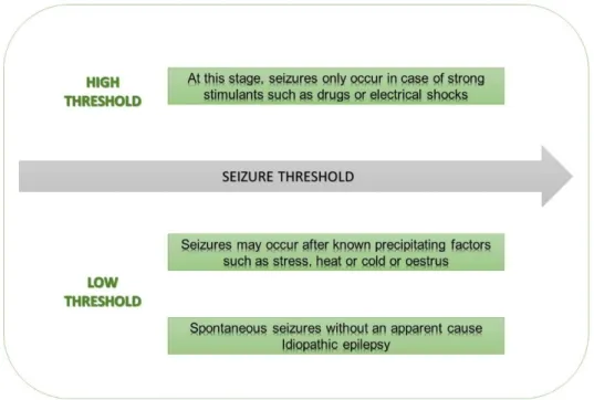
The concept of threshold is essential to understand the epileptogenesis. According to DeLahunta
et al., a seizure threshold is considered as the minimum level of inhibition that when exceeded,
may have the potential to originate an uncontrolled discharge of a particular group of neurons 51.
A seizure occurs whenever this threshold lowers after any disturbance 51. Factors related with
seizure threshold are illustrated in the Figure 1.
In most cases, the epileptic focus initiates in neurons localised in the forebrain but may be originated in other parts of the brain as well 51. The cerebral cortex and hippocampus englobe
groups of easily-firing neurons, which makes these regions the initial source for seizures in most of the cases 52.
There is an individual susceptibility for epilepsy and it varies among dogs 8. In fact, under the
same conditions, a dog with a lower threshold is more likely to experience an epileptic event 8 as
a lower threshold will mean that the mechanism that balances the excitatory and inhibitory responses in the brain is less stable. Dogs with a lower threshold are more vulnerable to factors such as stress, exhaustion, oestrus and weather conditions 53. The same dog may experience
ALTERATIONS IN THE NEURONAL ENVIRONMENT
ALTERATIONS IN..
Inhibitory/excitatory homeostasis Ions concentrations
Neuronal homeostasis (e.g. brain infections/tumours) Function of neurotransmitters
Neuronal transmission, leading to the spontaneous firing of large groups of neurons (e.g. trauma)
5
both focal and generalised seizures 8. Focal seizures arise from a specific area in the brain,
whereas generalised seizures start in an epileptic focus, evolving to other regions of the brain 54.
The neuronal environment is constituted by various elements that are intimately connected between each other and guarantee the normal homeostasis in the brain. If one of these elements is disturbed, it may trigger a potential seizure by lowering the threshold 51.
The sodium – potassium adenosine triphosphatase pump (Na+K+ ATPase) is the main enzyme
that composes this neuronal environment, responsible for the flow of sodium (Na+) and potassium
(K+) across the neuronal membrane in an energy-dependent process 51. All these ions have their
channels along the neuronal membrane, allowing them to freely move across it. Neurotransmitters may induce an excitatory or inhibitory response, being glutamate and GABA (Gamma-Amino-Butyric Acid) the most essential excitatory and inhibitory neurotransmitter, respectively. Nevertheless, astrocytes also play an essential role in this environment by metabolising neurotransmitters and allowing the flow of neurotransmitters and ions through capillaries 51.
Elements that constitute the neuronal environment are represented in the Figure 2.
Figure 1: Factors that may affect the seizure threshold (Adapted from DeLahunta et al.
6
Figure 2: Neuronal environment constituted by different elements (Adapted from DeLahunta et
al. 2015)
Understanding the physiology behind the neuronal transmission is essential to understand the mechanisms underlying the pathophysiology of the epileptic events. Neurons are capable of communicating between them, receiving and integrating signs received from other neurons and transmitting information in the form of electrical signs 55.
The neuronal membrane has a vital role in electrical transmission 55. This neuronal membrane
separates two spaces, the intra and extracellular space, each one mainly constituted by several ions differently charged 7,55. During the resting membrane potential, the inner membrane is
negatively charged, whereas the outer membrane is positively charged, mostly because of the presence of differently charged ions: Na+, K+ and chloride (Cl-) 55. In the absence of stimuli, the
resting membrane potential is maintained along the neuronal membrane at a difference of -70 mV between inside and outside spaces 55. Regarding the resting membrane potential, many factors
7
the presence of ions with different concentrations/voltages and the permeability of the neuronal membrane to these ions 7,55. The K+ flow throughout the neuronal membrane profoundly
influences the resting membrane potential because of the higher permeability to this ion 55. K+
moves quickly through K+ channels across the membrane by its concentration gradient and
enables a dynamic balance 55. Concerning the extracellular space, Na+ and Cl- are the primary
ions existing, whereas K+ ions are mostly concentrated in the intracellular space. Some anionic
proteins within the neuronal cell can also contribute to a negative charge 55. The regulation of
these ions is mainly controlled by astrocytes, which are the dominant subtype of glial cells that help in maintaining the normal homeostasis within the brain 56. These glial cells can work as
buffers, regulating ions levels and metabolising neurotransmitters 7.
The Na+- K+ ATPase pumps allows the flow of Na+ and K+ ions throughout the neuronal membrane
and against their concentration gradients 7,55,57. This process is energy-dependent, requiring ATP
as the primary source of energy that is provided by glucose metabolism and which represents the majority of the glucose consumption by the brain 55,58. Changes in the average concentration of
serum glucose could lead to seizures because neurons do not have appropriate mechanisms to maintain glucose for metabolism purposes 55.
As these pumps are a decisive factor to the normal function of neurons, many studies have been conducted to understand how alterations regarding Na+- K+ ATPase pumps can affect the
neuronal activity 57,59. One study reported that abnormalities in the functionality of these pumps
can trigger seizures 59.
In the presence of a stimuli, neurons will depolarise leading to an action membrane potential that turns the intracellular space more positively charged than the extracellular space 55.
Consequently, this action membrane discharge will progress along the neuron until it reaches its presynaptic terminal 55,57. Neurons are separated from each other by a synaptic cleft and in the
presynaptic terminal, it is possible to find vesicles that are neuronal structures responsible for the storage and releasing of chemical substances denominated neurotransmitters 7. These
neurotransmitters are an essential communication between consecutive neurons and depending upon the electric impulse generated, different neurotransmitters may be released 7. After the
electric impulse reaches the presynaptic terminal, the ions channels will open and subsequently it allows the bonding between synaptic vesicles and the neuronal membrane in the presynaptic terminal 7. After bonding, these vesicles are stimulated to release a particular neurotransmitter to
the synaptic cleft 7. Consequently, the released neurotransmitters will bond to specific receptors
localised in the nearest post-synaptic neuron 7.
The pathophysiology of seizures is complex and several factors have been credited as inducing-seizures factors. Some studies reported the relationship between neurotransmitters and epilepsy where glutamate seems the excitatory neurotransmitter more involved in epileptogenesis 60–62.
8
In a healthy brain, excitatory and inhibitory mechanisms are balanced, which maintains a regular neuronal synchronisation and avoids the triggering of neurons without previous stimulating factors provided by other surrounding neurons 7. When triggered alone, these neurons may initiate an
uncontrolled excitatory response that could lead to a seizure. An imbalance between inhibitory and excitatory responses may increase the likelihood of epileptiform activity if excitatory responses are over-represented 63. The simplest mode to inhibit an excitatory response is by
releasing inhibitory neurotransmitters like GABA 52. Typically, an inhibitory response precedes an
excitatory response and it is when inhibition mechanisms fail in controlling excitatory messages that neurons become hyper-synchronised, thus originating an epileptic focus 52.
Glutamate is the most important excitatory neurotransmitter that is recycled by astrocytes and is ubiquitously distributed in the central nervous system (CNS) 8,50,56,64,65. Glutamate is obtained from
glutamine that is converted into glutamate by glutaminase, a mitochondrial enzyme 52. Astrocytes
have an important role in metabolising glutamate into glutamine, which is carried back to the presynaptic terminal to be reused 52.
It is possible that glutamate is involved in the mechanism underlying epilepsy because there is a remarkable increase of glutamate’s concentration when a seizure occurs 8,56. The
overconcentration of glutamate might be toxic, contributing to neuronal death by inducing alterations in permeability to calcium (Ca2+) ions 13,52,64,66. Elevation of intracellular Ca2+
concentrations may lead to activation of certain enzymes such as proteases and phospholipases inducing proteolysis of proteins that constitute the neurons 62. Moreover, during neuronal death,
glutamate is released to the extracellular space as it is present in most of the neurons 67,68. The
resulting increase in extracellular glutamate overstimulates glutamate receptors especially the N-methyl-D-aspartate (NMDA) receptor, which allows the entrance of calcium ions into neuronal cells through this receptor 67,68. The overconcentration of Ca2+ inside the neuronal cells will also
contribute for oxidative stress and mitochondria’s damage 67,68. Neuronal death may expand the
seizure focus increasing the likelihood of a seizure occurring and entering into a vicious cycle
52,62. However, while it remains disputed as to whether neuronal death is a cause or a
consequence of seizures, some people believe neuronal death could be both a cause and consequence.
Several types of receptors differ by their agonist/antagonist function or their permeability to specific ions 7. According to Nakanishi, these receptors are involved in many cerebral functions
such as synaptic transmission, brain plasticity, learning, memory, brain development and differentiation 69.
Concerning glutamate, inotropic receptors are one type of receptor that work as a Ca2+ channel
that only opens when bonded to an affinity-neurotransmitter, causing an opening that allows ions to flow through the receptor 63,70,71. These ionotropic receptors are related to a direct and faster
9
ions 62. The 5-methyl-4-isoxazole propionate (AMPA) receptor is another subtype of ionotropic
glutamate receptor and it is related to fast excitatory responses 64.
On the other hand, metabotropic glutamate receptors (mGluR) are closely related to the membrane and coupled to a G-protein 70,71. These receptors function as a second messenger
system and when activated, they allow the flow of Ca2+ and Na+ ions which leads to depolarisation
of neurons 63,70,71. Because it depends on secondary messengers, the response is slower when
compared with ionotropic receptors 70. These receptors help to modulate the neuronal activity and
they also regulate the releasing of neurotransmitters 64. Contrary to what happens with ionotropic
receptors, mGluRs are activated in the presence of prolonged and increased concentrations of glutamate, which is relatively frequent after brain injury induced by seizures or trauma 72.
GABA is synthesized from glutamate and it also has main receptors which are known as GABAA
and GABAB51,63,73,74. The former is an ionotropic receptor and the latter a metabotropic receptor
and, as previously discussed, they have different mechanisms of actuation 73. The GABAA
receptors allow the flow of Cl- ions, leading to hyperpolarization of neurons whereas GABA B
receptors operate via second messenger system, increasing the conductance of K+ ions while
decreasing the conductance of Ca2+ ions 75. Activation of both types of receptors leads to a
hyperpolarization of neurons, therefore causing an inhibition of neurotransmission 63,75.
GABA is known to be neuroprotective against distressing situations such as brain hypoxia and status epilepticus (SE), having an essential role on ceasing seizures 76,77. A study by Loscher et
al., concluded that the neurotransmitter GABA was hardly found in cerebrospinal fluid (CSF) of
epileptic dogs when compared with healthy dogs 78. Another study found that dogs with a low
concentration of GABA in CSF appeared to develop antiepileptic drug (AED) resistance 79. Some
studies suggest that either inhibition of GABA receptorsor excessive activation of glutamate receptors may underlie the process of epileptogenesis 50,64,67,73.
Epileptic seizures may aggravate over time, especially without proper treatment. Some mechanisms that may worsen epileptiform events have been widely studied. Kindling is one of those events, where repeated lower-intensity stimulus may induce spontaneous seizures by stimulating non-hyperexcitable neurons that are converted into a group of hyperexcitable neurons
50,64. It is possible that dogs develop epilepsy after experiencing spontaneously seizures because
of this mechanism 80. The group of neurons that triggered the seizure will be considered as seizure
focus 80. A study conducted by Goddard et al., concluded that discrete and lower-intensity
electrical stimulation applied to some areas of mammalian brains overtime originated permanent alterations of brain function and lowered the seizure threshold, thereby increasing the likelihood of seizures with minimal stimulus 80. Another mechanism is known as mirroring and as stated by
Dewey and Thomas, it is the process where a group of neurons from the opposite brain hemisphere are drafted into the seizure focus via the corpus callosum 50. Secondary focus
10
consequence of the kindling process and this secondary focus has the potential to generate seizures just as the primary source 80.
Secondary brain abnormalities may result from seizures, mainly when occurring frequently and for an extended period. As referred previously, neurotoxicity resulting from overexcitation by high concentrations of glutamate may induce neuronal death 64,66,81. This disturbance of normal
neuronal function may induce brain oedema with subsequent increase of intracranial pressure (ICP), which may decrease the normal perfusion of the brain 81. Therefore, the regular supply of
energy and nutrients for neuronal cells is compromised, leading to anaerobic glycolysis, neuronal acidosis and neuronal dysfunction 81. In a study by Hasegawa et al., significant concentrations of
glutamate were found in the CSF of epileptic dogs which suggests the relationship between this neurotransmitter and the pathophysiology of epilepsy 82. Similarly, increased concentration of
glutamate was found in CSF of epileptic dogs 83, emphasising the theory that glutamate could be
a possible biomarker for the diagnosis of IE.
CLASSIFICATION OF EPILEPSY
Diagnosing epilepsy in dogs may be challenging for veterinarians considering the amount of different aetiologies and clinical manifestations involved. Therefore, an organised and detailed physical examination and signalment are necessary to ensure that noteworthy alterations are detected. A hemogram, routine serum chemistry and urinalysis are examples of simple and inexpensive exams that should be performed to exclude diseases that may mimic IE 11. IE should
only be considered when significant abnormalities are not detected throughout physical examination and with a normal interictal neurological examination 34,49. In human medicine it was
found that some types of epilepsy were possibly related to genetical abnormalities, resulting in changes of the existing classification regarding epilepsy by the International League Against Epilepsy (ILAE) 84. Many studies in veterinary medicine suggest genetical abnormalities
underlying IE 24,30,85–87. These findings led to a new classification adopting the terms of StE,
11
1.4.1 CLASSIFICATION OF EPILEPSY ACCORDING TO
ITS AETIOLOGY
Epilepsy can be classified according to its aetiology (Table 2). IE is classified as epilepsy of unknown cause when no other diseases are suspected 1 but recently IE has been divided into
subgroups. According to these subgroups IE can be defined as purely genetic, which is usually related to channelopathies that are characterised by mutations in genes that encode K+, Na+, Ca2+
and Cl- channels 1,12 but it can only be confirmed by specific genetic tests 1. When in the absence
of diagnostic tests that confirm genetic mutation, veterinarians should suspect of genetic-related epilepsy when the prevalence within a specific breed is higher than 2% 1. A causative gene has
been already confirmed as responsible for epileptic seizures 1.
Lastly, epilepsy can also be a consequence of any process that induces brain injury 1, being the
latter defined as StE1. Typically, StE includes all the intracranial pathologies, regardless of
aetiology. These pathologies can be either infectious, inflammatory, neoplastic, traumatic, vascular, degenerative or anomalous 1,7. The seizures associated with StE can also be defined
as secondary seizures, as they are secondary to other disorders that had inflicted direct or indirect damage to the brain 13.
Table 2: Epilepsy types defined by aetiology (Adapted from Berendt et al. 2015).
CLASSIFICATION OF EPILEPSY ACCORDING TO ITS AETIOLOGY
IDIOPATHIC EPILEPSY
Genetic Idiopathic Epilepsy Suspect Genetic Idiopathic Epilepsy Unknown Cause Idiopathic Epilepsy
STRUCTURAL EPILEPSY Intracranial Pathologies Vascular Anomalous Inflammatory/Infectious Neoplastic Traumatic Degenerative
12
1.4.2 CLASSIFICATION OF SEIZURES ACCORDING TO
LOCALISATION
The cause of seizures can be located inside or outside the CNS 11. Causes of seizures according
to its localisation are summarized in Table 3.
Intracranial causes directly affect the CNS and are intimately related to the brain 11. Progressive
brain disorders such as tumours, inherited diseases or inflammatory processes are defined as acquired epilepsy 11. Tumours are one of the leading reasons for seizures and most of the dogs
with brain neoplasia frequently experience seizures 88. IE is defined by the occurrence of repeated
seizures with an apparent unknown source and is also included in the intracranial causes 11.
Considering extracranial causes, toxins and metabolic disturbances are also responsible for epileptic episodes 11. Usually, epileptic episodes induced by intoxication or metabolic disorders
are classified as reactive seizures because they are a physiological response to a temporary disruption of inhibition and excitation neuronal processes that are provoked by systemic disorders
13,16,89.
Table 3: Causes of seizures in dogs (Adapted from Berendt et at. 2015 and Lorenz et al. 2011)
*Induce reactive seizures CAUSES OF SEIZURES IN DOGS
INTRACRANIAL CAUSES
Idiopathic epilepsy Genetic or unknown cause
Acquired epilepsy
Brain injury (e.g. trauma, cerebral vascular accident) Progressive brain disorders (e.g. tumours, inherited or
inflammatory diseases)
Brain Development disorders (e.g hydrocephalus)
EXTRACRANIAL CAUSES*
Toxins
External (e.g. carbamates, lead poisoning, ethylene
glycol, metaldehyde, permethrins, mycotoxins, chocolate, metronidazole, organophosphates)
Internal, associated with a metabolic disease or organ
failure (e.g. hepatic encephalopathy due to portosystemic shut, uraemia)
Metabolic Disorders
Hypoglycemia, hyperglycemia, electrolyte disorders, hypoxia, thiamine deficiency, cobalamin deficiency
13
1.4.2.1 BRAIN DEVELOPMENT DISORDERS
Some brain abnormalities may happen during the normal development of dogs and even though it is not as common as other causes of seizures, they can be included in the possible differential diagnosis, especially when in the presence of some breeds like toy breed dogs 63,90. For instance,
chihuahua dogs are known for having a higher predisposition for congenital hydrocephalus and this disease is the most commonly diagnosed brain development disease that may induce seizures 63,90. Most of the brain development diseases are severe, so generally it is possible to
find abnormalities on the neurological examination such as proprioceptive deficits and visual impairment 63. Ataxia is also a common sign observed in dogs with brain development diseases 90. These brain development disorders are normally seen in young dogs and imaging diagnostic
exams like MRI and computed tomography scan (CT-scan) are of great value to detect these structural dysfunctions in the brain 91.
1.4.2.2 BRAIN INJURY
In humans, acquired epilepsy is normally a result of brain injury 92. Brain injury is life-threatening
and usually is a consequence of direct head trauma caused by car accidents, gunshot, falls or blows inflicted to the head 92. In other circumstances, brain injury can be the result of a cerebral
vascular accident (CVA) that in most cases is a consequence of thrombosis, haemorrhage or embolism 93. Nevertheless, CVA is not as common in dogs as in humans 93. Brain injury may be
reversible or not, depending upon its severity. The likelihood of developing seizures after brain injury will be higher the more severe the injury 92. In situations where skull fractures are presented,
the chances of developing epilepsy as a result of brain injury are higher 92. Brain injury alters the
brain homeostasis, which deregulates the physiological mechanisms of inhibition and excitation and consequently originating seizures 93,94. Currently, it is difficult to predict whether a dog that
suffered a brain injury will develop seizures in the future, but owners must be aware that chances are higher in such cases. Seizures might occur within an extended period after brain injury episode and pharmacological therapy should be prescribed as soon as seizures appear 94.
1.4.2.3 PROGRESSIVE BRAIN DISORDERS
Brain tumours are known to be the most common causes of progressive brain disorders 92. Brain
tumours are a common condition that may affect dogs of all ages but usually is more common amongst elderly dogs 63. Brain tumours can be primary or secondary (e.g. metastasis from
14
tissues, which may disorganise the usual structures and also prevent the normal blood flow in the vessels 63. Veterinarians should suspect brain tumours whenever elderly dogs are presented with
seizures, especially when it is the first onset 95. In some cases, seizures are the first symptom
showing up in dogs with brain tumours 95. When a brain tumour is suspected, imaging diagnostic
exams should be performed to detect these abnormalities in the brain. MRI is usually the best diagnostic tool available for diagnosing tumours because of its high sensitivity and specificity for soft tissues 63. Veterinarians should be aware that dogs with brain tumours or other intracranial
lesions may have an unchanged neurological examination 96. According to Ghormley et al., the
neurological examination had 74% sensitivity and 62% specificity on previewing StE 96.
Inflammatory brain diseases are also a common cause of seizures, especially in non-vaccinated young dogs. All inflammatory processes have the potential to start seizures whenever neuroinflammation is presented. Many inflammatory diseases may induce brain inflammation, whether they are infectious or not. Inflammation-related with bacteria (e.g. ehrlichiosis), virus (e.g. distemper and rabies), parasites, protozoa (e.g. toxoplasmosis and neosporosis) or fungi (e.g. cryptococcosis) are relatively common in young animals, but these diseases may affect dogs of all ages 63. Generally, seizures are not the first sign of inflammatory processes induced by
infectious agents, but they are common in dogs in chronic stages 63. Nonetheless, seizures might
be the first and only clinical sign in diseases like toxoplasmosis or neosporosis 63. Veterinarians
should suspect inflammatory diseases whenever young dogs are presented with seizures, usually without a proper vaccination protocol, with other systemic signs and with remarkable alterations on the neurological examination 97. Systemic signs may vary depending upon the aetiology, but
gastrointestinal disturbances such as vomiting and diarrhoea, cough and anorexia are the most commonly observed 97. Generally, neurological deficits are non-specific, so further thorough
examination is necessary. As inflammatory diseases are progressive, it is expected that clinical signs worsen along time without treatment. For a dog who has been having seizures for an extended period, without any abnormalities detected in the interictal period, the cause of the seizures is unlikely to be inflammatory 97.
Non-infectious inflammatory diseases inducing seizures are less common than infectious inflammatory diseases 63. The most common non-infectious inflammatory disorders in dogs are
granulomatous meningoencephalitis (GME) and the necrotising meningoencephalitis (NME), affecting dogs of all ages. However, there is a higher prevalence of NME in younger dogs 63,97.
Ideally, analysis of CSF is advised when inflammatory processes are suspected and serology or polymerase chain reaction (PCR) should be performed whenever an infection is suspected 63,97.
Imaging diagnostic exams such as CT-scan or MRI should be performed as well, whenever appropriate. A definite diagnosis of non-infectious inflammatory diseases can be only made by histopathology, so a diagnosis in life is not possible 97.
15
1.4.2.4 TOXINS
One study reported that intoxications and hypoglycaemia were the most frequent causes leading to reactive seizures 98. Intoxications may lead to seizures by changing the normal
excitatory/inhibitory mechanisms or interfering with the metabolism of neurons 99. Toxins may
increase excitatory processes within the neuronal environment by acting on excitatory neurotransmitters 100. Conversely, toxins also act in inhibitory receptors, leading to an inhibition
of these same receptors 100.
1.4.2.4.1 EXTERNAL TOXINS
Intoxication may increase the risk of developing a SE and the risk is higher when compared with dogs with concomitant idiopathic epilepsy 14. Usually, intoxications have an acute onset and
systems like cardiovascular, gastrointestinal or urinary are affected, triggering systemic abnormalities together with neurological deficits 101. Depending upon the toxin, clinical
manifestations may vary but neurological and systemic abnormalities are typically presented at the same time 102. When left untreated, clinical signs may worsen within hours, leading ultimately
to death. Unless in the presence of an aggressive SE, dogs with IE will not experience their health deteriorating so rapidly compared to an intoxication 102. According to Zimmermann et al.,
intoxications with organophosphates and insecticides appeared to be the most common amongst dogs 101. These toxins inhibit the action of acetylcholinesterase in the cholinergic synapses, which
destabilise the normal homeostasis of the neuronal environment 101. Rodenticides are another
common cause of intoxications in dogs. Crimidine is a common rodenticide widely available and it is considered as a pro-convulsant because it acts as a vitamin B antagonist, which is essential for the synthesis of GABA and also inhibits the action of acetylcholinesterase 101,103. Intoxication
with permethrins is relatively common in cats because these insecticides are highly toxic for this species for reasons yet unknown 104. Permethrin toxicity mainly happens when topical spot-on
products formulated for dogs are applied in cats 105. Clinical signs related to toxicosis are mainly
neurological due to the effects of permethrin in the presynaptic nerve ending 105,106. Intoxication
with permethrins leads typically to an aggressive SE and most of the cats die when not treated.
1.4.2.4.2 INTERNAL TOXINS
Many metabolic disorders may be responsible for the occurrence of seizures in dogs, but most of the cases are reversible with the appropriate treatment. Metabolic-related disorders are a
16
common cause of seizures in dogs and are provoked by the accumulation of endogenous toxins in the organism 92,99.
Alterations in average blood glucose are one of the most common causes of reactive seizures in dogs and hypoglycaemia is more frequently seen in these same dogs 98. Seizures induced by
hypoglycaemia should be suspected whenever blood glucose levels are repeatedly low, especially after seizures and when seizures are reversed by glucose supplementation 98.
Hypoglycaemia may lead to a seizure due to the lack of glucose, which is the primary energy source for energy-dependent metabolisms occurring in the brain 99. Generally, neoplasia is the
most common cause for persistent hypoglycaemia and imaging exams such as abdominal ultrasound or CT-scan should be performed to rule-out this diagnosis. Insulinoma is the most common pancreatic neoplasia that affects beta cells which are responsible for insulin production, causing an overproduction of this hormone, therefore inducing hypoglycaemia 107. Insulinoma
should be suspected whenever an elderly dog is presented with seizures 107. Hypoglycaemia may
also be a consequence of inadequate production of glucose due to hepatic failure, so it should also be considered during the diagnosis 99.
Hepatic encephalopathy (HE) is a metabolic disorder normally provoked by the existence of a portosystemic shunt that is typically associated with either congenital or acquired hepatic abnormalities 108,109. HE develops when abnormal levels of ammonia are presented in the blood.
Ammonia is highly liposoluble and can easily cross the blood-brain barrier (BBB), therefore leading to alterations of normal brain homeostasis 108. When in the presence of a portosystemic
shunt, ammonia is not converted into urea, that is water-soluble and less toxic, going directly to the systemic circulation without being metabolised by the liver 108. HE is relatively common in
dogs and one of the most common metabolic causes of seizures in Yorkshire Terriers dogs 108
Uremic encephalopathy (UE) is another common metabolic disorder that is closely related to renal failure 99. Usually elderly dogs are the most affected however it can be seen in young dogs
especially in cases of acute renal failure. Seizures may be a sign of renal failure when in the presence of high levels of urea in the blood, due to a compromised excretion of this metabolite in the urine 99. An imbalance of acid-base homeostasis and electrolytes concentration as a
consequence of renal failure are also a cause for seizures 99.
1.4.3 CLASSIFICATION OF SEIZURES ACCORDING TO
THEIR PRESENTATION
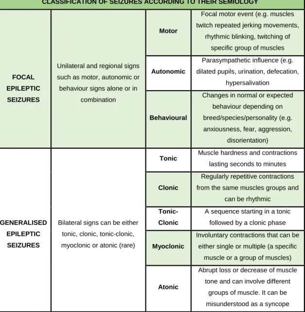
Seizures can be classified according to their presentation, which allows a better understanding of affected areas of the brain as well as their severity 63. Depending upon the affected areas of the
17
express different signs 63. Even though impairment of consciousness can be detected in some
situations, this evaluation is not as accurate as in humans because it relies on owners reports, which sometimes may not be precise 1. There is no reliable scientific evidence supporting a
relationship between clinical manifestations and aetiology of the source 8. Classification of
seizures according to its presentation may not influence the treatment but it may help not only identifying which region of the brain is possibly affected but also it allows the perception of whether there is improvement associated to treatment and progression of the disease 3.
1.4.3.1 FOCAL EPILEPTIC SEIZURES
Focal epileptic seizures (FES) are originated by abnormal electrical activity in a localised, specific group of neurons within one cerebral hemisphere and their clinical presentation may help to identify the affected areas of the brain 1. Focal seizure rises from an epileptic focus, but the
abnormal electrical activity is not propagated to the other cerebral hemisphere 63. Depending upon
the localisation of the epileptic focus, any region of the body may be affected 52. FES are more
likely to occur in the presence of StE as a consequence of other diseases, which emphasises the importance of searching for these diseases when in the presence of FES, especially if consciousness stays unchanged 4,35,37,63. This presentation usually manifests into a motor and
focal muscle activity that begins laterally 1. FES can also be present as autonomic or behavioural
and these presentations may coexist with motor presentation 1. FES may initiate after a sensory
experience like lights, smells, sounds or touch 102. These seizures may pass unnoticed by owners
or can be misinterpreted as behavioural abnormalities 110.
1.4.3.2 MOTOR FOCAL EPILEPTIC SEIZURES
This type of presentation is mainly characterised by involuntary, unilateral focal motor episodes that result in atypical movements of specific muscles, such as facial twitches, head movements (e.g. flexion/extension), contraction of masticatory muscles or facial jerking 1,9. Some of these
signs may be defined as hyperkinetic when there is an involuntary contraction of a group of muscles of one side of body, which may lead to an abnormal posture and dislocation of the midline
3.
On the other hand, hypokinetic signs are described as sudden atonia in a group of muscles, which may lead to falls 3. It is believed that this type of seizures might start from an epileptic focus that
18
1.4.3.3 AUTONOMIC FOCAL EPILEPTIC SEIZURES
In this type of presentation, it is possible to observe autonomic signs such as mydriasis, hypersalivation, urination, defecation or vomiting 1,110,111. Other less visible signs like salivary
gland enlargement, dysphagia or oesophageal spams may be associated with autonomic focal seizures 112,113. These autonomic signs may be associated to injured areas in the limbic system 3.
1.4.3.4 BEHAVIOUR FOCAL EPILEPTIC SEIZURES
In humans, this type of presentation is characterised by psychic and sensory episodes, where people experience sensations such as agitation, fear, anger or anxiety 114. According to the ILAE,
focal onset can be subclassified as non-impaired or impaired awareness, depending on whether consciousness is impaired or not 115. In dogs, the evaluation of behaviour changes and
consciousness is difficult to assess since it is based only on the owners reports or video-documentation 110.
For this reason, the International Veterinary Epilepsy Task Force (IVETF) decided that FES should not follow the same classification used in humans since it is difficult to do an accurate evaluation of these alterations 1. Nonetheless, some studies have reported behaviour changes in
dogs associated with focal seizures such as anxiety, seeking/attention or avoidance behaviour, confusion, fear, fatigue, bitting or licking imaginary objects and compulsive tail-chasing 110,111,116.
1.4.3.5 GENERALISED EPILEPTIC SEIZURES
Generalised epileptic seizures (GES) are described as the most common presentation in dogs and indicates that both cerebral hemispheres are affected 1,11,117. In contrast, other studies
suggest focal seizures as the most common 26,28. This opposition may be related to many factors
such as discrete clinical manifestation, non-agreement between veterinarians regarding seizure’s classification, lack of experience or population bias.
In GES, epileptic activity may propagate from an epileptic focus to other parts of the brain and other groups of neurons may be affected 1. This presentation usually reveals itself as a
tonic-clonic muscle activity that begins bilaterally, often symmetrical and usually consciousness is impaired 9,63. However, GES can also be present as only clonic, tonic, atonic and myoclonic but
these presentations are less frequent 1,63. There may be cases where consciousness stays
unmodified during GES 1. However, evaluation of consciousness in dogs is complicated because
19
electroencephalogram (EEG) recordings in veterinary medicine, the correct classification should rely on the differentiation between a focal and generalised seizure and more complex classifications are not useful and may be confusing 49. Normally, GES will follow a particular
pattern for a specific dog but may vary between different dogs 3.
Frequently, autonomic signs are noticed during GES and defecation, urination and hypersalivation the most commonly observed 1. According to the IVETF, GES is identified in the
presence of both clinical and EEG changes demonstrating possible affection of both sides of cerebral hemispheres 1. A generalised seizure is precepted by owners as a potentially
life-threating condition for their pets when compared with a focal seizure, which can remain unnoticed most of the time 32. Most of the owners believe that generalised seizures decrease the quality of
life (QoL) of their pets, which could lead to premature euthanasia if owners are not available to cooperate in the diagnosis and treatment 3. Furthermore, they can easily detect a generalised
seizure rather than a focal seizure 32.
1.4.3.6 GENERALISED TONIC-CLONIC EPILEPTIC SEIZURES
This type of presentation was formerly called grand mal seizures because of its generalised, aggressive muscle movements 111,117. There is a high variability on the duration and clinicalmanifestations of these episodes 9. Generalised tonic-clonic seizures may cease in less than five
minutes but the majority of them do not persist for more than one minute 3.
The tonic phase associated with this presentation is characterised by a generalised muscle contraction and increase of muscle hardness, which leads to opisthotonos that generally does not last more than one minute 1,9,63. Before this stage starts, there is a loss of consciousness 9.
According to Lorenz et al., consciousness may be measured through responsiveness, attentiveness and disorientation 63.
The clonic phase follows the tonic phase and is characterised by rhythmic, repetitive contractions of the muscles, that leads to jerking and chewing movements of limbs and jaw, respectively 9,11.
Some dogs may experience specific movements such as running or may vocalise 9. During the
seizure, tonic and clonic phase may alternate after one another 9. Autonomic signs are relatively
common during a generalised tonic-clonic seizure and hypersalivation, defecation or urination are normally observed 9.
In the post-ictal phase, dogs may experience abnormal behaviour such as confusion, aggressiveness, fear or anxiousness 9. Also, it has been reported that some dogs might have
proprioceptive deficits, temporary blindness and ataxia 9. If neuronal deficits persist for more than