Universidade de Lisboa
Faculdade de Farmácia
The effects of cage-divided housing on anxiety and
working memory in mice
Mónica Tenreiro Carona
Mestrado Integrado em Ciências Farmacêuticas
1 Universidade de Lisboa
Faculdade de Farmácia
The effects of cage-divided housing on anxiety and
working memory in mice
Mónica Tenreiro Carona
Trabalho de Campo de Mestrado Integrado em Ciências Farmacêuticas
apresentado à Faculdade de Farmácia da Universidade de LisboA
Orientador: Prof. Dr. Ilse Smolders, An Buckinx (phD student)
Co-Orientador: Dr. Bruno Sepodes,
Professor Auxiliar3
This work was developed at the Faculty of Medicine and Pharmacy, Vrije Universiteit Brussels (VUB), under the supervision of Prof. Ilse Smolders and An Buckinx, under the Erasmus program.
4 RESUMO
Os ratinhos são seres sociais e que no seu habitat natural vivem em grupo. Devido a requisitos de protocolo ou para proteção do animal em casos de agressão contra os companheiros de gaiola, os ratinhos de laboratório têm que ser alojados individualmente durante a realização das experiências. As conclusões acerca das consequências que podem advir do alojamento individual dos ratinhos são contraditórias, mas a sugestão geral é que o alojamento individual deste animal está associado a um aumento da ansiedade e prejudica a memória, podendo afetar assim a replicabilidade dos resultados. Na tentativa de diminuir os efeitos negativos associados ao alojamento individual, criámos um novo tipo de gaiola. Esta inclui um divisor que impede o contacto físico entre os ratinhos, mas que permite o contacto olfativo e visual. O objetivo deste estudo é, portanto, investigar o processamento da ansiedade e da memória nos ratinhos alojados neste novo tipo de gaiola comparativamente com os ratinhos alojados em pares e individualmente.
Ratinhos machos C57BL/6 de 8 semanas de idade forma divididos em três grupos que diferiam no modo de alojamento: alojamento individual, alojamento em pares e alojamento em pares na nova gaiola com divisor durante quatro ou dez semanas. Após esse período foram realizados testes comportamentais, “Fear Conditioning” e o teste de supressão de dexametasona. Primeiro, foi realizado o teste de Alternância Espontânea (Y-maze), onde se avaliou a memória de trabalho e as atividades locomotora e exploratória dos ratinhos. Os resultados não diferiam entre os grupos. O teste de campo aberto (Open Field) também não revelou quaisquer diferenças relativamente à atividade locomotora e comportamento ansioso. O teste de labirinto elevado (Elevated Plus Maze) foi também realizado. Neste, o tempo gasto nos braços abertos e a distância total percorrida também não diferiam entre os três grupos, confirmando os resultados obtidos no teste anterior. Relativamente ao “Fear Conditioning”, também podemos afirmar que não foram observadas diferenças significativas entre os grupos experimentais. Por último, o teste de supressão da dexametasona foi realizado para avaliar a responsividade do eixo hipotálamo-hipófise-adrenal (HPA). Alguns ratinhos foram tratados com dexametasona (análogo sintético da corticosterona), a qual consegue através de um mecanismo de feedback negativo suprimir a libertação de corticosterona nos ratinhos. Todos os grupos apresentaram níveis basais de corticosterona semelhantes entre eles. Dissecámos as glândulas supra-renais e a hipófise para análise posterior, pois o peso das glândulas supra-renais está associado à ansiedade e ao stress. O peso da glândula pituitária e adrenal normalizado em relação ao peso corporal não diferiu significativamente entre os grupos experimentais.
Assim, os nossos resultados sugerem que tanto as quatro como as dez semanas de alojamento individual não afetaram a ansiedade nem a memória dos ratinhos, um tópico muito debatido em vista do bem-estar animal. As observações comportamentais foram consistentes com os níveis séricos de corticosterona, pois o teste de supressão da dexametasona sugeriu que não existia desregulação do eixo HPA em nenhuma das condições experimentais. Ambas as guidelines Americana e Europeias continuam a defender a importância de manter os ratos e ratinhos alojados em grupo. No entanto, resultados obtidos não mostram qualquer influência do alojamento individual no bem-estar do animal. Mais estudos são necessários para confirmar estes resultados e para compreender melhor o modo como a ansiedade e a memória são afetadas pelas condições de alojamento dos ratinhos de laboratório.
5 ABSTRACT
Mice are social beings and they live in group in their natural habitat. Due to protocol requirements or for animal protection in case of aggression against cage mates, laboratory mice are individually housed during some experiments. The conclusions about the consequences that can result from the individual accommodation of mice are contradictory, but a general suggestion is that the individual accommodation of the mouse is associated with increased anxiety and impaired memory, what can influence the replicability of results. To lessen the effects associated with individual housing, we created a new type of cage. This cage includes a cage divider that separates mice and prevents physical contact between mice but allows visual and olfactory contact. The purpose of this study is, therefore, to investigate how housing in this new type of cage affects anxiety and working memory compared with mice that are paired and individually housed.
Male eight-week-old C57BL/6 mice were divided into three groups that differ in the mode of housing: individual housing, paired housed and paired in a new divider cage. They were housed for either four or ten weeks. After this period, several behavioural tests, fear conditioning and the dexamethasone suppression test were performed. First, the Y-maze Spontaneous Alternation test was performed, which assesses working memory, locomotive and exploratory activities of the mice. These parameters did not differ between groups. The open field test also revealed no significant differences in locomotor activity and anxiety-like behavior. The elevated plus maze test was also performed and revealed that the time spent in the open arms and the total distance travelled did not differ between the three groups, confirming the results obtained in the previous tests. Additionally, the Fear Conditioning test confirmed that housing conditions did not affect anxiety-like behavior in neither of the three groups. Finally, the dexamethasone suppression test was performed to assess the responsiveness of the hypothalamus-pituitary-adrenal (HPA) axis. Finally, mice were treated with the synthetic corticosterone analogue dexamethasone, which can, through a negative feedback mechanism, suppress corticosterone release in the mouse. There were no differences in corticosterone levels between the groups. We dissected the adrenal glands and pituitary gland for further analysis, as the weight of the adrenal glands is associated with anxiety and stress. The weight of the pituitary gland and adrenal gland normalized against body weight did not significantly differ between experimental groups.
Therefore, our results suggest that both four weeks and ten weeks of individual accommodation do not affect the anxiety or memory of mice, a much-debated topic in view of animal welfare. Results obtained in the behavioural tests corresponded with corticosterone levels, as the dexamethasone suppression test suggested that there is no HPA axis dysregulation under any of the experimental conditions. Both the American and European guidelines continue to defend the importance of keeping rats and mice group housed. However, the results obtained in this study indicate that individual housing up to ten weeks does not affect anxiety and working memory. Further studies are required to confirm these results and to gain a better and more in depth understanding on how anxiety-like behavior and working memory are affected by various housing conditions in laboratory mice.
6 ACKOWLEDGEMENTS
Firstly I would like to thank Professor Ilse Smolders and An Buckinx for all the support they gave me during the Erasmus period and even after I returned to Portugal. They consistently allowed this paper to be my own work, but steered me in the right direction whenever they thought I needed it. My special thanks go to AN Buckinx, who accompanied my work very closely and always gave me the orientation I needed and spends a lot of time helping me.
Secondly I want to express my deepest gratitude to Prof. Bruno Sepodes for all his support and advices and for being always available to help.
I want to express my thanks to my friends who have kept by my side and who never let me give up. Thanks for everything! Special thanks to Ana Sofia Tetino, Sofia Ferreira, Mariana Cabedal and Sara Dionísio for always accompanying me throughout these 5 years and for making this academic journey an unforgettable adventure. Thanks also to Vera and Filipe for being the people they are and for always having that special word that always helps to lift my head and move on.
Thanks also to Isabela, Jean-Pierre, Tozé, Paula, Pedro and Ourvashee for making me feel at home and for all the support they have given me since the beginning. Without you this work would not have been possible.
I must express my very profound gratitude to my parents and grandparents for providing me with unfailing support and continuous encouragement throughout my years of study and for their unconditional love, for their constant dedication and protection and for all the efforts made. To my sister there are no words to thank all she has done for me. Thanks to her for her infinite support and for never letting me lower my arms. Finally, I have to thank to my boyfriend Tiago Martinho for all the love and support he has given me. He never let me down. He was undoubtedly a fundamental pillar in this process. Without him none of this would be possible. He is one of the most important people and he made this come true.
Thank you all. This achievement is as much mine as yours.
I dedicate this work to my grandfather Aurélio Tenreiro. It is with great pride that I dedicate this thesis to one of the people who did the most to get me here. Thanks for everything. Without you this would never have been possible. Wherever you are this is for you!
7 CONTENTS LIST OF ABBREVIATIONS ... 8 LIST OF FIGURES ... 9 LIST OF TABLES ... 9 1. INTRODUCTION ... 10
1.1 - Single housing in mice ... 10
1.2 - What is stress? ... 10
1.3 - Stress Response ... 10
1.4 - Acute and Chronic Stress ... 12
1.5 - Consequences of chronic stress ... 12
1.6 - Fear and anxiety ... 13
1.7 - Behavior tests ... 14
1.8 - Objective: ... 17
2. MATERIALS AND METHODS ... 18
2.1 - Experimental set-up ... 18
2.2 – Animals ... 18
2.3 - Spontaneous Alternation Test (Y-Maze) ... 19
2.4 - Open Field Test ... 19
2.5 - Elevated Plus Maze ... 20
2.6 - Fear Conditioning ... 20
2.7 - Dexamethasone Suppression Test ... 20
2.8 - Pituitary Gland and Adrenal Gland Dissection ... 21
2.9 - Statistical analysis ... 21
3. RESULTS ... 22
3.1 - Body Weight ... 22
3.2 - Spontaneous Alternation Test (Y-Maze) ... 23
3.3 - Open Field ... 24
3.4 - Elevated Plus Maze ... 25
3.5 - Fear Conditioning ... 26
3.6 - Dexamethasone Suppression Test ... 27
3.7 - Pituitary and Adrenal Glands Weight ... 28
4. DISCUSSION ... 29
5. CONCLUSION ... 31
8 LIST OF ABBREVIATIONS
HPA axis - Hypothalamic-Pituitary-Adrenal axis SNS - Sympathetic Nervous System
PTSD - Post-Traumatic Stress Disorder ANS - Autonomic Nervous System
SAM system - Sympathetic Adrenomedullary system CRH - Corticotrophin Releasing Hormone
AVP - Arginine Vasopressin
ACTH - Adrenocorticotropic Hormone MR - Mineralocorticoid Receptor GR - Glucocorticoid Receptor
11β-HSD - 11-Beta-Hydroxysteroid Dehydrogenase mPFC - Medial Prefrontal Cortex
LTP - Long-term Potentiation LTD - Long-term Depression AHP - Afterhyperpolarization
CRF - Corticotrophin Releasing Factor
tPA - Tissue Plasminogen Activator
BLA - Basolateral Amygdala
EPM – Elevated Plus Maze FC - Fear Conditioning US - Unconditioned Stimulus CS - Conditioned Stimulus CR – Conditioned Response NMDA - N-Methyl-D-aspartate LA - Lateral Nucleus of the Amygdala BA - Basal Nucleus of the Amygdala CeA - Central Nucleus of the Amygdala PAG - Periaqueductal Grey Matter CORT - Corticosterone
9 LIST OF FIGURES
Figure 1 – HPA axis ... 10 Figure 2 – Fear conditioning neural circuit ... 15 Figure 3 – Time scheme of performed experiments ... 17 Figure 4 – Housing conditions according to which mice were housed during the
experiment……….17 Figure 5 – Spontaneous Alternation Test (Y-maze) scheme ... 18 Figure 6 – Body weight in single housed, paired housed and paired housed with grid mice . 21 Figure 7 – Spontaneous Alternation Test (Y-maze) in single housed, paired housed and paired housed with grid mice ... 22 Figure 8 – Open Field in single housed, paired housed and paired housed with grid mice .... 23 Figure 9 – Elevated Plus Maze in single housed, paired housed and paired housed with grid mice ... 24 Figure 10 – Fear Conditioning protocol performed in single housed, paired housed and paired housed with grid mice ... 25 Figure 11 – Results of dexamethasone suppression test ... 26 Figure 12 – Normalized weight of pituitary and adrenal glands for the three experimental groups ... 27
LIST OF TABLES
Table 1 – Results of Spontaneous Alternation Test ... 22 Figure 11 – Results of Open Field Test ... 24 Figure 12 –Results of Elevated Plus Maze ... 25
10 1. INTRODUCTION
1.1 - Single housing in mice
The mouse is the most widely used species in biomedical research. As mice are social beings, group housing is a better representation of their natural social life. Wild mice have a well-known social organization, where they are organized in a structure with one dominant male, several females, juveniles and a few subordinate males (1,2). For this reason, it is usually recommended to accommodate mice in group. However, for some experiments mice are isolated or grouped by gender, the opposite of their wild social organization (1,2). Some studies proved that a good social support system is essential for the well-being of an individual, so long-term social deprivation can represent a stressful situation and therefore have negative effects on physical and psychological health. Several studies show that there are physiological and behavioural differences between individually and group housed rodents. They defend that single housing is associated with changes in corticosterone levels, metabolism, growth, and behaviour (3). Although there are several conflicting studies, the overall conclusion is that individual housing of mice is associated with increased anxiety and memory impairments. An increase in emotionality and reactivity of the hypothalamic-pituitary-adrenal (HPA) axis has been observed in socially isolated mice. (4,5). Social isolation can also lead to metabolic disorders, such as an increase in adiposity and altered metabolic functions. This can be explained by the fact that the HPA axis and the sympathetic nervous system (SNS) are the main effector pathways of the stress system and both affect energy metabolism (4). Additionally, some studies conclude that the single house of mice can impair cognitive functioning, like memory and learning (1,6,7). Therefore, it is not surprising that replicability of experimental results in rodents may be influenced by stress related to inadequate housing conditions (2). In conclusion, several studies defend that social context influences both mouse welfare and experimental results, since laboratory mice are social animals and are, for this reason, motivated to interact with each other (2). The individual housing is described as a stressful situation (2).
1.2 - What is stress?
The organism attempts to preserve a predetermined steady state, essential for well-being and survival, called homeostasis. However, this state of equilibrium is constantly challenged by intrinsic or extrinsic adverse factors (stressors). Stressors can be emotional or physical adverse forces and their magnitude and chronicity can influence the stress response (1,2). Stress is an extremely personalized phenomenon, as it depends on the individual vulnerability and resilience. It’s a “non-specific response of the body to any demand for change” according to Hans Selye (10). The magnitude of the consequences of a stressing episode depends on the ability to deal with the stressor. Stress consequences include anxiety, fear, depression and post-traumatic stress disorder (PTSD), but it has also adverse effects on other major mental disorders like as schizophrenia (3,4).
1.3 - Stress Response
When a stressful event occurs, the body unconsciously activates physiological responses. To promote adaptation and survival to real or potential threats in the environment, the action of autonomic, neuroendocrine and behavioural processes is necessary. (5,6). The biological response to stress involves activation of two interrelated systems: the fast acting SNS and the slow acting HPA axis (7,8).First, the somatosensory cortex perceives the stressor. Here, the situation is evaluated and classified as being stressful or not. This decision is based on the
11
sensory input accompanying the stressor, on processing of this input and on stored memories (3).If the stressor is recognized as a threat, the Autonomic Nervous System (ANS) will be activated and therefore will activate the Sympathetic Adrenomedullary (SAM) System, which will lead to the release of catecholamines in the blood, predominantly epinephrine but also some norepinephrine. These will bind to various adrenoreceptors in multiple target organs and an increase in circulating catecholamines facilitates rapid mobilization of metabolic resources and prepares the body for a fight or flight response (15). Epinephrine leads to the stimulation of the SNS and reduces activity in the parasympathetic nervous system. Briefly, epinephrine will lead to an increase in cardiac output and blood pressure, vasodilatation in muscles and constriction of blood vessels in the skin and gut, to ensure blood supply to the brain and muscles. An increase in epinephrine is also responsible for the mobilization of energy sources by enhanced lipolysis and hepatic gluconeogenesis (3,8,9).(10) The brain simultaneously activates the HPA axis, which
results in the release of adrenal glucocorticoids: cortisol in man and corticosterone in rodents. The cascade of events that leads to the production of
glucocorticoids by the adrenal cortex begins with the release of corticotrophin releasing hormone (CRH) and arginine vasopressin (AVP) by cells in the paraventricular nucleus of the hypothalamus. These stimulate the release of adrenocorticotropic hormone (ACTH) in the pituitary gland, which will stimulate the production and release of glucocorticoids in the adrenal gland. The effect of glucocorticoids depends upon the receptors to which they bind: mineralocorticoid receptor (MR) or glucocorticoid receptor (GR). Outside the brain, glucocorticoids (GC) operate through the GRs since the 11-beta-hydroxysteroid dehydrogenase (11β-HSD) enzyme prevents GCs from binding to the MRs. In the brain, the 11β-HSD is minimally expressed so the GC binds to both MR and GR (8). Indeed, they have more affinity to MRs than to GRs, but both receptors are expressed at particularly high levels in limbic areas that are responsible for the modulation of the stress response. MRs are more involved in the process of
assessment and initiation of stress response, while the GRs are more involved in the mobilization of energetic substrates and in most stress-induced behavior changes, which includes anxiety-like behavior and facilitated learning and memory (in particular, consolidation of memories) (3,8,9).Many times, the GR-mediated effects oppose the MR-mediated effects. For example, meanwhile GRs impair the neural plasticity and the processes that are involved in learning and memory, basal levels of glucocorticoids acting via MR enhance the synaptic plasticity(8).
Thus, short-term responses are produced via SAM (The Fight or Flight Response), while the long-term stress response is controlled by the HPA system (8).
The actions of SAM and the HPA axis are regulated by different brain areas such as the limbic system, prefrontal cortex, amygdala, hypothalamus, and stria terminalis. This way, the contribution of these systems to the stress response will depend on the modality and intensity of the stressor. (11)
12 1.4 - Acute and Chronic Stress
In a normal response to stress, the activation of the HPA axis and the SAM System will return to the baseline, due to the negative feedback action of the corticosteroids at the level of the pituitary gland and hypothalamus. However, in situations of chronic stress, prolonged activation of the stress system may lead to a loss of this state of equilibrium (12,13). Two types of stressors and stress responses can be differentiated: acute (short-term) or chronic (long-term) stress. There are different ways to define chronic stress, because it can be defined considering the continued activation of the stress response, prolonged exposure to a stressor or even an allostatic overload. In general, stress is considered chronic in situations in which the stressor is overwhelming and cannot be resolved (14).
1.5 - Consequences of chronic stress
The consequences of the stress response will differ according to the duration of the stress. Long-term physical or mental consequences of stress would depend on long-term effects of allostatic load (15,16). In chronic stress, there is a dysregulation of the HPA axis, since the negative feedback mechanism is not working properly. The allostatic load present in long-term stress will culminate in a cognitive dysfunction, undermining memory and learning processes (16–18). This happens because stress influences synaptic plasticity, thereby reduces neuronal excitability and may cause atrophy of neurons and neurotoxicity, especially in the hippocampus, but also in amygdala and medial prefrontal cortex (mPFC). So, stress is a potent modulator of learning and memory processes, because in the rodent hippocampus, corticosterone regulates metabolic, physiologic and genomic functions of neurons (14,16–19). But glucocorticoids do not work alone. Stress mediators include adrenal steroids and catecholamines, the classic neuroendocrine hormones of the stress system, excitatory amino acids, cytokines and growth factors. These stress mediators are also involved in the adaptation to stressors, as well as in the negative impact of chronic stress and will act on different targets, such as cognitive, fear/anger, immune, cardiovascular and gastrointestinal systems. They also have effects on reproduction, growth and thyroid, as well on brain awakening-sleep centres (1,16,17).
Chronic stress is associated with macroscopic changes in some brain areas, such as hippocampus, amygdala and mPFC, with changes in volume and physical changes in the dendritic atrophy and decreased spine density (14). The hippocampus is one of the most sensitive and malleable regions of the brain and belongs to the limbic system. It is very important for cognitive function, because the hippocampus plays important roles in consolidation of information from short and long-term memory and in spatial memory. It contains a significant number of glucocorticoids, oestrogen and progesterone receptors, what makes the hippocampus particularly vulnerable to stress (13,16,20).The hippocampus is also critically involved in glucocorticoid-mediated negative feedback which is important to regulating the responsiveness of the HPA axis in stress response (21).
It is enriched with both types of adrenal steroid receptors: Type I receptors have a high affinity to aldosterone and corticosterone (MRs) and Type II receptors have a lower affinity (GRs). The MRs become fully occupied at low plasma corticosterone levels, while GRs only became saturated at very high levels (for instance during stress) (18,22). As we know, corticosterone is the main glucocorticoid synthesized by the adrenal cortex of rodents during the stress response, so in a stress situation corticosterone levels will be increased which might affect hippocampal neuroplasticity.(18) Some studies show that the two types of adrenal steroid
13
receptors produce potent and opposite effects on neuronal plasticity in the hippocampal formation. While the selective activation of MRs increases Long-term Potentiation (LTP), the activation of GRs attenuates LTP and enhances Long-term Depression (LTD), producing a potent suppression in neuroplasticity. This way, chronic stress and stress mediators impairs LTP in the hippocampus (CA1 field) (16,18,21). LTP is a persistent strengthening of the synapse, whereas LTD is a decrease in synaptic strength (23).
Stress is also responsible for retraction and simplification of dendrites in the CA3 region of the hippocampus and for changes in intrinsic properties of hippocampal neurons, because corticosterone will prolong the afterhyperpolarization (AHP) of CA1 neurons, by increasing the level of internal Ca2+. In this way, excitability will be lower and synaptic plasticity will be affected (16,18).
Chronic stress causes changes in other brain areas like the mPFC and amygdala (16). The mPFC, a subregion of the prefrontal cortex (PFC), is involved in different tasks which are influenced by chronic elevations of circulating glucocorticoids during a stress response. These stress-induced changes are characterized by a reorganization of neurons, with a decrease in apical dendritic branches and total dendritic length, which can contribute to changes in cognition (16,24,25). The PFC is involved in working memory functions, so these architectural changes in PFC and the release of catecholamines during the stress response, which reduces the neural activation of PFC, will impair working memory. Working memory plays an important role in reasoning and the guidance of decision-making and behavior. This refers to the capacity to maintain and manipulate information during short periods of time for goal-directed actions (26–28).
On the other hand, amygdala is considered one of the principal structures of limbic system and is important for processing memory, decision-making and emotional responses, including fear and anxiety. Amygdala is critically involved in mediating stress-related behaviors and modulating hippocampal function (18). While the hippocampus inhibits stress-induced HPA activation, the amygdala enhances glucocorticoid secretion in response to stress. So, we will have differential effects of stress on hippocampus and amygdala. In contrast to hippocampal effects, stress facilitates LTP and increases growth of dendritic length and spine density in amygdala neurons (in the basolateral nucleus), so there is stress-induced enhancement in amygdala memory tasks (e.g. aversive conditioning). Some studies indicate that amygdala neuronal activity is important for the emergence of stress effects in the hippocampus (16,18,19). In the amygdala, Corticotrophin Releasing Factor (CRF) is involved in control of behavior and autonomic responses to stress, like regulation of ACTH release and release of tissue plasminogen activator (tPA). The tPA is important to stress-induced anxiety and structural plasticity in the medial amygdala (16). It has been reported that chronic stress enhances anxiety and the risk for illness (development of psychological disorders) and causes changes in cognition (19,24)
1.6 - Fear and anxiety
Stress leads to a feeling of fear. Stress, fear and anxiety prepare the body to response to a threat. Fear and anxiety are closely inter-related but although both are alerting signals, they prepare the body for different actions. While anxiety is considered an anticipatory emotional response to an imprecise or unknown threat, fear is defined as an emotional response to a known or defined threat and is more often related with flight and fight response and escape actions
14
(4,29). Fear and anxiety are necessary to ensure survival, however when these are persistent they can have consequences in proper psychological functioning and lead to the onset of anxiety disorders (30).
The Basolateral Amygdala (BLA) contains the neural circuit responsible for the consolidation and extinction of fear memories, as will be explained after in detail. Stress exerts significant influence on the regulation of emotional memories, because in the stress response we have an increase in the release of glucocorticoids and these will have an action in the BLA, promoting the consolidation of fear memories. This happens because the elevation of glucocorticoids levels leads to a suppression of inhibitory synaptic inputs to BLA neurons, which leads to a LTP of excitatory inputs to the BLA neurons, i.e. an increase in excitability. This event results in an increase of anxiety-like behavior (31).
1.7 - Behavior tests
There are some well-established tests to assess anxiety-like behaviors in mice, like open field and elevated plus maze (EPM). These tests are based on innate tendencies of mice to explore new environments and to avoid bright light and open spaces, because these conditions simulate a predator risk situation (39,40).
The Open Field Test is one of the most widely used tests to measure anxiety in animal models. Many behavioral tests for assessing anxiety are based on the activity and locomotion of the subject animal. In this test an open field arena is used, usually a square arena, where the animal is placed and its behavior is assessed over a given period of time. In the open field, mice have an innate tendency to remain on the periphery or near the walls. This behavior is called thigmotaxis, which refers to the motion or orientation of an organism in response to a touch or physical stimulus. This behavior decreases gradually during the first minutes of exploration. Less time spent in the center area of the chamber indicates higher levels of anxiety-like behavior (39–42).
The EPM also considers the fact that mice have an aversion to open and illuminated spaces. This well-characterized behavioural paradigm is also one of the most often used tests to assess anxiety. The anxious animals will spend more time in closed arms than less anxious animals and make very few entries in the open arms (39,43).
It has been previously described that chronic stress adversely affects spatial memory and produces depressive-like symptoms, such as anxiety, which leads to a decrease in activity. The Y-maze is a behavioural test used to study different aspects of cognitive function, like the assessment of spatial learning and memory. The objective of the test is for rodents to remember which arm was last visited and alternate between the different arms, because rodents have a predisposition to explore new environments. An efficient alternation depends on a good working memory (44). Many parts of the brain are involved in this task, including the hippocampus, which is responsible for spatial memory. For this reason, the Y-Maze can be used to study hippocampal lesions. (45,46). The hippocampus is one of the most affected regions in chronic stress and is considered a critical structure for spatial learning (47).
In general, group housing is recommended to social animals, such as mice and rats, and the duration of single housing, due to the experiment, should be as short as possible. Several studies showed that the single housing conditions are considered as a stressful situation for the mice (1,7). As has been said, stress is associated with increased anxiety-like behavior and an
15
increased fear response. Thus, deficits in the ability to regulate fear can lead to anxiety disorders and depression (48,49). It has been shown previously that different areas of the brain, such as the amygdala, hippocampus and mPFC, play an important role in stress responses and may undergo changes with chronic stress (50). Moreover, these brain areas are critically important to the formation and acquisition of conditioned fear memories (48,51).
To test how housing conditions affect conditioned fear behaviour, Fear Conditioning (FC) can be used. FC is used as a model for emotional learning and memory process in animals and humans. It is a form of associative learning in which an Unconditioned Stimulus (US), usually an aversive stimulus, is associated with a Neutral Conditioned Stimulus (CS) (Conditioning of Pavlovian Fear). After the animal learns this association between the two stimuli, the animal will react to the neutral CS in the same way that it would react to the US (conditioned response - CR), that is, an autonomic and behavioural response conditioned by fear. For this reason, Pavlovian FC is used as a behavioural paradigm for the study of the cellular and molecular bases of long-term memory formation (50-54), since previously conducted studies indicate that the association between stimuli and aversive events is mediated by LTP (55).
The main neural circuits responsible for this form of learning are the sensory areas that process both stimuli, the amygdala regions that undergo plasticity during learning (LTP) and the regions that control the expression of specific CRs, such as freezing behavior. It is believed that FC occurs through the convergence of pathways that transmit the information from the CS and US. The convergence of these pathways occurs in the lateral nucleus of amygdala (LA), where synaptic plasticity happens (53–55). The amygdala is the structure of the temporal lobe that contains many nuclei that have extensive internuclear connections, as well as connections with cortical and subcortical regions. Among these nuclei are the basolateral (BLA) complex that is subdivided into the lateral (LA), the basal (BA) and the basal nuclei accessory, the central nucleus of the amygdala (CeA) and several other nuclei.
The amygdala is considered the primary area of the brain responsible for FC. The LA is the primary input nucleus of the amygdala. This one receives the sensory information from both cortical and subcortical (thalamic) regions (52,54). The main output nucleus of the amygdala is the CeA, which projects to several different structures, such as the paraventricular nucleus of the hypothalamus, where it will activate the HPA axis and trigger the release of cortisol. It also projects to the lateral hypothalamus, stimulating the ANS, and to the periaqueductal grey matter (PAG) which is a midbrain region implicated in several homoeostatic processes including fear, pain and analgesia. So, the PAG is responsible in mice for freezing behavior. In this way, by coordinating these downstream targets, the amygdala can produce many changes associated with the fear response, including increased heart rate, sweating, dilation of the pupils, reduction of pain sensitivity, freezing among others (54,56,57).
16
During the acquisition of fear, the US (foot shock) triggers sensory nerve ends in the paws, after which a signal is transmitted through the spinal cord to the thalamus and the cortex before this project to the LA. If the synaptic input, resulting from the foot shock, is strong enough to excite LA neurons, this will result in the activation of neurons in the CeA and it will produce a fear response. During the experiment, the neutral CS (tone) also reaches the thalamus and the auditory cortex and these projects to the LA too. However, the inputs of the neutral CS are not strong enough to depolarize the LA neurons. Therefore, there is no stimulation of the CeA and the CS does not produce a fear response. But when the CS is presented with the US (CS-US pairing) there is an increase in the strength of the synapse between the incoming neurons and lateral amygdala neurons, indicating the occurrence of synaptic plasticity. This will make the neutral CS have the ability to stimulate LA on his own. Then, the LA will excite the CeA and produce a CR (fear response), without the presence of US (52,54,55).
Amygdala interacts with other brain areas such as the hippocampus. This area is responsible for transmitting contextual information in which the CF occurs. Thus, mice also express fear when returning to the chamber where the experiment was performed. This means that mice not only learned fear CS but also the surrounding context (Contextual FC). Hippocampal neurons project to the BLA and in this way allow contextual cues to produce fear (58)
To evaluate the fear response and whether the animals learned the association between CS and US during the FC freezing behaviour is measured.In mice, freezing is a common and easily measured response used as an index of FC. Freezing is defined as a complete absence of any movement except for movements associated with respiration and tense body posture (56,57,59). Freezing is a universal fear response and is observed in reaction to a conditioned or unconditioned stimulus.
Figure 2 - Fear Conditioning Neural Circuit (53)
17
1.8 - Objective:
Laboratory mice are often single housed during the experiments due to protocol requirements, for post-surgical safety or in case of aggression towards cage mates. There are many conflicting studies on the exact effects of individual housing on mice, although the main idea is that individual housing of mice is associated with increased anxiety and memory impairment and with a tendency to show hyperactivity in behavioral tests (60). These behavioral consequences may affect scientific endpoints of experiments (6,61). To reduce negative effects associated with single housing, we investigate the potential use of a new type of cage that could improve animal welfare. This cage includes a cage divider that separates mice, avoiding physical contact while maintaining sensory contact with their cage mate. We believe this new set-up could be a solution for the effects of housing conditions on laboratory mice. With this new cage, mice can be separated from each other and this way accomplish with the protocol requirements and maybe not suffer from behavioral consequences associated with single housing. The aim of the study is to investigate anxiety and memory processing using this new cage set-up compared to mice that are paired and single housed.
18 2. MATERIALS AND METHODS
2.1 - Experimental set-up
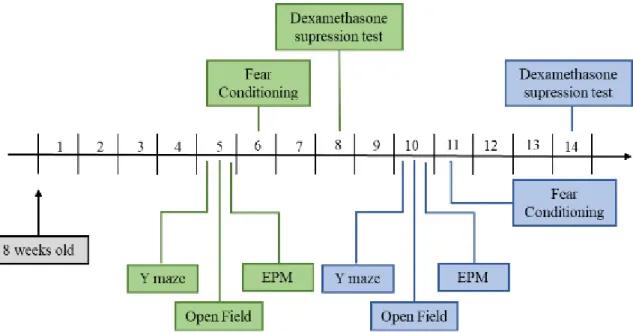
Figure 3 - Time scheme of performed experiments – Mice were housed under their
respective conditions for four (green scheme) or ten weeks (blue scheme) before behavioral testing began. After that period the Y-Maze Spontaneous Alternation Test, Open Field and EPM tests were performed. In the next week, mice were subjected to FC and two weeks later mice were sacrificed after performing the dexamethasone suppression test.
2.2 – Animals
A total of 60 male eight-week-old C57BL6/J (Janvier Laboratories, France) mice were used in this study, and were housed for either four or ten weeks before behavioral assessment of anxiety-like behavior and working memory. Mice were allocated to three different groups (n=10/group):
• Group 1: Single housed in Tecniplast 1264C Eurostandard Type II cages, with 215 x 268 mm floor area.
• Group2: Housing of two littermates in Tecniplast 1290D Eurostandard Type III cages with cage divider (mice are housed in sensory contact with a littermate, without direct physical contact), 212 x 266 mm floor area per mouse, and separate wire tops.
• Group 3: Paired housed in Tecniplast 1290D Eurostandard Type III cages, with a total floor area of 425 x 266 mm.
Figure 4 - Housing conditions according to which mice were housed during the experiment.
19
Animals were maintained in a 12/12 h light/dark cycle under stable temperature (19-25°C) and humidity (30-70% relative humidity) conditions and received food pellets and water ad libitum.
All procedures were in accordance with the National Rules on Animal Experimentation and were approved by the Ethical Committee for Animal Experiments of the Faculty of Medicine and Pharmacy of the Vrije Universiteit Brussel, Brussels, Belgium.
2.3 - Spontaneous Alternation Test (Y-Maze)
The Y-Maze consists of three identical and opaque arms, each with 35-cm long, 5-cm wide and 8-cm high walls, at an angle of 120 ° to each other. Each arm is identified by a different letter (A, B, C) (Figure 4). Each mouse was placed at the end of one of the arms (randomly selected and changing from mouse to mouse) in the Y-maze and was left to freely explore the three arms for a period of eight minutes. Mice were recorded with a video camera. After the trial, mice were placed back in their home cage. To eliminate odors, the labyrinth was cleaned with 70% ethanol in between trials. An entry in an arm was considered a true entry when all four paws were inside the arm runway, it means when all paws passed through the imaginary line originating from the floor of the Y-maze (44,62,63). Two parameters were calculated from the data: the total number of entries and the % spontaneous alternation performance (% SAP). %SAP (1) is defined as the ratio of the alternations performed to possible alternations ( = total arm entries–2) ×100 (62).
% SAP= Alternations
Total of possible alternations∗ 100 (1)
Figure 5 - Spontaneous Alternation Test (Y-Maze ) scheme (47) 2.4 - Open Field Test
The Open Field Test was the second behavioral test performed. This test was conducted in an open opaque cube with dimensions of 60 cm x 60 cm x 60 cm. The center of the arena was defined as the center 50 cm × 50 cm zone. At the beginning of the test, each mouse was placed in the same corner of the arena facing the wall. They were left to explore the arena freely for ten minutes after which they were placed back in their home cage. A camera mounted above the apparatus was used to record the movement of the animal. The experiment was recorded and later the total distance travelled (measure of locomotor activity) and the total time spent in the center zone (measure of anxiety-like behavior) were analysed with EthoVision® XT 11 software. Between each test the arena was cleaned with 70 % ethanol (38,39,64).
20 2.5 - Elevated Plus Maze
The third test was the elevated plus maze (EPM). The EPM consists of a platform, elevated 50 cm above the ground, with four perpendicular arms: two closed arms and two open arms, opposite each other. Each arm is 32,5-cm long and 6-cm wide from a central open square (6 x 6 cm). The two closed arms have 17 cm high walls. Mice were placed in the same enclosed arm, facing the wall and could explore the maze for five minutes. Between each test the arena was cleaned with 70 % ethanol. The duration and entries in each of the four arms were recorded with a video-camera and then analysed with EthoVision® XT 11 software (64). The total time spent in the open arms and in the center point (measure of anxiety-like behavior) and the total distance travelled (measure of locomotor activity) were calculated.
2.6 - Fear Conditioning
In the second week of testing, FC experiments were carried out in a FC apparatus comprising a test box (17 cm width × 17 cm length × 24 cm height) placed within a soundproof chamber (Ugo Basile, ANY-maze controlled Fear Conditioning System).
This procedure was performed in three consecutive days. There were two different contextual configurations: context A - white walls and white rubber floor washed with H.A.C., light intensity: 15 lux; and context B: patterned walls and a metal grid floor washed with 1 % acetic acid, light intensity: 125 lux. On day 1, mice were subjected to a habituation session in context A. First, they had 2 minutes of habituation where they could explore and took in the aspects of the chamber. After this period, mice were exposed to 5 presentations of one of two different tones (2.5 or 7.5 kHz, 80 dB, 30 s). The interval between tone presentations during the habituation session was randomized between 20–120 s. On day 2 discriminative FC was performed in context B and mice were exposed to five pairings of one tone (CS+: 2,5 or 7.5 kHz, 80 dB, 30 s) with an unconditioned stimulus (US: foot shock 0,6 mA). The foot shock was administered during the last 2 seconds of the tone presentation and co-terminates with the tone. Every time before each CS+ - US pairing, the other tone (CS-) was presented, but was never paired with the US. Like in day 1, during the conditioning session the interval between the CS+ and CS- presentations was randomized between 20–120 s. On day 3, mice were placed in context B for a fear retrieval, during which CS- and CS+ were presented sequentially in a block of 4 tones with a randomized interval between 20-120s (62). Freezing behavior, used as a measure of fear expression, was analysed using an automated video monitoring system (EthoVision® XT). This was defined as the absence of activity above a threshold (under 0,3% difference in movement between frames) for a duration of at least one second. Freezing levels for CS- were used as a measure for fear generalization.
2.7 - Dexamethasone Suppression Test
The Dexamethasone Suppression Test is used to evaluate the responsiveness of the HPA axis. Dexamethasone is a strong synthetic glucocorticoid analogue of the steroid hormone corticosterone, which can, through a negative feedback mechanism, suppress the release of corticosterone in the mouse. This occurs because dexamethasone will bind to GR in the hypothalamus and therefore it will inhibit the production of endogenous corticosterone. Mice received an intraperitoneal injection of dexamethasone or vehicle (0,05mg/kg body weight, diluted in 1% DMSO) between 11:00-12:00 AM. Six hours later, mice received an overdose of barbiturate (250mg/kg body weight of pentobarbital (Dolethal®) diluted in 0,9% NaCl) (65). After ensuring absence of the paw withdrawal reflex, trunk blood was collected to a tube
21
with an anticoagulant. Then, blood was centrifuged at 2500 x g for 15 min and plasma was kept and stored at -20 ºC.
After this procedure, a commercially available ELISA kit (ab108821, Corticosterone
ELISA kit, Abcam) was performed to determine the corticosterone levels in plasma. We
followed the protocol corresponding to the manufacturer’s instruction. The absorbance was read at a wavelength of 450 nm. Based on standard curves the corticosterone concentration was determined.
2.8 - Pituitary Gland and Adrenal Gland Dissection
Mice received an overdose of barbiturate (250mg/kg body weight of pentobarbital (Dolethal®) diluted in 0,9% NaCl) (65). After ensuring absence of the paw withdrawal reflex and collection of trunk blood, the pituitary gland and adrenal gland were dissected.
2.9 - Statistical analysis
Statistical analysis was performed using Graphpad Prism v6.1. For datasets consisting on multiple groups and multiple factors two-way ANOVA followed by Tukey post-hoc comparisons were used, for datasets consisting of multiple groups one-way ANOVA followed by Tukey post-hoc comparisons were used. Kruskal -Wallis test with Dunn’s multiple
22 3. RESULTS
3.1 - Body Weight
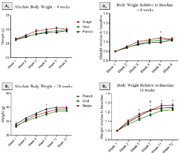
At the beginning of the experiment mice from the three different groups did not differ in body weight. During the 4 or 10 weeks of housing, mice were weighed several times (figure 5-A and B). Absolute body weight as well as body weight gained along the experimental procedure (body weight relative to baseline weight) were analysed.
Figure 6 – Body weight in single housed, paired housed and paired housed with grid mice A1) Absolute body weight of mice during the first short-term experiment. A2) Relative body weight of mice during the first short-term experiment. * = single housed versus paired housed mice. B1) Absolute body weight of mice during the second long-term experiment. * = paired with a grid housed versus paired housed mice. B2)– Relative body weight of mice during the second long-term experiment. * = single housed versus paired with a grid housed mice. # = paired housed versus paired with a grid housed mice. Repeated measures Two–way ANOVA with Tukey’s multiple comparisons test, p < 0,05; n=10 per group. Data are mean + SEM. * p < 0.05.
The results of both experimental procedures show some differences on the evolution of the weight during the experiment. On the second long-term experiment the differences are more significant. The paired housed mice with a grid show the lowest weight gain during the experiment.
23 3.2 - Spontaneous Alternation Test (Y-Maze)
Two different parameters were assessed to describe spatial working memory in the three different groups: number of arm entries that were made (fig 6- A1 and B1) and the %SAP (fig 6- A2 and B2). The total number of arm entries is a measure of locomotor activity, while the spontaneous alternation (%) is a measure of spatial working memory.
Figure 7 – Spontaneous Alternation Test (Y-maze) in single housed, paired housed and paired housed with grid A1) Number of arm entries during the first short-term experiment. A2)
%SAP during the first short-term experiment. B1) Number of arm entries during the second long-term
experiment. B2)– %SAP during the second long-term experiment. Repeated measures Two–way
ANOVA with Tukey’s multiple comparisons test, p > 0,05; n=10 per group. Data are mean + SEM. Table 1 – Results of Spontaneous Alternation Test
Single housed for 4 weeks Paired housed for 4 weeks Paired with a grid housed for 4 weeks Single housed for 10 weeks Paired housed for 10 weeks Paired with a grid housed for 10 weeks SPA (%) 62,57 + 1,939 63,70 +2,142 62,96 + 1,994 65,03 + 3,067 64,67 + 1,937 60,66 + 1,580 Number of arm entries 45,60 + 1,593 44,10 +1,773 42,70 + 2,176 37,70 + 1,212 45,00 + 2,049 37,40 + 2,363
After four or ten weeks of cage-divided housing conditions, no significant differences between the three groups were observed, implying that these housing conditions do not affect locomotor activity nor working memory.
24
As mentioned earlier, stress leads to a feeling of anxiety and cage-divided housing conditions are also associated with anxiety-like behavior, since these living conditions are considered a stressful situation for rodents (1,6,7) Therefore, the open field and the elevated plus maze tests were used to measure anxiety-like behavior.
3.3 - Open Field
In the open field, two different parameters were used to measure and compare anxiety-like behavior between the three different groups: the total distance travelled is a measure for locomotor activity (figures 7-A and 6-A) and total time spent in the central zone is a measure for anxiety-like behavior (figures 7-B and 6-B).
Figure 8 – Open Field in single housed, paired housed and paired housed with grid mice. A1) Total distance travelled during the first short-term experiment. A2) Time spent in center zone
during the first short-term experiment. B1) Total distance travelled during the second long-term
experiment. B2)– Time spent in the center zone during the second long-term experiment. Repeated
measures Two–way ANOVA with Tukey’s multiple comparisons test, p > 0,05; n=10 per group. Data are mean + SEM.
Analyses of the results revealed no difference between the three experimental groups regarding locomotor activity nor anxiety-like behavior (p>0,05). Mice were in the maze for ten minutes, but only the first five minutes were analysed. This occurs because, in the first five minutes, mice want to explore the area but after some time spent in the maze they start to know the area and stop the exploratory action. For this reason, the main effects on anxiety-like behavior are more evident in the begging of the test.
25 Table 2 – Results of Open Field Test
Single housed for 4 weeks
Paired housed with a grid for
4 weeks Paired housed for 4 weeks Single housed for 10 weeks Paired housed with a grid for
10 weeks
Paired housed for 10 weeks
Total distance
travelled (cm) 2810 + 207,2 2926 + 151,9 2328 + 179,5 2545 + 151.3 3391 + 377.9 2863 + 249,9
Time spent in the
center zone (%) 4,639 + 0,629 4,877 + 0,666 4,982 + 0,919 5,526 + 0,858 5,60 + 0,515 7,558 + 0,848
3.4 - Elevated Plus Maze
Anxiety-like behavior was measured in the elevated plus maze. Time spent in the open arms reflected anxiety-like behavior, and locomotor activity was measured by assessing total distance travelled (Fig.8).
Figure 9 – Elevated Plus Maze in single housed, paired housed and paired housed with grid mice A1) Time spent in open arms during the first short-term experiment. A2) The total
distance travelled during the first short-term experiment. B1) Time spent in open arms during the second
26 spent in open arms: Kruskal-Wallis test with Dunn’s multiple comparisons test, p > 0,05; The total distance travelled: Ordinary One-way ANOVA with Tukey´s multiple comparisons test, p > 0,05; N = 10 per group. Data are means + SEM.
Table 3 – Results of Elevated Plus Maze Single housed
for 4 weeks
Paired housed with a grid for
4 weeks Paired housed for 4 weeks Single housed for 10 weeks Paired housed with a grid for
10 weeks
Paired housed for 10 weeks
Time spent in
open arms (%) 8,206 + 1,571 8,502 + 1,334 6,028 + 1,472 5,634 + 1,165 7,068 + 1,451 8,761 + 2,064
The total distance travelled and the time spent in the open arms did not significantly differ between experimental groups, hereby confirming the obtained results in the previous test.
3.5 - Fear Conditioning
To measure the fear expression we measured freezing behaviour during the FC test. This test was performed over the course of three days and the freezing levels for CS- were used as a measure for fear generalization.
Figure 10 – FC protocol performed in single housed, paired housed and paired housed with grid mice A1) Habituation Procedure (day one) during the first short-term experiment.
A2) FC (day two) during the first short-term experiment. A3) Fear Retrieval (day three) during the second
long-term experiment. A1) A1) Habituation Procedure (day one) during the second long-term
27 during the second long-term experiment. Repeated measures Two-way ANOVA with Tukey’s multiple comparisons test, p > 0,05; n = 10 per group. Data are means + SEM.
The results showed no significant differences in freezing levels during habituation, FC and the fear retrieval test between the experimental groups.
3.6 - Dexamethasone Suppression Test
To measure plasma corticosterone levels in mice after the dexamethasone challenge, an ELISA was performed. Mice were divided in two groups: some received an intraperitoneal injection of dexamethasone and others of vehicle. This allows to measure the responsiveness of the HPA axis.
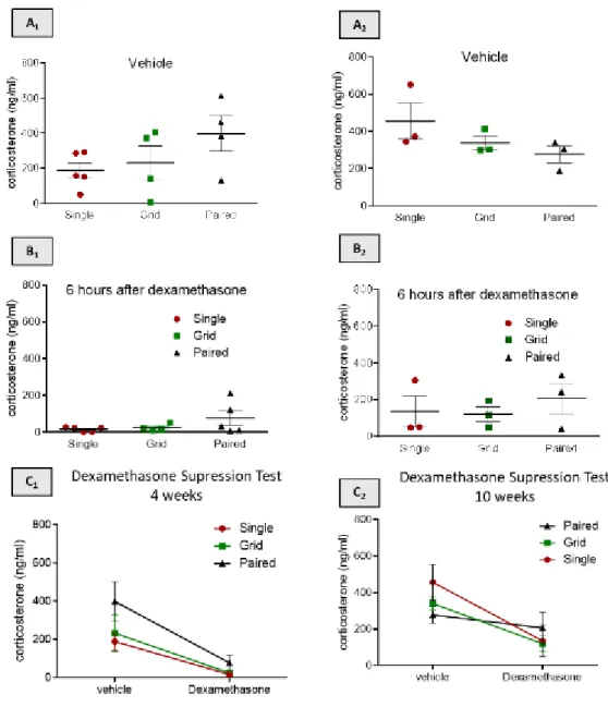
Figure 11 - Dexamethasone suppression test. A1)Plasma corticosterone levels in vehicle
injected or dexamethasone injected mice during the first short-term experiment. A2) Basal corticosterone
levels during the first short-term experiment. A3) Plasma corticosterone concentrations after
dexamethasone administration during the first short-term experiment; B1)Plasma corticosterone levels
in vehicle injected or dexamethasone injected mice during the second long-term experiment. B2) Basal
28 after dexamethasone administration during the second long-term experiment. A1 and B1: Two-way
ANOVA with Sidak’s multiple comparisons test; row factor p<0,0001; column factor p=0,0634. A2 and
B2: Kruskal-wallis test; p > 0.05; n = 4/5 per group. A3 and B3: Kruskal-wallis test; p > 0.05; n = 4/5 per
group. Data are mean ± SEM.
Analyses of the results revealed no difference in basal corticosterone levels neither in plasma corticosterone levels after dexamethasone administration between groups (p>0,05).
3.7 - Pituitary and Adrenal Glands Weight
Following the collection of trunk blood, adrenal glands and the pituitary gland were dissected to compare the weight of the pituitary and adrenal glands normalized against body weight.
Figure 12 – Normalized weight of pituitary gland and adrenal glands for the three experimental. A1)Normalized weight of adrenal gland for the mice of the first short-term experiment.
A2) Normalized weight of pituitary gland for the mice of the first short-term experiment; B1)Normalized
weight of adrenal gland for the mice of the second long-term experiment. B2) Normalized weight of
pituitary gland for the mice of the second long-term experiment. Ordinary one-way ANOVA with Tukey’s multiple comparisons test, p > 0,05; n = 10 per group. Data are means + SEM .
AG = Adrenal Gland; BW = Body Weight.
No difference was observed in normalized weight of pituitary and adrenal glands between the experimental groups.
29 4. DISCUSSION
The initial aim of this study was to investigate the effects of three different housing conditions on anxiety and working memory in mice and assess the advantages of the new cage set-up that we create. To our surprise, tests that assessed anxiety-like behaviour, working memory and locomotor activity, did not reveal any significant differences between groups. These results imply that individual housing up to ten weeks does not affect memory and behavior of male C57B/6J mice. First, we tried to explore the effects of housing on behavior and memory. For this, behavioral tests such as the OF, EPM and Spontaneous Alternation Test were performed. These tests are based on exploratory activity and involve conflicts between open and closed arms or between dark or light places. Mice show a natural curiosity to explore new territories and have an innate fear of open and bright spaces. Thus, mice with higher levels of anxiety spend more time in closed arms or in darkness. Our results show that there are no significant differences between the groups. Therefore, we concluded that housing conditions up to ten weeks do not contribute to an increase in anxiety-like behavior. Additionally, the Spontaneous Alternation Test revealed no differences in spontaneous alternation between the different groups of housing conditions, therefore implying that isolation of mice does not impair working memory.
However, there are several studies that conclude that replication of results may be influenced by different factors like the environmental or experimental conditions, like social isolation. These studies defend that when rodents are housed alone, they normally show a more ‘anxious’ reaction in behavior tests. These experiments showed that single housed mice have increased locomotor activity in OF, EPM an Y-maze and they also may show an impaired memory (FC). But there is still much controversy on this subject. However, the main idea defended is that individual housing is associated with behavioral consequences, such as increased anxiety and memory (6,60,61,66).
For this reason, the plasma corticosterone levels were measured as an indicator of the stress response. The results reveal no differences neither in basal corticosterone levels nor in plasma corticosterone levels after dexamethasone administration between groups, which means that housing conditions do not affect HPA-axis regulation in C57BL/6 mice. We also analysed the differences in the weight of adrenal and pituitary glands. An increase in the weight of adrenal glands can be associated with anxiety and stress, however no difference was observed in normalized weight of pituitary and adrenal glands between the experimental groups. Stress is usually associated with HPA hyperactivity, but some studies show that a lower adrenal activity can occur in a response to chronic stress. Some reports associate reduced basal levels of corticosterone to PTSD (66). In this study, the conclusion is that housing conditions do not affect the activity of HPA-axis.
The isolation in adult mice has been also associated with a delayed and incomplete contextual fear extinction (66). To assess the fear response and possible alterations in the ability to regulate fear due to social isolation in mice, FC was performed. The results conclude that housing conditions do not affect conditioned fear behavior. We can also conclude that neural plasticity is not affected by social isolation in male C57BL/6 mice. Our results show that in fear conditioning the results do not differ depending on the type of accommodation. Fear
30
conditioning is a process of learning and memory consolidation that is associated with a process of synaptic plasticity. This way, we can conclude that neural plasticity is not affected.
The analysis of the results shows that the new cage set-up does not have any advantage, since when comparing the results of the three groups there are no significant differences. The cage set-up in which mice are paired housed but divided by a grid could have the advantage of maintaining a sensory contact between mice without physical contact, hence improving the negative effects of single housing described in the literature. However, in this study, no differences were observed between the individual or group housed mice, which demonstrates no significant detrimental effects from the type of social accommodation to the animals. Hence the new cage set-up does not have any advantage or show any difference compared to other types of accommodation.
It is important to note that paired housing itself can be considered a stressful situation for male mice, as territorial fighting is not uncommon between cagemates. The presence of a dominant male can even pose a more stressful situation for the animal than the social isolation. Thus, there are probably male mice that may end up experiencing a more stressful environment when housed in a group with another male. It is also important to distinguish social isolation from individual housing. Social isolation occurs when the animal has no physical, visual, smell or auditory contact (3). In this study there is never true social isolation, because even if there is no physical contact, individually housed mice can still hear each other, see each other and maintain permanent contact with other animals that are housed in the same room.
It is important to study whether individual housing has effects on the behavior of mice as housing conditions may influence experimental results of studies relying on animal models. The general conclusion obtained from this study is that housing conditions up to ten weeks have no influence on the welfare of the mice, thus not undermining the memory and anxiety of the mice and therefore not altering the replicability of the experimental results. Although there are more studies that agree with the notion that single-housed mice are not more stressed than group-housed mice, both the American and European guidelines continue to defend the importance of keeping rats and mice group-housed (66,67).
31 5. CONCLUSION
In summary, housing conditions up to ten weeks do not affect working memory as was assessed in the y-maze, anxiety as assessed in the open field and elevated plus maze and the HPA-axis regulation as was assessed in the dexamethasone suppression test in C57BL/6 mice. We can also conclude that the novel type of cage with a divider does not have advantages regarding anxiety and working memory when compared to single or paired housing.
Although the conclusion of this study is that individual housing has no influence on anxiety and working memory in mice and is not considered a stressful environment, both the guidelines and many other studies continue to advocate group housing of mice. More studies are needed to try to ensure the ideal housing conditions for these animals and to establish universal conditions for all laboratories. Because, as group housing animals that are social in their natural habitat may be more beneficial to them, we cannot overlook the advantages that individual housing brings, such as the protection of the animal during the post-operative period. Housing male mice in groups may lead to territorial fighting in males which may lead to injury due to the presence of the dominant male, depicting a stressful environment for the mouse.
In conclusion, both individual and group housing have their advantages and disadvantages, therefore a risk-benefit analysis of accommodation of mice should be considered for each type of experiment. Further studies are required to confirm these results and to gain a better and more in depth understanding on how anxiety-like behavior and working memory are affected by various housing conditions in laboratory mice.
32 6. BIBLIOGRAPHY
1. Chrousos GP. Stress and disorders of the stress system. Nat Rev Endocrinol [Internet]. 2009;5(7):374–
81. Available from: http://dx.doi.org/10.1038/nrendo.2009.106
2. Tsigos C, Kyrou I, Kassi E, Chrousos GP. Stress, Endocrine Physiology and Pathophysiology. Endotext
[Internet]. 2000; Available from: http://www.ncbi.nlm.nih.gov/pubmed/25905226
3. Fink G. Stress, Definitions, Mechanisms, and Effects Outlined: Lessons from Anxiety. Stress Concepts,
Cogn Emot Behav. 2016;1(April):3–11.
4. American Psychiatric Association. Diagnostic and Statistical Manual of Mental Disorders [Internet]. 5th
ed. Vol. 25, CoDAS. American Psychiatric Association; 2013. 191-2 p. Available from: http://dsm.psychiatryonline.org//content.aspx?bookid=556§ionid=41101754
5. Davis GW. HOMEOSTATIC CONTROL OF NEURAL ACTIVITY: From Phenomenology to
Molecular Design. Annu Rev Neurosci. 2006;29(1):307–23.
6. Mcklveen JM, Myers B, Herman JP. The Medial Prefrontal Cortex: Coordinator of Autonomic,
Neuroendocrine, and Behavioral Responses to Stress. 2016;27(6):446–56.
7. Schwabe L, Joëls M, Roozendaal B, Wolf OT, Oitzl MS. Stress effects on memory: An update and
integration. Neurosci Biobehav Rev [Internet]. 2012;36(7):1740–9. Available from: http://dx.doi.org/10.1016/j.neubiorev.2011.07.002
8. Gunnar M, Quevedo K. The Neurobiology of Stress and Development. Annu Rev Psychol.
2006;58(1):145–73.
9. Saul McLeod. What is the stress response [Internet]. 2010. Available from:
https://www.simplypsychology.org/stress-biology.html
10. Lise Alschuler N. THE HPA AXIS [Internet]. 2016 [cited 2019 Sep 29]. Available from:
https://www.integrativepro.com/Resources/Integrative-Blog/2016/The-HPA-Axis
11. Ulrich-Lai YM, Herman JP. Effects of Work-Related Stress. Nat Rev Neurosci [Internet].
2014;10(6):397–409. Available from: http://www.admin.cam.ac.uk/offices/hr/policy/stress/effects.html
12. Lin E-JD, Sun M, Magee DJ, During MJ, Stets CW, Choi EY. Metabolic Effects of Social Isolation in
Adult C57BL/6 Mice. Int Sch Res Not. 2014;2014:1–9.
13. Joëls M, Krugers H, Karst H. Stress-induced changes in hippocampal function. In: Progress in Brain
Research [Internet]. 2007. p. 3–15. Available from:
https://linkinghub.elsevier.com/retrieve/pii/S0079612307670010
14. Mariotti A. The effects of chronic stress on health: new insights into the molecular mechanisms of brain–
body communication. Futur Sci OA. 2015;1(3).
15. Goldstein DS, Mcewen B, Section CN. Allostasis, Homeostats, and the Nature of Stress. Stress Int J Biol
Stress. 2002;5(786945523):55–8.
16. McEwen BS. Central effects of stress hormones in health and disease: Understanding the protective and
damaging effects of stress and stress mediators. Eur J Pharmacol. 2008;583(2–3):174–85.
17. Load A. Protective and damaging effects of stress mediators: allostasis and allostatic load. N Engl J