1 UNIVERSITY OF LISBON FACULTY OF MEDICINE
PHARMACOLOGY AND NEUROSCIENCES INSTITUTE
THE ROLE OF
BRAIN DERIVED NEUROTROPHIC FACTOR (BDNF)
ON EPILEPSY
Diogo Filipe Andrade Guimarães, nº12641
Professora Doutora Maria José Diógenes
6TH YEAR
INTEGRATED MASTER DEGREE IN MEDICINE 2016
3
Resumo
A epilepsia é habitualmente definida como uma condição de predisposição crónica para convulsões. É de destacar que nenhum dos “fármacos antiepiléticos” utilizados na prática clinica conseguem prevenir o desenvolvimento de epilepsia depois de um estímulo epilético, e por isso são cada vez mais denominados de
fármacos anticonvulsivantesem vez de antiepiléticos. Conjuntamente com o facto de que 50% dos pacientes
tratados continuarem a experienciar convulsões, as intervenções farmacológicas nesta doença estão ainda longe do desejável, e consequentemente novas estratégias terapêuticas são necessárias. Apesar de controverso, a modulação da sinalização das neurotrofinas, em particular do BDNF tem sido vista como uma estratégia terapêutica promissora.
Esta revisão tem como objetivo compilar a informação disponível relativamente ao papel da sinalização do BDNF na epilepsia, e os possíveis mecanismos envolvidos, tais como a modulação da transmissão neuronal excitatória e inibitória; “mossy fiber sprouting” e o neuropéptido Y.
Os dados apresentados demonstram uma clara discrepância entre as observações de vários estudos. Tal facto pode estar relacionado com as diferenças existentes entre: 1) modelos animais; 2) o protocolo de indução de convulsões espontâneas recorrentes; 3) esquema terapêutico de administração do BDNF exógeno; 4) modulação da sinalização em modelos genéticos de BDNF.
Conclui-se então que há necessidade de mais estudos para desvendar o papel desta neurotrofina na epilepsia e para aferir se a modulação das suas vias de sinalização poderá constituir estratégias terapêuticas.
Palavras-Chave
4
Abstract
Epilepsy is commonly defined as a condition of chronic predisposition for seizures. Importantly, none of the “antiepileptic dugs” used in clinical practice can prevent the development of epilepsy after an epileptic insult, and thus are increasingly referred as anti-seizure drugs rather than anti-epileptic. Together with the fact that about 50% of treated patients continue to experience seizures, the pharmacological intervention is far from the ideal, and consequently the need for new therapeutic alternatives emerges. Although controversial, the modulation of neurotrophin signaling, in particular of the brain-derived neurotrophic factor (BDNF), has been seen as promising therapeutic strategy.
The present review aims to compile the available data related to the role of the BDNF signaling on epilepsy and the possible mechanisms involved such as modulation of excitatory and inhibitory neuronal transmission; mossy fiber sprouting, and neuropeptide Y.
Taken together the presented data clear show a discrepancy among the available data. This could be related to differences in the 1) animal model; 2) induction of spontaneous recurrent seizures protocol 3) schedule of exogenous BDNF administration; 4) genetic models of BDNF signaling modulation.
More studies are needed to unveil the role of this neurotrophin upon epilepsy and whether the modulation of its signaling pathways could be considered a therapeutic strategy.
Keywords
5
Table of contents
Resumo ... 3 Abstract ... 4 Table of contents ... 5 List of figures ... 6 List of tables ... 7 Acknowledgements ... 8 Abbreviations ... 9 1. Introduction ... 10 2. Neurotrophins ... 11 3. Epilepsies ... 134. Endogenous BDNF and its receptors in epilepsy and in epilepsy models ... 16
4.1. From seizure to an increase in BDNF levels ... 18
4.2. From increased BDNF signaling to seizures ... 19
4.2.1. BDNF influence on neuronal excitability ... 20
5. Modulation of BDNF signaling as a therapeutic strategy ... 25
5.1. Is there a possible clinical application? ... 29
5.2. Is there a role for the A2A of adenosine receptors? ... 30
6. Final considerations ... 31
6
List of figures
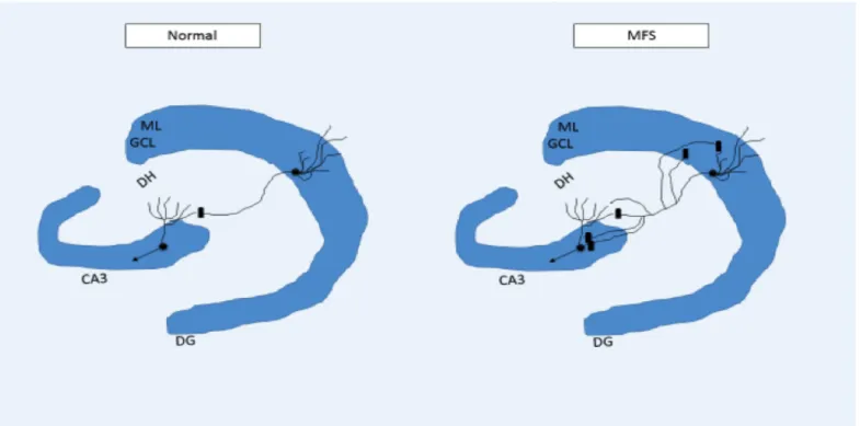
Figure 1 – This image illustrates MFS which occurs in the hippocampus. Neurons with cellular bodies in the GCL bifurcate
their axons in the hilar region of dentate gyrus, and project their collaterals o CA3 and to the ML.. Cornus ammonis 3 (CA3); dentate gyrus (DG); dentate hylus (DH); granular cell layer; mossy fiber sprouting (MFS); molecular layer (ML). Adapted from (Koyama and Ikegaya, 2004) ... 14
Figure 2 - Seizures lead to a fast and slow increase in BDNF. The Fast increase is dependent on: i) NMDA receptors
activation leading directly to BDNF release; ii) conversion of pro-BDNF to mature BDNF through tPA. The slow increase of BDNF is dependent of an increase in intracellular calcium which further increases transcription of BDNF in a AKT/Nf-kB and CREB dependent manner. Tissue plasminogen activator (tPA) ... 19
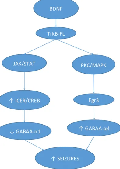
Figure 3 – BDNF may increases seizures through TrkB receptor signaling pathway. TrkB activation diminish the abundance
of the α1 subunit of GABA and increases the abundance of α4 subunit in GABA receptorsAdapted from (Grabenstatter et al., 2012) ... 22
7
List of tables
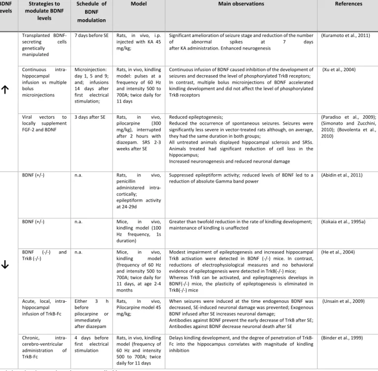
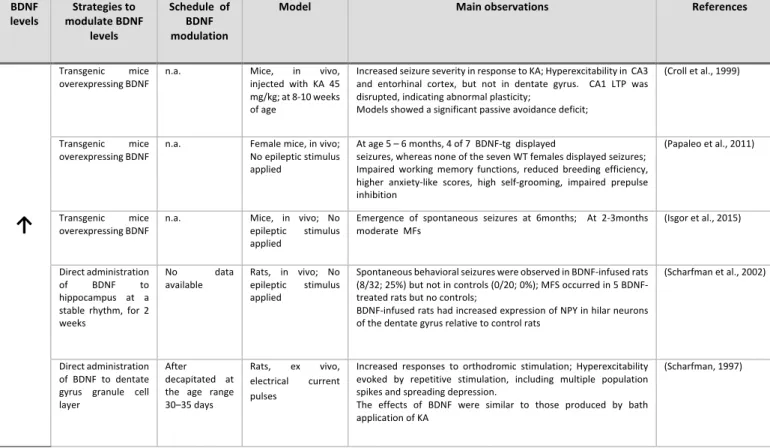
Table 1 - Studies suggesting that BDNF modulation inhibits epileptogenesis. ... 27 Table 2 - Studies suggesting that BDNF modulation do not inhibit epileptogenesis or increases it. ... 28
8
Acknowledgements
Se há premissa em que acredito é que nada na vida se consegue sem uma grande família e sem bons amigos. Estes são os grandes pilares da minha felicidade.
Ao longo dos últimos 3 anos tive a felicidade de estabelecer uma valiosa amizade com a principal responsável por todo o rigor científico, pertinência de tema e de conceitos, encadeação lógica de ideias, bem como de qualquer outra modesta genialidade de que esta tese de mestrado se possa valer. Apercebi-me da incontestável importância desta amizade recentemente, na sequência da leitura ocasional de uma frase:
“My best friend is the one who brings out the best in me” - Henry Ford
Por ter sempre acreditado em mim e nas minhas competências, por toda a dedicação, celeridade na resolução de qualquer problema, por ter fomentado em mim o espírito da investigação desde o 2º ano de faculdade e, acima de tudo, pela sua boa amizade, o meu muito obrigado Professora Maria José Diógenes. É para mim uma grande referência, e constitui um grande orgulho ter sido orientado por si nesta tese de mestrado. Desejava ser poeta e ter nas minhas palavras maior arte para um agradecimento mais justo face a toda a amizade e todo o auxílio prestado.
Não posso ainda deixar de agradecer ao Doutor André Jerónimo Santos. Apesar de não ter participado nesta tese, foi meu mentor no meu primeiro projeto de investigação. Nunca antes tinha tido a oportunidade de deixar por escrito o meu agradecimento por me teres ensinado praticamente tudo o que sei de procedimentos de laboratório, pela infindável paciência para a minha inexperiência e para as minhas perguntas básicas. Mais valioso ainda para mim, muito obrigado pela tua amizade.
Agradeço ainda á Professora Doutora Ana Sebastião que foi o meu primeiro contacto com a ciência e me encaminhou para porto seguro. Tenho sempre presente que toda a investigação que fiz até agora no laboratório e as excelentes relações inter-humanas que aqui existem só são possíveis por uma direcção firme, brilhante e bondosa, e por isso estou grato por todas as oportunidades de investigação que me tem concedido. Agradeço ainda à Professora Sandra Vaz pela disponibilidade em ser júri desta tese, e ao Dr. Hélder Viegas, pelas múltiplas reuniões e e-mails trocados, por me ter ensinado medicina, e especialmente por me alimentar os sonhos.
Aos meus pais e irmã, á minha namorada, á minha família, o meu mais sincero obrigado, por serem o meu suporte nesta vida, por serem a base sem a qual nada do resto seria possível. Sem vocês nunca teria chegado ao final deste curso. São os meus grandes ídolos, obrigado por terem feito de mim o homem que sou hoje.
9
Abbreviations
A2AR: adenosine A2A receptors
A1R: adenosine A1 receptors
BDNF: brain derived neurotrophic factor; Ca2+: calcium;
EEG: electroencephalogram; GABA: gamma-aminobutyric acid;
GFLs: glial cell line-derived neurotrophic factor family of ligands; K+: potassium;
KA: kainate acid;
MFS: mossy-fiber sprouting; Mg2+: magnesium
MMP7: metalloproteinase gene matrilysin; MTLE: mesial temporal lobe epilepsy; NGF: nerve growth factor;
NMDA: N-methyl-D-aspartate receptor; NPY: neuropeptide Y;
NT: neurotrophin; NT-3: neurotrophin-3; NT-4/5: neurotrophin-4/5; NTF: neurotrophic factors;
PDS: paroxysmal depolarizing shift; SE: status epilepticus;
SRS: spontaneous recurrent seizures;
TrkB-Fc: Tropomyosin receptor kinase B receptor body, scavenger for BDNF TrkB-FL: Tropomyosin receptor kinase B full-length
10
1. Introduction
Epilepsies are frequently devastating disorders, with a prevalence around 1% in the world’s population, being the third most common neurologic disorder, after dementia and stroke. There are about 40 distinct forms of epilepsy identified. Although 50% of this cases have a complete control of seizures and a further 25% have improved significantly under medication, remains 25% of patients whose disease has poor control although optimized therapy. Given the heterogeneous clinical presentation of the patients and the different responses to therapy, some authors prefer the designation epilepsies rather than epilepsy
The definition of epilepsy and epileptic seizure have been changing since the 1981 consensus. In 2014 a new consensus defined epileptic seizure as a transient occurrence of signs and/or symptoms due to abnormal excessive or synchronous neuronal activity in the brain; and epilepsy as a disorder of the brain characterized by an enduring predisposition to generate epileptic seizures and by the neurobiologic, cognitive, psychological, and social consequences of this condition, requiring the occurrence of at least one epileptic seizure (Fisher et al., 2014). Conversely, there are “non-epileptic” seizures, when evoked in a normal brain by treatments such as electroshock or chemical convulsants. The therapy for patients with seizure disorders is usually multimodal and it includes the treatment of underlying conditions, avoidance of precipitating factors and suppression of recurrent seizures with medication or surgery. However, whether any of the antiepileptic dugs being used in clinical practice can prevent the development of epilepsy (epileptogenesis) is uncertain, and so drugs are preferentially referred as anti-seizure rather than antiepileptic. Together with the evidence of about 20% of cases resistant to therapy, emerges the need of better therapeutic strategies. Neurotrophins, namely brain derived neurotrophic factor (BDNF) have been implicated in the physiopathology of epilepsy, and an increasing interest for a potential role in its therapeutics has been growing.
11
2. Neurotrophins
Neurotrophic factors (NTF) are a superfamily of proteins which are known to be essential in neuronal survival, growth and differentiation and to have acute effects on modulation of synaptic transmission and plasticity. Specifically, its effects on mature neuronal circuits include modulation of synaptic efficacy and plasticity as Long-term potentiation (LTP) and Long-term depression (LTD); and its effects on neuronal circuits development include modulation of neural stem cell survival and differentiation, survival of differentiated neurons, axon-dendrite differentiation, growth and guidance; synapse formation and maturation (Huang and Reichardt, 2001).
The NTF superfamily comprises four distinct major groups: (1) the neurotrophin (NT) family; (2) the glial cell line-derived neurotrophic factor family of ligands (GFLs); (3) neurotrophic cytokines (neurokines); and (4) the family of cerebral dopamine neurotrophic factor (CDNF) and mesencephalic astrocyte derived neurotrophic factor (MANF) (Rodrigues et al. 2014).
NT are a closely related group of secreted proteins that are initially synthesized as larger precursors (proneurotrophins), which undergo proteolytic cleavage to yield mature forms. In mammals, the neurotrophin family includes nerve growth factor (NGF), brain-derived neurotrophic factor (BDNF), neurotrophin-3 (NT-3), and neurotrophin-4/5 (NT-4/5). The biological actions of neurotrophins are mediated either by the activation of their cognate tropomyosin-related kinase receptor (TrkA, TrkB or TrkC), or by the activation of the common p75 neurotrophin receptor (p75NTR). TrkA is the high-affinity receptor for NGF (with low-affinity binding by NT-3 in some systems) (Kaplan et al., 1991; Klein et al., 1991), TrkB is the high-low-affinity receptor for BDNF with lower-affinity binding by NT-3 and NT-4 (Berkemeier et al., 1991; Squinto et al., 1991), and TrkC is the high-affinity receptor for NT-3 (Lamballe et al., 1991; Urfer et al., 1995).
In addition to the high-affinity NT receptors, all of the NTs bind to the low-affinity NT receptor, designated as p75NTR. (Chao and Hempstead, 1995). The p75NTR is a member of the tumor necrosis factor (TNF) receptor superfamily, and the p75NTR activation elicit distinct cellular responses, inducing cellular death when binding to pro-neurotrophins namely through JNK and NADE pathways (Casaccia-Bonnefil et al., 1996; Friedman, 2000), and modulating the affinity and selectivity of Trk activation when binding to mature neurotrophin (Teng et al., 2010). Although p75NTR in the highest levels are observed perinatally, then it declines with age, it is also overexpressed in various pathological conditions, including epilepsies, axotomy and neurodegeneration (Dechant and Barde, 2002).
In this work we review the specific role of the neurotrophin BDNF on epilepsy therefore BDNF signaling will be detailed in this section.
BDNF uncleaved form, the pro-BDNF is a 32-kDa precursor, comprising 247 amino acids with N-glycosylated and glycosulphated residues within the prodomain (Mowla et al., 2001). Following the initial
12
generation, most of the pro-BDNF is then packaged into vesicles in a regulated pathway and undergoes N-terminal cleavage by extracellular proteases, such as plasmin, metalloproteinase gene matrilysin (MMP7) (Lu, 2003; Pang et al., 2004), tPA/plasmin cascade and extracellular matrix-metalloproteinases (Lee et al., 2001). In trans-Golgi network, the ‘pro-region’ is cleaved resulting in the formation of mature BDNF (14 kDa), a biologically active form with C-terminal dimers (Seidah et al., 1996). This mature BDNF could be then released by the neurons through constitutive secretion or in an activity dependent manner (Mowla et al., 2001). But although BDNF is highly expressed in neurons, astrocyte and microglia are also capable of producing BDNF (Miklic et al., 2004).
Pro-BDNF selectively activates its high affinity receptor, the p75NTR, mainly inducing pro-apoptotic signaling pathways (Teng et al., 2005) while mature BDNF binds to TrkB receptors and as well as to the low-affinity neurotrophin receptor p75NTR (Lahteinen et al., 2003).
BDNF binding to the full length (FL) receptor TrkB-FL triggers autophosphorylation of the tyrosine residue in its intracellular domain, leading to ligand-induced dimerization in each receptor, which activates several intracellular signaling pathways. The major signaling pathways activated by the TrkB-FL receptors are; (1) PI3K/AKT/mTOR; (2) MAPK/ERK and (3) PLCγ pathways, and their downstream effectors. Conversely, these include PI3K stimulation of AKT, Ras stimulation of mitogen-activated protein MAPK cascades and PLC-γ dependent generation of inositol 1,4,5-triphosphate (IP3) and diacylglycerol (DAG) (Segal and Greenberg, 1996; Arévalo and Wu, 2006). Which one of the pathways leads do different cellular responses, specifically the PI3K/AKT signalling pathway is thought to be the main responsible by chronic effects of BDNF in neuronal circuits, as it is the survival, growth and differentiation. Conversely, it is thought that this pathway can halt apoptosis through phosphorylation and inhibition of pro-apoptotic proteins such as Bad and caspase-9 (Zhou et al., 2000).
13
3. Epilepsies
Epileptic seizures have been classified into partial seizures, those beginning focally in a cortical site, and generalized seizures, those that involve both hemispheres widely from the outset. Partial seizures account for around 60% of epileptic seizures and are further divided in simple, when there is preservation of consciousness and complex, when consciousness is impaired. Etiology of partial seizures is most commonly secondary to a lesion in some part of the cortex, such as a tumor, developmental malformation, damage due to trauma or stroke, alcohol or drug withdrawal, repeated episodes of metabolic insults, such as hypoglycemia (Berkovic et al., 2006; Longo, 2012). Alternatively, generalized epilepsies have usually a genetic etiology due to inheritance of multiple susceptibility genes, with rare exceptions as it happens with the spontaneous mutations in SCN1A which is responsible for catastrophic severe myoclonic epilepsy of infancy. Generalized epilepsies are usually categorized based on the seizure activity in absence, myoclonic and tonic-clonic seizures (Anon, 1981).
What concerns to the physiopathology, a seizure results of a sudden imbalance between the excitatory and inhibitory forces within the network of cortical neurons in favor of a sudden-onset net excitation (Fisher et al., 2005). When this imbalance happens, a group of neurons starts firing in an abnormal, excessive, and synchronized manner (Hammer and Hammer, 2010) which results in a wave of depolarization known as a paroxysmal depolarizing shift (PDS) (Somjen, 2004).
In a normal brain, there is a protection against PDS once after an excitatory neuron fires it becomes more resistant to firing for a period of time. This is due in part from the effect of inhibitory neurons and the electrical changes within the excitatory neuron. In epilepsy the resistance of excitatory neurons to fire during this period is decreased (Hammer and Hammer, 2010). Conversely, the mechanisms that are generally responsible for the focal-onset seizures are whether a decreased inhibition or an increased excitation of neurons (da Silva et al., 2003; Quyen et al., 2003).
While the mechanisms of decreased inhibition are mainly relate to defective GABAergic system, the mechanisms leading to increased excitation include increased activation of N-methyl-D-aspartate receptor
(NMDA) receptors, increased synchrony between neurons due to emphatic interactions and increased synchrony and/or activation due to recurrent excitatory collaterals (Goldberg and Coulter, 2013).
Importantly, neuropathological studies of patients with intractable focal-onset epilepsy have revealed frequent abnormalities in the limbic system, particularly in the hippocampal formation. The most common lesion is hippocampal sclerosis, which is CA1 and CA3 pyramidal neurons loss and end folium sclerosis (loss of neurons within the hilus) with relative sparing of transitional cortex measured at the mid-body of the anterior-posterior axis (Wieser, 2004).
The most prominent hippocampal sclerosis is frequently found in the mesial temporal lobe epilepsy (MTLE) (Rudge et al., 1998) which is the most common form of partial epilepsy in young adults. More subtle
14
and apparently more common than overt hippocampal sclerosis is granule cell axon (mossy-fiber) sprouting (MFS) (Sutula et al., 1989). The MFS, which create monosynaptic recurrent excitation circuits, consists of single granule cells that bifurcate their MF axons in the hilar region, projecting the collaterals to both the
cornus ammonis 3 (CA3) region and the molecular layer (ML). As a result some of the granule cells are
monosynaptically interconnected (Koyama and Ikegaya, 2004), providing a substrate for positive feedback monosynaptic recurrent excitation (Dudek and Shao, 2004) (figure 1).
Nonetheless, the role of MSF in epileptogenesis is controversial. The classical concept is that a precipitating brain insult trigger transient cascades of events that permanently enhance excitability of neuronal networks through mechanisms including MFS. Recent evidence suggests this permanent changes occur shortly after the insult, and that there is a continuous turnover of sprouted mossy fibers in epileptic patients and animal models. However, reversal of mossy fiber sprouting might not be antiepileptogenic, once blocking its development with rapamycin does not reduce seizure frequency in pilocarpine-treated mice (Buckmaster and Lew, 2011; Heng et al., 2013; Buckmaster, 2014). However rapamycin has side-effects (Swiech et al., 2008), including suppression of axon sprouting by inhibitory GABAergic interneurons and so it is not absolutely clear yet if MFS is just a epiphenomenon unrelated to seizure genesis as some authors recently suggest, or if in fact is part of epileptogenisis.
Several animal models have been developed during the last decades and there are indeed various animal models with chronic brain dysfunctions thought to reflect the processes underlying human epilepsy. The most used models to study epilepsy include (i) the kindling model and (ii) post-status epilepticus models. (i) The kindling model is induced by periodic administration of brief, low-intensity electrical stimulation of Figure 1 – This image illustrates MFS which occurs in the hippocampus. Neurons with cellular bodies in the GCL bifurcate their axons in the hilar region of
dentate gyrus, and project their collaterals o CA3 and to the ML.. Cornus ammonis 3 (CA3); dentate gyrus (DG); dentate hylus (DH); granular cell layer; mossy fiber sprouting (MFS); molecular layer (ML). Adapted from (Koyama and Ikegaya, 2004)
15
the amygdala or other limbic structures. Initial stimulations evoke a brief electrical seizure recorded on the EEG without behavioral change, but repeated (e.g. 10-20) stimulations result in progressive intensification of seizures, culminating in tonic-clonic seizures. Once established, the enhanced sensitivity to electrical stimulation persists for the life of the animal. The limitation of this model is that spontaneous seizures or a truly epileptic condition do not occur until 100-200 stimulations have been administered. In the (ii) post-status epilepticus models, animals experience a prolonged epileptic crisis (status epilepticus (SE)) after injection of pharmacological compounds (e.g. kainate, pilocarpine; intra-cortical penicillin injection; magnesium-free treatment) or via electrical stimulation of (limbic) brain regions. These post-SE models are the most widely used models in epilepsy research today. However, not all aspects of MTLE are reproduced and the widespread brain damage is often a caricature of the situation in the patient. As recently reviewed, more complex mammalian brains and genetic model organisms including zebrafish have been less studied but offer substantial advantages that are becoming widely recognized (Grone and Baraban, 2015).
These models allow exploring the three main intervention levels 1) anti-epileptogenic (prevention of development of transformation of the normal neuronal network into one that is chronically hyperexcitable (Lowenstein, 2015)), 2) anti-seizure (reduction of frequency and/or severity of seizures), and 3) disease-modifying strategies (alteration of the natural history of the disease).
ü The most used epilepsy models are kindling and post-SE (pilocarpine and kainate acid) models ü Three main interventions: anti-epileptogenic, anti-seizure, disease modifying
16
4. Endogenous BDNF and its receptors in epilepsy and in
epilepsy models
Seizures have been shown to stimulate the expression of a variety of genes, including transcription factors (Morgan and Curran, 1991; Kiessling and Gass, 1993), neuropeptides (Gall et al., 1991), proteases (Qian et al., 1993) and also neurotrophins (NT) (Gall, 1993).
What concerns to the regulation of the levels of NTs and specifically of BDNF in the epileptic brain,
in human, the evidence support they are increased in several cases. Accordingly, in resected hippocampal specimens from patients suffering from TLE compared to control there is an increased expression of BDNF protein (Takahashi et al., 1999) and mRNA (Mathern et al., 1997; Murray et al., 2000). Furthermore, resected specimens from the hippocampus of MTLE patients show an increased NTs expression in brain areas with MFS. Specifically, MFS correlates with increased granular layer nerve growth factor (NGF) immunoreactivity and seizure frequency (Kandratavicius et al., 2013). Similarly, glioneuronal tumors correlated with medically chronically intractable epilepsy also express increased BDNF protein expression (Villemure, 1996).
Recently it has been discussed the relevance of serum BDNF measurement in epileptic patients for a prognostic purpose. A recent study with 135 participants suggests that seizure frequency and epilepsy duration might be negatively correlated with serum BDNF levels independently of other factors (Hong et al., 2014).
Regarding TrkB receptors, western-blot analysis of human epileptic neocortex samples revealed that they are upregulated compared with control patients (Wyneken et al., 2003). However, it is not specified in this study what type of receptors were evaluated, the TrkB-FL or its truncated forms.
In Human:
ü BDNF mRNA and protein are increased in TLE patients and glioneural tumors associated with epilepsy ü TrkB receptors are upregulated in human epileptic neocortex
ü BDNF serum levels might negatively correlate with seizure frequency and epileptic duration
In animal model of epilepsies induced by kainate acid (KA) administration, it was shown that BDNF mRNA levels gradually increase in dentate granule cells of mice from 24h of status epilepticus onwards reaching a maximum in 7 days (Suzuki et al., 1995; Heinrich et al., 2011). Remarkably, the same evidence was observed performing an electroencephalogram (EEG) analysis that shows spontaneous recurrent discharges progressively developing in the mouse hippocampus during the first two weeks following KA treatment, within the same time-frame as the BDNF increase in dentate granule cells (Heinrich et al., 2011). Accordingly, in the kindling model, it has also been shown an increase in BDNF mRNA expression levels that occur predominantly in the hippocampus bilateral granule cell layer of the dentate gyrus 1-4h after kindled seizure in rat, and the maximum expression levels of BDNF mRNA occurs 2h after seizures (Lindvall et al.,
17
1994; Morimoto et al., 1998; Binder et al., 2001). Moreover, adult rats treated acutely with peripheral KA revealed that despite robust elevation in both mRNA and protein in multiple brain regions, in the same time frame plasma BDNF is unchanged and cerebrospinal fluid (CSF) BDNF levels remain undetectable (Lanz et al., 2012).
In KA model:
ü BDNF mRNA and protein expression levels are increased in a way directly correlated with spontaneous recurrent
discharges on EEG.
ü The increased brain levels of BDNF do not correlate with changes in the CSF or plasma
Interestingly, no evidences of epileptogenesis were detected in TrkB(-/-) mice even after 50 electrical focal stimulation of the amygdale. However, when exposed to electroshock they exhibit tonic-clonic an clonic seizure similar to wild-type controls. Interestingly, the available data revealed that the duration of seizure progressively increases with focal stimulations of the amygdala in TrkB (+/-) and TrkB (+/+) but not in TrkB (-/-) animals (He et al., 2004), which advances TrkB and its downstream signaling pathways as attractive targets for developing small molecule inhibitors for prevention of epileptogenesis.
Interestingly in BDNF knockout mice, which lack the most important endogenous ligand for TrkB-FL, the BDNF, there is still the promotion of epileptogenesis. This finding could be seen as a contradiction however, this might be explained by the activation of TrkB-FL receptors by other endogenous ligands such as NT-3 (He et al., 2004) and adenosine (Lee and Chao, 2001). And, indeed it was observed that BDNF knockout mice has an increased expression levels of NT-3 in the hippocampus which might contribute to TrkB-FL activation, promoting epileptogenesis progression even in the absence of BDNF (He et al., 2004). Conversely, in kindling model, NT-3 mRNA expression levels are usually decreased (Isackson et al., 1991; Rocamora et al., 1992; Kokaia et al., 1993; Suzuki et al., 1995; Mudo et al., 1996; Gall et al., 1997; Garcia et al., 1997) and it is also reported that the development of amygdala kindling is markedly retarded in mice heterozygous for a deletion of the NT-3 gene. These mice did not reach the fully kindled state (3rd grade 5 seizure) until after 28 +/- 4 days of stimulation compared to 17 +/- 2 days in the wild-type animals (Elmer et al., 1997).
In kindling:
ü BDNF mRNA and protein are increased in hippocampus ü TrkB gene knockout prevents epileptogenesis in kindling model ü BDNF gene knockout only impairs epileptogenesis in kindling model
18
Curiously, a great upregulation of truncated (Tc) TrkB and a decrease in TrkB-FL was observed in a model of epileptic seizures induced by magnesium-free treatment. In this experiment although calpain inhibitors have prevented the decrease in TrkB-FL receptors levels, the impairment on TrkB-FL mediated signaling pathways was not recovered (Xie et al., 2014). This fact was explained by the increased synthesis of TrkB-Tc which is known to act as a negative dominant receptor (Eide et al., 1996). This might constitute a protection mechanism against the possible pro-epileptogenic effect of TrkB-FL signaling pathway activation.
In Mg2+ free model:
ü TrkB-FL is decreased and truncated TrkB is increased compared to control. ü Truncated TrkB increase is mainly due to alternative splicing of TrkB mRNA. ü BDNF signaling is impaired in the presence of TrkB-T1.
4.1. From seizure to an increase in BDNF levels
What concerns to the seizure initiating mechanism, the bursting activity in individual neurons is caused by a relatively long-lasting depolarization of the neuronal membrane due to influx of extracellular calcium (Ca2+), and generation of repetitive action potentials. With sufficient activation, there is a recruitment of surrounding neurons via a number of synaptic and non-synaptic mechanisms, including: (1) an increase in
extracellular potassium (K+), which blunts hyperpolarization and depolarizes neighboring neurons; (2)
accumulation of Ca2+ in presynaptic terminals, leading to enhanced neurotransmitter release; (3)
depolarization-induced activation of the NMDA subtype of the excitatory amino acid receptor, which causes
additional Ca2+ influx and neuronal activation (Lowenstein, 2015)
The seizures being responsible for depolarization-induced activation of the synaptic NMDA subtype
of the excitatory amino acid receptor results in an influx in Ca2+ leading to the immediate release of BDNF
which in turn binds to and activates its high-affinity receptor, TrkB-FL (Marini et al., 2007). Moreover the high-frequency activity seen in seizures results in tissue plasminogen activator secretion, inducing the conversion of pro-BDNF to mature BDNF (Boutilier et al., 2008) (Figure 2).
19
Additionally, the influx of Ca2+ when NMDA receptor is activated leads to the activation of NF-kB
through the AKT and ERK 1/2 pathways. NF-kB is a major survival transcriptional factor (Glazner et al., 2000), that also plays an important role in learning and memory (Meffert et al., 2003). Activated NF-kB translocates to the nucleus where binds to promoter 4 of the BDNF gene increasing its transcription (Lipsky
et al., 2001; Marini et al., 2007). The influx of Ca2+ also leads to the phosphorylation of cAMP response
element-binding protein (CREB) in the nucleus which also binds to and activates promoter 4 of BDNF gene (Tao et al., 1998) (Figure 2).
4.2. From increased BDNF signaling to seizures
As a consequence of BDNF upregulation, there is an enhancement of fast excitatory transmission in pyramidal cells and dentate granule cells of adult rat hippocampus (Kang and Schuman, 1995; Levine et al., 1995; Tanaka et al., 1997), resulting in an increase of neuronal excitability in hippocampal networks, promoting the occurrence of epileptiform activities (Thoenen, 1995). It has already been shown that BDNF potentiates synaptic transmission ex vivo (Lohof et al., 1993) and in vivo (Messaoudi et al., 1998). It has also been showed that ex vivo rat hippocampal slices showed hyper-excitability after acute exposure to BDNF that it is blocked by the inhibitor of Trk receptor phosphorylation, K252a (Scharfman, 1997). Recent evidence also Figure 2 - Seizures lead to a fast and slow increase in BDNF. The Fast increase is dependent on: i) NMDA receptors activation leading directly
to BDNF release; ii) conversion of pro-BDNF to mature BDNF through tPA. The slow increase of BDNF is dependent of an increase in intracellular calcium which further increases transcription of BDNF in an AKT/Nf-kB and CREB dependent manner. Tissue plasminogen activator (tPA).
20
shows that in a rat model of massive neuronal activation upon kainate-induced seizures it was found that elevated neuronal expression of BDNF is associated with its detachment from the nuclear lamina, and translocation toward the nucleus center. In contrast, the position of stably expressed TrkB-FL remains unchanged after seizures (Walczak et al., 2013). Thus BDNF may interfere with gene expression after seizures. Also of note is that hippocampal epileptogenesis may develop as a continuous, dynamic and self-aggravating process, in which an initial insult promotes the increase in BDNF expression, that promotes neuronal excitability, including MF sprouting, leading to recurrence of epileptic activities which in turn further increase BDNF expression in the hippocampus (Heinrich et al., 2011).
4.2.1. BDNF influence on neuronal excitability
As stated above, the basic mechanisms responsible for a seizure are reduction of inhibitory synaptic activity or enhancement of excitatory synaptic activity. Consequently, antagonists of the GABAA receptor or agonists of different glutamate-receptor subtypes (NMDA, AMPA, or kainic acid) trigger seizures in experimental animals in vivo. Additional mechanisms leading to increased excitation include increased synchrony between neurons due to emphatic interactions and/or activation due to recurrent excitatory collaterals (Goldberg and Coulter, 2013). There is increasing evidence of BDNF role in some of this mechanisms.
Increased activation of NMDA receptors
BDNF through a mechanism dependent of intraneuronal Ca2+ levels increase, facilitates glutamatergic
synaptic transmission, mediated by increasing the efficacy of glutamate release from the presynaptic neuron (Drube et al., 1994; Leßmann and Heumann, 1998). Furthermore, BDNF dependent postsynaptic enhancement of NMDA receptor responsiveness has also been shown (Leßmann, 1998). BDNF enhances glutamate release (Canas et al., 2004) and increases activation (Levine et al., 1998) and phosphorylation of glutamate NMDA receptors (Suen et al., 1997) in the hippocampus.
GABAergic transmission
BDNF induce modulation of inhibitory transmission, and the most affected system is the GABAergic.
Gamma-aminobutyric acid (GABA) type A receptor (GABAARs) mediate most fast synaptic inhibition in the
forebrain. These receptors are constitute by different subunits, and it was observed that alteration in GABAAR
21
(1) BDNF activates early growth response factor (Egr3) synthesis via a protein kinase C
(PKC)/mitogen-activated protein kinase (MAPK)-dependent pathway, leading to an increase in GABAAR
containing α-4 subunit (Roberts et al., 2006; Lund et al., 2008) (Figure 3). Interestingly, after pilocarpine
induced SE, GABAAR α-4 subunit mRNA expression increases as the animal develops spontaneous recurrent
seizures (Brooks-Kayal et al., 1998, 2001). Moreover α-4 containing GABAAR have been shown to
desensitize rapidly (Lagrange et al., 2007). Additionally, they are very sensitive to zinc blockade (Buhl et al., 1996), which is physiopathology important once zinc containing mossy fibers terminals sprout from the
granule cells and into CA3, likely depositing zinc onto newly formed α-4 containing GABAAR, causing a
decrease response to GABA.
(2) BDNF activates the JAK/STAT pathway through TrkB-FL receptors activation, increasing inducible cAMP early repressor (ICER), a member of cAMP responsive element-binding protein (CREB) family which lead to a decrease in GABAAR containing α-1 subunit in in vivo model (Lund et al., 2008)
(Figure 3). . Interestingly, after pilocarpine induced SE, GABAAR α-1 mRNA expression also decreases (Brooks-Kayal et al., 1998, 2001). Given this evidence, one could advance that the blockade of JAK/STAT pathway would lead to a decrease in ICER, an increase in GABAα1R and a consequent decrease in seizures. However, it was shown that mutant mice lacking ICER have accelerated kindling (Kojima et al., 2008), developing more severe epilepsy following pilocarpine-induced SE (Porter et al., 2008). Reciprocally, ICER-overexpressing mice show delayed kindling acquisition (Kojima et al., 2008).
This results must be integrated with the multiple signaling pathways of JAK/STAT and ICER. As discussed above, ICER is responsible for increasing BDNF mRNA synthesis, which role in epileptogenesis is not only dependent in GABAergic neurons. Consequently, therapeutic strategy’s regarding GABA neurons subunits must aim more specific targets than CREB/ICER.
Moreover, in laboratorial models BDNF was also shown to influence GABAergic neurons in other ways, namely inhibiting GABA release from hippocampal synaptosomes (Canas et al., 2004) and enhancing its uptake from synaptic and extrasynaptic sites and this way influencing neuronal excitability. This uptake facilitation is mediated by GAT-1 molecules at the plasma membrane of astrocytes, which time span is increased as a consequence of BDNF presence in cultures of neurons (Vaz et al., 2011), opposite to what happens in nerve endings, in which GABA uptake seems to be impaired as a consequence of BDNF (Vaz et al., 2008).
22
ü BDNF modulates GABAAR subunits, increasing α4 and decreasing α1 in epileptic animal models
ü Mutant mice lacking ICER have accelerated kindling, and ICER overexpressing mice show delayed kindling
acquisition.
ü BDNF inhibits GABA release from hippocampal synaptosomes inhibits its uptake to synaptosomes and increases
its uptake for astrocytes.
BDNF mediates mossy fibers sprouting
The involvement of BDNF in MFS can be analyzed in two different perspectives: (1) the role of BDNF in developing MFS and (2) the influence of BDNF on excitability once MFS is installed.
(1) Role of BDNF in developing MFS
Figure 3 – BDNF may increases seizures through TrkB receptor signaling pathway. TrkB
activation diminish the abundance of the α1 subunit of GABA and increases the abundance of α4 subunit in GABA receptors Adapted from (Grabenstatter et al., 2012)
23
There are complicated, apparently contradictory observations that might be attributed to different experimental procedures or different animal models.
There is evidence that MFS is diminished in mice heterozygous for a deletion of the BDNF gene, in which BDNF expression level is substantially reduced (Vaidya et al., 1999); however, there is also a reported increase on MFS in the same mutant mice (Kokaia et al., 1995b).
There are also reports showing that infusion of BDNF into the hippocampus induces MFS (Scharfman et al., 2002) on contrary there are data revealing that BDNF infusion has no significant effect on MFS (Vaidya et al., 1999). In addition, the scavenger for BDNF, TrkB-Fc, failed to inhibit kainite-induced mossy fiber sprouting in hippocampal cultures (Routbort and Ryan, 1997). Moreover, transgenic mice overexpressing BDNF do not display significant MFS (Qiao et al., 2001). On the opposite, dispersed cultures of granule cells of rat hippocampus showed that MFS can be induced by locally implanted beads of BDNF and prevented with the coapplication of an inhibitor of TrkB phosphorylation or a functioning-blocking anti-BDNF antibody (Koyama et al., 2004).
In this context a suggested interpretation is that MFS is essentially a local phenomenon that is subject to systemic and extrahippocampal influences and that local BDNF action in the dentate hilus trigger MFS, but global application of BDNF mask this effect (Koyama et al., 2004), and this may explain the inconsistency in the literature.
(2) influence of BDNF on excitability upon MFS
MF contains high levels of BDNF protein under basal conditions, and this level of BDNF undergoes further up-regulation after seizure onsets, in KA epilepsy model (Rudge et al., 1998).
Electron microscopy experiments indicate that the vast majority of newly formed MF synapses terminates on dendritic spines of granule cells and that on average one MF forms >500 new synapses and only a minority (<25) contact GABAergic interneurons, being the greatest influence on excitatory neurons (Buckmaster et al., 2002). Given that BDNF facilitates excitatory transmission this can be a major contribution to excitability in epilepsy.
On the other hand, in the healthy hippocampus, more than 90% of MF synapses terminate on GABAergic neurons (Acsády et al., 1998), being MF capable of releasing either glutamate or GABA to synaptic cleft, and peculiarly an increase in GABA expression is observed with seizure activity (Gutiérrez and Heinemann, 2001; Gutiérrez, 2002). However, as already discussed GABA neurons activity is decreased, which might be responsible for increased excitability.
24
Influence of other Neuropeptides
Although the possible deleterious effects of BDNF in the epileptic brain already detailed in this review, robust evidence also support it beneficial effects in some epileptic models, particularly when administered for long periods of time and at low dosage. One of the evidences showing a protective role of BDNF is thought to be mediated by the regulation of neuropeptide Y (NPY) levels.
There are evidences that correlates the levels of BDNF with the levels of NPY: (1) the level of NPY is increased in the neocortex of TLE patients and positively correlated with the BDNF level (Takahashi et al., 1999); (2) In rat epilepsy models, BDNF up-regulation in the MF pathway is followed by an increase in NPY in the same brain region (Vezzani et al., 1999); (3) intrahippocampal infusion of BDNF for seven days induces NPY up-regulation one week after the end of the infusion (Reibel et al., 2000), a time course similar to the time course for the effect of BDNF on kindling model.
There is also robust evidence of the neuroprotector effects of NPY in epilepsy models and seizure control. Accordingly, mice lacking the NPY gene experience spontaneous seizures and are more susceptible to chemically induced seizures as compared to wild-type littermates (Erickson et al., 1996). In addition, transgenic rats overexpressing NPY show lower seizure susceptibility (Vezzani et al., 2002). Moreover, epileptic rats, exhibiting unprovoked spontaneous seizures, significantly lowered seizure frequency and exhibited delayed progression of spontaneous seizure severity following NPY overexpression (Noe et al., 2008).
25
5. Modulation of BDNF signaling as a therapeutic strategy
We aimed to group the experiments by BDNF modulation schedule in seizure or epileptogenesis properties (Simonato, 2014). However none of the experiments analyzed matched the anti-seizure criteria already defined. Consequently, only the anti-epileptogenesis properties of BDNF were evaluated because in all experiments BDNF modulation was performed before the epileptic model develops SRS.
Epileptogenesis is the process by which the previously normal brain is functionally altered and biased towards the generation of the abnormal electrical activity that subserves chronic seizures (Goldberg and Coulter, 2013). The available data show contradictory results. Tables 1 and 2 summarize the available information concerning the actions of BDNF upon epileptogenesis.
Epileptogenesis was considered whenever there were alterations in relation to control group of: 1) seizure stage; 2) rate of kindling development; 3) SRS either frequency or severity; 4) electrophysiological measurements in EEG; 5) MFS;
In summary there the available data show that 1) decreased levels of BDNF lead to an inhibition of epileptogenesis (table 1) while there are other that 2) demonstrate that increased levels of BDNF inhibits or increases epileptogenesis (table 2). The discrepant data collected might be due to the 1) heterogeneity of the animals used (rat vs mice); 2) heterogeneity of epileptic model (post-SE induced by kainate vs pilocarpine vs penicillin vs kindling model) and even for the same model are different protocols of induction; 3) heterogeneity of the modulation of BDNF levels (e.g. viral vector BDNF supplementation vs direct cannula administration in the brain); 4) different outcomes measured, sometimes only described as reduction of epileptogenesis without further description in the original article.
1) Decrease in BDNF levels lead to an inhibition of epileptogenesis. This conclusion arose from studies where the modulation of BDNF was conducted by several ways: by the knocking down of the BDNF gene (Kokaia et al., 1995a; He et al., 2004; Abidin et al., 2011); by the chronic intracerebro-ventricular or by the local acute administration of the BDNF scavenger, the TrkB-Fc (Binder et al., 1999; Unsain et al., 2009); In all this conditions there was evidence supporting a reduction in epileptogenesis.
2) Increase in BDNF levels might either inhibit or increase epileptogenesis. There are contradictory results
depending on the used models: i) different models of genetic overexpression of BDNF consistently show appearance of spontaneous seizures (Papaleo et al., 2011; Isgor et al., 2015) and/or increase of severity of seizures in response to an epileptic stimulus (KA) (Croll et al., 1999). This are particularly appealing results once seems that BDNF overexpression per si triggers SRS; ii) Viral vectors to supplement the brain with BDNF and FGF-2 (Paradiso et al., 2009; Bovolenta et al., 2010; Simonato and Zucchini, 2010)as well as
26
continuous administration through transplanted BDNF-secreting cells (Kuramoto et al., 2011) consistently inhibits epileptogenesis and reduce the frequency and severity of SRS compared to control. This apparently contradictory difference to genetic overexpression is an interesting object of study for which we could not find any consistent explanation in the literature; iii) direct administration of BDNF into the brain retrieved the most interesting results. Continuous intra-hippocampal infusion of BDNF have been shown to inhibit (Xu et al., 2004) and to potentiate (Scharfman et al., 2002) epileptogenesis. However, the same author also observed that multiple bolus injections in spite of continuous administration potentiates epileptogenesis (Xu et al., 2004). It was also observed that direct BDNF infusion to hippocampal slices in electrophysiological studies lead to increased hyperexcitability (Scharfman, 1997).
A possible interpretation of this results is that genetic overexpression of BDNF is related with increased epileptogenesis while BDNF gene manipulation (+/-) and (-/-) is related with the inhibition of epileptogenesis. The effects of BDNF supplementation in epileptic brain tissue remains relatively unclear, and contradictory results persist. Viral vector localized supplementation with BDNF show interesting results and might constitute a promising therapeutic strategy. However, better comprehension of the mechanism inherent to its anti-epileptogenic properties and possible side effects are needed prior to any clinical trial.
Also remarkable is the observation that administration of BDNF in continuous intra-hippocampal infusion produces distinct results from the ones obtained by the administration in multiple bolus microinjections (Xu et al., 2004). Together with the observation that in BDNF (-/-) mice only modest impairment of epileptogenesis is observed while in TrkB (-/-) mice epileptogenesis is eliminated (He et al., 2004), one might hypothesize that continuous administration of BDNF might downregulate the TrkB-FL receptors, justifying the inhibition of epileptogenesis.
27
Table 1 - Studies suggesting that BDNF modulation inhibits epileptogenesis. BDNF
levels modulate BDNF Strategies to levels
Schedule of BDNF modulation
Model Main observations References
↑
Transplanted BDNF-secreting cells genetically manipulated
7 days before SE Rats, in vivo, i.p. injected with KA 45 mg/kg;
Significant amelioration of seizure stage and reduction of the number of abnormal spikes at 7 days after KA administration. Enhanced neurogenesis (Kuramoto et al., 2011) Continuous intra-hippocampal infusion vs multiple bolus microinjections Microinjection: day 1, 5 and 9; and; infusions 14 days after first electrical stimulation; Rats, in vivo, kindling model: pulses at a frequency of 60 Hz and intensity 500 to 700A; twice daily for 11 days Continuous infusion of BDNF caused inhibition of the development of seizures and decreased the level of phosphorylated TrkB receptors; In contrast, multiple bolus microinjections of BDNF accelerated kindling development and did not affect the level of phosphorylated TrkB receptors (Xu et al., 2004) Viral vectors to locally supplement FGF-2 and BDNF
3 days after SE Rats, in vivo, pilocarpine (300 mg/kg), interrupted after 2 hours with diazepam. SRS 2-3 weeks after SE
Reduced epileptogenesis;
Reduced the occurrence of spontaneous seizures. Seizures were significantly less severe in vector-treated rats although, on average, they had the same duration in both groups;
All untreated animals displayed hippocampal sclerosis and SRSs. Animals treated had significant reduction of cell loss in the hippocampus;
Increased neuronogenesis and reduced neuronal damage
(Paradiso et al., 2009); (Simonato and Zucchini, 2010); (Bovolenta et al., 2010)
↓
BDNF (+/-) n.a. Rats, in vivo, penicillin
administered intra-cortically; epileptiform activity at 24-29d
Suppressed epileptiform activity; reduced levels of BDNF led to a
reduction of absolute Gamma band power (Abidin et al., 2011)
BDNF (+/-) n.a. Mice, in vivo, kindling model (100 Hz frequency, 1s duration) Greater than twofold reduction in the rate of kindling development; maintenance of kindling is unaffected (Kokaia et al., 1995a) BDNF (-/-) and
TrkB (-/-) n.a. Mice, kindling in model vivo, (frequency of 60 Hz and intensity 500 to 700A; twice daily for 11 days, at age 2-4 months
Modest impairment of epileptogenesis and increased hippocampal TrkB activation were detected in BDNF (-/-) mice. In contrast, reductions of electrophysiological measures and no behavioral evidence of epileptogenesis were detected in TrkB(-/-) mice; Whereas TrkB can be activated, and epileptogenesis develops in BDNF(-/-) mice, the plasticity of epileptogenesis is eliminated in TrkB(-/-) mice
(He et al., 2004)
Acute, local, intra-hippocampal infusion of TrkB-Fc Either 3 h before pilocarpine or immediately after diazepam
Rats, In vivo, Pilocarpine model 45 mg/kg;
When seizures were induced at the time endogenous BDNF was decreased, SE-induced neuronal damage was prevented; Exogenous BDNF infused after SE increases neuronal damage; Antibodies against BDNF prevent the early decrease of TrkB after SE; Antibodies against BDNF decrease neuronal death after SE (Unsain et al., 2009) Chronic, intra-cerebro-ventricular administration of TrkB-Fc 4 days before first electrical stimulation Rats, in vivo, kindling model (frequency of 60 Hz and intensity 500 to 700A; twice daily for 11 days
Delays kindling development, and the degree of penetration of TrkB-Fc into the hippocampus correlates with magnitude of kindling inhibition
(Binder et al., 1999)
28
Table 2 - Studies suggesting that BDNF modulation do not inhibit epileptogenesis or increases it. BDNF
levels modulate BDNF Strategies to levels
Schedule of BDNF modulation
Model Main observations References
Transgenic mice
overexpressing BDNF n.a. Mice, injected with KA 45 in vivo, mg/kg; at 8-10 weeks of age
Increased seizure severity in response to KA; Hyperexcitability in CA3 and entorhinal cortex, but not in dentate gyrus. CA1 LTP was disrupted, indicating abnormal plasticity;
Models showed a significant passive avoidance deficit;
(Croll et al., 1999)
Transgenic mice
overexpressing BDNF n.a. Female mice, in vivo; No epileptic stimulus applied
At age 5 – 6 months, 4 of 7 BDNF-tg displayed
seizures, whereas none of the seven WT females displayed seizures; Impaired working memory functions, reduced breeding efficiency, higher anxiety-like scores, high self-grooming, impaired prepulse inhibition
(Papaleo et al., 2011)
↑
Transgenic miceoverexpressing BDNF n.a. Mice, in vivo; No epileptic stimulus applied
Emergence of spontaneous seizures at 6months; At 2-3months
moderate MFs (Isgor et al., 2015) Direct administration
of BDNF to hippocampus at a stable rhythm, for 2 weeks
No data
available Rats, in vivo; No epileptic stimulus applied Spontaneous behavioral seizures were observed in BDNF-infused rats (8/32; 25%) but not in controls (0/20; 0%); MFS occurred in 5 BDNF-treated rats but no controls; BDNF-infused rats had increased expression of NPY in hilar neurons of the dentate gyrus relative to control rats (Scharfman et al., 2002) Direct administration of BDNF to dentate gyrus granule cell layer
After decapitated at the age range 30–35 days
Rats, ex vivo, electrical current pulses
Increased responses to orthodromic stimulation; Hyperexcitability evoked by repetitive stimulation, including multiple population spikes and spreading depression.
The effects of BDNF were similar to those produced by bath application of KA
(Scharfman, 1997)
29
5.1. Is there a possible clinical application?
So far it is not clear if the best therapeutic strategy for epilepsies is to down or up-regulate BDNF levels, or even if this might be a good approach for this disease in humans. Moreover, the clinical trials conducted so far aiming the BDNF modulation as a therapeutic strategy for other pathologies (amyotrophic lateral sclerosis and diabetic neuropathy) have shown inconclusive results (Lu et al., 2013).
In case of up-regulating BDNF levels, there is evidence that transposition of BDNF to target injury regions have demonstrated to be challenging in clinical trials. However, recent experiments showed that this problem can be circumvented by: (1) improving BDNF delivery to target zones using different administration routs such as intranasal administration (Jiang et al., 2011) and (2) using BDNF mimetics (Kingwell, 2010). Additionally, simple alternative interventions which are known to increase BDNF in the brain should might have an important role, such as physical exercise (Vaynman et al., 2004; Hopkins et al., 2011; Mattson, 2012), intellectual stimulation (Goshen et al., 2009; Cao et al., 2011), intermittent fasting (Johnson et al., 2007; Harvie et al., 2011; Mattson, 2012), antidepressants (Jiang and Salton, 2013) and electroconvulsive shocks (Mughal et al., 2011). These simple interventions might have an important role preventing epileptogenesis in genetically predisposed individuals.
Down-regulating BDNF levels, as a therapeutic strategy, has not been tried so far in clinical trials. Nevertheless there are observations that a human BDNF genetic polymorphism in which methionine (Met) substitutes for a valine (Val) amino acid impairs BDNF neural secretion (Egan et al., 2003) is associated with altered cognitive functions and predisposition to psychiatric illnesses such as schizophrenia, Alzheimer’s, depression, substance-related, and eating disorders (Egan et al., 2003; Dempster et al., 2005; Gratacòs et al., 2007; Soliman et al., 2010; Verhagen et al., 2010). These observations empower the concept that down-regulating BDNF with a therapeutic purpose might have serious adverse side effects.
Although all the challenges related to the modulation of BDNF system, this therapeutic strategy should not be dismissed. Nevertheless, it is still crucial to fully understand: (1) the differences observed in the different models; (2) the appropriate therapeutic schedule for BDNF signaling modulation; (3) the signaling pathways of BDNF responsible for decreasing seizures and epilepsy; (4) and very important, the consequences of modulating BDNF actions which are involved in so many different actions in the brain.
30
5.2. Is there a role for the A2A of adenosine receptors?
Adenosine exerts its effects through the activation of the specific G-protein-coupled receptors: A1,
A2A, A2B, and A3 receptors. The adenosine A1 receptors (A1R) and A2A receptors (A2AR) are the most sensitive
to adenosine and the most abundant in the central nervous system (Fredholm et al., 2011). During a seizure, extracellular adenosine levels rapidly rise and are believed to play an important role in the termination of the
seizure through binding to the inhibitory adenosine A1R (During and Spencer, 1992; Gouder et al., 2003).
Studies have shown that the adenosine A1R agonist promotes increased latency of seizure onset, decreased
seizure occurrence, shorter seizure duration, and reduced mortality rate in chemical-induced seizure models (Li et al., 2013).
Nevertheless, adenosine levels rise significantly during epileptic seizures, which could activate all adenosine
receptors, including the A2AR. These receptors, which are known for facilitating neuronal transmission
(Berman et al., 2000; Pedata et al., 2001), have also reports of anticonvulsant effects in some observations
(Huber et al., 2002). One might speculate this A2AR anticonvulsant effect might be related to interplay with
BDNF signaling (Sebastião et al., 2011). Functionally, it has been shown that most BDNF-mediated synaptic actions, such as in synaptic plasticity, transmission and neurotransmitter release, are fully dependent on
adenosine A2AR activation (Diógenes et al., 2004; Pousinha et al., 2006; Fontinha et al., 2008; Tebano et al.,
2008; Jerónimo-Santos et al., 2014; Rodrigues et al., 2014b)..
Nevertheless, recent observation suggest that both the A2A R agonist and antagonist, CGS 21680 and
ZM 241385, respectively, did not promote changes in seizure parameters, promoting a more determinant
31
6. Final considerations
Down regulation of BDNF signaling, including through regulation of TrkB receptors seems to be a possible clinical therapeutic strategy for epilepsy. It matters, however, to have a deep comprehension of the consequences of continuous upregulation of BDNF in animal models before any clinical trial. Targeting specific brain areas known to be the epileptic focus or targeting lower mediators in the cascade, such as CREB might reveal a promising strategy with less adverse effects. Also, deeper assessment of the influence of BDNF on MFS and NMDA neurons would help to identify possible therapeutic targets, other that interfering with the entire BDNF signaling.
It was also noted during this review that there are few studies of BDNF modulation after the epileptic model is displaying SRS, consequently it would be interesting some investigation on the anti-seizure properties of BDNF regarding the anti-seizure definition explored in this review.
Also remarkably is the large heterogeneity of variables in the studies of the effects of BDNF regulation in epileptic models. For that purpose it would be interesting to establish a protocol with standard outcomes to measure anti-epileptogenic and anti-seizure properties of BDNF as well as a standard animal and epileptic model for this analysis capable of closely reproduce human epilepsies characteristics.
32
References
Abidin I et al. (2011) Penicillin induced epileptiform activity and EEG spectrum analysis of BDNF heterozygous mice: an in vivo electrophysiological study. Brain Res Bull 86:159–164.
Acsády L, Kamondi A, Sík A, Freund T, Buzsáki G (1998) GABAergic cells are the major postsynaptic targets of mossy fibers in the rat hippocampus. J Neurosci 18:3386–3403.
Anon (1981) Proposal for Revised Clinical and Electroencephalographic Classification of Epileptic Seizures. Epilepsia 22:489–501.
Arévalo JC, Wu SH (2006) Neurotrophin signaling: many exciting surprises! Cell Mol Life Sci 63:1523–1537. Berkemeier LR, Winslow JW, Kaplan DR, Nikolics K, Goeddel D V., Rosenthal A (1991) Neurotrophin-5: A novel
neurotrophic factor that activates trk and trkB. Neuron 7:857–866.
Berkovic SF, Mulley JC, Scheffer IE, Petrou S (2006) Human epilepsies: interaction of genetic and acquired factors. Trends Neurosci 29:391–397.
Berman RF, Fredholm BB, Aden U, O’Connor WT (2000) Evidence for increased dorsal hippocampal adenosine release and metabolism during pharmacologically induced seizures in rats. Brain Res 872:44–53.
Binder DK, Croll SD, Gall CM, Scharfman HE (2001) BDNF and epilepsy: Too much of a good thing? Trends Neurosci 24:47–53.
Binder DK, Routbort MJ, Ryan TE, Yancopoulos GD, McNamara JO (1999) Selective inhibition of kindling development by intraventricular administration of TrkB receptor body. J Neurosci 19:1424–1436.
Boutilier J, Ceni C, Pagdala PC, Forgie A, Neet KE, Barker PA (2008) Proneurotrophins Require Endocytosis and Intracellular Proteolysis to Induce TrkA Activation. J Biol Chem 283:12709–12716.
Bovolenta R, Zucchini S, Paradiso B, Rodi D, Merigo F, Navarro Mora G, Osculati F, Berto E, Marconi P, Marzola A, Fabene PF, Simonato M (2010) Hippocampal FGF-2 and BDNF overexpression attenuates epileptogenesis-associated neuroinflammation and reduces spontaneous recurrent seizures. J Neuroinflammation 7:81. Brooks-Kayal AR, Shumate MD, Jin H, Rikhter TY, Coulter DA (1998) Selective changes in single cell GABAA
receptor subunitexpression and function in temporal lobe epilepsy. Nat Med 4:1166–1172.
Brooks-Kayal AR, Shumate MD, Jin H, Rikhter TY, Kelly ME, Coulter DA (2001) γ-Aminobutyric acidA receptor subunit expression predicts functional changes in hippocampal dentate granule cells during postnatal
development. J Neurochem 77:1266–1278.
Buckmaster PS (2014) Does mossy fiber sprouting give rise to the epileptic state? Adv Exp Med Biol 813:161–168. Buckmaster PS, Lew FH (2011) Rapamycin suppresses mossy fiber sprouting but not seizure frequency in a mouse
model of temporal lobe epilepsy. J Neurosci 31:2337–2347.
33 predominantly excitatory feedback circuit. J Neurosci 22:6650–6658.
Buhl EH, Otis TS, Mody I (1996) Zinc-Induced Collapse of Augmented Inhibition by GABA in a Temporal Lobe Epilepsy Model. Science (80- ) 271:369–373.
Canas N, Pereira IT, Ribeiro JA, Sebastião AM (2004) Brain-derived neurotrophic factor facilitates glutamate and inhibits GABA release from hippocampal synaptosomes through different mechanisms. Brain Res 1016:72–78. Cao L, Choi EY, Liu X, Martin A, Wang C, Xu X, During MJ (2011) White to brown fat phenotypic switch induced
by genetic and environmental activation of a hypothalamic-adipocyte axis. Cell Metab 14:324–338. Casaccia-Bonnefil P, Carter BD, Dobrowsky RT, Chao M V (1996) Death of oligodendrocytes mediated by the
interaction of nerve growth factor with its receptor p75. Nature 383:716–719.
Chao M V., Hempstead BL (1995) p75 and Trk: A two-receptor system. Trends Neurosci 18:321–326.
Croll SD, Suri C, Compton DL, Simmons M V, Yancopoulos GD, Lindsay RM, Wiegand SJ, Rudge JS, Scharfman HE (1999) Brain-derived neurotrophic factor transgenic mice exhibit passive avoidance deficits, increased seizure severity and in vitro hyperexcitability in the hippocampus and entorhinal cortex. Neuroscience 93:1491– 1506.
da Silva FL, Blanes W, Kalitzin SN, Parra J, Suffczynski P, Velis DN (2003) Epilepsies as Dynamical Diseases of Brain Systems: Basic Models of the Transition Between Normal and Epileptic Activity. Epilepsia 44:72–83. Dechant G, Barde Y-A (2002) The neurotrophin receptor p75NTR: novel functions and implications for diseases of
the nervous system. Nat Neurosci 5:1131–1136.
Dempster E, Toulopoulou T, McDonald C, Bramon E, Walshe M, Filbey F, Wickham H, Sham PC, Murray RM, Collier DA (2005) Association between BDNF val66 met genotype and episodic memory. Am J Med Genet B Neuropsychiatr Genet 134B:73–75.
Diógenes MJ, Fernandes CC, Sebastião AM, Ribeiro JA (2004) Activation of adenosine A2A receptor facilitates brain-derived neurotrophic factor modulation of synaptic transmission in hippocampal slices. J Neurosci 24:2905–2913.
Drube W, Lessmann A, Materlik G (1994) Resonant Anomalous X-ray Scattering. North-Holland, Amsterdam. Dudek FE, Shao L-R (2004) Mossy fiber sprouting and recurrent excitation: direct electrophysiologic evidence and
potential implications. Epilepsy Curr 4:184–187.
During MJ, Spencer DD (1992) Adenosine: a potential mediator of seizure arrest and postictal refractoriness. Ann Neurol 32:618–624.
Egan MF, Kojima M, Callicott JH, Goldberg TE, Kolachana BS, Bertolino A, Zaitsev E, Gold B, Goldman D, Dean M, Lu B, Weinberger DR (2003) The BDNF val66met Polymorphism Affects Activity-Dependent Secretion of BDNF and Human Memory and Hippocampal Function. Cell 112:257–269.
34 have dominant inhibitory effects on brain-derived neurotrophic factor signaling. J Neurosci 16:3123–3129. Elmer E, Kokaia M, Ernfors P, Ferencz I, Kokaia Z, Lindvall O (1997) Suppressed kindling epileptogenesis and
perturbed BDNF and TrkB gene regulation in NT-3 mutant mice. Exp Neurol 145:93–103.
Erickson JC, Clegg KE, Palmiter RD (1996) Sensitivity to leptin and susceptibility to seizures of mice lacking neuropeptide Y. Nature 381:415–418.
Fisher RS, Acevedo C, Arzimanoglou A, Bogacz A, Cross JH, Elger CE, Engel J, Forsgren L, French JA, Glynn M, Hesdorffer DC, Lee BI, Mathern GW, Moshé SL, Perucca E, Scheffer IE, Tomson T, Watanabe M, Wiebe S (2014) ILAE official report: a practical clinical definition of epilepsy. Epilepsia 55:475–482.
Fisher RS, van Emde Boas W, Blume W, Elger C, Genton P, Lee P, Engel J (2005) Epileptic seizures and epilepsy: definitions proposed by the International League Against Epilepsy (ILAE) and the International Bureau for Epilepsy (IBE). Epilepsia 46:470–472.
Fontinha BM, Diógenes MJ, Ribeiro JA, Sebastião AM (2008) Enhancement of long-term potentiation by brain-derived neurotrophic factor requires adenosine A2A receptor activation by endogenous adenosine.
Neuropharmacology 54:924–933.
Fredholm BB, IJzerman AP, Jacobson KA, Linden J, Müller CE (2011) International Union of Basic and Clinical Pharmacology. LXXXI. Nomenclature and classification of adenosine receptors--an update. Pharmacol Rev 63:1–34.
Friedman WJ (2000) Neurotrophins induce death of hippocampal neurons via the p75 receptor. J Neurosci 20:6340– 6346.
Gall C, Lauterborn J, Bundman M, Murray K, Isackson P (1991) Seizures and the regulation of neurotrophic factor and neuropeptide gene expression in brain. Epilepsy Res Suppl 4:225–245.
Gall CM (1993) Seizure-Induced Changes in Neurotrophin Expression: Implications for Epilepsy. Exp Neurol 124:150–166.
Gall CM, Lauterborn JC, Guthrie KM, Stinis CT (1997) Seizures and the regulation of neurotrophic factor expression: associations with structural plasticity in epilepsy. Adv Neurol 72:9–24.
Garcia ML, Garcia VB, Isackson PJ, Windebank AJ (1997) Long-term alterations in growth factor mRNA expression following seizures. Neuroreport 8:1445–1449.
Glazner GW, Camandola S, Mattson MP (2000) Nuclear factor-kappaB mediates the cell survival-promoting action of activity-dependent neurotrophic factor peptide-9. J Neurochem 75:101–108.
Goldberg EM, Coulter DA (2013) Mechanisms of epileptogenesis: a convergence on neural circuit dysfunction. Nat Rev Neurosci 14:337–349.
Goshen I, Avital A, Kreisel T, Licht T, Segal M, Yirmiya R (2009) Environmental enrichment restores memory functioning in mice with impaired IL-1 signaling via reinstatement of long-term potentiation and spine size