FACULDADE
DE
MEDICINA
DENTÁRIA
STUDY CONTRIBUTION FOR PERI-IMPLANT INFLAMMATORY RESPONSE AROUND DIFFERENT BIOMATERIALS IMUNOHISTOCHEMICAL STUDY SHEEP MODEL AND HUMAN RANDOMIZED CLINICAL CONTROL TRIAL
A
NDRÉ
T
SOU
C
HEN
ORIENTADOR:
Professor Doutor João Manuel Mendes Caramês
Tese orientada pelo especialmente elaborada para a obtenção do grau de doutor em Medicina Dentária especialidade: Medicina e Cirurgia Oral .
F
ACULTY OFD
ENTALM
EDICINESTUDY CONTRIBUTION FOR PERI-IMPLANT INFLAMMATORY RESPONSE AROUND DIFFERENT BIOMATERIALS IMUNOHISTOCHEMICAL STUDY SHEEP MODEL AND HUMAN RANDOMIZED CLINICAL CONTROL TRIAL
A
NDRÉ
T
SOU
C
HEN
Supervisor:
Prof. Doutor João Manuel Mendes Caramês
This thesis was undertaken as a requirement for the Degree of Doctor in Medicine with a specialization in Oral Surgery
This thesis is dedicated to the Chen clan.... my parents, Maria (Mã Aka. Vóvó) and Rui (Pá Aka Vôvô), my sister Sara (Titi), brother-in law Miguel (Miné), my beautiful wife Elena (mummy) and my three young “aprendices”
Sofia (Sofy), Henrique (Kiké) and Rodrigo (Rodry)
With love André (Papi)
ii
iii
It is a great honor for me to arrive at this stage of my professional career and, as in the path of life, it wouldn’t have been possible without the help and friendship of so many people.
To my dear friend João Caramês and teacher Prof Dr. João Caramês, thank you for all the inspiration and education over the years. The journey that we have had together over the past 15 years, sharing the same “home”, has been amazing with a capital “A”. I´m standing here today because of you, because you believed in me. Thank you for the honor of being my Thesis supervisor.
To my dear professor at NYU college of dentistry Dr. Dennis Tarnow who taught me how to stand tall, do a miracle at a time and care for every patient. Studying Implant Dentistry with him was a Big Inspiration and honor. Also, to Ken Beacham, who told me once that someday I would ride with the “stallions”.
I would like to include all my teachers at the University of Lisbon (FMDUL) who made me a graduate student. To the college director Prof. Doutor Luis Pires Lopes, thank you for supporting me and and giving me a proper education in the subjects that you lectured. To Prof. Paulo Coelho, for my Oral Surgery training, for allowing me to become a specialist and for the years of friendship that we had while he was director of the Oral Surgery program. We had a great time together and you truly made a difference in my life.
To Prof. Vasconcelos Tavares who always gave me wise advice in the brief moments that we had together.
And so many others that have helped me to become the dentist I am today.
To the staff of Estação Zootécnica de Santarém specially Dr. João Maria Nobre for all the friendship, teaching and help with the animal study.
To Professor Ramiro Mascarenhas for the kindness and open availability for doubts and problems, a big thank you for sharing your incredible knowledge.
iv
To the Instituto Superior Técnico (IST) Biotechnology Team, in particular Prof. Gabriel Monteiro, who received me at his unit with open doors and provided all the important knowledge on biochemistry issues. Some very special thanks also to Prof. Sofia Duarte for all the long days trying to see if interleukins were present or not, for all the friendship and input given.
To Professor and friend Joana Fialho for the long statistical work and prompt availability to correct all the issues in this thesis, warm thanks.
To all my “teammates” from the Oral Surgery and Implantology unit at FMDUL for supporting me and helping me to deliver this thesis. Dr João Lagrange for the long, long friendship since our college days, Dr. João Pedro Canta for the long discussion mornings on implant macrogeometry, and specially my dear friend Helena Francisco for all the friendship throughout the years, the help given during the creation of these thesis was amazing and wouldn´t be possible without her.
To all the staff from the Instituto de Implantologia, specially Ricardo, Manuela and “John” Ascenso who have been there since the beginning and walked side by side in the journey. To my recent “afilhado” Artur “Filipe” Simões for all the incredible input and recent friendship, thanks for all!
To my Dear Friend Sr. Eduardo Mota, for all the years that we had together, we really passed some great moments together.
To my dear friend and Ferrari colleague João Borges for all the friendship and support over the last 20 years.
To My roommate and friend from other battles João Sousa for all the nights in the old 3F talking about dentistry.
To Duarte Marques, a close friend that helped me a lot in guiding all the things to the correct track, thanks for all the support
v gave to help me.
To all my teachers at Colegio Moderno, Rui, Cidália, Liseta, Clara, Sofia just to name a few, they all have a special place in my heart, specially my director at the time Dra. Maria Barroso, who molded my personality during my teenage years through her wisdom and kindness and in the person of the current Director Dra.Isabel Soares. I send my warmest regards to all.
To my friends from Life, Ricardo, Daniel, Mike, Zé, Peralta, Bruno, Munhá, Granado, Santos, João Diogo, Tiago, Pipo, Barreiro and to many others that have walked beside me in this path of Life.
To my Family, my passion…
There are no words to describe family, you can only feel the joy of having them near you.
To Mum and Dad (AKA Vóvó e VôVô) who gave me all that I could possibly need and for encouraging me to be whatever I wanted and supporting me all the way through. A true inspiration for me. To my Sister Sara who walked with me along the years, a big thanks.
Finally, to my Wife Elena THE game changer of my life, without her nothing in my life would make sense and together with my three Kids Sofia, Henrique and Rodrigo we form the best family that I could possible have.
No matter what happens, when you read these lines, remember how happy we were in June 2018, and smile …
vi
vii
To study the inflammatory/autoimmune interleukin (IL) reaction of peri-implant tissues to three different computer-aided designed and manufactured (cad-cam) biomaterials: (zirconia (Z), acrylic (A) and titanium (T) at different time frames: at baseline – T0 (both animal and human study), at 1 month - T1 (animal study) and T3 (at 8 weeks in the human study and at 3 months in the animal study). To answer the question, “which material evokes a stronger inflammation response in peri-implant tissues” (measured in concentrations of interleukin 1β (IL-1β) and interleukin 6 (IL6)), two studies were undertaken.
The first, an animal trial study, whose primary goal was to determine IL concentration variations between time frames, refine cytokines extraction methodologies and calculate sample size for the second, A Human Randomized Clinical Trial. (RCT)
Part 1: Animal Study
Materials and methods:
Six adult male sheep each randomly received six mandibular titanium, platform-switch, implants (Biomet-Zimmer® 4.1/ 3.5x8,5 mm height), 1 mm below the crestal bone, three on each side, placed in the diastema (anatomical part of sheep mandible), between incisors and molars.
Peri-implant crevicular fluid (PICF) was analyzed for cytokines expression.
At the time of surgery each implant was randomly assigned a two-piece healing abutment made from milled CAD-CAM Titanium (T), milled CAD-CAM Zirconia (Z) or milled CAD-CAM Acrylic (A).
PICF samples were taken, on the day of surgery (1 hour after surgery was finished - baseline- T0), at 1 month (T1) and at three months (T3) with adsorbent paper (Periopaper®).
Samples were placed in Eppendorf tubes, transported in dry ice and stored at -80 ºC.
viii
Calibration Curve for IL6 and IL-1β and spectrophotometry at 450 nm readings were taken. In some samples concentration readings were randomly duplicated and triplicated for accuracy.
For the control group, the amount of IL-1β and IL6 was measured from periodontal crevicular fluid (PCF) sulcus of adjacent teeth (at T0 and T3), and the amount of IL from blood samples (BF) immediately taken after the first incision.
At each time point (baseline, T1 and T3) for IL6 and IL-1β, non-parametric tests were undertaken. p values<0,05 were considered statistically significant.
Results:
By Interleukin (IL6,IL-1β):
For IL6 at T0 (p=0,597), T1 (p=0,497) and T3 (p=0,481), and for IL-1β at T0 (p=0,857), T1 (p=0, 357) and T3 (p=0,237) there were no statistically significant differences between T, Z and A, regarding changes in IL expression (p values>0,05).
By Material (T, A, Z):
For each material (T (p=1,000); Z (p=0,857); A (p=0,095)), there were no statistically significant differences in IL6 (Mann-Whitney) between T0 and T3. For IL-1β there was also no significant difference, T (p=1,000); Z (p=0,905); A (p=0,286) respectively (p values>0,05).
By Time Frame (T0, T1, T3):
There were no statistical differences between (PCF) and (PICF), at baseline (p=0,688) and at day 3 (p=1,000) for IL6 (p values>0,05).
There were no statistical differences between (PCF) and (PICF), at baseline (T p=0,688, Z p=0,688, A p=1,000) and at day 3 (T p=1,000, Z p=1,000, A
ix
For T, Z and A, at T0 (corresponding to 1hour after surgery was over) all the parameters were equal, except that there was significantly less expression of IL6 (p=0,031<0,05) in PICF, compared to IL6 present in blood (BF), at the time of incision, for all abutments.
For IL-1β, all the parameters were equal with the exception of significantly less
expression for Z (p=0,031<0,05) in PICF at T0 (1 hour after), than IL-1β present in blood fluid (BF), but there was no difference for A (p=0,375>0,05) or T (p=0,219>0,05).
Conclusions Animal Study:
With regard to expression of IL6 and IL-1β from T0 to T3 in the sheep animal model, all biomaterials (T, Z and A) exhibited similar behavior over time.
All samples expressed the same amount of IL with no differences for PIC of the adjacent teeth (at the same time frames).
For IL6 at T0, T, Z and A expressed lesser amounts of IL than IL6 present in BF.
Z abutment had a significant inferior IL-1β and IL6 expression than that present in the BF at T0.
The lower reaction triggered by Z abutments (measured in osteoclastic inducer IL-1β) but not by A or T, may be the key to understanding if different materials have different inflammatory patterns in the first phases of healing.
Part 2: Human RCT Objective
To study the inflammatory interleukin reaction of peri-implant tissues to different Biomaterials (zirconia, acrylic and titanium) at different time frames (baseline T0, and at 8 weeks T2).
x
Clinical Trials Registry was done at http://clinicaltrials.gov under the registered name Implantology Institute, Portugal, and was assigned the number NCT01961635 for free consultation.
The Clinical trial was reported according to the CONSORT statement ® for a parallel randomized non-inferiority clinical controlled trial.
Three Arms were used (60 subjects - 20 in each arm) with a common surgical phase – place platform-switch dental Implants (Biomet-Zimmer® 4.1/3.5) subcrestally, and then three (with different materials) randomly placed, two-piece healing abutments (CAD-CAM zirconium oxide (Z), CAD-CAM commercially pure titanium IV (T) or polimetacrilate CAD-CAM processed Acrylic (A)).
The primary objective was to evaluate changes in inflammatory levels (measured in IL6 and IL-1β) from T0 (baseline) to T2 (8 weeks).
Secondary outcomes such as marginal bone loss (MBL), gingival height (GH) levels, osseointegration, gender, age, time of surgery, anatomical position and implant stability were also taken into account in the evaluation.
All p values<0,05 were considered statistically significant.
Results
IL6 and IL-1β inflammatory results by time frame (T0, T2)
At T0, IL-1β (5,24 ± 3,91 pg/ml), IL6 (6,20 ± 5,43 pg/ml) and total IL-1β + IL6 (11,44 ± 7,62 pg/ml), did not differ significantly with the material. (Z, T or A). Only IL-1β (55,41 ± 49,85 pg/ml) differed significantly, with the material at T2. Analyzing pairwise comparisons at T2, IL-1β differed significantly between T and Z (29,94 ± 54,0764 pg/ml for Z vs 75 ± 55,24 pg/ml for T), with IL-1β being, on average, significantly higher in T. In the other 2 pairs (Z -29,94 ± 54,07 pg/ml vs A -31,44 ± 33,40 pg/ml and T-A) there were no statistically significant differences.
xi
For T (4,65 ± 4,57 pg/ml at T0 and 4,06 ± 7,99 pg/ml at T2), and A (7,63 ± 6,58 at T0 and 8,56 ± 14,82 pg/ml at T2) the results were very similar, showing that for IL6 there were no statistically significant differences between T0 and T2. For IL-1β, concentrations were significantly higher at T2, for both material T (6,35 ± 5,37 pg/ml at T0 and 64,75 ± 55,24 pg/ml at T2) and A (5,31 ± 3,16 pg/ml at T0 and 31,44 ± 33,40 pg/ml at T2).
Using Z, IL-1β (4,11 ± 2,7 pg/ml at T0 and 29,94 ± 54,07 pg/ml at T2) was significantly higher at T2, but the IL6 values were not (6,17± 4,64 pg/ml at T0 and 4,76 ± 13,83 pg/ml at T2).
Results PICF (peri-implant crevicular fluid) vs PCF (periodontal crevicular) vs BF (blood fluid)
PCF extracted from the adjacent control teeth was on average -2,4 pg/ml for IL6 (which as a biological measure was considered 0 or absence of IL) and 15,15 pg/ml for IL-1β.
In relation to PICF for IL6 concentrations in all implants (54 readings), independent of the material (A, Z or T) it was concluded that at T0, the total IL6 present in PICF was, on average, significantly higher than the value of the PCF. At T2, the IL6 of PICF was again, on average, also significantly higher than the value of PCF.
In relation to PICF for IL-1β concentrations in all implants (54 readings) independent of the material (A, Z or T), it was concluded that at T0, IL-1β was, on average, significantly lower than the value of IL-1β of PCF of the adjacent teeth, but at T2, IL-1β present at PICF, was on average, significantly higher than the value of IL-1β present in PCF. In terms of the other control group, the blood fluid at the time of incision (BF), results showed that, when analyzed by time frame, at T0, IL6 was, on average, significantly higher than the BF, but IL-1β was not.
At T2, IL6 and IL-1β present in the PICF were, on average, significantly higher than the BF. Analyzing BF and compared to PICF of different material, the
xii in the same time frame.
At T2 for the Z abutment, all IL levels of the PICF were higher than IL levels of BF.
For T and A at T0, PICF IL6 expression was higher than BF and IL-1β showed no statistical differences. At T2, for A and T, the IL6 and IL-1β expression was higher in PICF than in the BF values.
Results: MBL (Marginal Bone Loss) vs Inflammation
Athough there was a tendency for the Z healing abutment to have less MBL, no statistical differences between MBL on the three healing abutments were found. In our results, there was a tendency for there to be less MBL when there was less expression of PICF IL-1β on the Z healing abutment. (8,79 ± 13,13 mm for T, 8,67 ± 9,04 mm for A and 5,65± 7,91 mm for Z).
No correlation between MBL (measured at T2) and the concentration of IL measured at T0 or at T2 was found, leading to the conclusion that none of the three biomaterials was more or less correlated with marginal bone resorption.
Secondary Results Outcomes: to relate inflammatory levels to MBL and Height of gingiva
Initial gingival height did not significantly influence MBL. In terms of inflammation, height significantly influenced the values of IL6 at T0 (2,87 ± 4,03 pg/ml for 2 mm and 7,41 ± 5,40 pg/ml for 3 mm) and IL-1β (4,25 ± 4,68 pg/ml for 2 mm vs 5,50 ± 3,53 pg/ml for 3mm), where inflammatory markers were on average significantly higher in a 3 mm tissue height than in those of 2 mm. In all indicators, at T2, height did not significantly influence IL-1β, IL6 and total indicators, indicating that MBL and inflammatory levels did not correlate with the height of pre-existing tissue in our study sample.
xiii
In relation to age, results showed that at T2 (8 weeks), age did not significantly influence IL-1β, IL6 and IL6+IL-1β.
At T0, IL6 differed significantly with age, and, on average, IL6 was significantly higher at ≥65 years (4,45 ± 4,54 pg/ml vs 8,57 ± 5,71 pg/ml). The same conclusions apply for IL-1β (4,16 ± 2,67 pg/ml vs 6,69 ± 4,83 pg/ml), indicating that at T0 patients older or equal to 65 tended to experience more inflammation (IL6, IL-1β and IL-1β +IL6) at early stages of implant placement than patients under 65 years old.
Gender (male vs female)
Inflammatory indicators at T2, IL-1β, IL6 and IL6+IL-1β do not differ significantly with gender. At T0, IL6, was on average, significantly higher in males (4,36 ± 4,23 pg/ml vs 45 ± 5,85 pg/ml.). The other indicators did not differ significantly with gender.
MBL is significantly influenced by gender: on average, women experienced more bone loss than men (mean 0,8 mm Vs 1,3 mm).
Anatomical position (maxilla vs mandible)
Correlating the 3 variables, MBL inflammation and biomaterials, results showed that MBL differed significantly with the position, and in the maxilla, bone loss was on average significantly higher (mean 0,92 vs 1,08 mm).
None of the 3 inflammatory indicators (IL6, IL-1β and total IL6 + IL-1β) at T2 differed significantly with position. The same conclusion was drawn for T0 (baseline).
Duration of surgery
Duration does not significantly influence marginal bone loss and there were no instances where the duration influenced the indicated inflammatory variables.
xiv
One of the first conclusions, was that at T0 (baseline), implant stability was not significantly related to MBL nor was it related to inflammation, namely to IL6, IL-1β and IL6+IL-IL-1β.
When comparing anatomical position, we found that at T0, stability differed with position being, on average, significantly higher in the mandible than in the maxilla.
Conclusion
The autoimmune / inflammatory response exists moderately in dental implants. Inflammatory indexes that are present in the PICF sulcus may be responsible for marginal bone loss, among other problems that can affect a dental implant. IL-1β was expressed in greater quantity in titanium abutments at T2 (end of osseointegration process). However, in all implants, without exception, the concentration of IL at T2 was statistically higher than at T0.
This expression was not found in the control groups blood values (BF) nor in PCF of healthy teeth.
Placement of a dental implant into the oral cavity triggers a local inflammatory reaction that remains in a chronic form over time, very similar to a low density foreign body reaction.
The attribution of marginal bone loss solely to a bacterial phenomenon is to be seen, in the light of this research thesis, as a highly reductive explanation. The host response to a foreign body may play a pivotal role, as or more important than the microbiological theory of biological width formation.
Keywords: Inflammation, Auto-immune response, IL6, IL-1β, dental implants, CAD-CAM Zirconia, CAD-CAM Acrylic, CAD-CAM Titanium, Marginal Bone Loss.
xvi Objetivos:
Estudar a reação inflamatória (medida em IL-1β e IL6) de tecidos peri-implantares, a diferentes biomateriais (cad-cam dióxido de zircónia, cad-cam acrílico e cad-cam titânio) em diferentes períodos de tempo (dia da cirurgia T0 a T1 – 1mês, a T3- 3 meses). Num estudo animal em modelo ovino.
Para responder á questão, qual o biomaterial, que em contacto com o tecido conjuntivo peri-implantar provoca menos inflamação (medida na concentração de IL-1β e IL6) elaborámos dois estudos.
Um estudo experimental animal, para determinar as variações das concentrações de IL entre estadios (T0, T1, T3), com objetivo de refinar as metodologias de extração de citoquinas e calcular o tamanho da amostra para o segundo estudo desta tese: o ensaio clínico aleatorizado humano (RCT)
Parte 1: Estudo animal em modelo Ovino Materiais e métodos:
Seis ovelhas adultas, receberam cada uma, aleatoriamente, seis implantes de titânio, com plataforma discrepante (Biomet-Zimmer ® 4.1 / 3.5x8,5 mm), 1 mm abaixo do osso crestal, três de cada lado, colocados na zona denominada de “diastema” (parte anatómica da mandibula) que se situa entre incisivos e pré-molares da ovelha. O fluido crevicular peri-implantar (PICF) foi analisado para caracterizar a expressão de citoquinas.
No momento da cirurgia (T0), para cada implante, foi distribuído aleatoriamente, um pilar de duas peças de titânio, zirconia ou acrílico. Foram colhidas amostras de PICF, no dia da cirurgia (1 hora após a conclusão da cirurgia – T0), 1 mês (T1) e três meses (T3) com papel adsorvente (Periopaper®). As amostras foram colocadas em tubos de eppendorf, transportadas em gelo seco e armazenadas em -80º C.
As concentrações de interleucinas 1β (IL-1β) e 6 (IL6) presentes em cada amostra, foram medidas a partir de kits ELISA validados (Raybiotech®)
xvii precisão.
Para o grupo de controlo, medimos a quantidade de IL-1β e IL6 presente no fluido crevicular periodontal (PCF) do sulco de dentes adjacentes (em T0 e T3) e a quantidade de IL em amostras de sangue (BF), imediatamente tomadas após a primeira incisão, no dia da cirurgia.
Em cada ponto de medição para IL6 e IL-1β, um teste não paramétrico foi utilizado. p <0,05 foram considerados estatisticamente significativos.
Resultados:
Considerando a variação de interleucinas por tempo, para a IL6 em T0 (p = 0,597), T1 (p = 0,497) e T3 (p = 0,481), e para a IL-1β em T0 (p = 0,857), T1 (p = 0,357) e T3 (p = 0,237) não houve diferenças estatisticamente significativas entre T, Z e A, em relação á variações na expressão de interleucinas (p> 0,05). Para cada material, não houve diferenças estatisticamente significativas (Mann-Whitney) entre T0 e T3 para IL6, T (p = 1.000); Z (p = 0,857); A (p = 0,095) e para IL-1β, T (p = 1,000); Z (p = 0,905); A (p = 0,286) respetivamente. (p> 0,05) Não houve diferenças estatisticamente significativas, entre fluido crevicular periodontal (PCF) e perimplantar (PICF), em T0 (p = 0,688) e em T3 (p = 1,000) para IL6 (p> 0,05)
Não houve diferenças estatisticamente significativas entre PCF e PICF, em T0 (T p = 0,688, Z p = 0,688, A p = 1.000) e em T3 (T p = 1.000, Z p = 1,000, A p = 0,125) para IL-1β (p> 0,05)
Para T, Z e A, em T0 houve menor expressão de IL6 (p = 0,031 <0,05) em PICF, do que a IL presente no sangue (BF), no momento da primeira incisão. Para IL-1β houve menor expressão de concentração no pilar de Z (p = 0,031 <0,05) no PICF em T0 (1 hora depois da cirurgia ter acabado), do que a concentração de IL presente no sangue (BF) no momento da incisão. O mesmo não se passou para os pilares de A (p = 0,375> 0,05) e de T (p = 0,219> 0,05) que se mantiveram com concentrações semelhantes.
xviii
Em relação à expressão de IL6 e IL-1β de T0 para T3, os pilares de T, Z e A têm um comportamento semelhante, expressando a mesma quantidade de IL ao longo do tempo.
Expressam também a mesma quantidade de IL do que o grupo de controlo (PIC de dentes adjacentes), num período de tempo similar.
Para IL6 em T0, os pilares de T, Z e A expressam menor quantidade de IL, do que a mesma IL presente no sangue colhido aquando da primeira incisão. O pilar de Z tem uma expressão inferior de IL-1β e IL6 (estatisticamente significativa) do que a IL presente no sangue (BF) em T0.
A reação inflamatória menor provocada pelos pilares de Z (medida em IL-1β), mas não de A ou T, pode ser a chave para entender, se diferentes materiais possuem diferentes padrões inflamatórios nos primeiros dias de cicatrização peri-implantar.
Parte 2: Ensaio Clínico Aleatorizado (RCT) humano Objetivo
Estudar a reação inflamatória (medida em IL-1β e IL6) de tecidos peri-implantares, a diferentes biomateriais (cad-cam zircónia, cad-cam acrílico e cad-cam titânio) em diferentes períodos de tempo (dia da cirurgia T0 a T2- 8 semanas após a cirurgia). Em humanos, num ensaio clínico aleatorizado.
Materiais e métodos
O ensaio clínico foi registado em Clinical Trials Registries, http://clinicaltrials.gov sob o nome Implantology Institute, Portugal, recebendo o número NCT01961635 para consulta livre.
Ensaio clínico elaborado de acordo com a declaração CONSORT ® para ensaios clínicos paralelos aleatorizados, de não inferioridade.
Três grupos de estudo (60 pacientes - 20 em cada grupo) com uma fase cirúrgica comum - colocar implantes dentários de plataforma discrepante
xix CAM Acrílico (A).
Avaliar as alterações nos níveis inflamatórios de T0 (baseline) para T2 (8 semanas após cirurgia). Avaliar também os resultados das variáveis secundárias: perda óssea marginal (POM), níveis de altura gengival (GH), osteointegração, género, idade, tempo de cirurgia, posição anatómica e estabilidade do implante.
Resultados com p <0,05 foram considerados estatisticamente significativos.
Resultados
Resultados da resposta inflamatória por tempo (T0, T2)
Em T0, a IL-1β (5,24 ± 3,91 pg / ml), a IL6 (6,20 ± 5,43 pg / ml) e o valor total (IL6 + IL-1β) 11,44 ± 7,62 pg / ml, não diferiram significativamente com o material. (Z, T ou A).
Apenas a IL-1β (55,41 ± 49,85 pg / ml) diferiu, em T2, significativamente, com o material. Analisando as comparações em pares, em T2, a IL-1β diferiu significativamente entre T e Z (29,94 ± 54,0764 pg / ml versus 75 ± 55,24 pg / ml), sendo a IL-1β, em média, significativamente maior em T. Os outros 2 pares (Z-29,94 ± 54,07 pg / ml vs A- 31,44 ± 33,40 pg / ml e T-A) não houve diferença estatisticamente significativa.
Resultados por Material (Z, A, T)
Para o pilar de T (4,65 ± 4,57 pg / ml T0, 4,06 ± 7,99 pg / ml T2) e A (7,63 ± 6,58 T0, 8,56 ± 14,82 pg / ml T2) os resultados foram muito semelhantes, no que concerne à IL6 não existiram diferenças entre T0 e T2.
Para a IL-1β, a concentração foi significativamente maior em T2 para o material T (6,35 ± 5,37 pg / ml a T0-64,75 ± 55,24 pg / ml em T2) e A (5,31 ± 3,16 pg / ml a T0 e 31,44 ± 33,40 pg / ml em T2).
xx
Resultados PICF (fluido crevicular peri-implantar) vs PCF (fluido crevicular periodontal) vs. BF (fluido sanguíneo)
O PCF foi em média -2,4 pg / ml para IL6 (o que considerámos como valor biológico 0, ou seja, ausência de interleucina) e 15,15 pg / ml para IL-1β.
Considerando o PICF das concentrações de IL6, em todos os implantes (54 leituras) independentes do material (A, Z ou T), concluímos que em T0, a IL6 total foi, em média, significativamente maior que o valor do PCF.
Em T2, a IL6 do PICF foi, em média, novamente, significativamente maior do que o valor de PCF.
Considerando o PICF das concentrações de IL-1β, em todos os implantes (54 leituras) independentes do material (A, Z ou T), concluímos que em T0, a IL-1β foi, em média, significativamente menor do que o valor do PCF do dente, mas em T2, o PICF da IL-1β, foi, em média, significativamente maior que o valor de PCF.
Para as amostras de sangue (BF) e analisando pelos intervalos de tempo (T0 para T2) verificamos que, em T0, a IL6 foi, em média, maior do que os valores de BF.
Em T2, os valores de concentração de IL6 e da IL-1β foram, em média, significativamente maiores que o BF.
Analisando BF por material, os resultados mostram que, no pilar de Z a concentração de IL6 no PICF em T0, foi, em média, significativamente maior do que a IL6 presente no BF, em relação á IL-1β para a mesma comparação não existiram diferenças estatisticamente significativas. Em T2 todos os valores de inflamação PICF são mais elevados do que a concentração no BF.
Para T e A em T0, IL6 teve valores de concentração mais elevados do que BF, mas a IL-1β não teve diferença estatisticamente significativa. Em T2 a IL6 e a IL-1β também foram estatisticamente maiores do que os valores BF.
xxi
Existiu tendência para o pilar de cicatrização de Z induzir menor perda óssea marginal, apesar dessa tendência, não encontrámos diferenças estatisticamente significativas entre eles.
Os resultados mostraram uma tendência para o pilar de Z induzir uma inferior POM, ao mesmo tempo que expressa menor quantidade de IL-1β. (8,79 ± 13,13 mm para T, 8,67 ± 9,04 para A e 5,65 ± 7,91 para Z)
Neste trabalho experimental, não encontrámos correlação entre POM (medida em T2) e a concentração de IL medida em T0 e T2. Nenhum dos três biomateriais correlacionou-se mais ou menos, à reabsorção óssea marginal.
Resultados das variáveis secundárias (impacto nos mediadores inflamatórios e de remodelação óssea marginal)
Espaço Biológico Residual (altura gengival)
A altura gengival não influenciou significativamente a POM. Quando se trata de inflamação, em T0, a altura influenciou significativamente os valores de IL6 (2,87 ± 4,03 para 2 mm e 7,41 ± 5,40 para 3 mm) e IL-1β (4,25 ± 4,68 para 2 mm vs. 5,50 ± 3,53 para 3mm), em média, esses indicadores foram significativamente maiores em 3 mm de altura de tecido gengival do que nos casos onde existia apenas 2 mm.
Em todos os indicadores, em T2, a altura não influenciou significativamente a IL-1β ou a IL6. Significando que POM e níveis inflamatórios não se correlacionaram com a altura do tecido pré-existente neste estudo.
Idade (≥65 e <65)
Como resultados, verificámos que em T2 (8 semanas), a idade não influenciou significativamente os valores de IL-1β, IL6. Em T0, a IL6 diferiu significativamente com a idade e, em média, a IL6 foi significativamente maior em ≥ 65 anos (4,45 ± 4,54 pg / ml versus 8,57 ± 5,71 pg / ml).
xxii
tendem a expressar maior inflamação (IL6, IL-1β e IL-1β+IL6) nos estágios iniciais da colocação do implante, que as pessoas com menos de 65 anos de idade
Género (masculino vs. feminino)
Em relação aos indicadores inflamatórios em T2, os indicadores da IL-1β, da IL6 e da IL6+ IL-1β não diferiram significativamente com o género. Em T0, a IL6 foi, em média, significativamente maior no género masculino (4,36 ± 4,23 pg / ml, 45 ± 5,85 pg / ml). Os outros indicadores não diferiram significativamente com o género.
A POM foi significativamente influenciada pelo gênero: em média, as mulheres apresentaram maior perda óssea do que os homens (média de 0,8 mm Vs. 1,3 mm).
Posição anatómica (maxila vs. mandíbula)
Apenas a POM diferiu significativamente com a posição, na maxila, a perda óssea foi, em média, significativamente maior (média de 0,92 vs. 1,08 mm). Nenhum dos 3 indicadores inflamatórios (IL6, IL-1β e IL6 + IL-1β total) em T2 diferiu significativamente com a posição. A mesma conclusão para T0 (baseline)
Duração da cirurgia
A duração não influenciou significativamente a perda óssea marginal e, em nenhum caso, a duração influenciou as variáveis inflamatórias indicadas.
Valores de Estabilidade ISQ
Uma das primeiras conclusões é que em T0 (baseline), a estabilidade do implante não esteve significativamente relacionada à POM nem esteve
xxiii
Ao comparar a posição anatómica, descobrimos que, em T0, a estabilidade diferiu com a posição, sendo, em média, significativamente maior na mandíbula do que na maxila.
Conclusões
A resposta autoimune/inflamatória existe de uma forma moderada nos implantes dentários. Os índices inflamatórios que estão presentes no sulco PICF podem ser responsáveis por perda óssea marginal e outros problemas biológicos.
A IL-1β é expressa em maior quantidade nos pilares de titânio em T2, correspondendo á etapa final da osteointegração. No entanto em todos os implantes, sem exceção, a concentração de IL em T2 é estatisticamente superior ao encontrado em T0.
Essa expressão não foi encontrada nos valores sanguíneos de controlo nem no periodonto são.
A colocação de um implante dentário na cavidade oral despoleta uma reação inflamatória local que se mantêm de uma forma cronica muito semelhante a uma reação de corpo estranho de baixa densidade.
A atribuição de perda óssea marginal unicamente a um fenómeno bacteriano é á luz desta tese de investigação redutor. A resposta do hospedeiro a um corpo estranho pode desempenhar um papel fulcral tão ou mais importante do que que a teoria microbiológica de formação de espaço biológico livre.
Palavras-chave: Inflamação, resposta autoimune, IL6, IL-1β, implantes dentários, Zirconia CAD-CAM, Acrílico CAD-CAM, Titânio CAD-CAM, Perda óssea marginal
xxiv
LIST OF ABBREVIATIONS ... xxxii CHAPTER1.INTRODUCTION ... iii
1.1. Historical Perspective ... 3 1.2. Modern implantology... 5 1.3. Hard tissue integration events ... 6 1.4. Osteogenesis ... 7 1.5. Tooth Formation and Periodontal Bone Formation ... 7 1.6. Osteoblast ... 9 1.7. Osteoclast ... 9 1.8. Bone Biology and Phenotypes ... 10 1.9. Clinical Factors in Implant Dentistry ... 11 1.9.1. Primary Stability ... 11 1.9.2. Stability Measurements ... 11 1.9.3. Insertion torque measurement ... 13 1.9.4. Percussion test ... 13 1.9.5. Periotest ... 14 1.9.6. Resonance frequency analysis RFA ... 14 1.10. Mechanical Factors in Dental Implants ... 17 1.10.1. Macrogeometry ... 17 1.10.2. Tapered dental implant ... 18 1.10.3. Progressive thread dental implant ... 19 1.10.4. Implant Platform ... 19 1.11. Success and survival definition in dental implants ... 21 1.12. Biological width formation on teeth and dental implants ... 22 1.13. Marginal bone remodeling/loss ... 25 1.14. Platform Matched Vs Platform Switch ... 26 1.15. One Abutment One time concept ... 27 1.16. Perimplantitis ... 28 1.17. Dental Implant Inflammation ... 30 1.18. The role of Interleukins ... 31 1.18.1. Inflammation cascade ... 31 1.18.2. Inflammation and dental implants ... 31 1.18.3. Interleukin-2 (IL-2) ... 32
xxv
1.18.6. IL6 ... 34 1.18.7. IL-1β and IL6 in the oral cavity ... 36 1.18.8. Interleukin influence on periodontal health and disease ... 36 1.18.9. Interleukins in Other Oral Cavity Pathologies ... 40 1.18.10. Inflammatory Differences in Peri-implant tissues and Periodontal Tissues ... 41 1.18.11. Inflammatory PICF Levels in a Healthy State and in Active Disease (perimplantitis and mucositis) ... 42 1.18.12. The inflammatory reaction arising from the fitting the abutment several times . 45 1.18.13. The inflammatory reaction of Placing the implant at different crestal levels (subcrestal, equicrestal and supracrestal) ... 46 1.18.14. The effect of Oral Hygiene on the Inflammatory levels ... 46 1.18.15. Interleukin influence on Different abutment material ... 47 1.18.16. Interleukin influence on Oral pathology ... 49 1.18.17. Interleukin influence on Partial removable dentures ... 51
CHAPTER2.OVERALL OBJECTIVE OF THE RESEARCH ... 53
CHAPTER3.RESEARCH PROJECT –PART 1-ANIMAL STUDY... 57 Section 3.1 Objectives and Hypothesis of the Animal experimental model ... 59
3.1.1. Animal Study (AS) Objectives ... 59 3.1.2. AS Hypothesis ... 59 3.1.3. AS Specific aim 1: Study Inflammatory reaction at Implant Placement (T0 Baseline) ... 59 3.1.4. AS Specific aim 2: Study Inflammatory reaction at T1 (1Month) ... 60 3.1.5. AS Specific aim 3: Study Inflammatory reaction at T3 (3 Month) ... 60 3.1.6. AS Specific aim 4: Study Inflammatory reaction from T0 to T3 ... 60 3.1.7. AS Study Experimental Design ... 61 3.1.8. AS Research Unit Location - Estação Zootécnica Nacional Santarém ... 63 3.1.9. AS and Human Study (HS) base center - Lisbon University, School of dentistry (FMDUL) ... 63 3.1.10. AS and HS sample readings - Instituto Superior Técnico - University of Engineering (IST) ... 65 Section 3.2 Materials and methods ... 66 3.2.1. Animal experimental model ... 66 3.2.2. Implant and Abutment In Vitro Mechanical Characterization ... 67
xxvi
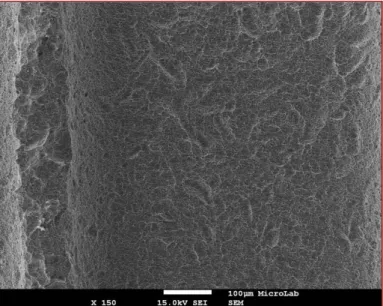
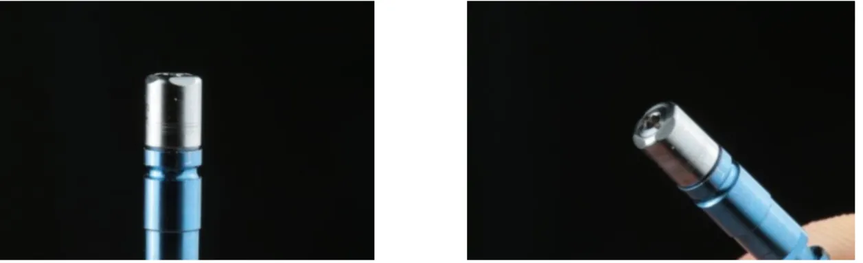
3.2.2.3. CAD-CAM Healing Abutment SEM - Scanning Electron Microscopy
Characterization and fabrication ... 71 3.2.2.4. CAD-CAM Titanium Healing Abutment ... 72 3.2.2.5. CAD-CAM Zirconia Healing Abutment ... 74 3.2.2.6. CAD-CAM Acrylic Healing Abutment ... 77 3.2.3. Sheep Anatomical Study Pre-Surgical procedure ... 79 3.2.4. Surgical procedure Description ... 82 3.2.5. Experimental animal diet, oral hygiene protocol and post-op. ... 89 3.2.6. PICF and PCF Cytokines Extraction Method at T0 ... 89 3.2.7. PICF and PCF Cytokines Extraction Method from Biological Sheep Tissues at T1 and T3 ... 92 3.2.8. Sample Transportation Methodology ... 94 3.2.9. Processing Samples and ELISA protocol ... 95 3.2.10. Elisa Essay Methodology ... 96 3.2.11. Step-By-Step Elisa Procedures (short resume) ... 97 3.2.12. Standard Preparation for Calibration Curve... 98 3.2.13. Extraction method and Reagent preparation ... 99 3.2.14. Sample reading ... 103 3.2.15. Calculation of results ... 103 3.2.16. Statistical Methodology ... 103 Section 3.3 Results of animal experimental model ... 104 3.3.1. Results of Calibration Curve for IL6 and IL-1β ... 104 3.3.1.1. For IL6 ... 104 3.3.1.2. For IL-1β ... 105 3.3.2. Interleukin IL6 and IL-1β Results ... 106 3.3.2.1. IL6 inflammatory performance results: ... 106 3.3.2.1.1. IL6 inflammatory behavior of different materials (Z, A, T) at Baseline T0 period ... 106 3.3.2.1.2. IL6 inflammatory behaviour of different materials (Z, A, T) at one month (T1) ... 108 3.3.2.1.3. IL6 inflammatory behavior of different materials (Z, A, T) at 3 months T3 .. 110 3.3.2.1.4. Statistical evaluation of the results at T0, T1 and T3 ... 112
xxvii
3.3.2.1.6. Compare Peri-implant Inflammatory Levels (IL6) with Sheep blood levels (BL) at baseline T0 ... 115 3.3.2.1.7. Compare Peri-implant Inflammatory Levels (IL6) with Sheep Periodontal Crevicular Fluid (PCV) at baseline T0 and T3 ... 115 3.3.2.1.8. PCF Basal Values (Baseline T0) compared to PICF at T0 ... 117 3.3.2.1.9. AS 3 Month Values comparing PICF with PCF ... 118 3.3.2.2. AS IL-1β Results ... 119 3.3.2.2.1. IL-1β Interleukin behavior in different materials (Z, A, T) at each time frame (Baseline T0) ... 119 3.3.2.2.2. Behavior of IL-1β in different materials (Z, A, T) at each time frame (T1 one month) ... 121 3.3.2.2.3. IL-1β behavior of different materials (Z, A, T) in each time frame (3 Month) ... 122 3.3.2.2.4. IL-1β Statistical evaluation of the Results at T0, T1 and T3 ... 124 3.3.2.2.5. IL-1β Behavior of Different materials over time (from Baseline T0 to 3 month T3) ... 125 3.3.2.2.6. Compare Peri-implant Inflammatory Levels (IL-1β) with Sheep blood levels (BL) at baseline T0 ... 127 3.3.2.2.7. Compare Peri-implant Inflammatory Levels (IL-1β) with Sheep Periodontal Crevicular Fluid (PCV) at baseline T0 ... 128 3.3.2.2.8. AS 3 Month Values of PCF vs PICF at T3 ... 129 Section 3.4 Discussion of animal experimental model ... 130 Section 3.5 Conclusion and clinical implications of animal experimental model ... 141
CHAPTER 4.RESEARCH PROJECT PART2-RANDOMIZED CLINICAL TRIAL (RCT)
STUDY ... 143 Section 4.1 Materials and Methods ... 145
Task 1: Study Outline ... 145 A - Introduction: Previous Experience with inflammation Protocols ... 145 B - Human Subject review: Ethical Committee Protocol Clearance ... 146 C - Study Outline ... 147 The RCT will follow the outline shown in table 27 to 29. ... 147 D - Study Registration as Randomized Clinical Control Trial ... 148 E - Study Design ... 148 F - Study Clinical Locations ... 149 G - Study Sample Reading Location ... 149
xxviii
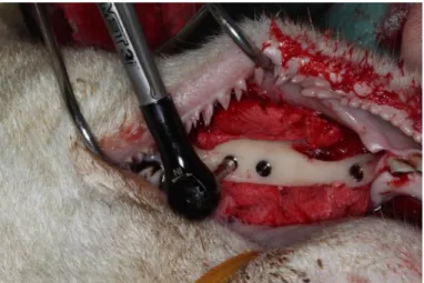
J - Patient Organization - Baseline Formation ... 151 K - Recruitment ... 151 L - Control and monitoring patient adhesion ... 151 M - Informed Consent ... 151 N - Randomization Process... 151 O - Blinding ... 153 P - Sample size calculation ... 153 Q - Patient appointment control (“Flow Chart”) ... 154 R - Drop out ratio and patient control ... 154 S - Software ... 154 T - Adverse effects related to Implantology ... 154 U - Oral Hygiene flow chart and Patient control ... 155 Task 2: Observer/investigator Calibration ... 155 A - Inter-observer agreement ... 155 B - Inter-observer agreement in radiographic Marginal Bone Loss (MBL) reading ... 155 C - Inter-observer agreement - Clinical torque the abutment ... 156 D - Inter-observer agreement - Clinical protocol in implant preparation ... 156 Task 3: Biomaterial Characterization and quality control ... 156 A - Radiographic Characterization of the Implant-Abutment complex ... 156 B - Description of the healing abutments sterilization procedure ... 157 Task 4: RCT Intervention Phase ... 158 A - Formation of 3 Groups (3 Arms) ... 158 B - Description of Surgical/prosthodontic procedure (intervention equal to all groups) ... 158 B1 - Anaesthesia ... 158 B2 - Surgical Technique measuring crest dimensions ... 159 B3 - Surgical Technique ... 159 B4 - First Surgical Phase-Implant Placement ... 159 B5 - 1st Stage Surgery: Implant Placement ... 159 B6 - Technique and types of suture ... 161 B7 - Description of medication protocol Intra / pre-and post-operative ... 161 B8 - Prosthodontic flow chart (not part of the study) ... 162
xxix
F - Extraction of Blood Samples ... 167 G - Periodontal Cytokines extraction method ... 167 H - Peri-implant Cytokine Extraction of IL-1β and IL6 at T0 (baseline) and at T2 (8weeks) ... 168 I - Post-op Instructions ... 169 J - Post-op/Follow-up ... 169 Task 5: Interleukin Sample handling and process ... 169 A - Specimens Treatment from surgery to storage ... 169 B - Interleukin (IL6 and IL-1β) Extraction Method (common to
periodontal/peri-implant/blood samples) ... 170 C - Processing Peri-implant, Periodontal and Blood IL-1β, IL6 Samples ... 170 D - Assay Principle for IL6 and IL-1β ... 172 E - Interleukin 6 (IL6) ELISA Preparation and Testing ... 173 F - IL6 Sample Dilution Guideline ... 173 G - IL6 ELISA ... 173 H - ELISA Kit Validity and Inter/intra Assay Precision for IL6 ... 173 I - Reagent Preparation and Storage - Human Standard IL6 ... 174 J - Preparation of biotinylated anti-Human IL-6 antibody working solution... 175 K - Preparation of Avidin-Biotin-Peroxidase Complex (ABC) working solution for IL6 . 175 L - Preparation of PBS washing buffer for IL6 ... 175 M - Interleukin IL-1β ELISA Preparation and Testing ... 176 N - Sample Dilution Guideline For IL-1β ... 176 O - For Interleukin IL-1β ... 176 P - Kit Validity and Inter/intra Assay Precision for IL-1β ... 176 Q - Reagent Preparation and Storage - Human Standard IL-1β ... 177 R - Other Reagent Preparation ... 177 S - Calibration curve IL6 ... 178 T - Calibration curve IL-1β ... 178 U - Calibration curve IL6 ... 179 V - Assay procedure (common for both IL6 and IL-1β) ... 179 W - Sample Reading Well Distribution ... 182 X - Software and Data analysis ... 184 Y - Database ... 185
xxx
AB - Statistical Methodology ... 188 AC - Results Inter-Observer agreement ... 189
CHAPTER5.HYPOTHESIS AND RESULTS ... 191 Section 5.1. Primary Outcome Measures: Inflammation Levels of IL6 and IL-1β in three different healing abutments Zirconia (Z) Acrylic (A) and Titanium (T) - Hypothesis and Results ... 193
Section 5.1.1. Hypothesis ... 193 Section 5.1.2. Results ... 196 Section 5.2. Primary Outcome Measures: Inflammation Levels of IL6 and IL-1β on implants compared to Inflammation Levels of IL6 and IL-1β in Periodontal Crevicular Fluid (PCF) - Hypothesis and Results ... 224
Section 5.2.1. Hypothesis ... 224 Section 5.2.2. Results ... 230 Section 5.3. Primary Outcome Measures: Inflammation Levels of IL6 and IL-1β compared to Inflammation Levels of IL6 and IL-1β in Blood Samples (BF) at Baseline (T0)- Hypothesis and Results ... 239
Section 5.3.1. Hypothesis ... 239 Section 5.3.2. Results ... 243 Section 5.4. Primary Outcome Measures: Zirconia, Acrylic and Titanium Inflammation Levels of IL6 and IL-1β and correlation to Marginal Bone Loss (MBL) - Hypothesis and Results .... 252
Section 5.4.1. Hypothesis ... 252 Section 5.4.2 Results ... 263 Section 5.5 Secondary Outcome Measures: Zirconia, Acrylic and Titanium Inflammation Levels of IL6 and IL-1β and correlation to marginal bone loss and height of existing tissue at the time of surgery (biological width height)- Hypothesis and Results ... 277
Section 5.5.1. Hypothesis ... 277 Section 5.5.2. Results ... 279 Section 5.6. Secondary Outcome Measures: Zirconia, Acrylic and Titanium Inflammation Levels of IL6 and IL-1β and correlation to marginal bone loss and Age - Hypothesis and Results ... 288
Section 5.6.1. Hypothesis ... 288 Section 5.6.2. Results ... 290 Section 5.7. Secondary Outcome Measures: Zirconia, Acrylic and Titanium Inflammation Levels of IL6 and IL-1β and correlation to marginal bone loss and Gender- Hypothesis and Results ... 299
xxxi
Section 5.8. Secondary Outcome Measures: Zirconia, Acrylic and Titanium Inflammation Levels of IL6 and I IL-1β and correlation to Marginal Bone Loss (MBL) and Anatomical
Position (Maxilla vs Mandible)- Hypothesis and Result ... 311 Section 5.8.1. Hypothesis ... 311 Section 5.8.2. Results ... 313 Section 5.9. Secondary Outcome Measures: Zirconia, Acrylic and Titanium Inflammation Levels of IL6 and IL-1β and correlation to Marginal Bone Loss (MBL) and the duration of surgery - Hypothesis and Results ... 322
Section 5.9.1. Hypothesis ... 322 Section 5.9.2. Results ... 324 Section 5.10. Secondary Outcome Measures: Zirconia, Acrylic and Titanium Inflammation Levels of IL6 and IL-1β and correlation to marginal bone loss implant stability (ISQ)-
Hypothesis and Results ... 334 Section 5.10.1. Hypothesis ... 334 Section 5.10.2. Results ... 339
CHAPTER 6.DISCUSSION... 352 6.1. Section 5.1, 5.3, 5.4 Discussion of the impact of different biomaterials on the overall inflammatory response to dental implants (PCF, PICF and BF measure) ... 354 6.2. Inflammatory levels in PICF compared to PCF ... 367 6.3. Section 5.5 Marginal Bone Loss (MBL) and inflammatory parameters Discussion ... 371 6.4. Section 2.6 Discussion on biological width formation and correlation with marginal bone loss and inflammatory levels ... 381 6.5. Section 5.7, 5.8, 5.9 Secondary Outcome Measures Discussion on the impact of Age, Gender and anatomical position on MBL and Inflammation ... 385 6.6. Section 5.10 Discussion of the Time of surgery, inflammatory levels and MBL. ... 387 6.7. Section 2.11 Discussion of Primary stability, inflammatory levels and MBL. ... 387
CHAPTER7.FINAL REMARKS AND CONCLUSION ... 392
CHAPTER8.REFERENCES ... 400
APPENDIXA.LICENSEFORTHEANIMALEXPERIMENT ... 428 APPENDIXB.ETHICALCOMITTEPERMISSION AND INFORMEDCONSENT
... 430 APPENDIXC.RCTREGISTERSHEET ... 435 APPENDIXD.LISTOFFIGURES ... 442 APPENDIXE.LISTOFTABLES ... 451
xxxii % Percentage # - Number SD - Standard deviation vs. - Versus T - Titanium Z - Zirconia
CAD-CAM - Computer aid design Computer aid manufactured A - Acrylic IL-1β - interleukin 1β IL6 - Interleukin 6 IL - Interleukin mm - Millimeters pg - Picogram ml-Milliliter ul-Microliter I - Implant min - Minutes
PCF - Periodontal Crevicular fluid PICF - Peri-implant crevicular fluid BF - Blood fluid levels
T0 – Baseline (both animal and RCT study) T1 - 1 month (animal study only)
T2 - 2 month (RCT study only) T3 - 3 Month (animal study only) RCT – Randomized clinical control MB - Marginal bone
MBL- Marginal bone loss
MBL1 - Marginal bone loss considering all implants
MBL2 - Marginal bone loss considering only the implants that had exposed surface
xxxiii ISQ - Implant stability quotient
AS - Animal study
IST-Instituto Superior Técnico
FMDUL- Faculdade de Medicina Dentária da Universidade de Lisboa ABC - Avidin-Biotin-Peroxidase Complex
BW- Biological width
R - Different time frames on sample readings PBS - Buffer Solution
HRP-Conjugate
TMB – Termination solution [ ] – Concentration
n - Number of cases
p - Statistical value of significance PhD - Doctor of Philosophy
OD- Optical diffraction H - Higher L- Lower S - Same T - Time M - Mesial Ma - Material D - Distal Ave - Average ML - Male FM - Female Psb - Primary stability SS - Secondary stability PS - Platform switching
3
1.1. HISTORICAL PERSPECTIVE
PI Brånemark’s 25 years of osseointegration research in 1977(Brånemark et al. 1977) marked a revolution in dentistry, defining two separate eras of implant dentistry. (Adell et al. 1981a).
Before Brånemark, implant dentistry, had been characterized by clinicians using metal devices for fixed anchorage, with limited knowledge and investigation to support the use of dental implants.
The development of these types of devices for edentulous patients was taking place mainly in Europe and North America. Leonard Linkow from North America (Brinks, Kuyl, and Zeegers 1988) created several devices for bone anchorage to solve the retention problems of fully edentulous patients. His first work on implants was published in 1954, and was followed by 24 others, before the paper in which he presented his blades. (L. Linkow 1966)
Linkow was the first to produce a subperiosteal implant (L. I. Linkow 1967), a device that was placed between bone and periosteum to allow for the support of dentures and multiple elements.
Linkow’s original subperiosteal implant was tailored to different shapes according to the anatomical site of insertion. (L. I. Linkow 1986)
The result was fair but this kind of approach had both poor and low survival rates which were highly prone to infection (L. I. Linkow and Ghalili 1998; L. I. Linkow 1967) and which didn’t meet the full criteria for a fixed long term stability oral rehabilitation.
Motivated by the low survival rate of the subperiosteal implants, endosseous implants were making headway and later Linkow blades were created and published in 1966 (L. I. Linkow 1966), making it possible to treat partial or total edentulism.
The technique consisted of opening an access in bone to insert a titanium blade for single, partial or full mouth rehabilitation. This invention helped pave the way for the innovations in implant dentistry of the 1960s.
4
blade implant, which could be modelled to meet the most common anatomical configurations and which, with its screw abutment, offered a solution to the problem of tongue-thrust during swallowing (causing the majority of post-surgical failures) for the first time.
Tramonte in Italy and Cherchève (Linkow co-wrote a two-volume work with the Frenchman Cherchève: Theories and Techniques of Oral Implantology) (Cherchève 1966) were also inventing screw-like implants that were inserted in bone to allow for fixed, stable restoration. (Passi et al. 2017)
Tramonte (S. Tramonte 1965) used screws, casted in chrome-cobalt, making them with a thinner profile and honing their threads to make them sharper. These were machined dental implants with a higher survival rate than the subperiosteal or the blade implants of Linkow. (S. M. Tramonte 1989)
This period of research was characterized by strong personal experience, unpredictability of results and low scientific support.
In the 1960s and 1970s, implant-supported prostheses based on subperiosteal or blade implants had a poor reputation, mainly because of questionable clinical outcomes and lack of scientific documentation, encouraging the transformation into a scientifically sound discipline initiated by the two scientific pioneers of modern implant dentistry, Professor P. I. Brånemark from the University of Gothenburg in Sweden and Professor André Schroeder from the University of Bern in Switzerland. Together with their teams, and independently of each other, they laid the foundation for the most significant development and paradigm shift in dental medicine.
P.I. Brånemark was an experienced orthopedist studying bone physiology with titanium microcameras in the bone of several dogs when he found that the cameras had remained osseointegrated.(Brånemark et al. 1977)
Focussing on this phenomena Brånemark and his team decided to lay down the foundations of the first comprehensive study of the integration of titanium to bone and its application in oral rehabilitation. (T Albrektsson et al. 1981a; Adell et al. 1981b; Brånemark et al. 1983)
5
publication on osseointegration of dental implants in 1977 (Brånemark et al. 1977) opening the way to a new era.
Originally direct bone-to-implant contact (i.e. osseointegration) was referred to as direct bone deposition on the implant surface without interposition of fibrous or connective tissue (Brånemark et al. 1977), a term also called “functional ankylosis” (Schroeder et al. 1981) in contrast to the idea of Fibrointegration that was the accepted methodology at that time. (L. I. Linkow and Rinaldi 1987)
1.2. MODERN IMPLANTOLOGY
What came to be known as the post- Brånemark era, was based on a biological approach, the foundations of which can be found in the experimental methods reported by P.I. Brånemark who defined osseointegration as “the formation of a direct interface between an implant and bone, without intervening soft tissue”. (Brånemark 1983a)
One of the first definitions of survival and success was formulated by Albrektsson who stated that, for an implant to be successful over the years, it had to be free of infection, mobility and with marginal bone resorption of no more than 1,5 mm in the first year and 0,1mm in the following years. (T Albrektsson et al. 1981b)
In 1982 the Toronto Osseointegration Conference in Clinical Dentistry was held, introducing and validating the concept of osseointegration. The Toronto conference would provide a springboard for dental implantology out of an era of unpredictable and often short-lived treatment outcomes based upon limited research, to an evidence-based and predictable procedure, providing long-term replacement of failing and missing teeth. (Norkin 2012).
Implant dentistry was mainly being performed in Europe to treat fully edentulous patients, but with the Toronto conference, Professor George A. Zarb of the Faculty of Dentistry in Toronto introduced the concept of osseointegration, along with its application in treating edentulous patients in North America. (Zarb and Symington 1983).
6
first application for commercial use of dental implants in the United States and in 1986 the American Academy of Implant Dentistry, which fostered the advancement of the field, was created.
1.3. HARD TISSUE INTEGRATION EVENTS
For osseointegration to occur when a dental implant is inserted a number of physiological and biochemical events take place.
Osseointegration is considered a healing process of bone in reaction to an alloplastic material in a foreign body reaction type. (van Steenberghe 1988; Brånemark et al. 1983)
Titanium is usually the material of choice but zirconia (Pieralli et al. 2017) and gold (Ingemar Abrahamsson and Cardaropoli 2007) have also proven osseointegration qualities.
Just a few minutes following implant installation, a blood clot is formed around the passivated titanium shell, produced by air contact from the implant surface to oxygen (passivation layer). (John E Davies 2003)
In rough surface implants (implants that undergo surface treatment after milling) the phenomena is called “contact osteogenesis” (J E Davies 2017), meaning that the first response of the clot is to adhere to the implant wall, in contrast to “distant osteogenesis” a characteristic of the “machined implants”. (the clot contracts first, and only a few hours later the implant wall is found).
After a blood clot is formed in the first 48 hours, inflammatory cells and biochemically induced proteins are released into the environment.
The auto immune response starts to release unspecific components to the area, mainly in the form of macrophages, neutrophils and lymphocytes.
Macrophages have a key function in wound healing and presumably also in bone regeneration, since the regulatory release of interleukins and other cell mediators are first released by these molecules.
After the first week, granulation tissue is formed and numerous blood capillaries that are embedded in the loose connective tissue are produced by fibroblasts.
7
Woven bone is a rapidly (approximately 10 μm/day) growing mineralized tissue characterized by means of haphazardly oriented collagen fibers and many large osteocyte lacunae.
Lamellar bone forms at approximately 1-2 μm/day, also on the surface of previously formed woven bone. (Shah et al. 2014; Brånemark 1983b)
Bone remodeling, which is a synonym for bone turnover, causes a mineralized structure of exclusively lamellar bone, thus creating a sound and hard tissue around the titanium surface.
1.4. OSTEOGENESIS
Skeletal basal bone formation may have two different origins: the direct conversion of mesenchymal tissue into bone, is called intramembranous ossification and the process by which a cartilage intermediate is formed and replaced by bone cells, is called endochondral (they are formed through apposition or as part of an endochondral matrix). (Dirckx, Van Hul, and Maes 2013)
The upper maxilla has an endochondral formation, on the first branchial arch. Endochondral ossification involves the formation of cartilage tissue from aggregated mesenchymal cells, and the subsequent replacement of cartilage tissue by bone.
The mandible has an intramembranous aetiology. Intramembranous ossification is the characteristic way in which the flat bones of the skull mineralize. (Ornitz and Marie 2015)
1.5. TOOTH FORMATION AND PERIODONTAL BONE FORMATION
The basic steps of tooth morphogenesis were well described over 100 years ago and are basically similar in all vertebrates.
The establishment of the dental lamina, the area that forms teeth, precedes the initiation of individual teeth. Teeth become visible during the following stages of
8
development, called the bud and the cap stages, with the appearance of the initial epithelial invagination and the tooth crown area, respectively. The cap stage is followed by the bell stage, during which species-specific cusp patterns emerge. (Soukup et al. 2008)
After the formation of the cusp pattern, the tooth grows to its final size, and mesenchymal odontoblasts and epithelial ameloblasts differentiate at the epithelial-mesenchymal interface to form dentin and enamel, respectively. These hard-dental tissues, together with cementum, which is made by cementoblasts, have largely similar compositions in all vertebrates, with enamel comprising up to 98% hydroxyapatite.
Periodontal bone (alveolar crest/bundle bone/lamina dura) came from the ectomesenquimal cells that form the dental papilla, precursor of the tooth germen and the periodontal tissues. (Jernvall and Thesleff 2012)
The formation of periodontal bone is different from the formation of endochondral or intramembranous bone. In terms of the formation of the Hertzwig sheath, there is a biochemical and molecular signalling interaction, inducing the creation of a row of osteoblasts and cementoblasts that create structures capable of achieving a true adhesion by means of the Sharpey fibbers. Thus, on the side of the tooth we have cement and on the side of the bone, a cortical structure called the bone crest, lamina dura, alveolar or bundle bone.
The interaction of these tissues is of utmost importance since the regulation in health or in pathology is made by means of several biochemical signs.
Interleukins are of critical importance to the cortical remodelling of this periodontal bone. Released by inflammatory mechanisms through macrophages and neutrophils IL-1β and IL6, they are able to determine the loss of insertion and bone remodelling, epithelial transformation factors such as IGF and FTβ released by fibroblasts which also serve as regulatory mechanisms. The importance of this tooth-alveolar crest interaction is so critical that the extraction of teeth induces a remodelling pattern that eventually leads to bone
9
resorption and deformity of the area. (Araujo and Lindhe 2005).
1.6. OSTEOBLAST
The skeletal structure is constantly renewed by the balance osteoblast (responsible for laying down bone) and osteoclast (responsible for bone resorption).
The osteoblasts secrete a collagen-proteoglycan matrix that can bind calcium salts. Through this binding, the prebone (osteoid) matrix becomes calcified. In most cases, osteoblasts are separated from the region of calcification by a layer of the osteoid matrix they secrete. Occasionally, though, osteoblasts become trapped in the calcified matrix and become osteocytes—bone cells. As calcification proceeds, bony spicules radiate out from the region where ossification began. (Dirckx, Van Hul, and Maes 2013).
1.7. OSTEOCLAST
Destruction of bone tissue is due to osteoclasts, multinucleated cells that enter the bone through the blood vessels (Kahn and Simmons 1975; Manolagas and Jilka 1995). Osteoclasts are probably derived from the same precursors as macrophage blood cells, and they dissolve both the inorganic and the protein portions of the bone matrix (Blair et al. 1986). Each osteoclast extends numerous cellular processes into the matrix and pumps out hydrogen ions onto the surrounding material, thereby acidifying and solubilizing it.
In osteoclast differentiation, hormones regulate production and the hormonal changes of aging and may cause osteoporosis by increasing the number of osteoclasts. The conversion of a macrophage stem cell into an osteoclast is regulated by osteoprotegerin and its ligand. It is through these,s that signals on osteoblasts instruct the progenitor cell to become an osteoclast. (Boyce 2013). Osteoclasts resorb bone and are responsible for numerous pathologies in relation to bone tissue, but also play a key role and factors in a number of bone metastases. (Ishii and Kikuta 2013).
10
They are a key target for oncology drugs such as bisphosphonates and other antiresorptive drugs. (Pelaz et al. 2015)
Osteoclasts are mainly responsible for marginal bone loss on dental implants and are key to any discussion of long term survival rate and dental implant complications. (Bang et al. 2014)
A number of implant researchers are including bisphosphonate in implant surfaces to try to decrease bone resorption around dental implants and thus secure a more stable complex. (Pyo et al. 2017)
Several investigations have tried to control the role of the interleukins on osteoclast activation on a biochemical level. We know that interleukin 1β and interleukin 6 in particular are potent chemical activators of these cell lineages. (Farhat et al. 2017a)
1.8. BONE BIOLOGY AND PHENOTYPES
Lekholm and Zarb maintain the classification system of bone as follows: Bone quality has been classified in four categories based on radiographic appearance and the resistance to drilling: Type 1 bone, in which almost the entire bone is composed of homogenous compact bone; Type 2 bone, in which a thick layer of compact bone surrounds a core of dense trabecular bone; Type 3 bone, in which a thin layer of cortical bone surrounds a core of dense trabecular bone; and Type 4 bone characterized as a thin layer of cortical bone surrounding a core of low density trabecular bone of poor strength. (Brånemark et al. 1983)
These differences in bone quality can be associated with different areas of anatomy in the upper and lower jaw. Mandibles are generally more densely corticated than maxillae and both jaws tend to show a decrease in their cortical thickness and an increase in their trabecular porosity as they move posteriorly. It has been shown, although there are have been counter studies, that there is a decrease in success rates as the bone type increases. (Cobo-Vazquez et al. 2017)
11
There have been a range of statistics on implant survival that have been reported according to bone quality, from a 2% difference from type 1 (98% in 36 months) to type 4 (96% in 36 months) to a 14% difference in another group (90% type 1 vs. 76% type 4 in 36 months). (Marquezan et al. 2012)
These are important statistics, as this indicates that bone quality is significant when considering an implant placement site, and secondly there appear to be other factors in the success rates of implants as one considers the vast discrepancy between the results. (Shadid, Sadaqah, and Othman 2014)
1.9. CLINICAL FACTORS IN IMPLANT DENTISTRY
1.9.1. Primary Stability
Osseointegration or secondary stability is the ability of an implant to be integrated into the surrounding bone structures. Secondary stability offers biological stability through bone regeneration and remodeling.
Primary stability mostly occurs from mechanical attachment with cortical bone and is affected by bone quality and quantity, surgical technique and implant geometry (length, diameter, surface characteristics). (Turkyilmaz and McGlumphy 2008)
Secondary stability is affected in part by primary stability but not entirely, since osseointegration is a multifactorial equation where stability is only one factor. (Esposito et al. 2013)
1.9.2. Stability Measurements Invasive/destructive methods
The following methods are reported in the literature (the most important ones): Histologic/histomorphologic analysis
Tensional test
Push-out/pull-out test and Removal torque analysis.
12 Histomorphometric analysis
This is obtained by calculating the peri-implant bone quantity and bone-implant contact (BIC) from a dyed specimen of the implant and peri-implant bone.
Tensional test
The Tensional test is measured by detaching the implant plate from the supporting bone. It was later modified by Bränemark by applying the lateral load to the implant fixture.
Push-out/pull-out test
Push-out/pull-out test investigates the healing capabilities at the bone implant interface. It measures interfacial shear strength by applying load parallel to the implant-bone interface. The typical push-out or pull-out test checks tensile or compressive stresses. (Brunski et al. 2000, Chang et al. 2010).
There are Noninvasive/nondestructive methods for assessing implant stability including:
Surgeon’s point of view
Radiographical analysis/imaging techniques Insertion torque measurement
Reverse torque Seating torque test Percussion test Periotest
Resonance frequency analysis (RFA): Electronic technology Magnetic technology.