Carborane-Based Design of
Active Ligands of Nuclear
Receptors for BNCT
Beatriz Brito
Mestrado em Bioquímica
Departamento de Química e Bioquímica 2018
Orientadores
Antonio Mouriño Mosquera, Facultad de Química, USC
Jose Enrique Rodriguez Borges, FCUP
Coorientadora
Todas as correções determinadas pelo júri, e só essas, foram efetuadas. O Presidente do júri,
Acknowledgments
Foremost, I would like to express my sincere gratitude to my advisors Professor Dr. Antonio Mouriño and Professor Dr. Enrique Borges for introducing me to the field of Organic Chemistry research and for giving me the opportunity to integrate this research group. Their guidance and teachings were invaluable.
I would like to thank Dr. Rita Sigüeiro for guiding me, often with big doses of patience, through this journey. For all her teachings and suggestions.
I am grateful to Professor Dr. Miguel Maestro and all my laboratory colleagues for the support, teachings and good environment provided. Special thanks to Patri and Xiao for their tireless support, infinite encouraging words and for the immensurable fun times we had together.
I want to thank the University of Porto for giving me the opportunity to broaden my horizons through the Erasmus+ program. I also want to gratefully acknowledge the University of Santiago de Compostela for hosting me during this year.
Special thanks to my friends, Alfredo, Ana Beatriz, Bárbara, Gabriel, Gabriela, João, Rita, Ricardo and Tiago, for always keeping things lighthearted, if a little strange. And to Hélio, for always pushing me forward and never letting me drift astray.
Finally, I would like to express my deepest appreciation to my family, specially my mother, sister and grandmother, for supporting me unconditionally by means that words cannot explain.
FCUP | ICBAS | USC
Abstract
v
Abstract
Icosahedral carboranes are highly stable boron-carbon clusters that have been used as unique pharmacophores in biological active compounds. Carboranes have promising applications in Boron Neutron Capture Therapy (BNCT), a technique based on the nuclear capture of neutrons by 10B to produce high energy particles that selectively destroy tumors without affecting healthy tissues. The application of BNCT depends on the availability and selectivity of boron compounds which bind receptors in the tumors. The vitamin D nuclear receptor (VDR) is a potential target for cancer therapy, because it is expressed in many tumors and its ligand, 1α,25-dihydroxyvitamin D3 (1,25D or
calcitriol), can induce anticancer actions. To combine these properties with BNCT application, new VDR agonists based on the skeleton of the hormone 1,25D that contained ortho, meta and para-carboranes at the end of its side chain were designed. Docking studies revealed several promising carborane-containing calcitriol analogs. Three analogs were chosen as synthetic targets. The synthetic approach features a Pd-catalyzed construction of the triene system and an SN2 displacement of a tosylate
group by a carboranyllithium reagent to afford three carborane-containing vitamin D analogs.
Key words: Carboranes, vitamin D, 1,25D, BNCT, analogs, docking, organic
vi FCUP | USC
Abstract
Resumo
Carboranos icosaédricos são complexos de boro-carbono altamente estáveis que têm sido usados como farmacóforos únicos em compostos biologicamente ativos. Os carboranos têm aplicações promissoras na Terapia de Captura de Neutrões pelo Boro (BNCT), uma técnica baseada na captura nuclear de neutrões pelo 10B para produção de partículas de alta energia que destroem selectivamente tumores sem afetar tecidos saudáveis. A aplicação da BNCT depende da disponibilidade e selectividade dos compostos de boro que ligam a recetores tumorais. O recetor nuclear da vitamina D (VDR) é um potencial alvo para a terapia cancerígena, porque é expresso em vários tumores e o seu ligando, 1α,25-dihidroxivitamina D3 (1,25D ou calcitriol), é capaz de
induzir ações anticancerígenas. De modo a combinar estas propriedades com a aplicação da BNCT, foram desenhados novos agonistas do VDR cuja estrutura se baseia no esqueleto da hormona 1,25D que contivessem ortho, meta e
para-carboranos no final da cadeia lateral. Estudos de docking revelaram promissores
análogos do calcitrol com carboranos na cadeia lateral. Três compostos foram escolhidos como objetivo sintético. A abordagem sintética engloba a construção do sistema triénico catalisada por paládio e a substituição de um grupo tosilato por um reagente carboranilítio para se obter três análogos da vitamina D com unidades carborânicas diferentes.
Palavras-chave:Carboranos, vitamina D, 1,25D, BNCT, análogos, docking, síntese,
FCUP | ICBAS | USC Index vii
Index
ACKNOWLEDGMENTS ... iii ABSTRACT ... v RESUMO ... vi INDEX ... vii SCHEME INDEX ... ix FIGURE INDEX ... xi ABBREVIATIONS ... xiii INTRODUCTION ... 1 1. Vitamin D3 ... 31.1. Structure and nomenclature of vitamin D ... 3
1.2. Biotransformation of vitamin D ... 5
2. Mechanism of action of calcitriol ... 6
2.1. Genomic actions of calcitriol ... 6
2.2. Non-genomic actions of calcitriol ... 10
3. Biological activity of calcitriol ... 11
3.1. Calcium and Phosphate Homeostasis ... 11
3.2. Cellular Differentiation and Proliferation ... 12
3.3. Immune System Regulation ... 13
3.4. Transcaltachia ... 13 3.5. Pharmacological Endeavours ... 13 4. Carboranes ... 14 4.1. Structure of carboranes ... 15 4.2. Electronic Properties ... 16 4.3. Hidrophobicity ... 16 4.4. Acidity ... 17 4.5. Molecular interactions ... 17 4.6. Chemistry ... 18
4.7. Boron Neutron Capture Therapy (BNCT) ... 18
5. Background ... 20
OBJECTIVES ... 23
RESULTS AND DISCUSSION ... 27
1. Design and Docking of carborane-containing vitamin D analogs ... 29
viii FCUP | ICBAS | USC
Index
1.1. Design of carborane-containing vitamin D analogs ... 29
1.2. Building of the proposed carborane-containing vitamin D analogs ... 30
1.3. Docking of the proposed carborane-containing vitamin D analogs ... 30
1.4. Docking Results ... 30
1.5. Choice of target analogs for synthesis ... 36
2. Synthesis of Carborane Analogs A1, A17 and A33 ... 39
2.1. Retrosynthesis ... 39
2.2. Synthesis of benzoate 6 ... 40
2.3. Synthesis of boronic ester 2 ... 41
2.4. Synthesis of enol-triflate 3 ... 43
2.5. Synthesis of tosylate 1 ... 44
2.6. Synthesis of analogs A1, A17 and A33 ... 46
CONCLUSIONS ... 53 EXPERIMENTAL SECTION ... 56 1. Docking calculations ... 57 2. General procedures ... 59 REFERENCES ... 71 APPENDIX ... 77 1. DOCKING RESULTS ... 79 1.1. Ortho series ... 81 1.2. Meta series ... 87 1.3. Para series ... 93 2. NMR Spectra ... 99 3. IR Spectra ... 115 4. Structure Index ... 121
FCUP | ICBAS | USC
Scheme Index
ix
Scheme Index
Scheme 1. Biosynthesis of vitamin D3 in the skin ... 5
Scheme 2. Biological activation of vitamin D3 ... 5
Scheme 3. Catabolism of calcitriol ... 6
Scheme 4. Mechanism of genomic actions of calcitriol ... 9
Scheme 5. Mechanism for the non-genomic actions of calcitriol ... 10
Scheme 6. Schematic illustration of the effects of calcitriol on the calcium and phosphate homeostasis ... 12
Scheme 7. Synthesis of ortho, meta and para-carboranes ... 18
Scheme 8. Synthesis of target carborane RO. Reagents and conditions. ... 21
Scheme 9. Retrosynthetic analysis planned for the synthesis of compounds A1, A17 and A33. P = protecting group ... 39
Scheme 10. Synthesis of benzoate 6 by metalation of diol 4 ... 40
Scheme 11. Selective synthesis of benzoate 6... 40
Scheme 12. Synthesis of boronic ester 2 ... 41
Scheme 13. Proposed mechanism for the Miyaura borylation reaction in the synthesis of boronic ester 2 ... 42
Scheme 14. Synthesis of enol triflate 3 ... 43
Scheme 15. Synthesis of benzoate 13 via carbociclization and Suzuki-Miyaura cross-coupling of 2 and 3 ... 44
Scheme 16. Proposed mechanism of action of the Suzuki Miyaura cross-coupling to afford 13 ... 44
Scheme 17. Synthesis of tosylate 1 ... 45
Scheme 18. Synthesis of analog A1 ... 46
Scheme 19. Synthesis of analog A17 ... 48
FCUP | ICBAS | USC
Structure Index
xi
Figure Index
Fig. 1. Structure of vitamin D3 and calcitriol ... 3
Fig. 2. Structure of calcitriol and other steroid hormones ... 3
Fig. 3. Conformational flexibility of the triene system, side chain and the A ring of calcitriol... 4
Fig. 4. Schematic representation of the structure of human VDR ... 7
Fig. 5. A) Crystal structure of the 1,25D-LBD-VDR complex; B) Representation of polar interaction in the ligand pocket of VDR (PDB code: 1DB1) ... 8
Fig. 6. Representation of non-polar interaction between the residues of LBD-VDR and calcitriol (PDB code:1DB1) ... 8
Fig. 7. Commercially available calcitriol analogs ... 14
Fig. 8. Commercially available boron-containing drug Velcade ... 15
Fig. 9. Representation of the three closo-carborane isomers ... 15
Fig. 10. Representation of an o-carborane, adamantane and benzene ... 16
Fig. 11. Two-dimensional representation of intermolecular interactions of o-carborane ... 17
Fig. 12. Selective neutron capture of neutrons by 10B in a tissue47 ... 19
Fig. 13. A) Representation of analog RO. B) Structure of the binding pocket of zVDR LBD bound to analog RO. ... 20
Fig. 14. Target compounds ... 25
Fig. 15. Representation of carborane-containing analogs to analyze by docking in human VDR ... 29
Fig. 16. Conformation of the most promising docked analogs ... 31
Fig. 17. 1H-NMR and 13C-NMR main signals for compound 6 ... 40
Fig. 18. Main 1H-NMR and 13C-NMR signals for boronic ester 2 ... 42
Fig. 19. 1H-NMR and 13C-NMR signals of enol-triflate 3 ... 43
Fig. 20. Main 1H-NMR and 13C-NMR signals of tosylate 1 ... 45
Fig. 21. 1H-NMR spectrum of analog A1 ... 47
Fig. 22. 11B-NMR spectrum of analog A1 ... 47
Fig. 23. 1H-NMR spectrum of analog A17 ... 48
Fig. 24. 11B-NMR spectrum of analog A17 ... 49
Fig. 25. 1H-NMR spectrum of analog A33 ... 50
FCUP | ICBAS | USC
Abbreviations
xiii
Abbreviations
1,25D 1α,25-Dihidroxyvitamin D3 or calcitriol
1,25D-MARRS 1,25D membrane associated rapid response steroid binding
11B-NMRac Coupled Boron-11 Nuclear Magnetic Resonance
11B-NMRdec Decoupled Boron-11 Nuclear Magnetic Resonance
13C-NMR Carbon-13 Nuclear Magnetic Resonance
1H-NMR Proton Nuclear Magnetic Resonance
25(OH)D3 25-Hidroxyvitamin D3
7-DHC 7-Dehydrocholesterol
AF-2 Activation function 2
APCI Atmospheric-pressure chemical ionization
APCs Antigen presenting cells
aq aqueous
Arg Arginine
B2pin2 Bis(pinacolato)diboron
BNCT Boron Neutron Capture Therapy
Cav-1 Caveolin-1
CDKIs Cyclin-dependent kinase inhibitors
CoA Coactivators
c-Src Non-receptor tyrosine kinase
CYP24A1 Cytochrome P450 family 24 subfamily A member 1
CYP27B1 Cytochrome P450 family 27 subfamily B member 1
CYP2R1 Cytochrome P450 Family 2 Subfamily R Member 1
d doublet
DBD DNA binding domain
xiv FCUP | ICBAS | USC
Abbreviations
DCs Dendritic cells
dd Doublet of doublet
ddd Doublet of doublet of doublets
ddt Doublet of Doublet of Triplets
DEPT Distortionless Enhancement by Polarization Transfer
DIBAL-H Diisobutylaluminium hydride
DMAP 4-Dimethylaminopyridine
DMP Dess-Martin periodinane
DNA Deoxyribonucleic acid
dt Doublet of triplets
EtOAc Ethyl acetate
ESI Electrospray ionization
FGF23 Fibroblast growth factor 23
GA Genetic algorithm
GMB Glioblastoma multiform
H12 Helix 12
Hex Hexane
His Histidine
HPLC High-performance liquid chromatography
HPT Hyperparathyroidism
HRMS High-Resolution Mass Spectrometry
Hz Hertz
J Coupling constant
LDA Lithium diisopropylamide
LET Linear energy transfer
Leu Leucine
m multiplet
FCUP | ICBAS | USC
Abbreviations
xv
MHz Mega Hertz
mRNA Messenger Ribonucleic acid
MS Mass Spectrometry
MTBE Methyl tert-butyl ether
n
BuLi n-Butyllithium
nm Nanometers
PCy3 Tricyclohexylphosphine
Pdia3 Protein Disulfide Isomerase Family A Member 3
Ph Phenyl
Phe Phenylalanine
ppm Parts per million
PTH Parathyroid hormone Py Pyridine q quartet Rf Retention Factor rt Room temperature RXR Retinoid X receptor s singlet Ser Serine
SN2 Bimolecular nucleophilic substitution
t triplet td Triplet of doublets THF Tetrahydrofuran TLC Thin-Layer Chromatography TOF Time-of-flight Trp Tryptophan
UVB Ultraviolet light B
xvi FCUP | ICBAS | USC
Abbreviations
VDR Vitamin D receptor
FCUP | ICBAS | USC
Introduction
3
Introduction
1. VITAMIN D
3Vitamin D3, through its hormonal form, the 1α,25-dihydroxyvitamin D3 (calcitriol, 1,25D,
Fig. 1) is essential for the maintenance of health. Indeed, calcitriol influences a large range of physiological processes, including calcium and phosphate metabolism, bone maintenance, cellular proliferation, differentiation, apoptosis and immune regulation.1
Fig. 1. Structure of vitamin D3 and calcitriol
Calcitriol plays an important role in several pathologies, like psoriasis, multiple sclerosis, rheumatoid arthritis, diabetes and cancer,which has increased the interest in the development of vitamin-D based therapies.2
1.1. Structure and nomenclature of vitamin D
The structure and mode of action of calcitriol are closely related to that of classical steroid hormones, like cortisol, testosterone and progesterone (Fig. 2).
4 FCUP | ICBAS | USC
Introduction
The structure of steroids has a common cyclopentaneperhydrophenanthrene skeleton composed of four rings (A, B, C and D). The cleavage of one of these rings is designed by the term “seco”. The vitamin D is classified as a secosteroid because its skeleton derives from the photochemical cleavage of the C9-C10 bond of the B ring of a steroid precursor.
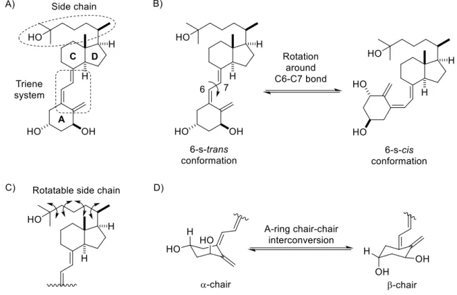
The vitamin D and its metabolites are named and numbered according to their steroid precursor. They have more conformational flexibility than other steroid hormones, due to the presence of:
1) a triene system that can rotate around the C6-C7 bond, to afford two planar conformations: 6-s-cis and 6-s-trans (Fig. 3B);
2) a rotatable side chain with 5 single C-C bonds, that can freely rotate 360 º (Fig. 3C); 3) an A-ring that can undergo a chair-chair interconversion, which changes the orientation of the 1α and 3β hydroxyl groups between the equatorial and axial orientation (Fig. 3D).3
Fig. 3. Conformational flexibility of the triene system, side chain and the A ring of calcitriol
FCUP | ICBAS | USC
Introduction
5
1.2. Biotransformation of vitamin D
The predominant form of vitamin D in humans is vitamin D3 , which can be obtained
through diet, dietary supplements or by endogenous production from 7-dehydrocholesterol (7-DHC, Scheme 1).4
The biosynthesis of vitamin D3 is initiated in the skin by the photochemical ring opening
of 7-DHC under UVB radiation (Scheme 1). The resulting previtamin D3 undergoes a
sigmatropic rearrangement [1,7]-H to afford the vitamin D3.4-5
Scheme 1. Biosynthesis of vitamin D3 in the skin
Vitamin D3 goes through two enzymatic transformations to give the active form,
calcitriol. These two hydroxylations occur first in the liver and then in the kidneys or extra-renal sites (Scheme 2).
Scheme 2. Biological activation of vitamin D3
The first hydroxylation is predominantly catalyzed by vitamin D-25-hydroxylase CYP2R1.2 The resulting 25-hydroxyvitamin D3, 25(OH)D3 is the main circulating form of
the vitamin in the serum. The generated 25(OH)D3 associates with vitamin D binding
protein (DBP),6 to reach the kidneys or extra-renal sites.2
A cytochrome P450 enzyme, CYP27B1, is responsible for the second hydroxylation, in the 1α position, to produce the hormonal form of vitamin D, calcitriol.7
6 FCUP | ICBAS | USC
Introduction
In extra-renal tissues, locally synthetized 1,25D works in an autocrine or paracrine manner, while calcitriol synthetized in the kidneys performs its functions in an endocrine manner, via association with DBP in the serum.
Calcitriol also undergoes metabolic degradation, mainly through an inactivating 24-hydroxylation, which is catalyzed by another cytochrome P450 enzyme, CYP24A1 (Scheme 3).2
Scheme 3. Catabolism of calcitriol
This enzyme catalyzes three consecutive oxidations of calcitriol at C-24 and C-23, leading to the formation of more water-soluble and less active metabolites. This ends with the production of the inactive calcitroic acid, which can easily be eliminated through urine.5
2. MECHANISM OF ACTION OF CALCITRIOL
Vitamin D, in consonance with other nuclear steroids, induces both genomic and non-genomic actions. The slow classical genomic functions of vitamin D occur via transcription activation triggered by nuclear vitamin D receptor (VDR). The fast non-genomic actions of the hormone involve second messengers generated by membrane-initiated signaling pathways.
2.1. Genomic actions of calcitriol
The genomic actions of calcitriol mediate hormone secretion, calcium and phosphate homeostasis and even regulate cellular proliferation, differentiation and apoptosis. These actions involve binding of calcitriol to the vitamin D receptor (VDR). Since the discovery of this nuclear receptor, in 1974,8 a lot of explorative studies were undertaken to collect information on the VDR’s distribution, structure and mechanism of action.
FCUP | ICBAS | USC
Introduction
7
The VDR (427 amino acids, 48 kDa) is a member of the superfamily of ligand-activated nuclear receptors that include receptors for steroid, retinoid and thyroid hormones. VDR is expressed in a multitude of tissues, including malign cells from breast, prostate and colon cancer, as well as acute myeloid leukaemia.9 This receptor modulates gene expression by association with its ligand, calcitriol, and its heterodimer partner, retinoid X receptor (RXR).
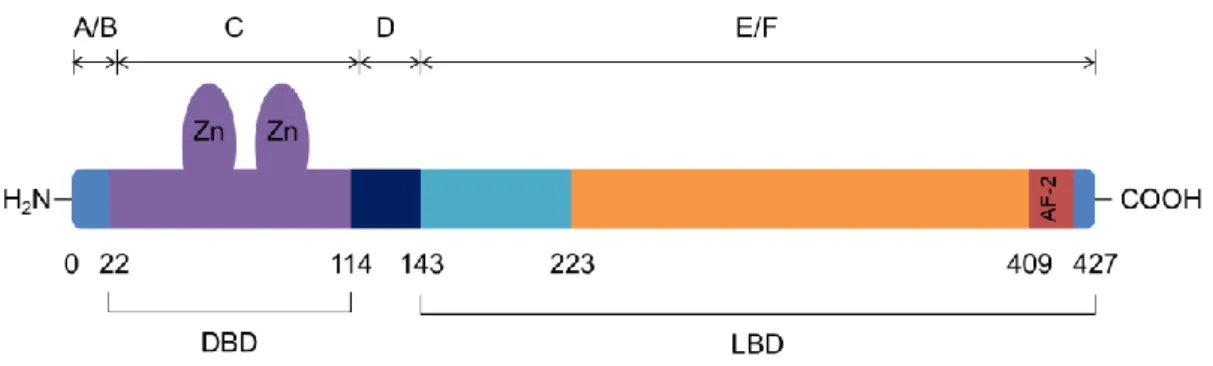
Structurally, it is composed of six regions (A to F), numbered from the N-terminal extension. Additionally, VDR is also divided in two distinct domains, based on functional aspects: a DNA binding domain (DBD) and a ligand binding domain (LBD) (Fig. 4). These domains are bound together by an unstructured linker.
Fig. 4. Schematic representation of the structure of human VDR
The crystal structure of the entire VDR is not yet available. However, the crystal structures of the individual domains (DBD and LBD) have been published and are useful in the study of the function of calcitriol and VDR.10-11
The DBD is a highly conserved domain that contains two zinc fingers. Structurally, this domain corresponds to the C region of the protein. This domain anchors VDR to specific DNA sequences, the vitamin D response elements (VDREs), through the zinc fingers.10 These VDREs are usually repeats of hexanucleotides with a spacing of 3 nucleotides between half sites, called DR3 VDR motifs.
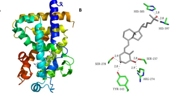
The LBD is a highly variable C-terminal domain that corresponds to the E and F regions of the receptor. This domain confers the VDR high specificity and affinity towards its ligand, calcitriol. The three dimensional structures of the modified human, rat and zebrafish VDR(LBD) have been established, with the receptor bound to calcitriol and other ligands.11-13 These structures show that the VDR(LBD) structure is comprised of 13α helices sandwiched in 3 layers and 3 stranded β sheets that, together, form a highly hydrophobic ligand binding pocket (Fig. 5A).11 This ligand
8 FCUP | ICBAS | USC
Introduction
binding cavity is large (697 Å) and calcitriol only occupies 56% of the total volume. Indeed, there is additional space around the aliphatic chain and near position C2 of the A-ring.14
The 1,25D is anchored to this binding pocket by six hydrogen bonding interactions (Fig. 5B).11 In the human VDR(LBD), the C1α-OH of calcitriol interacts with Ser237 and Arg274, while the C3β-OH binds to Tyr143 and Ser278. The C25-OH group at the side chain of calcitriol interacts with His305 and His397.
Fig. 5. A) Crystal structure of the 1,25D-LBD-VDR complex; B) Representation of polar interaction in the ligand pocket
of VDR (PDB code: 1DB1)
Calcitriol is also anchored to the LBD by non-polar interactions, which greatly influence the conformation adopted by this hormone inside the binding pocket (Fig. 6). The conjugated triene system of the ligand accommodates a trans conformation tightly fitted in a hydrophobic channel made of Ser275 and Trp286 on one side and Leu233 on the other.
Fig. 6. Representation of non-polar interaction between the residues of LBD-VDR and calcitriol (PDB code:1DB1)
FCUP | ICBAS | USC
Introduction
9
The aliphatic side chain of calcitriol is surrounded by hydrophobic residues and adopts an extended conformation parallel to the C13-C18 bond. The methyl group of the side chain of calcitriol makes two important van der Waals contacts with residues from the helix 12 (H12), Val418 and Phe422 (Fig. 6).
This interaction of calcitriol with the residues of the C-terminal H12 is particularly important for interaction of VDR with transcriptional machinery proteins. This helix contains a seven amino acid region called the ligand dependent activation function (AF-2) that acts as a molecular switch in a ligand-dependent manner.
Regarding the mechanism of action of VDR, it is known that this receptor induces gene expression via association with calcitriol, RXR and co-activators (Scheme 4).
Scheme 4. Mechanism of genomic actions of calcitriol
Calcitriol or 25(OH)D3 are transported in the blood stream by DBP and enter target
cells by passive diffusion. Inside the cell, 25(OH)D3 is locally hydroxylated to calcitriol
by CYP27B1. Calcitriol interacts with cytoplasmic VDR, inducing the conformational shift of helix 12. This molecular switch closes over the hormone in a mouse-trap mechanism that anchors calcitriol to VDR and facilitates the formation of protein interaction interfaces.
Upon calcitriol binding, VDR is rapidly translocated to the nucleus, where it forms a heterodimer with another nuclear receptor, RXR, to become transcriptionally active. Once the VDR-RXR heterodimer is formed, it associates with VDREs and recruits
10 FCUP | ICBAS | USC
Introduction
various coregulatory complexes to target gene promotors,15-16 resulting in activation of target gene expression. 17
These highly specific changes in gene expression combine to orchestrate complex and tissue-specific responses. Indeed, using transcriptome-wide analysis, calcitriol was found to modulate the expression of 229 genes.18
2.2. Non-genomic actions of calcitriol
Besides the classical genomic actions, it is presently recognized that calcitriol also exerts fast, non-genomic actions. These actions work in a nuclear VDR independent manner, as they involve membrane-triggered processes. The mechanism of non-genomic action is gradually being unraveled and is thought to impact a wide range of cellular processes,19 such as transcaltachia, insulin secretion and phospholipid metabolism.15
These rapid responses involve two distinct membrane-bound receptors: the membrane associated rapid response steroid (MARRS), also known as Pdia3,20 and membrane-bound VDR (Scheme 5). Although VDR is typically a nuclear receptor, it has been co-localized in caveolae-enriched plasma membranes, through association with a scaffolding protein, caveolin-1 (Cav-1).21-23 MARRS also binds to Cav-1 in these caveolae. The presence of Cav-1 is then essential for the non-genomic actions of 1,25D.
FCUP | ICBAS | USC
Introduction
11
Once VDR or MARRS are associated with Cav-1, calcitriol is able to bind to either one of these receptors. The interaction between calcitriol and the VDR/Cav-1 complex or the MARRS/Cav-1 complex triggers the initiation of different signaling pathways, through a not fully understood mechanism. Presumably, the binding of calcitriol to either of these complexes leads to the activation of G proteins or non-receptor tyrosine kinases, like Src kinase (c-Src),24 which leads to the initiation of different signaling cascades. These signaling pathways are summarized in Scheme 5 and are responsible for the induction of changes in protein activity and even changes in gene expression, in a cell-type, cell-stage and gene specific manner.19, 25
3. BIOLOGICAL ACTIVITY OF CALCITRIOL
Calcitriol, through both genomic and non-genomic mechanisms, is responsible for a multitude of biological functions that are divided in classic and non-classic actions. The role of calcitriol in the maintenance of calcium levels in extracellular fluids is a classic function of this hormone. On the other hand, the non-classic biological functions of calcitriol include the regulation of cellular proliferation and differentiation and modulation of the immune response.
3.1. Calcium and Phosphate Homeostasis
The maintenance of physiologic levels of extra and intracellular levels of calcium and phosphate is a highly complex and regulated process. This homeostasis is dependent on different enzymes and hormones, and involves a coordinated action of the parathyroid glands, kidneys, intestine and bone.
Calcitriol is responsible for the absorption of dietary calcium and phosphate from the gastrointestinal tract, the renal calcium reabsorption and the calcium release from bone (Scheme 6). These effects are regulated by a negative feedback mechanism, in which calcitriol inhibits the synthesis and secretion of the parathyroid hormone (PTH).
The parathyroid glands are responsible for the synthesis of the PTH. In response to low calcium levels (hypocalcemia), the glands secrete PTH, which stimulates the activation of renal CYP27B1 and renal degradation of CYP24A1 mRNA, leading to an increase of calcitriol production (Scheme 6).26-28
The calcium and phosphate homeostasis is also regulated by fibroblast growth factor 23 (FGF23).26 The production of FGF23 by osteoblasts and osteocytes in the bone is stimulated by an increase of calcitriol levels. FGF23 participates in the
12 FCUP | ICBAS | USC
Introduction
regulation of calcium and phosphate homeostasis, as it inhibits the synthesis and secretion of PTH and decreases renal CYP27B1 in response to elevated calcitriol and phosphate levels.29
Scheme 6. Schematic illustration of the effects of calcitriol on the calcium and phosphate homeostasis
These classic actions of calcitriol can be applied clinically, through the use of calcitriol and its analogs in the control of secondary hyperthyroidism in renal failure and osteoporosis.
3.2. Cellular Differentiation and Proliferation
Calcitriol is known to have anti-proliferation and pro-differentiation effects in several cell types mainly through a genomic mechanism.9
Regarding its anti-proliferative function, calcitriol is known to increase the expression of cyclin-dependent kinase inhibitors (CDKIs) p21 and p27. These CDKIs are responsible for regulating the transition from the G1 phase (responsible for cell growth) to the S phase (responsible for DNA replication) of the cell cycle. Calcitriol increases the expression of these inhibitors, which prevents the progression of the cell cycle to the S phase, thus inhibiting cell proliferation.30
FCUP | ICBAS | USC
Introduction
13
Calcitriol has also been found to induce differentiation in several cells, from keratinocytes to breast and prostate cancer cells.31-33
Given these functions, it was postulated that calcitriol and its analogs could have applications in cancer therapy. Additionally, taking into account that calcitriol induces differentiation and inhibits proliferation in keratinocytes, several calcitriol analogs are commercially available for the treatment of moderate forms of psoriasis.32
3.3. Immune System Regulation
Calcitriol plays a role in immune regulation because it can be synthesized in and have effect on T cells,34-35 B cells36 and antigen presenting cells (APCs).37-38 Therefore, calcitriol has immunomodulatory effects that could be used in the treatment of autoimmune diseases. Indeed, vitamin D-based therapies have been applied in the control of autoimmune disorders like systemic lupus erythematosus, rheumatoid arthritis, multiple sclerosis and type I diabetes.
3.4. Transcaltachia
Transcaltachia refers to the non-genomic rapid (2-10 minutes) stimulation of intestinal calcium transport. Although this process can have a useful role in calcium homeostasis, it greatly hinders the application of vitamin D-based therapies. Calcitriol and some of its analogs have poor clinical application due to side effects like hypercalcemia, mainly caused by transcaltachia. As such, there has been a major effort to synthesize non-calcemic calcitriol analogs.39-41
3.5. Pharmacological Endeavors
The elucidation of the crystallographic structure of the LBD of VDR11 has facilitated the rational design of new analogs.
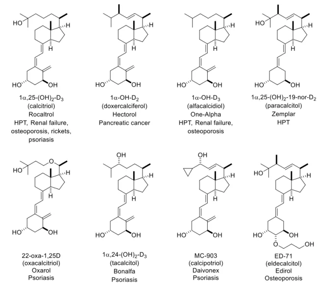
Although more than 3500 analogs have been synthesized, only a few, are currently commercially available and are clinically applied (Fig. 7).42 Some of these drugs are used in the treatment of psoriasis, osteoporosis, multiple sclerosis, and bone and pancreatic cancer.
14 FCUP | ICBAS | USC
Introduction
Fig. 7. Commercially available calcitriol analogs
The design of innovative VDR ligands with unique functional groups was propelled. This search for interesting novel leads has unveiled the great promise of a previously unexplored element in drug design: boron.
4. CARBORANES
Recently, the pharmaceutical industry and an increasing number of medicinal chemistry researchers are using boron as a substitute for carbon in different classes of drug molecules. Molecules with boron atoms are expected to be less complex than their carbon counterparts, reducing costs in drug research and manufacturing. Also, the possibility of new interactions of these molecules with biological targets unaffected by classical ligands makes the use of these boron moieties very alluring. A boron-containing drug, Velcade® is commercialized (Fig. 8) and several others have entered human clinical trials.43-45
FCUP | ICBAS | USC
Introduction
15
Fig. 8. Commercially available boron-containing drug Velcade
Boron tends to form compounds with covalent B-H and B-B bonds called boranes. These boranes exist preferentially in polyhedral clusters with globular architectures.46 One important class of polyhedral borane clusters are carboranes, in which two BH -units of the closo-B12H122− cluster are replaced by 2 CH vertices.
The recent interest in applying carboranes to medicinal chemistry has arisen due to their particular electronic properties, geometry and versatility, together with the newest development in carborane chemistry.
4.1. Structure of carboranes
Carboranes, or carbaboranes, are polyhedral carbon-containing boron clusters. Among these, the dicarba-closo-dodecaboranes (C2B10H12) are the most extensively studied
and are usually referred to as simply carboranes. They are rigid molecules with an icosahedral structure, containing 10 BH vertices and 2 CH vertices. The carbon and boron atoms are hexacoordinated to compensate the low electron density, forming 20 triangular faces.
Carboranes exist in three isomeric forms, depending on the positioning of their 2 CH vertices: ortho- (or 1,2-), meta- (or 1,7-) and para- (or 1,12-) carborane (Fig. 9).
Fig. 9. Representation of the three closo-carborane isomers
The ortho- and meta-carboranes have C2v symmetry, while the p-isomers have a D5d
symmetry. The overall size and the core volume of carboranes slightly decrease from
16 FCUP | ICBAS | USC
Introduction
size and volume of carboranes is similar to that of adamantane and almost twice the spherical volume occupied by a phenyl ring (Fig. 10).
Fig. 10. Representation of an o-carborane, adamantane and benzene
4.2. Electronic Properties
Carboranes have non-classical bonding interactions, resulting in a very complex overall electronic structure. They have 26 electrons (2n+2 skeletal e-) divided between the 12 vertices, which are three-dimensionally delocalized, meaning that carboranes are characterized as three-dimensional aromatic compounds. As such, these compounds are not easily defined by usual organic bond diagrams and the bonds between atoms represent only geometry.
In terms of electronegativity, the carbon and hydrogen atoms are more electronegative than the boron atoms, which explain some of the carborane properties. Additionally, boron-carbon bonds are stronger than carbon-carbon bonds, meaning that the more stable isomer has carbon atoms in nonadjacent positions.
4.3. Hidrophobicity
In terms of hydrophobicity, carboranes are usually more hydrophobic than their carbon counterparts. The hydrogens atoms of the BH vertices have partial negative charges, making them similar to hydrides, which explain the high hydrophobicity of carboranes. The different carboranes isomers show differences in hydrophobicity, as the hydrophobicity increases in the order ortho, meta to para isomers.
The hydrophobicity of carboranes and carborane-containing drugs can be fine-tuned by choosing to substitute the different vertex positions. Free CH groups of carboranes, which are acidic, decrease the hydrophobicity of the carborane. On the contrary, C-substituted carboranes are more hydrophobic. B-substituted carboranes are less hydrophobic than their counterparts.
FCUP | ICBAS | USC
Introduction
17
4.4. Acidity
The protons of the C atoms are relatively acidic, depending on the cluster isomer. The acidity of the carborane protons decreases in the order ortho-, meta- to
para-carborane. Organometallic bases, such as nBuLi can remove a proton from C-H, generally creating a carboranyl nucleophile. B or C substituents influence both the acidity of the carborane protons and the acidity of the cluster protons.
4.5. Molecular interactions
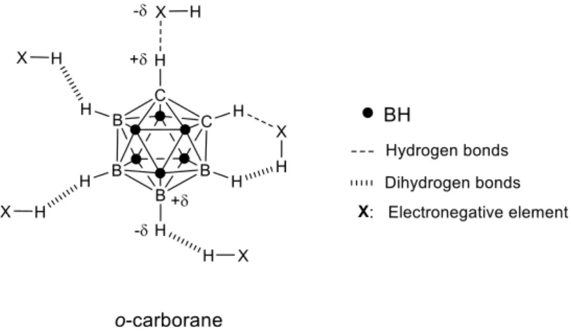
The outer sphere of carboranes is composed of hydrogens, meaning that these hydrogen atoms are the main mediators of intermolecular interactions in unsubstituted carboranes.
Carboranes have two different types of hydrogen atoms: acidic hydrogens from CH groups and hydride hydrogens from BH groups.
These two types of hydrogen atoms are involved in distinct intermolecular interactions (Fig. 11). The CH moieties form classic hydrogen bonds (C-H • • •X) with electronegative atoms (O, N, S or F).
Fig. 11. Two-dimensional representation of intermolecular interactions of o-carborane
On the other hand, the hydride BH units can form dihydrogen bonds (B-H • • •H-X) with protons bound to electronegative atoms. These interactions occur with small molecules, as well as in the active site of proteins and enzymes, which makes carboranes very interesting pharmacophores.
18 FCUP | ICBAS | USC
Introduction
4.6. Chemistry
The ortho-carborane can be obtained by reaction of decaborane (B10H14) with
acetylenes in the presence of a Lewis base (Scheme 7). Thermal isomerization of
ortho-carboranes, in an autoclave, leads first to the meta isomer (465-500 ºC) and
finally to the para isomer (615-700 ºC).
These carboranes are chemically stable and are inert to water and atmospheric oxygen.
Scheme 7. Synthesis of ortho, meta and para-carboranes
They can be derivatized on the boron vertices, by electrophilic substitution; and on the carbon vertices, by removal of acidic CH protons with strong bases.
Carboranes can undergo metalation with nBuLi to afford the carboranyllithium derivative, which can react with an electrophile to form a C-substituted carborane. Thus, from a synthetic point of view, carboranes are interestingly versatile and can be used to create wide libraries of pharmacophores for distinct purposes.
4.7. Boron Neutron Capture Therapy (BNCT)
As most of their properties are prone to be fine-tuned, carboranes may prove to be superior bioactive molecules than their organic counterparts. Also, these
closo-carboranes are great candidates for boron neutron capture therapy (BNCT).
This experimental cancer treatment uses boron compounds in the treatment of several cancers, mainly glioblastoma multiform (GBM). In this case, clinical studies have
FCUP | ICBAS | USC
Introduction
19
demonstrated that BNCT is not only safe but at least as efficient as conventional radiotherapy.
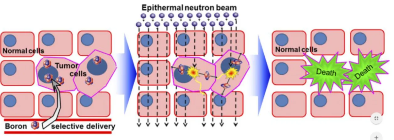
This technique is based on the nuclear capture of neutrons by 10B to produce high energy particles that selectively destroy tumors without affecting healthy tissues. It works as a two-component, or binary, therapy, since it uses two independent key components: the boron clusters and low energy thermal neutrons.
When irradiated, the non-radioactive 10B isotopes present in boron clusters capture the low-energy neutrons and then fissure into kinetic energy and into high linear energy transfer (LET) particles: alpha particles (two protons and two neutrons) and lithium ions.47 These high LET particles have path-lengths of about 10 µm, roughly the diameter of a single cell, which makes the cytotoxic effect largely confined to cells that accumulated the boron compound (Fig. 12).
Fig. 12. Selective neutron capture of neutrons by 10B in a tissue48
The application of BNCT depends on:
1) the selectivity of the compounds for a target highly expressed in tumors, as that would mean that surrounding healthy tissue would be less affected;
2) the number of boron atoms accumulated in tumor cells, as a high number accumulated of boron atoms in tumor cells would increase the probability of neutron capture events.
As such, highly selective compounds that contain boron clusters like carboranes (1 moiety = 10 B) should be very efficient BNCT agents. Indeed, carboranes are currently being studied for that purpose, as they are being used in the synthesis of novel DNA metallointercalators that could bind to tumor cell DNA and of hypoxia-selective agents that could enter low-oxygen regions in tumors.45
20 FCUP | ICBAS | USC
Introduction
Another tumor-selective target that should be considered for BNCT therapy is the VDR. The VDR is a good target for selective application of BNCT because its expression is increased in some tumors, such as breast cancer. The next obvious step is, then, the design of VDR agonists that contain closo-carboranes, an endeavor explored in this work.
5. BACKGROUND
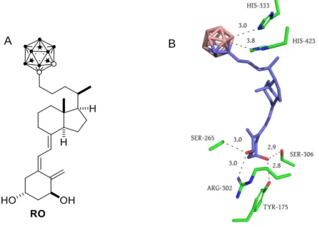
Recently, an unique secosteroid vitamin D analog with an o-carborane moiety at the side chain (Fig. 13A) has been designed, synthetized and biologically tested.49 This compound (RO) binds efficiently to VDR and induces similar biological activities with reduced calcemic effects, in comparison to 1,25D.
Fig. 13. A) Representation of analog RO. B) Structure of the binding pocket of zVDR LBD bound to analog RO.
Analog RO was crystallized in the zebra-fish wild-type VDR LBD at a 2.4 Å resolution (Fig. 13B). This compound adopts the canonical active conformation of the calcitriol-VDR complex. The carborane unit mimics the interaction of the C25-OH in the binding pocket of VDR. It is even postulated that the carborane cage interacts with His333 and His423 (His305 and His397 in human VDR, respectively) through non-conventional dihydrogen bonds (BH•••HN).
The synthesis of analog RO was accomplished in 9 steps and is illustrated in Scheme 8.
The ortho-carborane moiety of analog RO was formed late in the synthesis, to avoid complications regarding carborane chemistry and characterization. The carborane was
B A
FCUP | ICBAS | USC
Introduction
21
introduced in the side chain by reaction of an alkyne with decaborane (B10H14), in the
presence of N,N-dimethylaniline.
These results proved that carborane-containing vitamin D analogs can be biologically active and have less calcemic effects than calcitriol itself. As such, the synthesis of
analog RO paved the way for the rational design of new
carborane-containing vitamin D analogs that could have medical and pharmaceutical application.
Scheme 8. Synthesis of target carborane RO. Reagents and conditions: a) pTsCl, py, 0 ºC, 12 h; b) Et2O/THF, Li2CuCl4,
THF, -78 ºC / rt, 12 h; c) PDC, CH2Cl2, rt, 5 h; d) (Ph3PCH2Br)Br, KO
t
Bu in THF, tol, -15 ºC / rt, 3 h; e) tBuLi, B(OiPr)3,
pinacol, tol/THF, -78 ºC / rt, 4 h; K3PO4 (2 M), PdCl2(PPh3)2, THF, 2 h; (g) K2CO3, MeOH, rt, 14 h; (h) PhNMe2, tol,
FCUP | ICBAS | USC
Objectives
25
Objectives
This work was developed to improve the properties of analog RO and explore potential application of other carborane-containing calcitriol analogs in BNCT. As such, the design and synthesis of new carborane analogs was planned. The objectives of this work were:
1) Docking of new carborane-containing vitamin D analogs into the 1,25D-VDR(LBD) complex to study their in silico affinity for the VDR.
2) Synthesis of the target analogs A1, A17 and A33 (Fig. 14).
FCUP | ICBAS | USC
Results and Discussion
29
Results and Discussion
1. CHAPTER I: Design and Docking of carborane-containing vitamin
D analogs
1.1. Design of carborane-containing vitamin D analogs
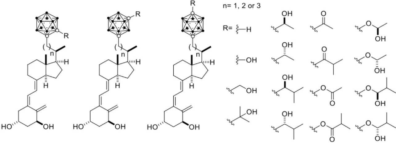
New carborane-containing calcitriol analogs were designed and docked into the LBD of VDR (Fig. 15). In order to find the ligands that bind better to this receptor, several different parameters were investigated: 1) the carborane isomer connected to the side chain (ortho-, meta- or para-carborane); 2) the C-substituents attached to the carborane unit (R groups); 3) the length of the side chain connecting the carborane unit to the CD-ring system (n=1, n=2 or n=3).
Fig. 15. Representation of carborane-containing analogs to analyze by docking in human VDR
Justification for the choice of parameters:
1) It is unknown whether the slight differences in electronic properties, hydrophobicity and acidity46 between carborane isomers could influence the analogs’ binding affinity. The orientation of the attached R groups could also affect the binding.
2) Analogs with different R groups could interact differently with the VDR, thus altering their binding affinity. The size and nature of the R groups were expected to have an impact on the chemical and physical properties of each analog inside the VDR.
3) Given the size of the carborane moiety49 and depending on the size of the different R groups attached, the length of the side chain (n= 1, 2 or 3) could influence the analogs’ binding affinity for the VDR.
30 FCUP | ICBAS | USC
Results and Discussion
1.2. Building of the proposed carborane-containing vitamin D analogs
The ligands were built using the PyMol program.50 The vitamin D part was obtained from the crystal structure of the complex of 1,25D-hVDR LBD (PDB code: 1DB1).11 The carborane cage part of the ligands was obtained from the crystal structure of the corresponding ortho (PDB code: 5E7V),49 meta (PDB code: 4MDL)51 and
para-carboranes (PDB codes: 3VJS).52 The ligands were further treated by adding hydrogen atoms and checking atom valence using Chem3D. Energy minimization of the ligands was performed using the MM2 function of Chem3D.
1.3. Docking of the proposed carborane-containing vitamin D analogs
Docking studies to predict the affinity of the new ligands for the VDR were carried out using the GOLD program (version Suite 5.2). A modified crystal structure (addition of hydrogens, reconstitution of gaps and correction of His tautomers) of the complex between 1,25D-hVDR(LBD) was chosen as protein (PBD code: 1DB1).11
The Ligand Binding Pocket of the mutant LBD was defined as Binding Site with the automatic active-site detection on, and the radius was set to 10 Å. The conformations for each ligand in the active site of the protein were sampled using a genetic algorithm (GA). The ligands were docked in 25 independent genetic algorithm (GA) runs, for each of which a maximum of 125000 GA operations were performed on a single population of 100 individuals. Operator weights for crossover, mutation, and migration in the entry box were used as default parameters (95, 95, and 10, respectively), as well as the hydrogen bonding (4.0 Å) and van der Waals (2.5 Å) parameters. The different poses were ranked according to the force-field-based CHEMPLP scoring function and GoldScore was used as a re-scoring function. The 3 best solutions for each ligand were obtained with an associated score and these results were compared with the solutions for 1,25D.
1.4. Docking Results
The best obtained poses for each ligand were analyzed in terms of score, adopted conformation inside the receptor and interactions with the different residues.
In appendix 1 (page 83), the chosen conformation for each ligand was superimposed to the natural ligand 1,25D (represented in grey) into the ligand binding pocket of VDR. The interactions with His305 and His397 are shown for all analogs. The interactions
FCUP | ICBAS | USC
Results and Discussion
31
between the C3-OH and C1-OH of the analogs and Tyr143, Ser237, Arg274 and Ser278 were only shown when the interactions of the A ring of the analog changed significantly.
In summary, analogs B2, C2, B4, B8, A13, A14, C17, B18, C18, A19, B19, A20, A21, A23, B30, A31, A32, B33 and C33 are the most promising in terms of adopted conformation, interactions with the VDR and score. Therefore, they are good candidates for synthesis and biological assays (Fig. 16).
32 FCUP | ICBAS | USC
Results and Discussion
Fig. 16. Conformation of the most promising docked analogs (continuation)
The different conditions tested also provided some interesting information, which is explored next.
1.4.1. Docking results of analogs depending on the carborane isomers
Regarding the distinct conditions tested, it is possible to conclude that, overall, analogs with meta-carboranes appear to fit better in the binding pocket of VDR, as the connected R groups impart a favorable orientation. On the contrary,
para-carborane containing analogs fit worse in the binding pocket, as most R groups
do not interact with residues His305 and His397. This preference is detected in regards to both ligand poses, interaction with the VDR and docking scores.
For example, when comparing analogs B3 (ortho-carborane), B19 (meta-carborane) and B35 (para-carborane, Table 1), it is possible to see that analog B19 fits better, has a better score and its R group forms more stable hydrogen bonds with the receptor.
FCUP | ICBAS | USC
Results and Discussion
33
Table 1. Comparison of docking scores for analogs containing different carborane isomers. First line shows a
representation of the docked analog. Last line shows the docking result for each ligand superimposed with calcitriol inside the VDR binding pocket. Calcitriol is represented in grey.
B3 (109%) B19 (117%) B35 (103%)
From these docking results, it is possible to observe that the theoretical binding affinity for the VDR depends on the carborane isomer attached to the side chain. Thus, these
in silico results postulate that similar compounds containing distinct carborane isomers
could have different biological properties. Therefore, we decided to synthesize
ortho-, meta- and para-carborane analogs.
1.4.2. Docking results of analogs with different side chain sizes and R groups
Regarding the size of the side chain, it was concluded that the preferred length of the side chain (n) greatly depends on the size of the R groups. Analogs with smaller R groups (-H or -OH) benefit from larger side chains (n=3), while analogs with intermediate to large R groups (alcohols, esters or hemiacetals) fit better inside the VDR when combined with smaller side chains (n=1 or n=2).
34 FCUP | ICBAS | USC
Results and Discussion
For example, with regard to small R groups (Table 2), analog C17 (n=3) has a better score than the other two analogs, A17 (n=1) and B17 (n=2), as it fits better inside the binding pocket.
Table 2. Comparison of docking score for analogs with small R groups. First line shows a representation of the docked
analog. Last line shows the docking result for each ligand superimposed with calcitriol (grey) inside the VDR binding pocket.
A17 (102%) B17 (113%) C17 (119%)
Intermediate sized groups, such as small alcohols, are better in combination with an intermediate side chain length (Table 3). For example, analog B22 (n=2) has a better score than analogs A22 (n=1) and C22 (n=3), which contain smaller and larger side chains, respectively. Analog B22 is able to adopt a conformation similar to that of calcitriol, while forming similar hydrogen bonds with His305 and His397.
FCUP | ICBAS | USC
Results and Discussion
35
Table 3. Comparison of docking score for analogs with intermediate R groups. First line shows a representation of the
docked analog. Last line shows the docking result for each ligand superimposed with calcitriol (grey) inside the VDR binding pocket
A22 (105%) B22 (112%) C22 (111%)
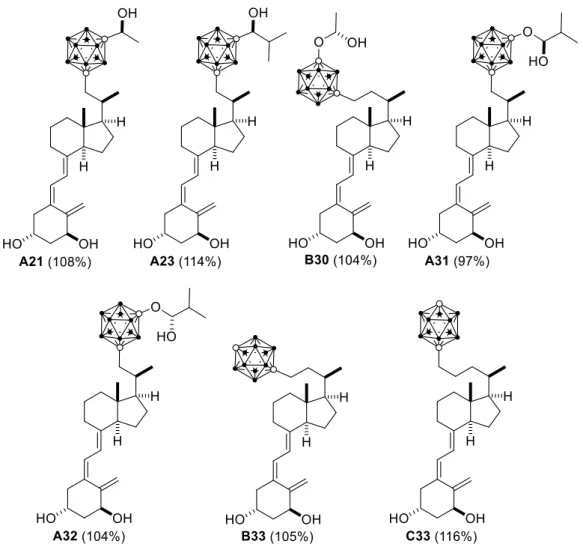
When it concerns larger R groups, such as large ketone, acetal and hemiacetal substituents, a smaller side chain (n=1) is often favourable (Table 4). In this case, analogs A32 (n=1) and B32 (n=2) have similar docking results. However, analog A32 binds to both His305 and His397 and adopts a more preferable conformation inside the pocket. On the contrary, analog C32 is clearly too large to fit the binding pocket, as seen from its distorted side chain and CD bicycle.
36 FCUP | ICBAS | USC
Results and Discussion
Table 4. Comparison of docking score for analogs with large R groups. First line shows a representation of the docked
analog. Last line shows the docking result for each ligand superimposed with calcitriol (grey) inside the VDR binding pocket
A32 (104%) B32 (107%) C32 (98%)
These results demonstrate that the choice of side chain size depends on the R group.
1.5. Choice of target analogs for synthesis
Compounds A1, A17 and A33 (Table 5) were investigated as possible targets to study the biological role of the ortho-, meta- and para-carboranes. Since they have a different side chain length (n=1) than the previously synthesized RO analog (n=3), these compounds would also be useful to study the effect of the side chain length in the analogs’ biological behavior.
FCUP | ICBAS | USC
Results and Discussion
37
Table 5. Target analogs for synthesis. . First line shows a representation of the docked analog. Last line shows the
docking result for each ligand inside the VDR binding pocket
A1 (98%) A17 (102%) A33 (97%)
In addition, these analogs could be used as precursors of promising hemiacetal analogs. One of the long term goals of this research group is the synthesis of carborane-containing calcitriol analogs with stable hemiacetal substituents on the carborane, to test their stability and biological properties. From the hemiacetal analogs tested, compounds A13, A14, A31 and A32, which have ortho- or meta-carboranes and a side chain length of n=1, were the most promising (Fig. 16). Since analogs A1, A17 and A33 have the same side chain length and carborane isomers, they could be used as precursors to these hemiacetal compounds.
38 FCUP | ICBAS | USC
Results and Discussion
In terms of docking results, these analogs have docking scores similar to 1,25D. They adopt a favorable conformation inside the receptor and bind to the same amino acids as calcitriol. Therefore, they should have binding affinity towards the VDR and should induce
FCUP | ICBAS | USC
Results and Discussion
39
2.
CHAPTER II: Synthesis of Carborane Analogs
A1, A17 and A33
2.1. Retrosynthesis
The retrosynthesis analysis for the preparation of the target compounds A1, A17 and A33 is depicted in Scheme 9.
Scheme 9. Retrosynthetic analysis planned for the synthesis of compounds A1, A17 and A33. P = protecting group
The key step of the synthesis is the introduction of the carborane moiety at the end of the synthesis by reaction of the corresponding carboranyllithium on tosylate 1.
The triene system would be built by a stereoselective Pd-catalyzed ring closure on enol-triflate 3 and subsequent Suzuki-Miyaura coupling with alkenyl-boronic ester 2. The enol-triflate 3 would arise from epoxide 5, following procedures previously developed in this laboratory.53 The required boronic ester 2 would be prepared from Inhoffen-Lythgoe diol 4 by oxidation, Wittig chemistry and Pd-catalysed Br-B interchange.54
40 FCUP | ICBAS | USC
Results and Discussion
2.2. Synthesis of benzoate 6
The synthesis started with known Inhoffen-Lythgoe diol (4), prepared from reductive ozonolysis of commercial vitamin D2,55 which was converted to benzoate 6 (Scheme
10).
Diol 4 was treated with nBuLi, at low temperature, followed by addition of benzoyl chloride to give compounds 6 (14%), 7 (8%) and 8 (21%), together with starting material 4 (52%).
Scheme 10. Synthesis of benzoate 6 by metalation of diol 4
We next tried to improve the yield of the desired benzoate 6. Thus, treatment of diol 4 with benzoyl chloride in the presence of 2,6-lutidine, in dichloromethane, provided benzoate 6 in a good yield (Scheme 11).
Scheme 11. Selective synthesis of benzoate 6
Fig. 17 shows the most significant 1H-NMR and 13C-NMR signals of benzoate 6
FCUP | ICBAS | USC
Results and Discussion
41
2.3. Synthesis of boronic ester 2
Boronic ester 2 was then prepared in 50% overall yield, following previously developed procedures of this laboratory, which involve Dess-Martin oxidation, Wittig chemistry and Miyaura borylation (Scheme 12).56-57
Scheme 12. Synthesis of boronic ester 2
The oxidation of alcohol 6 with Dess-Martin periodinane in CH2Cl2 afforded ketone 9 in
excellent yield.
Wittig chemistry between ketone 9 and ylide Ph3P=CHBr, generated in situ by
treatment of (bromomethyl)triphenylphosphonium bromide with potassium tert-butoxide in toluene, provided vinyl bromide 10 in a 74% yield.
The alkenyl bromide 10 was then converted to boronate 2 in 68% yield by a Pd(0) catalyzed cross-coupling reaction employing bis(pinacolate)diboron and PdCl2(dppf)•CH2Cl2, as the catalyst, in the presence of tricyclohexylphosphine and
KOAc in DMSO.57
42 FCUP | ICBAS | USC
Results and Discussion
Scheme 13. Proposed mechanism for the Miyaura borylation reaction in the synthesis of boronic ester 2
The reaction starts with the in situ formation of the Pd(0) catalyst, which is then inserted in the C-Br bond of the vinyl halide through oxidative addition. Then, transmetallation with bis(pinacolate)diboron, followed by reductive elimination, furnishes boronate 2 and leads to the regeneration of Pd(0).
Fig. 18 shows the most significant 1H-NMR and 13C-NMR signals of boronic ester 2.
FCUP | ICBAS | USC
Results and Discussion
43
2.4. Synthesis of enol-triflate 3
The enol-triflate 3 was prepared from the diprotected epoxide 5, following previously developed procedures (Scheme 14).58
Scheme 14. Synthesis of enol triflate 3
Epoxide 5 was converted to dibromide 12 by a two-step sequence. Periodic acid-oxidative cleavage of the epoxide 5 in Et2O gave aldehyde 11, which was
transformed into dibromide 12 by Corey-Fuchs’ conditions.59
Finally, the enol-triflate 3 was obtained in moderate yield from dibromide 12, by treatment with lithium diisopropylamide (LDA), followed by reaction of the resulting enolate with N-(5-chloro-2-pyridyl)triflimide (Comins’ reagent).
Fig. 19 shows the most significant 1H-NMR and 13C-NMR signals of enol-triflate 3.
44 FCUP | ICBAS | USC
Results and Discussion
2.5. Synthesis of tosylate 1
With the upper and lower fragments in hand, we proceeded to build the vitamin D triene system by a Pd-catalyzed ring closure of enol-triflate 3, followed by Suzuki Miyaura cross-coupling with boronate 2, in the presence of aqueous K3PO4, to give 13 in 68%
yield (Scheme 15).
Scheme 15. Synthesis of benzoate 13 via carbociclization and Suzuki-Miyaura cross-coupling of 2 and 3
The proposed mechanism for this reaction is shown in Scheme 16.
FCUP | ICBAS | USC
Results and Discussion
45
The Pd(0) complex coordinates with the alkyne group of enol-triflate 3, which is followed by oxidative addition to the triflate group to furnish intermediate compound a. Then, a Pd-catalyzed Heck-type addition produces the Pd(II) intermediate (b), which undergoes a transposition with an activated boronate to produce intermediate c. Finally, this intermediate is transformed into benzoate 13 by reductive elimination, which is accompanied by the regeneration of the Pd(0) complex.
To facilitate the introduction of the carborane moiety in the side chain, tosylate 1, which contains a good leaving group at C-22, was prepared from benzoate 13 in a two-step sequence (71% yield, Scheme 17).
Scheme 17. Synthesis of tosylate 1
Ester 13 was reduced with DIBAL-H to alcohol 14, which upon tosylation afforded the desired compound 1 in good yield.
Fig. 20 shows the most significant 1H-NMR and 13C-NMR signals of tosylate 1.
46 FCUP | ICBAS | USC
Results and Discussion
2.6. Synthesis of analogs A1, A17 and A33
As mentioned in the introduction, the carborane unit was previously introduced in the side chain of a vitamin D analog via reaction of an alkyne with decaborane (B10H14), in
the presence of N,N-dimethylaniline.49 However, this methodology had some drawbacks, including low reaction yields and high toxicity of decaborane. Additionally, this reaction only allows the production of ortho-substituted derivatives. Since one of the purposes of this work is the synthesis of analogs with ortho, meta and
para-carborane units at the side chain, we focused on an alternative method to directly
introduce the carborane moiety. A SN2 reaction between tosylate 1 and a
carboranyllithium was found suitable for this purpose.
Thus, direct displacement of the tosylate group of 1 with ortho-carboranyllithium, generated by reaction of the ortho-carborane with nBuLi, provided the ortho-carborane derivative 15, which was deprotected with aqueous HF to furnish the desired vitamin D analog A1 in good yield (54%, 2 steps, Scheme 18).
Scheme 18. Synthesis of analog A1
The ortho-carborane analog A1 was obtained from the Inhoffen-Lythgoe in 9 steps and 12% overall yield.
FCUP | ICBAS | USC
Results and Discussion
47
Fig. 21. 1H-NMR spectrum of analog A1
The well-defined singlet at 3.53 ppm corresponds to the C-24 proton of the
ortho-carborane unit. The 10 H of the carborane moiety, present at the 10 BH vertices,
appear as a wide band between 3.0-1.0 ppm, as seen by the increase of the baseline of the spectrum.
The 11B-NMR spectrum of analog A1 (Fig. 22) shows that the 10 BH protons of the carborane unit are divided in 5 signals that integrate in the ratio 1:1:2:4:2 (-2.3, -5.6, -9.3, -11.6 and -13.1 ppm). This spectrum is similar to the known spectrum of free
ortho-carborane, which confirms the successful introduction of this moiety in the target
analog.46, 60
48 FCUP | ICBAS | USC
Results and Discussion
Following the same methodology, analog A17 was synthesized from tosylate 1 in a 60% yield (2 steps, Scheme 19).
Scheme 19. Synthesis of analog A17
The meta-carborane analog A17 was obtained from the Inhoffen-Lythgoe in 9 steps and 13% overall yield.
Fig. 23 shows the 1H-NMR spectrum of vitamin D analog A17
Fig. 23. 1H-NMR spectrum of analog A17
The well-defined singlet at 2.90 ppm corresponds to the proton of C-24. The wide band between 3.00 and 1.00 ppm, observed by the increase of the baseline of the spectrum, corresponds to the 10 BH protons.
FCUP | ICBAS | USC
Results and Discussion
49
The 11B-NMR spectrum of analog A17 (Fig. 24) shows that the 10 BH protons of the
meta-carborane unit of the analog A17 are divided in 4 different sets in the ratio 2:4:2:2
(-3.8, -10.7, -13.7, -15.1 ppm), which is similar to the spectrum obtained for the free
meta-carborane.46
Fig. 24. 11B-NMR spectrum of analog A17
Following the same methodology, analog A33 was prepared from tosylate 1 in a 48% yield (2 steps, Scheme 20).
Scheme 20. Synthesis of analog A33
The synthesis of A33 was accomplished in 9 steps and a 12% overall yield from the Inhoffen-Lythgoe diol.
50 FCUP | ICBAS | USC
Results and Discussion
Fig. 25. 1H-NMR spectrum of analog A33
The proton of C-24 of the para-carborane moiety appears as a singlet at 2.59 ppm. The 10 H of the BH units appear as a wide band between 3.0-1.0 ppm, similarly to the other analogs.
The decoupled 11B-NMR spectrum of analog A33 (Fig. 26) shows that the 10 BH protons of the para-carborane unit of the analog A33 are divided in 2 different sets in the ratio 5:5 (-12.3; -15.2 ppm). One of the signals is assigned to the 5 boron atoms closer to the free CH group and the other signal is assigned to the BH vertices closer to C-23.