Universidade de Lisboa
Faculdade de Medicina Dentária
Small-Diameter Implants for Definitive Prosthodontic
Treatment - A Literature Review
Lisa Steinhausen
Dissertação
Mestrado Integrado em Medicina Dentária
II
Universidade de Lisboa
Faculdade de Medicina Dentária
Small-Diameter Implants for Definitive Prosthodontic
Treatment - A Literature Review
Lisa Steinhausen
Dissertação orientada
Pelo Dr. André Chen
Mestrado Integrado em Medicina Dentária
2016
III
“It’s not what you are that holds you back, it’s what you think you are not.”
- Denis Waitley
Agradecimentos
Ao meu orientador, Dr. André Chen, pelo apoio e disponibilidade na realização deste trabalho e pela transmissão dos seus conhecimentos.
Ao Dr. Henrique Luís, pela disponibilidade e ajuda na realização da análise estatística.
Aos meus colegas e amigos pelo companheirismo e toda a paciência, e à minha família pela incansável motivação e apoio.
IV Table of Contents
List of Tables and Figures... V Abbreviates and Symbols... VII Abstract... VIII Resumo... IX
I. Introduction... 1
II. Materials and Methods... 5
- Data Assessment... 7 III. Results... 9 IV. Discussion... 15 V. Conclusions... 22 VI. Appendices... A VII. References... J
V List of Tables and Figures
Table 1 – Various small-diameter implant definitions available in the literature
2
Table 2 – Inclusion and exclusion criteria 5 Table 3 - Relation between inserted and failed implants restored with
overdentures, depending on the loading protocol
14
Table 4- Relation between inserted and failed implants restored with fixed single restorations, depending on the loading protocol
14
Table 5 - Survival rate data A
Table 6 - Success rate data A
Table 7 - Frequency data of bone loss A Table 8 - Frequency data of follow-up time B Table 9 - Frequency data of upper or lower jawlocation B Table 10 - Frequency data of anterior or posterior location B Table 11 - Frequency data of function/restoration type C Table 12 - Frequency data of surgical access technique C Table 13 - Frequency data of surgical approach C Table 14 - Frequency data of loading protocols D Table 15 - Pearson correlation between survival rate and loading protocol D Table 16 - Data collected from all included studies E
VI
Figure 2 – Frequency of different article types 9 Figure 3 – Percentage of failed implants according to failure reasons 10 Figure 4 – Distribution of bone loss across studies 11 Figure 5 – Percentage of reported complications 11 Figure 6 – Follow-up time distribution across studies 11 Figure 7 – Distribution of implant location in upper and lower jaw across
studies
12
Figure 8 – Distribution of implant location in anterior or posterior sites across studies
12
Figure 9 – Function/restoration type of SDIs across studies 12 Figure 10 – Distribution of surgical access technique across studies 13 Figure 11 – Different surgical approaches followed by studies 13 Figure 12 – Different loading protocols followed by studies 13
VII Abbreviates and Symbols
SDI Small-diameter implant/ Implante de pequeno diâmetro SDIs Small-diameter implants/ Implantes de pequeno diâmetro mm Millimeters/ milímetros
Ti Titanium/ Titânio Zr Zirconium/ Zircónia
RCT Randomized Controlled Trial/ Ensaio Clínico Randomizado ≤ smaller or equal /menor ou igual
≥ bigger or equal/ maior ou igual < smaller/ menor
> bigger /maior = equal/ igual
Ti6Al4V Titanium alloy composed mostly of titanium, aluminum and vanadium
VIII Abstract
Introduction: Dental implants are available in a wide range of diameters. Although there is still no clear definition, in this review small-diameter implants (SDIs) were considered ≤3.5mm in width. SDIs are mainly used when the placement of a larger implant is difficult due to insufficient bone substance. Due to structural weakness and a smaller contact area with the bone, they have been associated with biomechanical risk factors, especially in high occlusal loads, which might lead to peri-implant bone resorption or fatigue fracture of the implant. Their survival and success rates in short-term follow-up seem to be comparable to regular-diameter implants (>95%). The aim of this study was to review the survival and success rates of SDIs in definitive prosthetics, and report complications and failure reasons associated.
Materials and Methods: An electronic search was undertaken in the PubMed database until March 2016, for small-diameter, narrow-diameter and mini implants with a diameter ≤3.5mm, used for definitive prosthetics with a follow-up time after loading of ≥1month.
Results: The electronic search resulted in 907 publications and 79 met the eligibility criteria. The mean survival rate was 95,2% with a mean follow-up time of 2,7years (6weeks to 12years). Most common failures were of early biological origins. Complications were biologic or prosthetic related. SDIs were mainly inserted in anterior regions of the mandible or both jaws, restored with overdentures or fixed single restorations, following a one-stage surgical approach and immediate loading protocols.
Conclusions: Within the limitations of this review, survival of SDIs seems to be comparable to regular-diameter implants in short-term. Early biologic failures could be related to immediate loading protocols. More RCTs with longer follow-up times are necessary to address these speculations, confirm SDIs long-term survival, study their ideal loading protocol related to restoration type and investigate their use in more occlusive demanding situations.
IX Resumo
Introdução: Implantes dentários estão disponíveis numa vasta gama de diâmetros. Embora não haja um consenso acerca da sua definição, implantes de pequeno diâmetro (SDIs) são geralmente relatados com um diâmetro ≤ 3.5mm. Estão maioritariamente indicados quando a colocação de implantes de maior diâmetro se torna difícil devido à quantidade insuficiente de osso disponível, evitando desta forma a necessidade de procedimentos cirúrgicos complicados de regeneração óssea.
Devido à sua fraqueza estrutural e reduzida área de contato com o osso comparativamente com os implantes de diâmetro regular, SDIs têm sido associados a potenciais fatores de risco biomecânicos, especialmente em situações de elevada carga oclusal, podendo levar a uma reabsorção óssea peri-implantar ou à fratura por fadiga do implante. Consequentemente a sua colocação tem sido restringida a locais de menor carga oclusal e sobredentaduras.
As suas taxas de sucesso e sobrevivência para um tempo de follow-up de curta duração parecem ser comparáveis às de implantes de diâmetro regular (> 95%).
Objetivo: O objetivo desta revisão foi de analisar a literatura disponível acerca das taxas de sobrevivência e sucesso dos SDIs reabilitados com prostodontia definitiva, e reportar as complicações e motivos de insucesso associados.
Materiais e Métodos: Foi realizada uma pesquisa eletrónica na base de dados PubMed até Março de 2016. As palavras-chave utilizadas foram “small diameter dental
implant”, “narrow diameter dental implant” e “dental mini implants”. Os critérios de
inclusão foram: implantes com diâmetro ≤ 3.5mm colocados para fins de reabilitação prostodontica definitiva que mencionassem ou subentendessem a taxa de sobrevivência dos implantes, associados a um tempo de follow-up após carga oclusal de pelo menos 1 mês em todos os implantes e escritos em inglês, português ou alemão. Os artigos foram excluídos quando: os implantes eram usados para fins ortodônticos, transitórios, ou outros fins que não fossem reabilitação prostodontica definitiva; fossem realizados deliberadamente em indivíduos doentes, em animais ou em pacientes pediátricos; fossem estudos do tipo revisão ou experimentais/laboratoriais e impossibilidade de identificar o diâmetro utilizado no estudo.
X
O objetivo principal foi definir a taxa de sobrevivência e sucesso dos SDIs; objetivos secundários foram motivos de insucesso, complicações, tipo de reabilitação/ função dos implantes, localização dos implantes, técnica cirúrgica, acesso cirúrgico e protocolos de carga oclusal. Uma análise estatística descritiva foi realizada e uma correlação de Pearson entre taxa de sobrevivência e protocolos de carga.
Resultados: A pesquisa eletrónica obteve 907 resultados e 79 foram incluídos para análise de resultados. Foram englobados 4882 pacientes e 13982 SDIs, com um diâmetro médio de 2,58mm. Os estudos incluídos foram maioritariamente séries de casos, apresentando um alto risco de viés.
A taxa de sobrevivência média foi de 95,2% (desvio-padrão de 12,2), com um tempo de follow-up médio de 2,7anos (6 semanas até 12 anos). Apenas 34 artigos mencionaram a taxa de sucesso.
Os implantes falharam maioritariamente por razões biológicas, principalmente por dificuldades de atingir a osteointegração ou por desenvolverem peri-implantites, e não por fratura. Os insucessos biológicos precoces foram os mais comuns. As principais complicações relatadas foram prostodonticas ou biológicas. Os implantes foram maioritariamente colocados em zonas anteriores da mandíbula ou de ambas as maxilas, reabilitados com sobredentaduras ou restaurações prostodonticas unitárias fixas, recorrendo maioritariamente a procedimentos de uma fase cirúrgica e a protocolos de carga imediata.
Sobredentaduras receberam maioritariamente carga oclusal imediata, e muitos dos insucessos ocorreram neste protocolo. Restaurações unitárias fixas receberam maioritariamente “restauração” imediata e o maior número de insucessos ocorreu em carga adiantada.
Apesar de não haver significância estatística, parece haver uma ligeira tendência de a sobrevivência geral aumentar à medida que se adia o tempo de colocação em carga dos implantes.
Discussão: Apesar de SDIs já estarem no mercado há um tempo considerável, a literatura carece estudos de elevada qualidade de evidência científica, tal como estudos
XI
de longo follow-up. Deste modo os dados têm de ser interpretados com cautela devido ao elevado risco de viés.
A informação recolhida acerca da função/ tipo de restauração e localização destes implantes parece indicar que os SDIs continuam a ser maioritariamente colocados dentro dos limites das suas indicações, permanecendo o seu uso em situações de pouca carga oclusal e sobredentaduras.
Apesar de o desvio-padrão da taxa de sobrevivência ser relativamente elevado, a literatura disponível sobre SDIs corrobora a taxa de sobrevivência calculada nesta revisão (95,2%). Para além disso, a maioria dos estudos afirma que a taxa de sobrevivência dos SDIs pode ser comparada à taxa de sobrevivência dos implantes de diâmetro regular para períodos de curto follow-up, parecendo ser possível apoiar esta hipótese.
Numa tentativa de entender as complicações e os insucessos associados aos implantes, os fatores etiológicos têm vindo a ser estudados. Geralmente insucessos e complicações podem dever-se a infeção, incorreta cicatrização ou cargas oclusais excessivas. Perda óssea, que ocorreu na maioria dos estudos, parece ser aceitável quando associada a um certo padrão. Relacionado com o sistema do implante existem algumas opções que se podem ter em conta na escolha dos SDIs.
Uma macro geometria do corpo do implante em forma de parafuso permite uma maior área de contacto entre o implante e o osso, e consequentemente mais estabilidade primária. Implantes mais compridos têm mostrado melhores taxas de sucesso, tal como superfícies rugosas parecem oferecer melhor osteointegração. Os SDIs são geralmente fabricados com ligas de Ti por apresentarem melhor resistência à fadiga que titânio puro. Mais recentemente surgiu a liga de Ti-Zr. Os SDIs podem também ser de peça única ou duas peças, sendo que os de peça única apresentam melhor resistência e possivelmente menos perda óssea devido à ausência de micro-gap.
Apesar de os insucessos mecânicos terem sido pouco reportados, as fraturas dos implantes que ocorreram pós-cirurgia podem ter sido por excesso de carga funcional ou hábitos parafuncionais.
XII
O maior número de insucesso no entanto ocorreu devido a causas biológicas precoces, e existem várias fatores que se podem ter em consideração para tentar explicar estes resultados.
Atualmente implantes podem ser restaurados com diferentes protocolos de carga, sendo necessário elevada estabilidade primária e boa qualidade e quantidade ósseas para protocolos imediatos. O protocolo mais seguido pelos estudos nesta revisão foi de carga imediata.
Alguns autores afirmam que restaurações unitárias fixas podem ser restauradas com protocolos imediatos de carga sem grandes alterações das taxas de sobrevivência e sucesso, enquanto sobredentaduras possam ter taxas ligeiramente inferiores de sucesso com protocolos de carga imediata. Nesta revisão um elevado número de insucessos nas sobredentaduras ocorreu no protocolo de carga imediata.
Apesar da relação entre taxa de sobrevivência e carga oclusal não ter sido estatisticamente significativa, existe uma ligeira tendência da sobrevivência aumentar em casos carregados mais tardiamente.
Com tudo isto parece ser possível considerar a carga oclusal imediata uma hipótese explicativa da ocorrência destes insucessos biológicos precoces. Não se pode no entanto excluir outras hipóteses para explicar estes acontecimentos.
Conclusão: Dentro das limitações deste estudo, a taxa de sobrevivência de implantes de pequeno diâmetro parece comparável à de implantes de diâmetro regular, para períodos de follow-up curtos. Os insucessos biológicos precoces podem dever-se a protocolos de carga imediata oclusal. Mais ensaios clínicos randomizados com tempos de follow-up mais longos são necessários para lidar com estas especulações e confirmar a sobrevivência dos SDIs em follow-ups mais longos, estudar o seu protocolo de carga oclusal ideal nos diferentes tipos de reabilitação e investigar o seu uso em situações de maior stress oclusal.
1 I- Introduction
Since the antiquity, the desire has always been to replace missing teeth with something similar to the root of a tooth (C. E. Misch, 2008).
Modern dental implantology developed out of the bone healing and regeneration studies conducted by Brånemark in the 1950s and 1960s, and is based on the discovery, that titanium can be successfully fused with bone when osteoblasts grow on and into the rough surface of the implanted titanium. This forms a structural and functional
connection between the living bone and the implant, known as osseointegration (Brown & Babbush, 2011; Gaviria et al., 2014).
Therefore, whilst earlier implants used to be fibrointegrated, the most widely accepted and successful implant today is the osseointegrated implant (Brown et al., 2011; Gaviria et al., 2014).
Dental implants are designed to achieve primary stability and to promote a strong bone-implant interaction over time. In other words, implants are primarily anchored in bone by means of mechanical interlocking, and therefore the quantity and quality of bone that contacts the implant determine its initial stability. This stability must be maintained to allow for sufficient bone to form at the implant surface, so the immobility of the implant is necessary for its successful osseointegration (Gaviria et al., 2014; Tagliareni & Clarkson, 2015).
The surgical approach (and loading protocol) will be influenced by this and can be divided into different categories depending on the need of a second surgery (two-stage or one-(two-stage), or placement of a healing or prosthetic abutment (one-(two-stage or immediate restoration). The one-stage approaches requires adequate primary stability and a two-stage surgical approach is indicated when initial stability is less than adequate (C. E. Misch, 2008; Tagliareni et al., 2015).
Implants are available in a wide range of diameters (measured from the widest point of a thread to the opposite point on the implant (Gaviria et al., 2014)) mainly documented with a diameter between 3.75mm and 4.1mm (Klein et al., 2014; C. E. Misch, 2008). These types of implants are widely regarded as regular-diameter implants (Klein et al., 2014).
2
Small-diameter implants have first been introduced in the literature as the “miniplant” by Barber and Seckinger in 1994, with a diameter of 2.9mm. In 1996, Sendax published a report of a one-piece 1.8mm wide implant. The initial intentions for these implants were of temporary nature, such as provisional prosthesis, since it was expected these implants would be easily removed, or up until now as orthodontic anchorage (Ali et al., 2014; Bidra & Almas, 2012; Gleiznys et al., 2012).
There is still no clear definition available in the literature to define or describe a small-diameter implant. The most common terminologies encountered were “mini implants”, “narrow diameter implants” and “small diameter implants”, and the terms narrow and small diameter seem to be used similarly. The terminology used in this review was “small-diameter implant” (SDI), and definition of a SDI was considered an implant with a diameter equal or smaller than 3.5mm.
The different definitions encountered are summarized in Table 1.
Mini Implant >1.8 and <3mm (Bidra et al., 2012)
≤ 2.7mm (Gleiznys et al., 2012) 1.8 - 2.4mm (Hasan et al., 2014)
1.8 - 3.3mm (Flanagan & Mascolo, 2011)
Small/ Narrow Diameter Implant ≤3.5mm (Klein et al., 2014) (Sohrabi et al., 2012) 1.8-3.3mm (Ortega-Oller et al., 2014) 3 - 3.5mm (Altuna et al., 2016) (Sierra-Sánchez et al., 2014) 2.75 - 3.3mm (Hasan et al., 2014) 3-3.4mm (Davarpanah et al., 2000). 3 - 3.3mm (Gleiznys et al., 2012) <3.6mm (Pabst et al., 2015) <3.75mm (Mohamed et al., 2012) (Arsan et al., 2010)
3
Even though there seems to be some sort of difference between mini- and small-diameter implants, this review considered all implants ≤3.5mm equally and did not address this division.
Despite having a wide range of indications (Ortega-Oller et al., 2014), the placement of regular-diameter implants can be challenging in cases where the quantity or quality of available bone is insufficient to accommodate the width of the implant (Bidra et al., 2012). Since bone remodeling after tooth loss is inevitable, and loss of horizontal ridge width occurs more frequently and to a greater extent after tooth extraction compared to vertical bone loss, regular-diameter implant placement holds some limitations (Ortega-Oller et al., 2014).
The solutions to overcome these kind of situations include mostly advanced surgical procedures (such as, for example, ridge split or ridge expansion procedures or guided bone regeneration (Bidra et al., 2012; Ortega-Oller et al., 2014)) to augment the bone available. Small-diameter implants were introduced to overcome these bone-quantity problems and avoid this surgical invasiveness (Degidi et al., 2009b; Klein et al., 2014), but they might however still be associated to bone grafting procedures (Altuna et al., 2016).
SDIs are consequently indicated in situations with reduced crestal width (narrow alveolar ridge), reduced mesio-distal space and reduced amount of interradicular space (Altuna et al., 2016; Davarpanah et al., 2000). Up until now their use has been restricted to low occlusal loading sites or as retaining elements for overdentures (Klein et al., 2014) in locations such as the maxillary lateral incisors and the mandibular incisor region (Jackson, 2011; C. E. Misch & Wang, 2004).
Besides having already been used in different clinical situations, further research is still necessary before they can be recommended in a broader clinical setting (Klein et al., 2014).
The usefulness of small-diameter implants has to be discussed with an awareness of its potential limitations. SDIs are structurally weaker compared to regular-diameter implants (Comfort et al., 2005). Also, the reduced contact area with the bone compared to regular-diameter implants, increases the level of stress to the crestal bone under functional load (Jackson, 2011), and may compromise the short- and long-term
4
survival rates (Altuna et al., 2016). Since the load-bearing capacity of the integrated implant has to be greater than the anticipated load during function, when the functional loads exceed the load-bearing capacity of the implant-bone interface or the implant itself, biological or mechanical failure can occur. An early indication of biological failure might be bone loss around the implant, which may progress around the entire implant resulting in complete biological failure and loss of the implant. Mechanical failure may present as a complete fracture of the implant or a component from the restorative prosthesis (Tagliareni et al., 2015).
SDIs have been associated with potential biomechanical risk factors regarding the resistance and possible fatigue strength of the implant, especially when used in areas with high occlusal loads (such as posterior areas) or in patients with parafunctional habits (Altuna et al., 2016). As explained above, an inadequate overload of the SDI might lead to peri-implant bone resorption or result in fatigue fracture of the implant (Klein et al., 2014).
In implant dentistry, survival and success rates can be measured in relation to a certain follow-up time when considering definitive prosthodontic treatment. This can be of short-term (1 to 5 years), medium-term (5 to 10 years), or long-term (beyond 10 years) (Bidra et al., 2012).
Although it is complicated to compare the survival and success rates of implants between different studies (due to all the individual study related factors), it seems that in short-term follow-up, the survival and success rates of SDIs are comparable to regular-diameter implants (>95%) (Altuna et al., 2016) (Klein et al., 2014).
The aim of this study was to review the literature regarding the survival and success rates of small-diameter implants in definitive prosthetics, and report complications and failure reasons associated with them.
5 II- Materials and Methods
This review was conducted consulting the PRISMA guidelines (Knobloch et al., 2011). The focus question was developed according to the PICO format (Santos et al., 2007). The Population was considered partially or fully edentulous patients, the Intervention was considered the insertion of small-diameter implants for definitive prosthetics, no specific Control group was considered and the Outcome was implant survival or success. The focus question was: what are the survival and success rates of small-diameter implants in definitive prosthetics?
An electronic search was undertaken in January 2016 via the PubMed database. The last complementary check-up for newly published papers within the topic was performed on 4th March 2016. No filters from the referred website were selected and no time restriction was applied. The following key words were used: “small diameter dental implant” (155 articles), “narrow diameter dental implant” (121articles) and “dental mini implants” (631 articles). A total of 907 publications were identified. An initial search in the Cochrane Library database was performed as well, however no literature has yet been published about this matter.
Inclusion and exclusion criteria are summarized in Table 2.
Inclusion Criteria Exclusion Criteria
Implant diameter ≤ 3.5mm Orthodontic implants, implants used for temporary purposes or any other reason other than definitive prosthetics
Mention (or possibility to identify) the survival rate of SDIs
Studies done deliberately on evidently sick patients
Written in English, Portuguese or German
Animal, experimental or laboratorial studies
Follow-up after loading of all the implants, at least 1 month after loading
Any type of revision article or studies done in pediatric patients
Restored with definitive prosthetics Impossibility to identify the implant diameter used in the study
Table 2 – Inclusion and exclusion criteria.
Clinical studies of all levels of evidence were included,ultimately because of the small amount of clinical studies with high level of evidence available. Many studies did
6
not mention the study type leaving the examiner to determine whether those were case series or cohort studies. Following the explanation given by Suresh et al. (Suresh et al., 2012), this classification was made to the best knowledge of the examiner. When multiple publications were published on the same study population, only the study with the longest follow-up time was included (the previous publications were only assessed for additional information).
All the articles were analyzed by title and abstract (when present) to evaluate if they met the eligibility criteria. The screening was done independently by one reviewer. A total of 79 articles were included for data extraction.
The primary outcome was implant survival and success. Secondary outcomes were failure reasons, complications, restoration type/function of implants, location of implants, surgical approach, surgical access technique and loading protocols.
Due to each study reporting their own criteria for implant survival and success, the definitions presented in this review are not necessarily the same used by each included study, even though all of them were considered valid.
In this review, survival was defined as in situ or not planned for removal at the time of clinical control, since this was the most common definition applied by the investigated studies. Implant success was defined according to Albrektsson et al. and included the immobility of the implant when clinically tested, the absence of radiographic signs of peri-implant radiolucency, an annual vertical bone loss <0.2mm after the first year in function and no implant related complications such as pain, infection, neuropathies, paresthesia or violation of mandibular canal (Albrektsson et al., 1988).
One reviewer assessed the general methodological quality of all included studies. Since most of them were considered of low level of evidence and therefore high risk of bias, no individual bias risk assessment was performed for the other studies. Consequently the data in this review is mostly of high risk of bias.
The data assessment followed a descriptive purpose and the available data was therefore inadequate for a statistical analysis and only reported descriptively. Only the loading protocol data assessment followed a chronological order, meaning, that a Pearson correlation could be carried out between survival rate and loading protocol. The
7
significance level was set at: p<0.05. The descriptive analysis was performed using MS Excel, 2007 and IBM SPSS Statistics, Version 23.0.
Data Assessment
Data was collected from all the included studies and organized into a table according to specific parameters (table 16, appendix E). The way data was collected and the different parameters will be presented in this section or together with the results.
Most of the data was considered as occurring once per article and was not multiplied by the number of implants used in each study. Only specific data was counted by the number of implants and will be identified as such (general failures and complications, as well as the specific number of failures in overdentures or fixed single restorations; or for the calculation of the mean diameter in this review).
However, if the studies divided their population in various groups, with for example different survival rates or different loading protocols, that data was considered individually.
For mean diameter and mean follow-up time assessment, a mean value had to be considered for those studies that only provided a range instead of a fixed value.
Since follow-up time can be considered of short-, medium- and long-term, the different studies were organized into different intervals to make the data assessment and examination easier. For fixed follow-up times this categorization was easy, since all the implants in the study were observed after the same follow-up time. However many studies mentioned a follow-up time range, meaning, that implants within the study were observed at different follow-up times, and therefore additional intervals had to be considered to include these results. These time ranges were classified according to the shortest follow-up time.
If not specified by the study, biological complications and failure reasons were divided into early or late according to Manor et al. (Manor et al., 2009). Early was considered occurring before or at abutment connection and late after occlusal loading. Mechanical implant failures were considered early if fracture occurred at implant insertion, and late if it occurred after loading.
8
Implant location was divided into mandible, maxilla or both jaws, and anterior, posterior or both anterior/posterior. Locations between the mental foramen were considered anterior.
Function data assessment was divided into the different rehabilitation types: overdentures (overdentures with partial palate coverage were considered separately); fixed single restorations (including single splinted restorations); fixed partial restorations; various fixed protocols (meaning that implants within the study were inserted following different fixed protocols such as single, partial or total fixed restorations, but the study did not specify them); or both fixed/removable prosthesis.
Surgical access technique was divided into flapless or with tissue flap. Tissue flaps were mentioned as mucoperiostal, subperiostal, full-thickness, split-thickness or soft tissue flaps.
Bone loss was assessed and labeled considering the mean value reported by the studies. If values for different sites, mesial and distal, were reported, only the highest value was considered to label the study.
Loading Protocol and associated healing times were divided into 5 different groups according to Atieh et al.: Immediate restoration (restoration placement within 48h of implant placement, out of occlusal contact - provisional restorations were included in this category), immediate loading (oclusal loading of restoration within 48h of implant placement- relieved overdentures or use of softliner in overdentures were included in this category); early loading (between 48h and 3 months after implant placement), conventional loading (between 3-6months) and delayed loading (after 6months) (Atieh, Payne, Duncan, & Cullinan, 2009).
The category “unreported” means that the study did not mention anything about a certain variable; “various” generally indicates the presence of various protocols/variables followed by the implants in the study, but without further information; the term “unspecified” or “undefined” means that the study mentions the existence of a certain variable but does not explain specifically, how it was used.
9 III- Results
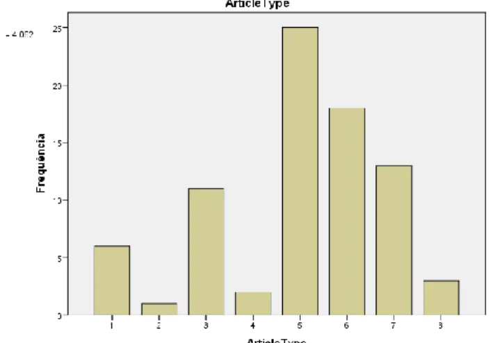
This review incorporated 79 articles, 4882 patients (most of the articles had under 100 patients each), and 13 982 small-diameter implants (most of the articles had under 200 implants each). The mean diameter was 2,58mm, ranging from 1.8 to 3.5mm. The relation between publication year and number of studies is given in figure 1, showing that most of the studies included were recent. The review included 7 RCTs, of which one was double blind, 13 cohort studies of which 2 were retrospective, 43 case series, of which 18 were retrospective, 13 case reports and 3 pilot studies (figure 2),
The main outcomes in this review were survival and success rates of small-diameter implants. Considering all the different survival rates gathered for this review, the mean survival rate was 95,16% (with a standard deviation of 12,21). Only 34 articles reported success rates, and a mean success rate of 96,4% was calculated, with a standard deviation of 4,21 (see tables 5 and 6, appendix A).
Complications and failure reasons were also recorded (figure 3). Implants failed mainly due to biological reasons, either, because they failed to achieve osseointegration or because they developed peri-implantitis, instead of failing due to fracture. Within the biological failures, early failures due to unsuccessful osseointegration seemed to be
Figure 1- Distribution of studies according to their publication year.
Figure 2- Frequency of different article types. Code: 1-RCT; 2-double-blind RCT; 3-Prospective Cohort Study; 4-Retrospective Cohort Study; 5-Case Series; 6-Retrospective Case Series; 7-Case Report; 8-Pilot Study.
10
more common than late failures. However the unrelated biological failure data was relatively high. The biological early failures were mainly reported as failed at 2nd stage surgery, failed to osseointegrate and failed due to insufficient healing or infection. Late biological failures were reported as failed at follow-up, failed due to late peri-implantitis, or due to excessive bone resorption over time.
Implant fractures occurred mostly in fixed single restorations (5 late fractures, 4 early fractures, and 6 unspecified fractures) and overdentures (5 early fractures and 3 late fractures). One unspecified fracture occurred with a partial restoration.
Although the data of general unreported failures was high, they occurred all in one study (Shatkin & Petrotto, 2012) and were reported as failed mainly due to mobility and some due to fracture, as well as mostly occurring in the first 6 months after implantation, seeming to follow the results of this review.
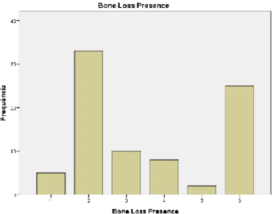
General bone loss was reported across more than half of the studies analyzed (53 studies 63,8%) and mainly under 1mm (figure 4, as well as table 7 - appendix A). However, 25 studies (30,1%) did not mention whether there was or wasn’t any bone loss. Aside from that, prosthetic related and biological complications occurred (figure 5). Prosthetic complications like loosening and fracture of prosthetic components/prosthesis were the most commonly reported, but biological complications like periimplantitis or mucositis occurred as well. Biologic complications ocurred normally once per implant, while prosthetic related complications were sometimes multiple for one implant.
Figure 3- Percentage of failed implants according to failure reason. Code: BE- Biologic Early failure; BL- Biologic Late; BU- Biologic Undefined; MIE- Mechanical Implant Early; MIL- Mechanical Implant Late; MIU- Mechanical Implant Undefined; U- Undefined failures.
11
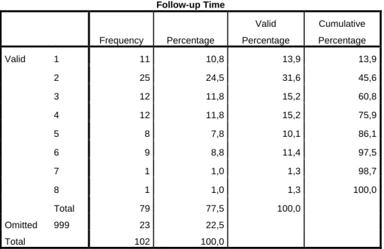
The mean follow-up time across studies was 2,65 years, ranging from 6 weeks up until 12 years. Considering only studies with a fixed follow-up time, the most common range was between 1 and 2 years (31,6%) (figure 6, as well as table 8 – appendix B). It is important to refer that 8,9% of the studies were under 6 months and 5,1% between 6 months and a year. Studies with a follow-up time interval (or mean value) represented 24,1% of all studies, and the most common reported started between ≥1 and ≤2 years (11,4%), followed closely by the ones beginning < 1 year (10,1%).
The most common implant location across studies was in the mandible (36,6%), followed closely by location in both jaws (35,4%). Regarding the division in anterior
3,6 8,1 12,7 75,6 Complications % BE BL BU PR
Figure 4- Distribution of bone loss across studies. Code: 1- No bone loss; 2- Mean bone loss <1mm; 3- Mean bone loss ≥1 and ≤5 mm ; 4- Unspecified bone loss; 5- Mean bone loss >5mm; 6- Unreported.
Figure 5 – Percentage of reported complications. Code: BE- Biologic Early Complications; BL- Biologic Late; BU- Biologic Undefined; PR- Prosthetic Related.
Figure 6- Follow-up time distribution across studies (years). Code: 1: <1year; 2: ≥1 and ≤2 years; 3: >2 and <5years; 4: ≥5years; 5: Time interval starts <1year; 6: Time interval starts ≥1 and ≤2 years; 7: Time interval starts >2years; 8: Undefined time interval.
12
and posterior, the anterior area was clearly the most commonly reported in the studies (44,3%). Figures 7 and 8 show these results (as well as tables 9 and 10, appendix B).
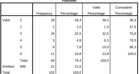
The most common reported functions/restoration types reported in the studies were overdentures (37,6%) and fixed single restorations (32,5%). These findings are shown in figure 9 (and table 11, appendix C).
Figure 9- Function/restoration type of SDIs across studies. Code: 1-overdenture; 2-overdenture (partial palate coverage); 3-fixed single; 4- fixed partial; 5- fixed various; 6- both fixed and removable prosthesis
Studies with both overdentures and fixed single restorations reported the insertion of their implants mainly in the anterior regions. However, studies with overdentures reported the implant insertion mainly in the mandible, while studies with fixed single restorations mostly reported implant insertion in both jaws.
Figure 7: Distribution of implant location in upper and lower jaw across studies. Code: 1-mandible; 2-maxilla; 3- both jaws; 4- unreported.
Figure 8: Distribution of implant location in anterior or posterior sites across studies. Code: 1- anterior; 2- posterior; 3-both anterior and posterior; 4-unreported.
13
The most common surgical access technique reported in each study seems to have been with raising a tissue flap, although the unreported data is very high, and the most common surgical approach was the one-stage approach. Data is shown in figures 10 and 11 (and tables 12 and 13, appendix C).
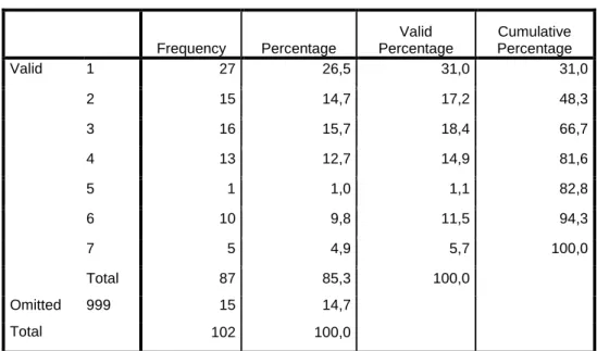
The most common loading protocol in each study was the immediate protocol (48,2%) (immediate loading more common than immediate restoration). Figure 12 shows this data (as well as table 14, appendix D).
Figure 12- Different loading protocols followed by studies. Code: 1- immediate loading (until 48h, with occlusion); 2- immediate restoration (until 48h, without occlusion); 3- early loading (>48h and <3months); 4- conventional loading (≥3months and ≤6months); 5-delayed loading (>6months); 6-various/unspecified; 7-unreported.
Figure 10- Distribution of surgical access technique across studies. Code: 1- flapless; 2- with flap; 3- various; 4-unreported.
Figure 11- Different surgical approaches followed by studies. Code: 1- one-stage; 2- two-stage; 3- various; 4- unreported.
14
Implants restored with overdentures were mostly inserted following the immediate loading protocol (57,1%) and the “various” loading protocol (22,6%). Depending on implants inserted initially in each loading protocol, the most failures occurred within the “various” protocol (18,8%) and the immediate loading protocol (16,8%), as seen in table 3.
Table 3- Relation between inserted and failed implants restored with overdentures, depending on the loading protocol.
Implants restored with fixed single restorations were inserted mainly following the immediate restoration (30%), the “various” (29,3%) and the early loading protocol (24,2%). Depending on implants inserted initially in each loading protocol, the most failures occurred however within the early loading protocol (5,1%) and the conventional loading protocol (2,5%), as shown in table 4.
Fixed Single Restorations Loading time code Number of inserted implants % of inserted implants Number of biologic failures % of failed implants depending on loading protocol 1 33 4,1% 0 (0/33)*100 = 0% 2 243 30% 3 1,2% 3 196 24,2% 10 5,1% 4 79 9,8% 2 2,5% 5 1 0,1% 0 0% 6 237 29,3% 3 1,3% 7 21 2,6% 0 0% Total 810 100% 18 2,2%
Table 4- Relation between inserted and failed implants restored with fixed single restorations, depending on the loading protocol.
There was a higher percentage of failures with overdentures than with fixed single restorations
The Pearson linear correlation between survival rate and loading protocol showed a very weak positive correlation, meaning that, when loading time increases, survival rate has a tendency to also increase. Nevertheless, this data showed no statistical significance, as seen in table 15, appendix D.
Overdentures Loading time code Number of inserted implants % of inserted implants Number of biologic failures % of failed implants depending on loading protocol 1 672 57,1% 113 (113/672)*100 = 16,8% 3 171 14,5% 18 10,5% 4 67 5,7% 6 9% 6 266 22,6% 50 18,8% Total 1176 100% 187 15,9%
15 IV- Discussion
Although SDIs have been introduced in the literature over 20 years ago and researched for a long time, the clinical data available lacks not only studies with high levels of evidence but also comprehends mainly short-term follow-ups. Moreover,there is a high incidence of studies comprehending a time interval for follow-up, instead of a fixed time period, which makes the interpretation of data more difficult. Consequently the possibility to draw conclusions for SDIs appears to be susceptible to high risk of bias, and the data must be interpreted with caution.
The data collected about the function/type of rehabilitation used with SDIs seems to indicate that SDIs are still being used within the range of their indications, which were previously described. Implants were mainly inserted in the anterior region, either in the mandible alone mostly restored with overdentures, or in both jaws mostly restored with fixed single restorations. Although some SDIs are already being used in posterior locations or for partial fixed restorations, the majority remains in lower occlusal bearing sites and overdentures.
Since SDIs have been introduced in the world of definitive prosthetics to minimize the surgical invasiveness associated with bone augmentation, it was considered interesting to find out if the SDIs are being inserted following a flapless or tissue flap approach, as well a one-stage or two-stage surgical approach. Besides the data collected indicating a slightly higher incidence for the tissue flap surgical access, no conclusions could be made due to the high incidence of unreported data. The one-stage surgical approach however, was the most commonly followed for SDIs placement, meaning that most of the studies avoided the need of a second surgery.
In this review the mean survival rate for SDIs was 95,16% (with a standard deviation of 12,21). The standard deviation encountered was relatively high, which could be related to considering the survival rate of a study without taking into consideration the number of implants reported within that study. For example, a case report of a failure of one implant ended up having the same significance as a case series with 43 implants reporting a 100% survival rate.
Regardless of that, the available literature (about SDI used for definitive prosthesis) seems to support the survival rate data: Klein et al. reviewed the survival of
16
implants ≤3.5mm and reported a survival rate of mostly > 95% with no study reporting survival rates below 88%, for a follow up between 1 and 8 years (Klein et al., 2014). Bidra et al. reviewed the survival rate of implants with a diameter <3mm, and reported a survival rate of 94,7% for a follow-up of up to 1 year (Bidra et al., 2012). Altuna et al. reviewed implants with a diameter between 3 and 3.5mm made of Ti-Zr, stating that both survival and success rates were high (Survival: 98.4% - 12 months, 97.7% - 24months; Success: 97.8% - 12 months, 97.3% - 24months) (Altuna et al., 2016). Sohrabi et al. reviewed implants with a diameter ≤3.5mm stating a survival rate of over 90%, for a follow-up time between 5months and 9 years (Sohrabi et al., 2012). Sierra-Sánchez et al. reviewed implants with a diameter between 3 and 3.5mm, and reported a survival rate above 90%, for a follow-up between 1 and 12 years (Sierra-Sánchez et al., 2014). Gleiznys et al. reviewed implants with a diameter ≤3.3mm reporting a survival rate of more than 91% for a follow-up between 4months and 8years (Gleiznys et al., 2012).
Almost all of these reviews considered the survival rate of SDIs to be comparable to regular-diameter implants in short-term follow-up (Altuna et al., 2016; Klein et al., 2014; Sierra-Sánchez et al., 2014; Sohrabi et al., 2012).
Since regular-diameter implants have a longer history of use, they have been studied over a longer period of time, and therefore more clinical data is available for long-term follow-up. Pjetursson et al. reviewed implant therapy outcomes between studies published before and after the year 2000, and stated that for studies published after the year 2000 their 5year survival rate was 98,1% (Pjetursson et al., 2014). Srinivasan et al. reviewed implant therapy in elderly patients, reporting a survival rate of 97,7%, 96,3%, 96,2% and 91,2% , for a 1, 3, 5 or 10 year follow-up, respectively (Srinivasan et al., 2016). Moraschini et al. reviewed implant therapy in longitudinal studies of at least 10 years, and reported an implant survival rate of 94,6% (and success of 89,7%) for a mean 13,4years (Moraschini et al., 2015).
Taking all of this into consideration, it seems to be possible to consider SDI’s survival comparable to regular-diameter implants, at least for the short-term follow-up. Longer follow-up studies are necessary to confirm these findings.
Implant survival is however not the only important factor to define the clinical indications and attention to other factors must be drawn as well. Complications and
17
failure reasons are of significant matter and generally reported in studies. Success rate is also important, but unfortunately not even half of the studies included in this review reported their implant success rate and therefore no conclusions could be drawn about success.
In an attempt to understand and hopefully lessen complications and failures, one must be aware of the several associated factors related either to the implant system itself, to the patient, or the clinician. Implant system failures might be associated to poor body design, insufficient implant size or number of implants, a large micro-gap and abutment/implant precision, or the implant surface. Patient related failures might be associated to parafunctional habits, smoking, insufficient oral hygiene or a preexisting medical condition. Clinician related failures can be either pre- or intra-operative, post-surgical or restorative. Pre-operative factors might be related to poor quality/quantity of hard or soft tissue; intra-operative factors could be excessive drill speed and/or pressure, insufficient irrigation or oversized/undersized osteotomy; post-surgical factors might be related to follow-up wound care or infection control and restorative factors could be related to prosthesis materials, occlusion or cantilever length (Babbush, 2011).
As stated in the introduction, SDIs have always been associated to potential biomechanical risk factors, due to their structural weakness and reduced contact area with the bone (compared to regular-diameter implants). To overcome these difficulties, a few options have been described in the literature, such as different implant body designs, surfaces, materials or implant types.
The screw body design is the most commonly reported in literature, and offers improved initial stability as well as an increased bone-implant surface area (C. E. Misch, 2008) (Gaviria et al., 2014). In addition, rough titanium surfaces offer faster bone deposition (Silvasan, 2010), and research in implant dentistry has shown, that longer implants guarantee better success rates and prognosis than shorter implants (Gaviria et al., 2014).
Implants have been manufactured with many materials over the years (Gaviria et al., 2014). SDIs have been mainly manufactured with titanium alloys, such as Ti6Al4V, since they are stronger and more fatigue resistant than pure Ti. However, despite strengthening the implant, these alloys have been shown to have relatively poor bone-to-implant contact (Altuna et al., 2016; Gaviria et al., 2014; Klein et al., 2014).
18
Recently a new titanium-zirconium alloy (Roxolid; Institut Straumann AG) has been developed, with increased fatigue resistance and unimpaired biocompatibility compared to commercially pure Ti (Klein et al., 2014). Its properties seem to allow the use of SDIs even in clinically challenging situations (Müller et al., 2015), but the clinical evidence regarding the use of Ti–Zr SDIs is still limited (Altuna et al., 2016).
Implants are also available as one- or two-piece implants. The one-piece implant design may be beneficial when using SDIs, because the unified structure of implant and abutment increases the strength of the implant, compared to the two-piece design (Prithviraj et al., 2013; Raviv et al., 2013). Also, as the one-piece design does not have a micro-gap between the implant body and abutment connection, the crestal bone loss may be reduced (C. E. Misch et al., 2004; Prithviraj et al., 2013).
Unfortunately the data collected in this review about SDIs anatomy, surface, material and type was insufficient to be reported and is therefore not further discussed. However the availability of these options, and the fact, that this review reported a relatively low mechanical failure rate, might perhaps indicate that the implant related factors are being accounted for when planning a SDI treatment.
Nonetheless, some early and late implant failures did occur. Early implant fracture may have occurred due to insufficient preparation of the bone or forced placement of the implant through excessive torque, and late implant failures might be related to overload during function or undiagnosed parafunctional patient habits (Babbush, 2011).
Also, a high incidence of prosthetic related complications was reported. These complications were obviously higher than other complications because one implant may report several different prosthetic complications, nonetheless their general prevalence was still high. These complications might have occurred due to material difficulties or overload factors.
The failures encountered in this review were however mostly related to biological reasons, such as failure to achieve or maintain osseointegration and not fracture of the implant. An attempt to better understand the biological failures and complications was made.
19
quality of bone available and can be divided into different categories. These categories are directly related to the loading protocol.
As also mentioned before, the load bearing capacity of an implant has to be greater than the anticipated load during function and when these loads exceed the load-bearing capacity of the implant-bone interface or the implant, biological or mechanical failure can occur.
Conventional loading protocols have been achieved with a two-stage surgical technique, and an unloaded healing period of 3 to 6 months (Silvasan, 2010) (Atieh, Payne, Duncan, de Silva, et al., 2009). Nowadays, it is possible to immediate and early load implants with outcomes comparable to conventional protocols. However, a correct treatment planning is necessary, as well as high primary stability of the implant at the insertion time and good bone quantity and quality (Silvasan, 2010). In this review, the immediate protocol was the most commonly reported across studies, meaning, that most of the implants were loaded or restored out of occlusion within 48h.
Most of the fixed single restorations in this review followed an immediate restoration or “various” loading protocol. Contrary to what one may expect, the most biologic failures occurred however within the early loading protocol, followed by the conventional loading protocol. According to Benic et al., immediately and conventionally loaded single-implant crowns are equally successful regarding implant survival and marginal bone loss (Benic et al., 2014). Degidi et al. showed in his RCT, that for small-diameter implants rehabilitating a maxillary lateral incisor, no statistical difference was found between immediately and one-stage restoration protocols, regarding implant survival, bone loss and probing depth (Degidi et al., 2009b). Although further investigation is necessary, it might be a possibility to speculate that, when SDIs are restored with fixed single restorations within the range of their indications, they can be loaded within the protocol best suited for the patient.
Most of the overdentures were inserted following an immediate loading as well as a “various” loading protocol, and most of the biologic failures also occurred in the “various” and immediate loading protocols. According to Schimmel et al., even though all loading protocols provide high survival rates for implant-supported overdentures, early and conventional loading protocols are still better documented than immediate loading and seem to result in fewer implant failures during the first year (Schimmel et
20
al., 2014). Moreover, according to a clinical report by Maryod et al. regarding the analysis of immediate versus early loading of mini-implants supporting mandibular overdentures, even though both loading protocols show good clinical results, early loading appears to be preferable to immediate loading (Maryod et al., 2014). Although further research is also required, it might be necessary to consider early or conventional loading of overdentures, whenever immediate loading isn’t ideal.
Biologic failures occurred to a higher percentage with overdentures than with fixed single restorations. This could be related to the fact, that overdentures generally receive higher functional loads than fixed single restorations in low occlusive bearing sites, however, this hypothesis was not further investigated.
Even though the Pearson correlation between general loading time and survival rate did not have any statistical significance and considering, that these findings might be related only to chance/coincidence, there was reported a slight tendency for survival rates being higher, the later the occlusal load was applied.
Considering this, as well as all the findings previously discussed, the higher biological failures encountered in this review might be related to a potential overload of the implants (or more precisely the implant-bone interface) during the healing phase. Further research is necessary to address these speculations.
Nonetheless other factors related to general implant complications cannot be excluded, such as patient related factors like infection or healing difficulties due to inappropriate oral hygiene or a preexisting medical condition, as well as preoperative factors such as poor quality or quantity of available bone.
The biological complications reported might be related to the same possibilities mentioned for failure reasons.
Bone loss also had a high prevalence across studies. Nevertheless bone loss until 2 mm around the implant neck during the first year after functional loading has been assumed normal by the dental community. Tissue stability is expected 1 year after placement, and only then a bone loss of more than 0.2 mm per year is regarded as undesirable (Galindo-Moreno et al., 2015).
regular-21
diameter implants was made, and even though most of the studies did not consider complications and failure reasons separately, complications were also reported with regular-diameter implants. Biological complications such as mucositis and peri-implantitis were mentioned in all of the studies, as well as technical/mechanical complications such as loosening or fracture of prosthetic components (although seemingly to a lower extent) (Moraschini et al., 2015; Pjetursson et al., 2014; Srinivasan et al., 2016). Moraschini et al. also stated that the main failures occurred after loading, and Pjetursson et al. considered implant fractures a rare complication with regular-diameter implants (Moraschini et al., 2015; Pjetursson et al., 2014).
It is difficult to establish a correlation between complications and failure reasons in regular- and small-diameter implants, since the data is mostly given in a descriptive way and frequencies across different studies cannot be easily compared. Consequently it was only possible to conclude, that the reported complications with SDIs also occur with regular-diameter implants, only maybe to other extents, which cannot be assessed.
To find out the current availability of small-diameter implants in the market, the offered SDIs were investigated, and it was concluded that most of the commercially available brands still restrict the indications of their SDIs to the ones previously explained, with only few brands extending their indications.
Nonetheless, due to less surgical invasiveness, simpler surgeries and showing results in poor bone quantity and quality, for being more economical and a vaster amount of clinicians being able to place them, the popularity of small-diameter implants has been increasing over the years and is expected to continue to do so (R. Choi & Campbell, 2006).
This review is presented with some limitations such as: the vast amount of studies included with low levels of evidence and therefore high risk of bias in included data; the fact that most of the data wasn’t counted for number of implants but for number of studies, creating a risk of bias especially when relating one variable with another; the fact that data was collected in a descriptive way and no statistical analysis was performed.
22 V- Conclusion
Within the limitations of this review, the survival of small-diameter implants can be considered comparable to regular-diameter implants in a short-term follow-up. More randomized clinical trials with longer follow-up times are necessary to confirm the long-term survival of SDIs.
SDIs seem to have a low fracture rate within their range of indications, showing higher biological failures. Early biological failures could be related to the high incidence of immediate loading protocols, however more research is necessary to address these speculations.
More research is also necessary to study the SDIs behavior in different loading protocols especially considering different restoration types, as well as to investigate their behavior in higher occlusive demanding situations, such as posterior locations.
A VI- Appendices
Descriptive Statistic
N Minimum Maximum
Mean/
Average Standard Deviation
% Survival (%) 86 0 100 95,16 12,207
N valid (listwise) 86
Table 5 – Survival rate data.
Descriptive Statistic
N Minimum Maximum
Mean/
Average Standard Deviation
% Success (%) 34 81 100 96,40 4,208
N valid (listwise) 34
Table 6- Success rate data.
Bone Loss Presence
Frequency Percentage Valid Percentage Cumulative Percentage Valid 1 5 4,9 6,0 6,0 2 33 32,4 39,8 45,8 3 10 9,8 12,0 57,8 4 8 7,8 9,6 67,5 5 2 2,0 2,4 69,9 6 25 24,5 30,1 100,0 Total 83 81,4 100,0 Omitted 999 19 18,6 Total 102 100,0
B Follow-up Time Frequency Percentage Valid Percentage Cumulative Percentage Valid 1 11 10,8 13,9 13,9 2 25 24,5 31,6 45,6 3 12 11,8 15,2 60,8 4 12 11,8 15,2 75,9 5 8 7,8 10,1 86,1 6 9 8,8 11,4 97,5 7 1 1,0 1,3 98,7 8 1 1,0 1,3 100,0 Total 79 77,5 100,0 Omitted 999 23 22,5 Total 102 100,0
Table 8- Frequency data of follow-up time.
Location: mandible/maxilla Frequency Percentage Valid Percentage Cumulative Percentage Valid 1 30 29,4 36,6 36,6 2 14 13,7 17,1 53,7 3 29 28,4 35,4 89,0 4 9 8,8 11,0 100,0 Total 82 80,4 100,0 Omitted 999 20 19,6 Total 102 100,0
Table 9 – Frequency data of upper or lower jaw location.
Location: anterior/posterior Frequency Percentage Valid Percentage Cumulative Percentage Valid 1 35 34,3 44,3 44,3 2 11 10,8 13,9 58,2 3 19 18,6 24,1 82,3 4 14 13,7 17,7 100,0 Total 79 77,5 100,0 Omitted 999 23 22,5 Total 102 100,0
C Function Frequency Percentage Valid Percentage Cumulative Percentage Valid 1 29 28,4 36,3 36,3 2 1 1,0 1,3 37,5 3 26 25,5 32,5 70,0 4 5 4,9 6,3 76,3 5 8 7,8 10,0 86,3 6 11 10,8 13,8 100,0 Total 80 78,4 100,0 Omitted 999 22 21,6 Total 102 100,0
Table 11 – Frequency data of function/restoration type. Surgical Access Technique
Frequency Percentage Valid Percentage Cumulative Percentage Valid 1 19 18,6 24,4 24,4 2 28 27,5 35,9 60,3 3 6 5,9 7,7 67,9 4 25 24,5 32,1 100,0 Total 78 76,5 100,0 Omitted 999 23 22,5 System 1 1,0 Total 24 23,5 Total 102 100,0
Table 12 – Frequency data of surgical access technique. Surgical Approach Frequency Percentage Valid Percentage Cumulative Percentage Valid 1 58 56,9 71,6 71,6 2 10 9,8 12,3 84,0 3 4 3,9 4,9 88,9 4 9 8,8 11,1 100,0 Total 81 79,4 100,0 Omitted 999 21 20,6 Total 102 100,0
D Loading Protocol Frequency Percentage Valid Percentage Cumulative Percentage Valid 1 27 26,5 31,0 31,0 2 15 14,7 17,2 48,3 3 16 15,7 18,4 66,7 4 13 12,7 14,9 81,6 5 1 1,0 1,1 82,8 6 10 9,8 11,5 94,3 7 5 4,9 5,7 100,0 Total 87 85,3 100,0 Omitted 999 15 14,7 Total 102 100,0
Table 14- Frequency data of loading protocols. Correlations
% Survival (%)
Loading Protocol
% Survival (%) Pearson Correlation 1 ,085
Sig. (bilateral) ,434 N 86 86 Loading Protocol (Code) Pearson Correlation ,085 1 Sig. (bilateral) ,434 N 86 87
E Study Article Code N° Patients (n) N° Implants (n) Diameter (mm) Length (mm) Location Code Ant/ Post Code Surgical Access Code Surgical Approach Code Loading Protocol Code Follow- up time Follow-up Time Code Success (%) Survival (%) Function Code Bone Loss Code Failures (n) Biologic Failure (n) Mecanical Implant Failure (n) Un reported Failures (n) Complications (n) Biological Complic. (n) Prosthetic Related Complic. (n) Total (n) E L U E L U Total (n) E L U All (King et al., 2016) 5 38 62 3 11 - 15 3 1 2 1 3 36 months 3 - 96,8 3 2 2 2 16 1 15 (Schwindling & Schwindling, 2016) 6 25 99 1.8, 2.1, 2.4 10 - 18 3 4 1 1 1 2-87 months 5 - 92 1 6 10 8 2 26 26 (Alan et al., 2015) 3 25 25 2.7 9 - 15 3 4 2 1 4 90 days 9 - 100 5 4 0 - (Mundt, Schwahn, Biffar, et al., 2015) 6 133 738 1.8, 2.1, 2.4 10 - 18 3 3 3 1 6 0,3-5 years 5 - 95,9 1 2 30 26 2 2 - (de Souza et al., 2015) 1 38 152 2 10 1 1 4 1 1 12 months 2 - 89 1 6 16 6 10 505 14 102 389 42 84 82 15 6 9 (Moustafa Abdou Elsyad, 2015) 5 28 112 1.8 12 - 18 3 3 1 1 1 5 years 4 - 97,9 1 6 2 2 413 413 (Banu R et al., 2015) 8 10 112 3.3 13 1 1 4 1 1 4 months 9 100 100 1 6 0 (Müller et al., 2015) 2 47 94 3.3 8 - 14 1 1 4 1 3 5 years 4 95,8 98,9 1 2 3 3 4 3 1 92,6 97,8 2 (Flanagan, 2015) 6 50 79 2 10 - 13 4 4 4 4 7 7 – 92 months 5 92 96,2 6 6 5 4 1 3 3 7 2.5 46 3 (Al-Nawas et al., 2015) 3 233 409 3.3 8 -14 4 3 2 3 6 2 years 2 97,4 97,6 6 4 10 9 1 - (Ioannidis et al., 2015) 1 20 17 3.3 8 -14 3 3 2 2 4 3 years 3 - 100 3 2 0 23 20 3 (Lambert et al., 2015) 5 20 39 3.3 - 4 3 3 1 3 1 year 2 94,7 94,7 4 2 2 2 - (Zweers et al., 2015) 4 75 75 3.3 8 -14 1 4 2 1 3 3 years 3 - 100 1 2 0 - (F. G. Mangano et al., 2015) 5 54 57 2.7 10 - 13 1 1 2 1 1 1-4 years 6 - 96,9 1 2 6 2 4 25 3 2 7 13 174 3.2
F (Miodrag Šćepanović et al., 2015) 3 30 120 1.8 13 1 1 3 1 1 18 months 2 - 98,3 1 2 2 2 - (Mundt, Schwahn, Stark, et al., 2015) 6 133 738 1.8, 2.1, 2.4 10 - 18 2 4 1 1 6 7.2-61.6 months 5 - 94,3 1 6 15 15 97 97 1 95,7 13 9 2 2 (El-Sheikh & Shihabuddin, 2014) 8 20 40 3.3 8 - 12 4 2 2 1 3 12 months 2 - 100 4 2 0 1 1 (Karl et al., 2014) 7 1 1 3.3 12 2 1 4 4 3 11 months 1 - 0 6 6 1 1 - (F. Mangano et al., 2014) 5 279 324 3.3 8 - 14 3 3 4 2 4 1-10 years 6 - 98,7 6 2 1 1 27 2 1 24 (Tolentino et al., 2014) 3 21 21 3.3 8 - 12 3 2 2 1 3 12 months 2 95,2 95,2 3 6 2 2 30 30 21 21 3.3 (Kolerman et al., 2014) 5 9 7 3.3 13 - 16 2 3 2 2 4 6-108 months 5 - 100 6 3 0 38 18 20 23 3.5 (Maryod et al., 2014) 3 30 112 1.8 15 1 1 1 1 1 36 months 3 - 91,7 1 4 5 5 1 1 96,7 2 2 (Preoteasa et al., 2014) 5 23 110 1.8, 2.1, 2.4 10 - 18 3 4 4 1 1 3 years 3 - 92,7 1 4 8 3 3 2 87 72 15 (Ashmawy et al., 2014) 5 12 48 1.8, 2.1 15 1 1 1 1 1 3 months 9 - 100 1 6 0 - (Mohan et al., 2014) 7 1 4 2.4 13 1 1 2 1 3 5 years 4 100 100 3 6 0 0 (Persic et al., 2014) 5 23 61 1.9 - 2.5 10 - 15 3 3 1 1 3 3 months 9 - 100 5 6 0 - (F. Mangano et al., 2013) 5 16 22 2.7 10 - 13 3 2 2 1 2 2 years 2 94,6 100 4 2 0 2 2 15 3.2 (Patel, 2013) 7 1 7 2.5, 3 >10 2 3 1 1 1 9 months 1 100 100 1 1 0 0 (Cordaro et al., 2013) 6 10 40 3.3 10, 12 2 4 4 4 3 12-16 months 2 97,5 100 1 2 0 5 3 2 (Jorge Jofre et al., 2013) 1 15 30 1.8 15 1 1 1 1 1 1 year 2 - 100 1 6 0 1 1 (Lee et al., 2013) 6 338 200 3.3 8 - 15 3 3 4 4 7 1-12 years 6 91,8 98,1 5 2 9 7 2 47 4 43 119 3.4