Universidade Trás-os-Montes e Alto Douro
Immunohistochemical expression of αB-crystallin and L1CAM in
canine mammary tumors
Masters Dissertation in Veterinary Medicine
Francisco de Assis Teixeira Mendes
Advisor: Prof. Dr. Adelina Gama Quaresma
Co-advisor: Prof. Dr. Laura Peña Fernandez
ii
iii
A
CKNOWLEDGMENTSFirst of all, I would like to thank both my advisors for all of their time, patience, support, knowledge and availability to help me throughout this dissertation:
To my co-advisor Laura Peña, for having me in Madrid and making me feel like home. Thank you for all the time you were able to spend with me, even with your (super) busy schedule, for all of the knowledge you gave me and for the amazing 3 months I spent in the the beautiful city of Madrid! I will never be able to thank you enough! Besides her, a special thanks to everyone on the Veterinary Pathology Department of the UCM, specially to Lucia and Angela, for all the kindness and support!
To my advisor, the first person who approached me saying that this is what I should be doing in life, Prof. Adelina. All of the “thank you’s” in the world wouldn’t be enough to thank you for all the work you had with me. You were always available to help me, even when you didn’t have to. You were one of the best choices I made in life, I would recommend you to anyone in the world! Your professional ethics, your knowledge, your humbleness… thank you for sharing all of that with me! Also, a special thanks to everyone in our laboratory, at the Trás-os-Montes e Alto Douro University, for all the moments and knowledge shared throughout my stay either during my internee or my degree.
I would also like to thank all of the team in the Baixo Vouga Veterinary Hospital, for an amazing 4-month stay. A special thanks to Dr Carla Marrinhas, for the guidance and expertise in cytology, even when not present; to my fellow internees, and, obviously, to my girl, my friend, Daniela Ribau. Thank you so much for everything!
Obviously, I have to thank all of my closest and dearest friends: the ones in Guimarães, my hometown, Raquel, Nó, Queirós, Filipa and Joana, thank you for being with me (since forever) even when I know I got so distant from you! I would forever be thankful for your love and support. And to the ones I made in Vila Real: Bia, Helga, Rufino, Barbosa, Raquel, Pilão, Portugal, and more recently, Laura and João, and to everyone that crossed my way in my journey in this city, including, of course, my two pupils Bessa and Chica, which I’m sure will make me even more proud of them than I already am! I would also like to thank Galiza for all the friendship regained and all the advices and company shared in Madrid. A special thanks to Bia, for never leaving my side, even when I felt like everyone else did, and to Helga, for all the life changing advices, for all the times I cried in your shoulder and for the amazing 3 months in Madrid, with all the ups and downs (that I would never, ever, forget) included. You all are the best friends I could have asked for!
iv To Vila Real and UTAD, for everything it gave to me, for being my second home.
To Madrid, for a life changing experience. To Zurich, for representing a second chance.
To all of my family, for all the support during the difficult times, for all the genuine love and friendship.
To my brother in law, Miguel, for always supporting my family in every possible way. For welcoming me in his home, in his family (which I would also like to thank) and for all the help in all of the aspects he could have helped. You will never know how much grateful I am for everything. To my goddaughter, Ritinha, for showing me what true, genuine and innocent love is. For always making me smile. For being so beautiful, so individual, so mine… I will love you forever my little thing! Don’t ever forget that.
To both of my sisters, so different and yet so equals. To Diana, for showing me that even in the most difficult times our inner strength is always enough to get ourselves together, for showing me that there’s nothing wrong with being and expressing yourself, in being different. You are a true inspiration. To Carina, for always being there for me and for welcoming me in her home. For all the endless “stupid” conversations, for all the advices and for being a mom to me when she didn’t have to, for almost 6 years. I admire you so much, as a sister, as a mother, as a woman. To both of you, the biggest thank you I can give. I love you so much you cannot imagine.
And last, but definitely not least, to my foundation, my parents. And this is where I get without words, because there’s truly no words to describe what I feel for you, what I think of you, what I want to say to you. Life was never easy for us, yet you always got a smile on your faces so that we got one in ours. We were always so happy, besides everything that was going on, and that was because we knew we could trust you, we knew you would do whatever it takes to make things right again. Thank you so much for allowing me to follow one of my dreams, even when it made you give up on yours, even when it got you away from your home, your friends, your family. I love you with all of my strength and there’s nothing in this world I wouldn’t do to see you smile. You are my inspiration, you are the reason I never gave up on anything, even when, apparently, there was no choice. Thank you. Thank you for being my heroes.
A thank you will never be enough for any of you, but here it is: thank you for being an important part of my life. I love you all.
v
S
IDEN
OTEThis masters dissertation was elaborated as an integrant part of my internship of the degree in Veterinary Medicine.
My internship consisted in a 3 month stay in Madrid, accompanying the work of Prof. Dr. Laura Peña in the Veterinary Pathology Laboratory of the Department of Animal Medicine, Surgery and Pathology at the Complutense University of Madrid. During this stay, besides our research in L1 CAM, I participated in weekly joint sessions for the exam of the European College of Veterinary Pathology, as well as monthly journal sessions. I also did necropsies, biopsy diagnosis and helped around the normal routine of the laboratory.
I also had a small stay in Vila Real, at the Trás-os-Montes e Alto Douro University, of less more than 1 month, were I accompanied the work of Prof. Dr. Adelina Gama Quaresma, where we did our research with CRYAB.
Besides the work of this dissertation, I also did a 4-month internship in the Baixo-Vouga Veterinary Hospital, where I got the chance to develop my medical skills. At this time I was also able to be involved in the normal routine of the laboratory of the hospital, where blood and urine samples and cytologies were available to evaluation every day, so I also developed my clinical pathology skills.
vii
A
BSTRACTHuman breast cancer is one of the most common neoplasms found in women, being the female dog the most accurate animal model to study it. Research on this field has been growing largely for the past years.
αB-Crystallin (CRYAB) is a member of the small heat shock proteins superfamily, implicated in cellular homeostasis and anti-apoptotic features, while L1 cell adhesion molecule (L1CAM) is a surface glycoprotein of the Ig’s superfamily interacting with the extracellular matrix and other cells and more recently implicated in angiogenesis. In the present work, two separate immunohistochemical studies were performed in canine mammary tumors, one evaluating CRYAB and the other L1CAM expression.
To evaluate CRYAB expression in canine mammary tumors (CMT’s) we used a series of 79 samples, composed by normal/hyperplastic tissues (n=9), benign tumors (n=15) and malignant tumors (n=55). Immunohistochemical evaluation was based on a semiquantitative analysis, according to the immunolabeling percentage (PS) and staining intensity (SI). A final score was obtained, based on the product PSxSI, with tissues classified as low (PSxSI<4) or high scores (PSxSI≥4). The goal was to evaluate the immunolabeling characteristics and to accomplish if there was any relation of CRYAB expression and clinicopathological characteristics, such as lymph-node metastasis or histological groups. Results showed a low expression of CRYAB by the myoepithelium of normal/hyperplastic tissues and a tendency to a higher expression in tumors (either benign or malignant), although not on a significant level. We found significant differences between histological groups regarding CRYAB expression (p=0.022). Positive expression was associated with the absence of lymph-node metastasis (p=0.014). CRYAB was mainly expressed by the myoepithelial cells of tumor samples, although not exclusively. Differences between histological groups suggest CRYAB may be a future factor of prognosis and probably an interesting therapeutic target. Further studies with a larger series are required to explain the differences obtained.
L1CAM expression was evaluated on 27 malignant, grade 3 CMT’s, including inflammatory carcinomas (n=12) and non-inflammatory carcinomas (n=15). Immunohistochemical evaluation was based on a quantitative analysis, according to the immunolabeling percentage (PS) and the staining intensity (SI). A final score was obtained, rising from 0 or 2 to 8, by adding PS+SI. The goal was to evaluate the differences between inflammatory and non-inflammatory carcinomas. We also evaluated L1CAM expression by endothelial cells. Results showed significant differences between the inflammatory and the non-inflammatory group (p=0.003), with an overexpression
viii within the inflammatory group. The staining pattern was also significantly different (p=0.036). Regarding the endothelial cells, a positive correlation was found between the total score of L1CAM and the positivity of tumor and embolized vessels, but not on a significant level (p=0.05). Endothelial cells of normal vasculature showed an overexpression when compared to tumor ones. The latter showed 56% negativity in L1CAM expression (n=14). These results reveal, for the first time in CMT’s, that L1CAM may be a potential therapeutic target in inflammatory carcinomas in female dogs, giving its differences between the two groups. Further studies should be performed in larger series to accomplish more information regarding this marker in the canine species.
ix
R
ESUMOOs tumores de mama representam um dos tipos de cancro mais comuns na mulher, sendo que a cadela é apontada, atualmente, como o melhor modelo animal para o seu estudo. Assim sendo, a investigação neste ramo tem crescido a um ritmo elevado.
A αB-cristalina (CRYAB) pertence à superfamília das small heat shock proteins, influenciando a homeostasia celular e tendo funções anti-apoptóticas, enquanto a molécula de adesão celular L1 (L1CAM) é uma glicoproteína de superfície da família das imunoglobulinas, interagindo com a matriz extracelular e com outras células. Recentemente foi descrito o seu envolvimento na angiogénese. Neste trabalho, foram realizados dois estudos imunohistoquímicos independentes em tumores mamários caninos, tendo um avaliado a expressão do CRYAB e o outro a expressão do L1CAM.
Para avaliar a imunoexpressão do CRYAB em tumores mamários caninos, foram utilizadas 79 amostras, constituidas por tecidos normais/hiperplásicos (n=9), tumores benignos (n=15) e malignos (n=55). A avaliação foi feita com base num método semi-quantitativo, multiplicando a percentagem (PS) pela intensidade da imunomarcação (SI), obtendo o score total (PSxSI), posteriormente dividido em score alto (TS≥4) e baixo (TS<4). Os resultados revelaram uma baixa expressão do CRYAB ao nível do mioepitélio das amostras normais/ hiperplásicas e uma maior expressão em amostras tumorais. Encontrámos diferenças estatisticamente significativas relativamente à expressão do CRYAB entre diferentes grupos histológicos (p=0.022) e entre os diferentes tipos histológicos de neoplasias malignas, sendo mais frequentemente expresso em neoplasias com proliferação mioepitelial. A expressão do CRYAB encontrava-se também associada à ausência de metástases nos linfonodos (p=0.014). Consideramos necessários estudos com uma amostragem maior para confirmar as diferenças obtidas.
A expressão do L1CAM foi avaliada em 27 carcinomas mamários de alto grau histológico, incluindo 12 carcinomas inflamatórios e 15 não inflamatórios. A avaliação foi efetuada baseando-se na percentagem e intensidade de células coradas (PS e SI, respetivamente). O score final foi obtido somando PS+SI, obtendo valores 0 ou 2 a 8. O objetivo foi avaliar se existiam diferenças na expressão do L1CAM entre carcinomas inflamatórios e não inflamatórios. Os resultados revelaram diferenças entre o grupo inflamatório e o não inflamatório (p=0.003), com uma sobrexpressão no grupo inflamatório. O padrão de coloração apresentado pelas células também se revelou significativamente diferente (p=0.036). Relativamente às células endoteliais, encontrámos uma correlação positiva entre a expressão do L1CAM e a positividade de células endoteliais do tumor e do endotélio com êmbolos, mas não a níveis significativos (p=0.05).
x Na vasculatura normal, observou-se sobrexpressão do L1CAM relativamente às células endoteliais do tumor, com 56% (n=14) das mesmas com expressão negativa. Estes resultados revelam, pela primeira vez em tumores mamários caninos, que o L1CAM poderá representar um alvo terapêutico em carcinoma inflamatório, dadas as diferenças encontradas entre os dois grupos. São necessários estudos posteriores, em séries mais numerosas, para reunir mais informação acerca desta molécula.
xi
I
NDEX 1. Introduction ... 1 2. Alpha B-Crystallin ... 5 2.1. Review ... 5 2.1.1. Structure ... 5 2.1.2. Expression ... 5 2.1.3. Interactions/function ... 6 2.1.4. αB-Crystallin in cancer ... 72.1.5. αB-Crystallin as a marker of prognosis ... 9
2.2. Objectives ... 9
2.3. Material and Methods ... 10
2.3.1. Tumor specimens ... 10
2.3.2. Clinicopathological parameters evaluation ... 10
2.3.3. Immunohistochemistry ... 11
2.3.4. Quantification of immunolabelling ... 11
2.3.5. Statistical analysis ... 12
2.4. Results ... 13
2.4.1. Clinicopathological characteristics ... 13
2.4.2. Immunolabelling of CRYAB in the normal mammary gland and hyperplasia ... 15
2.4.3. Immunolabeling of CRYAB in benign neoplasms ... 16
2.4.4. Immunolabeling of CRYAB in malignant neoplasms ... 17
2.4.5. Comparative study of CRYAB immunoexpression with the clinicopathological characteristics ... 18
2.4.6. Immunolabeling and comparative study of CRYAB in the mammary gland adjacent to neoplastic tissues ... 20 2.5. Discussion ... 23 3. L1CAM ... 26 3.1. Review ... 26 3.1.1. Structure ... 26 3.1.2. Expression ... 27 3.1.3. Interactions/Function ... 27 3.1.4. L1CAM in Cancer ... 28
3.1.5. L1 CAM as a marker of prognosis ... 30
xii
3.3. Material and Methods ... 31
3.3.1. Tumor specimens ... 31
3.3.2. Histopathological parameters evaluation ... 31
3.3.3. Immunohistochemistry ... 31
3.3.4. Quantification of Immunolabeling ... 31
3.3.5. Statistical methods ... 32
3.4. Results ... 33
3.4.1. Immunolabeling of L1CAM in malignant neoplasms ... 33
3.4.2. Comparative study of L1CAM expression between the inflammatory and non-inflammatory mammary cancer groups ... 36
3.4.3. Comparative study of L1 CAM expression in ECs of the inflammatory and non-inflammatory mammary cancer groups ... 38
3.4.4. Comparative study of L1CAM TS with the expression of L1CAM by ECs in tumor and embolized vessels ... 38
3.4.5. Other comparative studies ... 39
3.5. Discussion ... 41
4. Conclusions ... 43
xiii
F
IGUREI
NDEXFigure 1 – The Hallmarks of Cancer (Hanahan & Weinberg, 2011) ... 3
Figure 2 - Immunohistochemical expression of CRYAB in canine mammary tissues ... 22
Figure 3 – L1 CAM structure and cleavage sites (adapted from Kiefel et al., 2012) ... 26
Figure 4 - Immunohistochemical expression of L1CAM in canine mammary tissues ... 40
T
ABLEI
NDEX Table 1 - Quantification of CRYAB immunolabeling ... 12Table 2 - Clinicopathological characteristics of the present series. ... 14
Table 3 - Histological diagnosis and respective frequencies ... 15
Table 4 - Immunolabeling of CRYAB in normal/hyperplastic mammary gland ... 16
Table 5 - Immunolabeling of CRYAB in benign neoplasms ... 17
Table 6 - Immunolabeling of CRYAB in malignant neoplasms ... 18
Table 7 - Comparative study of CRYAB GS with the clinical characteristics ... 19
Table 8 - Comparative study of CRYAB GS with the histopathological characteristics ... 20
Table 9 - CRYAB immunolabeling of the adjacent mammary gland of benign and malignant tumors... 21
Table 10 – Quantification of L1CAM immunolabeling (adapted from Allred et al., 1998) ... 32
Table 11 - Immunolabelling characteristics of L1 CAM in epithelial and endothelial cells ... 34
Table 12 - Comparative study of L1 CAM expression in epithelial cells with the inflammatory and non-inflammatory mammary cancer groups ... 37
Table 13 - Comparative study of L1 CAM expression in endothelial cells of the inflammatory and non-inflammatory mammary cancer groups ... 38
Table 14 - Comparative study of L1CAM TS with the expression of L1CAM by ECs in tumor and embolized vessels ... 39
xiv
A
BBREVIATIONS,
ACRONYMS AND SYMBOLS™ - Trademark
® - Registered trademark
ADAM – A desintegrin and metalloproteinase AG – Adelina Gama
ALCAM – Activated leucocyte cell adhesion molecule AMG – Adjacent mammary gland
Anapl. – Anaplastic AR – Androgen receptor Bcl-2 - B-cell lymphoma 2 CA – Complex Adenoma
CAMs – Cell adhesion molecules Carc. – Carcinoma
COX-2 – Cyclo-oxygenase 2 CRYAB – αB-Crystallin
DAB – 3,3'-diaminobenzidine tetrahydrochloride ECM – Extracellular matrix
ECs – Endothelial cells
EGFR – Epidermal growth factor receptor EMT- Epithelial to mesenchymal transition ER – Estrogen receptor
ERK – Extracellular signal-regulated kinases FAK – Focal adhesion kinase
FGFR – Fibroblast growth factor receptor FM – Francisco Mendes
GS – Global Score
HE – Hematoxylin and eosin HPF – High power fields Hsps – Heat shock proteins ICD – Intracellular domain Ig – Immunoglobulin
IMC – Inflammatory mammary cancer INF-Ɣ – Gamma interferon
xv kDa- kiloDalton
L1CAM – L1 Cell adhesion molecule LP – Laura Peña
MAPK – Mitogen-activated protein kinases MBT – Mixed benign tumor
Myoep. – Myoepithelioma
N-CAM – Neural cell adhesion molecule NF-1A – Nuclear factor 1A
NF-ĸB – Nuclear factor kappa B
NIMC – Non-inflammatory mammary cancer OHE - Ovariohysterectomy
PBS – Phosphate Buffered Saline PR – Progesterone receptor PS – Percentage of stain
REST – RE1 Silencing transcription factor RGD-motif – Arginyl-glycyl-aspartic acid motif RNA - Ribonucleic acid
sHsps – Small heat shock proteins superfamily SI – Staining intensity
Src – Proto-oncogene tyrosine-protein kinase Src TAG-1 – Transient axonal glycoprotein 1
TGF-β1 – Transforming growth factor betta 1 TNF-α – Tumor necrosis factor alpha
TNM – Method of classification of malignant tumors: T-Tumor; N-Lymph Node; M-Metastasis
TRAIL - TNF-related apoptosis-inducing ligand TS – Total score
UCM – Universidade Complutense de Madrid (Complutense University of Madrid) UV – Ultraviolet
VEGF-A – Vascular endothelial growth factor A WHO – World Health Organization
1. I
NTRODUCTIONCanine mammary tumors (CMTs) are the most common neoplasm found in intact female dogs, representing around 50% of all tumors diagnosed (Merlo et al., 2008; Vascellari et al., 2009; Sleeckx et al., 2011; Salas et al., 2015). Around 50% of these are malignant, being most of them of epithelial origin. This makes carcinomas the most frequent malignant mammary neoplasms diagnosed in female dogs, with sarcomas (malignant mesenchymal tumors) representing less than 5% of the diagnosed neoplasms (Sleeckx et al., 2011). Lately, the incidence of CMTs is decreasing in countries where ovariohysterectomy (OHE) is being performed at an early age as a preventive measure to avoid CMTs (Sleeckx et al., 2011).
Histopathological classification of CMTs is highly important, since different histological subtypes may have a different prognosis. For instance, an anaplastic carcinoma has the worst prognosis of all subtypes of CMTs and a complex carcinoma is usually associated with a good prognosis, when we are strictly talking about histopathological classification by itself (Goldschmidt et al., 2011). Nowadays, the most globally accepted classification system to CMTs was published in 1999 by the World Health Organization. Since then, research in CMTs has been arising interest and numerous investigations have increased the knowledge in this area. With that in mind, Goldschmidt et al. (2011) have proposed a new classification system, introducing new subtypes of CMTs. Throughout this dissertation, this updated classification will be used to classify CMTs.
Besides classification, grading of malignant CMTs is also of high importance, since it can influence prognosis and treatment protocols. The grading system relies in 3 major features of malignancy: tubule formation, nuclear pleomorphism and number of mitoses per 10 high power fields (HPF). These features are classified from 1 to 3 points and then summed up to obtain a total score (from 3 to 9 points). The higher the total score, the higher the grade and, consequently, the higher the malignancy (Peña et al., 2012).
CMTs are now divided in 8 major categories: malignant epithelial neoplasms (carcinomas), special types of carcinomas, malignant mesenchymal neoplasms (sarcomas), malignant mixed mammary tumors (carcinosarcomas), benign neoplasms, hyperplasia or dysplasia, neoplasms of the nipple and hyperplasia/dysplasia of the nipple (Goldschmidt et al., 2011). Sometimes, histologic evaluation by itself is not enough to distinguish between different types of tumors and immunohistochemistry must be performed to classify the CMT(Goldschmidt et al., 2011; Peña et al., 2014).
Risk factors to develop CMTs include age, breed, genetics, hormones, diet, among others (Sleeckx et al., 2011). Older animals have an increased risk of developing CMTs, probably
2
because of their longer exposition to the ovarian hormones that are, by themselves, a risk factor too. These hormones stimulate the growth of the mammary tissue under physiologic conditions (steroids induce epithelial proliferation), and they probably also induce tumorigenic growth. Since this hormonal exposure occurs in each oestrus, it is easily understandable that female dogs tend to get more sensitive to this exposure as they get older. Knowing this, preventive spaying at an early age is a key factor to fight the high prevalence of CMTs. It is now known that female dogs spayed before the first oestrus have a risk of 0,5% of developing CMTs, while dogs spayed before the second oestrus or after the second oestrus have a risk of 8 or 26% of developing CMTs, respectively (Sleeckx et al., 2011). Breed or genetic predisposition is also thought to influence the development of CMTs, but studies are contradictory. OHE by the time of tumor removal was found to have positive influence in the prognosis of dogs with estrogen receptor positive mammary carcinomas, grade 2 CMT’s or with increased peri-surgical serum E2 concentration (Kristiansen et al., 2016).
Treatment of CMTs usually implicates surgery (except those cases of inflammatory carcinoma, where surgery is not indicated) and can involve post-surgical chemotherapy (Sleeckx et al., 2011; Tran et al., 2016). Radiotherapy, hormonal therapy, antiangiogenic strategies, among others, are not routine treatment protocols for CMTs, and surgery remains the gold standard of treatment, especially in early stage, low grade tumors (Sleeckx et al., 2011). Other treatment protocols are more useful in malignant cases, when metastasis are present, or in which surgery cannot be performed, either by anesthetic precautions or by surgical planning (Sleeckx et al., 2011; Tran et al., 2016).
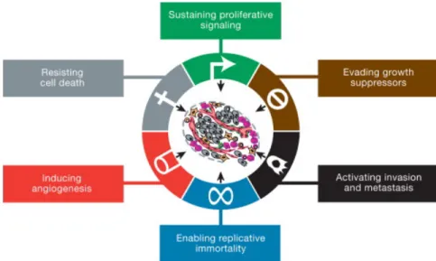
Mechanisms responsible for the conversion of a regular cell into a cancerous one are very complex but have been subject of deep investigation from cancer researchers. We are now able to identify 6 major key events/features that a cell needs to achieve malignancy. These 6 features (summarized in Figure 1) represent the result of years of cancer investigation, either in humans, animals and in vitro, and resume all the features that researchers believe that are responsible for the tumor formation and proliferation (Hanahan & Weinberg, 2000). These features are the following, including some examples of each of them (Hanahan & Weinberg, 2000):
1. Self-sufficiency in growth signals – The overexpression of HER-2 in mammary carcinomas; 2. Insensitivity to anti-growth signals – The phosphorylation of the retinoblastoma protein,
suppressor of growth;
3. Evading apoptosis – The inactivation of the gene p53 which is a tumor suppressor; 4. Limitless replicative potential – Activation of telomerase;
3 5. Tissue invasion/metastasis – Changing properties of the cell-cell adhesion molecules
(CAM’s) and integrins (cell-extracellular matrix adhesion); 6. Sustained angiogenesis – Increased expression of VEGF.
More recently two more features have been proposed, and are now classified as “emerging hallmarks”, until further prove is acquire that they are transversal to almost every type of cancer, such as the previous six. These two new features are (a) the capability of a cell to deregulate the cell metabolism and energetics and (b) to avoid destruction by the immune system (Hanahan & Weinberg, 2011).
Each of these features represents a further step in the pathway to the cancerous capacity of cells, which is believed to happen in all (or almost all) types of cancer. At the molecular level, these features are way too complex to be the subject of study on this dissertation. Since we could not talk about all of these, and considering the material we had in our power, we decided to investigate two different molecules that could fit in some of the categories of the hallmarks of cancer.
αB-Crystallin (CRYAB) is a member of the small heat chock protein superfamily (sHsps),
which is a group of molecules responsible for the maintenance of the cellular homeostasis (Sun & MacRae, 2005). It is expressed in response to multiple stress factors including heat shock, cytokines, and others (Ilhan et al., 2010; Guvenc et al., 2012). It was considered as a marker of poor prognosis (Moyano et al., 2006), as well as a marker for basal-type and metaplastic breast carcinomas in women (Moyano et al., 2006; Sitterding et al., 2008; Chan et al., 2011).
4
L1 cell adhesion molecule (L1CAM) is a transmembrane glycoprotein involved in
cell-cell interaction as well as cell-cell to extracell-cellular matrix interactions (Homrich et al., 2015; Samatov et al., 2016) discovered in mice brain in 1984 (Rathjen & Schachner, 1984). Its expression is regulated by a variety of genes namely RE1-Silencing Transcription factor (REST) (Kiefel et al., 2012; Samatov et al., 2016) and others. Overexpression of this molecule has been correlated to poor prognosis, chemotherapy resistance, shorter disease-free interval, high grades of malignancy, epithelial to mesenchymal transition (EMT), among others (Kiefel et al., 2012; Kiefel et al., 2012; Zhang et al., 2015; Altevogt et al., 2016; Samatov et al., 2016).
5
2. A
LPHAB-C
RYSTALLIN2.1. Review
2.1.1. Structure
αB-Crystallin (CRYAB) is a heat shock protein, belonging to the small heat shock proteins superfamily (sHsps). The heat shock proteins are divided in 6 major families according to their molecular weight, being those the Hsp 100, Hsp 90, Hsp 70, Hsp 60, Hsp 40 and, for last, the small heat shock proteins (Bakthisaran et al., 2015). The latter represent a group of molecules responsible for the maintenance of the cellular homeostasis (Sun & MacRae, 2005), preventing cell damage and promoting cell survival (Clark & Muchowski, 2000; Moyano et al., 2006). CRYAB belongs to a protein family composed by three classes, including alpha, beta and gamma crystallins. The group of alpha crystallins includes both acid and basic crystallins, giving rise to either αA-Crystallin or αB-Crystallin, respectively (Ilhan et al., 2010). Composed by 175 amino acids residues (Wistow, 1985), CRYAB has a molecular weight of 22 kDa, but its tridimensional structure is not well defined yet (Ilhan et al., 2010). However, sHsp are in general composed by a conserved α-crystallin core domain, a N-terminal domain and a C-terminal extension domain, with typical weights between 12 and 43 kDa (Ghosh et al., 2005; Bakthisaran et al., 2015; Tikhomirova et al., 2017).
CRYAB is expressed in response to multiple stress factors including heat shock, cytokines, and others (Ilhan et al., 2010; Guvenc et al., 2012). It is considered as a marker of poor prognosis in breast cancer (Moyano et al., 2006), as well as a marker for basal-type and metaplastic breast carcinomas in women (Moyano et al., 2006; Sitterding et al., 2008; Chan et al., 2011).
2.1.2. Expression
First reported in the human lens (Delaye & Tardieu, 1983), where it is responsible for maintaining the lens transparency (Delaye & Tardieu, 1983), it was readily proved that its expression was extended to the heart, brain, spleen, kidney, lung and almost all vertebrate cells (Dubin et al., 1989). In humans, the gene encoding CRYAB is located on chromosome 11 (Ngo et al., 1989).
In human breast, αB-Crystallin was shown to be expressed by the myoepithelial cells in normal tissue samples, proliferative diseases and myoepithelial cells surrounding the area of in situ carcinomas (Moyano et al., 2006; Sitterding et al., 2008). It is also expressed by adipocytes (Moyano et al., 2006; Sitterding et al., 2008; Chan et al., 2011) and the vascular smooth muscle wall (Chan et al., 2011). Luminal epithelium of breast samples was always negative in these
6
situations (Moyano et al., 2006; Sitterding et al., 2008; Chan et al., 2011). Expression by tumor cells is further discussed below.
2.1.3. Interactions/function
In human medicine, the small heat shock proteins are involved in cataracts formation, desmin-related myopathy, neurological diseases (such as Alzheimer, Parkinson or Huntington’s Syndrome), and cancer formation/progression (Sun & MacRae, 2005).
Regarding αB-Crystallin, it is responsible for maintaining cellular homeostasis by acting as a molecular chaperone (class of proteins responsible for maintaining the original structure of cell proteins (Tikhomirova et al., 2017)), preventing protein aggregation/denaturation and consequent cellular damage. This type of damage occurs because of the sHsps response to oxidative stress, heat shock or other stress factors (Sun & MacRae, 2005; Ilhan et al., 2010; Guvenc et al., 2012; Bakthisaran et al., 2015; Tikhomirova et al., 2017). It is also responsible for conceding antiapoptotic features to cells (Malin et al., 2016).
As stated before, CRYAB is responsible for maintaining the lens transparency, since it avoids protein aggregates formation and inhibits apoptosis and consequent protein degradation in the lens. It is also reported as having an important role in maintaining the heart muscle functional integrity and preserving its contractility. In the brain, its overexpression is related to neurodegenerative diseases such as Alzheimer, Parkinson, lateral amyotrophic sclerosis and Alexander’s disease, but its role in these diseases is not the scope of the present dissertation. As a curiosity, CRYAB was overexpressed in the brain of patients with Alzheimer, and an association was found between CRYAB and the deposit of the amyloid plaques in astrocytes, a characteristic feature of this disease (Boelens, 2014).
It was linked to the inhibition of the myogenic apoptosis through inhibition of the protease caspase-3 (Kamradt et al., 2002). This finding is consistent with the findings of the same authors regarding CRYAB expression in cancer cell lines, as discussed on 2.1.4. (Kamradt et al., 2005).
Moyano et al. (2006) proved that the overexpression of αB-Crystallin was responsible for augmenting the tumor invasiveness and cell motility in vitro, as well as in promoting the emergence of mammary carcinomas in nude mice, in vivo. The first was further verified to be dependent of the activation of the MAPK/ERK pathway, since the blockage of this pathway highly inhibited motility and invasiveness of tumor cells. RNA silencing of αB-Crystallin also inhibited the phenotypical changes responsible for this increased motility and invasiveness.
7
2.1.4. αB-Crystallin in cancer
Hsps are overexpressed in a variety of tumors, and their role in tumor development has been linked to both the promotion of autonomous cell proliferation and inhibition of mechanisms of cellular death (reviewed by Calderwood et al., 2006). Although this review does not encompass our marker, CRYAB, it does show the important role of Hsps, in general, in cancer progression. Their function in breast cancer has been reported both in women and in female dogs back in 2005, where a variety of markers were studied in breast/mammary tumors, including the HSPs 90 and 70, caspase 3 and 8, Bcl-2 and others. The authors proved that dysbalanced antiapoptotic and proapoptotic features, resulting in cellular death “escape” from cancer cells represents an important step in malignancy (Kumaraguruparan et al., 2005), as stated in the world acclaimed article “The hallmarks of cancer” (Hanahan & Weinberg, 2000).
With regard to CRYAB, several authors have described its overexpression in a variety of tumors, such as renal carcinomas (Pinder et al., 1994), breast carcinomas (Chelouche-Lev et al., 2004), gliomas and others (reviewed by Gruvberger-Saal et al., 2006). Authors do not believe that its role in cancer is due to a mutation in the gene that regulates its expression. Instead, it’s thought that the response of Hsps (including CRYAB) to multiple stress factors results in an overexpression, which will represent a key factor in tumor development, aggressiveness and, consequently, prognosis (Malin et al., 2016).
It was also proved that CRYAB could inhibit apoptosis in certain cell lines. First, it was reported that it was able to inhibit the Tumor Necrosis Factor alpha (TNFa) cytokine, through inhibition of caspase 3 (Kamradt et al., 2001). Further, it was reported that the same happens in myogenesis, during myoblasts differentiation into myocytes (Kamradt et al., 2002) and finally it was proved that the previously reported inhibition of caspase-3 happens through the TNF-related apoptosis-inducing ligand (TRAIL) (Kamradt et al., 2005). TRAIL is a member of the TNFα family, known for selectively inducing apoptosis of cancer cells, but preserving the normal ones, making it a promising cancer therapeutic agent. The same authors proved, for the first time, that CRYAB could actually promote tumor growth in vivo (Kamradt et al., 2005) when they tested mice baring anti-apoptotic CRYAB activity (using a CRYAB mutant), which had significantly reduced tumor growth when compared to athymic nude mice with wild type CRYAB.
Presently, it is known that CRYAB is capable of inhibiting apoptosis either by interfering with the intrinsic/mitochondrial or with the extrinsic/death receptor pathway. Both pathways culminate in activation of caspase-3, either by caspase 9 or caspase 8, respectively (Malin et al., 2016). CRYAB is capable of inhibiting these pathways, resulting in no activation of caspase 3,
8
which leads to a resistance to multiple apoptotic stimuli, such as the TNFα, TRAIL, hypoxia, chemotherapy agents, UV radiation, among others (Malin et al., 2016).
In a recent review, CRYAB was described as a metastasis enabler, by a proposed multifactorial role in cancer development. The authors concluded that CRYAB has a role in angiogenesis, EMT, extravasation, apoptosis resistance, migration, invasion, among other important factors in metastasis formation (Malin et al., 2016).
In breast cancer, CRYAB has shown to be an excellent marker for basal-like breast tumors and for metaplastic carcinomas, which are characterized by an overexpression when compared to other types of breast cancer (Moyano et al., 2006; Sitterding et al., 2008; Chan et al., 2011). Basal-like breast tumors are commonly referred to as triple negative breast tumors, since they usually lack expression of ER, PR and HER-2 (Reis-Filho et al., 2006) and express basal markers, such as cytokeratin 5 and 14 and p63, characteristic of myoepithelial/basal cells, such as in canines (Peña et al., 2014). Metaplastic carcinomas are a rare form of breast carcinomas, characterized by having spindle and/or metaplastic features and are believed to be a subgroup of basal-like tumors (Reis-Filho et al., 2006).
To our knowledge, there is only one article regarding canine mammary cancer and CRYAB, which refers its expression in luminal epithelial cells and not in the myoepithelium, contrarily to the previously reported findings in normal human breast samples. They also concluded that the higher the malignancy of the tumor, the higher the expression of the protein, by comparing hyperplastic tissue, benign and malignant tumors expression, which made the authors link this marker with poor prognosis (Guvenc et al., 2012).
9
2.1.5. αB-Crystallin as a marker of prognosis
In terms of prognosis, the protein is linked to poor clinical outcome and shorter patient survival in breast cancer (Moyano et al., 2006; Sitterding et al., 2008) since its expression is linked to higher tumor growth and chemotherapy resistance (Kamradt et al., 2001, 2002, 2005). In basal-like breast tumors, CRYAB was proved to be an independent prognostic factor (Moyano et al., 2006). It was also tested as a marker for lymph node involvement in breast carcinomas, where a strong correlation was found, giving rise to the hypothesis that CRYAB can be a marker of breast cancer progression and prognosis (Chelouche-Lev et al., 2004). An association between brain metastasis formation and CRYAB overexpression in breast cancer was also found (Malin et al., 2014), as well as an association with chemotherapy resistance (Ivanov et al., 2008) and tumor growth (Kamradt et al., 2005).
2.2. Objectives
The goals of our study were:
- to evaluate the immunohistochemical expression of CRYAB in a series of canine mammary gland tissues;
- to determine an association between its expression with clinicopathological parameters, namely between different histological groups.
10
2.3. Material and Methods
2.3.1. Tumor specimens
Canine mammary gland tumor specimens were obtained from the archives of the Histopathology Laboratory of the University of Trás-os-Montes and Alto Douro, Vila Real. Tumor samples were surgically removed from 79 female dogs by lumpectomy or mastectomy (regional or radical). From the available archival material obtained between 1999 and 2015, selected normal/hyperplastic (n=9), benign (n=15) and malignant (n=55) mammary tumors were studied. The material had been fixed in 10% neutral buffered formalin, routinely processed and embedded in paraffin wax. Sections (3 μm) were cut for histological examination and for immunohistochemistry.
2.3.2. Clinicopathological parameters evaluation
Clinical data included animal breed, age, reproductive status (intact/ ovariohysterectomized with mastectomy or prior to tumor development), previous administration of oestrus-prevention medications and tumor characteristics (location, size, skin ulceration). Tumor size was defined as the maximum diameter, with tumors grouped according to the TNM World Health Organization (WHO) staging of canine mammary tumors in: tumors with less than 3 cm; tumors with 3-5 cm and tumors larger than 5 cm (Rutteman et al., 2001).
All tumor samples were revised in haematoxylin and eosin (HE) stained sections, according to the new proposed classification for canine mammary neoplasms (Goldschmidt et al., 2011). Other histopathological parameters evaluated included: histological grade, lymphovascular invasion (presence vs. absence) and lymph node metastases (presence vs. absence).
Histological grade was evaluated in malignant neoplasms, according to the method for canine mammary tumors (Goldschmidt et al., 2011), which is based on Elston and Ellis (1988) criteria by the assessment of three morphological features: tubule formation, nuclear pleomorphism and mitotic counts. Each of these features was scored as 1, 2 or 3 to indicate whether it was present in slight, moderate or marked degree, respectively, giving a putative total of 3-9 points. Grade was allocated by an arbitrary division of the total points as follows: grade I (well differentiated), 3, 4 or 5 points; grade II (moderately differentiated), 6 or 7 points; and grade III (poorly differentiated), 8 or 9 points. Mitotic counts were assessed as the number of mitoses per 10 high power fields (40x) at the most mitotically active areas.
For the grading and immunolabeling quantification, a Nikon Labophot microscope was used (HPF area=0,152 mm2).
11
2.3.3. Immunohistochemistry
Immunohistochemistry was performed with a mouse monoclonal antibody raised against αB-crystallin (Clone 1B6.1-3G4, 1:200, Stressgen Biotechnologies/Enzo Life Sciences).
Slides were deparaffinized for 30 minutes and then hydrated with solutions of alcohol with consecutively higher concentrations (70%, 80%, 90% and finally 100%), for 5 minutes. Antigen retrieval was carried out by microwave treatment in 10 mM citrate buffer, pH 6.0, in 3 cycles of 5 minutes. It was then chilled down for 30 minutes at room temperature. Slides were washed with Phosphate Buffered Saline (PBS) and endogenous peroxidases were blocked by a 3% Hydrogen Peroxide solution for 30 minutes. After another wash with PBS, protein blocking was performed using the Novacastra™ Protein Block solution for 5 minutes. Primary antibody was added (concentration 1:200) and incubated overnight in a humid chamber at 4ºC. After washing with PBS, a polymeric labeling methodology was used as a detection system (Novolink Polymer Detection System, Leica Biosystems®, Newcastle, United Kingdom), following the manufacturer’s instructions. Briefly, slides were incubated for 30 minutes with the Post-Primary solution, followed by another 30 minutes incubation with the Polymer solution. Washing with PBS was performed between these 2 steps. Finally, the color was developed with 3,3'-diaminobenzidine tetrahydrochloride (DAB) and slides were counterstained with Gill’s hematoxylin, dehydrated, and mounted for evaluation by light microscopy.
2.3.4. Quantification of immunolabeling
Quantification of immunolabeling was performed by two observers (FM and AG). To evaluate CRYAB expression in canine mammary tissues we adapted the method used by Kim et al. (2015). We classified the stain intensity (SI) in 0, 1, 2 or 3 corresponding to no stain, weak, moderate or strong epithelial staining, respectively. We also evaluated the percentage of staining (PS), which was divided in 5 categories: 0 (0%), 1 (1-25%), 2 (26-50%), 3 (51-75%) and 4 (76-100%) (Kim et al., 2015). A total score (TS) was obtained, by multiplying the SI for the PS (TS= SI*PS), with total scores ranging from 0 to 12. Finally, we defined two major categories: low score, for values from 0 to 3, and high score, for values of 4-12. This last division was denominated as Global score (GS) (Table 1).
12
Table 1 - Quantification of CRYAB immunolabeling
2.3.5. Statistical analysis
To the comparative study of CRYAB immunolabeling with the multiple variables, the Pearson’s Chi-Square and Fisher’s exact test were used when appropriate. Analysis was performed by IBM SPSS Statistics 24 software, with p values < 0.05 considered statistically significant. Stain Intensity (SI) Percentage of Staining (PS) Total Score (TS=SI*PS) Global Score (GS) 0 (Absent) 0 (0%) 0 to 12 Low (0-3) High (4-12) 1 (Weak) 1 (1-25%) 2 (Moderate) 2 (26-50%) 3 (Strong) 3 (51-75%) 4 (75-100%)
13
2.4. Results
2.4.1. Clinicopathological characteristics
The clinicopathological characteristics of the present series and respective frequencies are presented on Table 2.
Animal age (n=70) ranged between 4 and 16 years (medium = 9.79 ± 2.62 years old). Tumor size (n=65) varied between 0,4 cm and 20 cm (medium = 4,63 ± 4.15 cm). Most of the tumors were localized on the most caudal mammary glands (M4 and/or M5), representing 47.2% (25/53) of the samples. Several breeds were represented in our study, with Poodle and Cocker Spaniels being the more representative ones; however, female dogs of undetermined breed were the most common (n=35; 47.9%). Most tumors were not ulcerated (n=55; 80.9%) and had less than 3 cm of diameter (n=27; 41.5%); however, tumors larger than 5 cm in diameter did represent a significant part of the samples studied (n=24; 36.9%). Most animals were not submitted to ovariohysterectomy (n=38; 79.2%), and did not receive contraceptive drugs (n=29; 76.3%).
Malignant neoplasms were the most represented group (n=55; 69.6%), with most carcinomas classified as grade 3 (poorly differentiated) tumors (n=22; 40.0%). Most of the cases were already metastatic to the lymph nodes (n=17; 65.4%) and vascular invasion was present in nearly 50% of the cases (n=28).
14 Table 2 - Clinicopathological characteristics of the present series.
Clinicopathological Characteristics n (%) Age (n=70) <10 years 34 (48.6%) ≥10 years 36 (51.4%) Total 70 (100%) Breed (n=73) Undetermined 35 (47.9%) Poodle 10 (13.7%) Cocker Spaniel 7 (9.6%) Others 6 (28.8%) Total 73 (100%) Tumor Size (n=65) <3 cm 27 (41.5%) 3-5 cm 14 (21.5%) >5 cm 24 (36.9%) Total 65 (100%) Location (n=53) M1 and/or M2 7 (13.2%) M3 13 (24.5%) M4 and/or M5 25 (47.2%) Multiple 8 (15.1%) Total 53 (100%) Ulceration (n=68) Absent 55 (80.9%) Present 13 (19.1%) Total 68 (100%) OHE (n=48) Not performed 38 (79.2%) Performed 10 (20.8%) Total 48 (100%) Contraception (n=38) Not performed 29 (76.3%) Performed 9 (23.7%) Total 38 (100%) Histological group (n=79)
Hyperplasic/normal mammary gland 9 (11.4%)
Benign neoplasm 15 (19.0%) Malignant neoplasm 55 (69.6%) Total 79 (100%) Histological grade (n=55) 1 16 (29.1%) 2 17 (30.9%) 3 22 (40.0%) Total 55 (100%) Vascular invasion (n=55) Absent 28 (50.9%) Present 27 (49.1%) Total 54 (100%)
Lymph node metastasis (n=26)
Absent 9 (34.6%)
Present 17(65.4%)
15
The histological diagnosis of tumor samples is presented in Table 3. The most common neoplasm was the tubulopapillary carcinoma (n=15; 19.0%), being also the most common within the malignant type. The most common benign neoplasm was the complex adenoma (n=11; 13.9%).
Table 3 - Histological diagnosis and respective frequencies
Histological diagnosis n (%) Hyperplastic mammary gland (n=6) 6 (7.6%)
Benign tumors (n=15)
Ductal Adenoma 1 (1.3%)
Intraductal papilloma 1 (1.3%)
Benign mixed tumor 2 (2.5%)
Complex Adenoma 11 (13.9%) Malignant tumors (n=55) Anaplastic Carcinoma 1 (1.3%) Malignant myoepithelioma 1 (1.3%) Carcinosarcoma 4 (5.1%) Complex Carcinoma 5 (6.3%)
Carcinoma and malignant myoepithelioma 8 (10.1%) Carcinoma in Complex Adenoma/Benign
mixed tumor 9 (11.4%)
Solid Carcinoma 12 (15.2%)
Tubulopapillary Carcinoma 15 (19.0%)
Total 76 (100%)
2.4.2. Immunolabeling of CRYAB in the normal mammary gland and
hyperplasia
With regard to the immunolabeling of the normal and hyperplasic mammary gland (n=3 and n=6, respectively), the results are described on Table 4.
CRYAB staining in normal/hyperplastic glands was mainly observed in luminal epithelial cells, with focal myoepithelial positivity. Normal mammary gland tends to obtain a low GS score (n=2; 66.7%), usually associated with week staining intensity, while hyperplastic mammary gland
16 had a 50% equal distribution in terms of GS, with strong intensity representing the most common SI (n=3; 50%) (Fig. 2a). Regarding PS, the normal tissue usually showed 26-50% of positive cells (n=2; 66.7%), while hyperplastic mammary gland was more frequently characterized by less than 25% of stained cells (n=3; 50.0%), with a heterogeneous pattern of expression. Ductal hyperplasia (epitheliosis) was usually strongly positive. No significant differences were found between normal and hyperplastic glands (p=1). Consistently, CRYAB expression was also observed in muscle, nerves and vessels.
Table 4 - Immunolabeling of CRYAB in normal/hyperplastic mammary gland
2.4.3. Immunolabeling of CRYAB in benign neoplasms
The CRYAB immunolabeling of benign neoplasms is presented on Table 5. All benign samples analysed were positive, with a predominance of moderate intensity (n=8; 53.3%) and a percentage of more than 75% (n=9; 60.0%). Consequently, GS was usually high (n=13; 86.67%). Complex adenomas always obtained high GS (n=11; 100%) (Fig. 2b), as well as mixed benign tumors (n=2; 100%). Ductal adenoma and intraductal papilloma were classified as low GS. Although few cases were analysed, a p value of 0.019 regarding GS revealed that these
CRYAB immunoexpression Normal mammary gland n(%) Hyperplastic mammary gland n(%)
Staining Intensity Absent 0 (0%) 0 (0%) Weak 2 (66.7%) 2 (33.3%) Moderate 1 (33.3%) 1 (16.7%) Strong 0 (0%) 3 (50.0%) Percentage of Staining 0% 0 (0%) 0 (0%) 1-25% 1(33.3%) 3 (50.0%) 26-50% 2 (66.7%) 1 (16.7%) 51-75% 0 (0%) 2 (33.3%) 76-100% 0 (0%) 0 (0%) Global Score Low 2 (66.7%) 3 (50.0%) High 1 (33.3%) 3 (50.0%) Total 3 (100%) 6 (100%)
17
differences are statistically significant. CRYAB expression was observed in both neoplastic luminal epithelial and myoepithelial cells, with these ones usually showing strong intensity and high percentage of staining in the complex adenoma and mixed benign tumor cases.
Table 5 - Immunolabeling of CRYAB in benign neoplasms
2.4.4. Immunolabeling of CRYAB in malignant neoplasms
The results regarding CRYAB expression in malignant tumors are shown on Table 6. Out of the 55 malignant neoplasms available, 34 of them showed moderate SI (61.8%) and 26 showed 1 to 25% of PS (47.3%); thus, the GS was mostly low (n=29; 52.7%).
Of note, only two carcinomas were completely negative (tubulopapillary carcinoma subtype), with simple carcinomas (anaplastic carcinoma, solid carcinoma and tubulopapillary carcinoma) predominantly characterized by a low GS (Fig. 2c). In contrast, non-simple carcinomas (complex carcinoma, carcinoma and malignant myoepithelioma and carcinoma in CA/MBT) and the only analysed malignant myoepithelioma obtained a high GS, with both epithelial and myoepithelial neoplastic cells positive (Fig. 2d). A p value of 0.039 was found regarding the GS, meaning significant differences were found between different histological types.
CRYAB immunolabeling Adenoma Complex n (%) Ductal Adenoma n (%) Intraductal papilloma n (%) Mixed Benign Tumor n (%) SI Absent 0 (0%) 0(0%) 0 (0%) 0 (0%) Weak 0 (0%) 1 (100%) 1 (100%) 0 (0%) Moderate 6 (54.5%) 0 (0%) 0 (0%) 2 (100%) Strong 5 (45.5%) 0 (0%) 0 (0%) 0 (0%) PS 0% 0 (0%) 0 (0%) 0 (0%) 0 (0%) 1-25% 0 (0%) 1 (100%) 1 (100%) 0 (0%) 26-50% 1 (9.1%) 0 (0%) 0 (0%) 0(0%) 51-75% 2 (18.2%) 0 (0%) 0 (0%) 1 (50.0%) 76-100% 8 (72.7%) 0 (0%) 0 (0%) 1 (50.0%) GS Low 0 (0%) 1 (100%) 1 (100%) 0 (0%) High 11 (100%) 0 (0%) 0 (0%) 2 (100%) Total 11 (100%) 1 (100%) 1 (100%) 2 (100%)
18 Table 6 - Immunolabeling of CRYAB in malignant neoplasms
2.4.5. Comparative study of CRYAB immunoexpression with the
clinicopathological characteristics
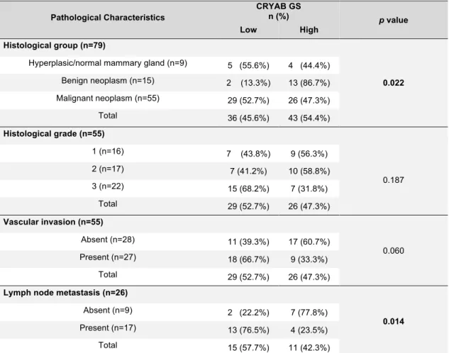
When comparing CRYAB GS with the clinicopathological characteristics, significant differences were found in tumor size (p=0.039), histological group (p=0.022) and presence of lymph node metastasis (p=0.014). Larger tumors were usually characterized by a low GS (n=14; 58.3%). With regard to the histological group, benign neoplasms showed higher scores (n=13; 86.7%), while normal/hyperplastic and malignant neoplasms tended to obtain lower scores (n=5; 55.6% and n=29; 52.7%, respectively). Regarding metastasis, CRYAB GS was higher (n=7; 77.8%) in cases with no lymph node metastasis. When present, the score tended to be low (n=13; 76.5%). These results are shown on Tables 7 and 8.
CRYAB Anapl carc. Complex carc. malignant Carc. and myoep.
Carc. in
CA/MBT Solid carc.
Tubulo- papillary carc. Carcino sarcoma Malignant myoep. SI Absent 0 (0%) 0 (0%) 0 (0%) 0 (0%) 0 (0%) 2 (13.3%) 0 (0%) 0 (0%) Weak 1 (100%) 0 (0%) 0 (0%) 2 (22.2%) 1 (8.3%) 2 (13.3%) 1 (25.0%) 0 (0%) Moderate 0 (0%) 5 (100%) 5 (62.5%) 4 (44.4%) 7 (58.3%) 11 (73.3%) 1 (25.0%) 1 (100%) Strong 0 (0%) 0 (0%) 3 (37.5%) 3 (33.3%) 4 (33.3%) 0 (0%) 2 (50.0%) 0 (0%) PS 0% 0 (0%) 0 (0%) 0 (0%) 0 (0%) 0 (0%) 2 (13.3%) 0 (0%) 0 (0%) 1-25% 1 (100%) 1 (20.0%) 2 (25.0%) 2 (22.2%) 7 (58.3%) 10 (66.7%) 3 (75.0%) 0 (0%) 26-50% 0 (0%) 1 (20.0%) 1 (12.5%) 2 (22.2%) 3 (25.0%) 2 (13.3%) 0 (0%) 0 (0%) 51-75% 0 (0%) 1 (20.0%) 4 (50.0%) 4 (44.4%) 1 (8.3%) 1 (6.7%) 1 (25.0%) 0 (0%) 76-100% 0 (0%) 2 (40.0%) 1 (12.5%) 1 (11.1%) 1 (8.3%) 0 (0%) 0 (0%) 1 (100%) GS Low 1 (100%) 1 (20.0%) 2 (25.0%) 3 (33.3%) 7 (58.3%) 12 (80.0%) 3 (75.0%) 0 (0%) High 0 (0%) 4 (80.0%) 6 (75.0%) 6 (66.7%) 5 (41.7%) 3 (20.0%) 1 (25%) 1 (100%)
19 Table 7 - Comparative study of CRYAB GS with the clinical characteristics
Clinical Characteristics CRYAB GS n (%) p value Low High Age (n=70) 0.641 <10 years (n=34) 15 (44.1%) 19 (55.9%) ≥10 years(n=36) 18 (50.0%) 18 (50.0%) Total 33 (47.1%) 37 (52.9%) Breed (n=73) 0.279 Indetermin. (n=35) 17 (48.6%) 18 (51.4%) Poodle (n=10) 3 (30.0%) 7 (70.0%) Cocker Spaniel (n=7) 2 (28.6%) 5 (71.4%) Others (n=21) 13 (61.9%) 8 (38.1%) Total 35 (47.9%) 38 (52.1%) Tumor Size (n=65) 0.039 <3 cm (n=27) 7 (25.9%) 20 (74.1%) 3-5 cm (n=14) 8 (57.1%) 6 (42.9%) >5 cm (n=24) 14 (58.3%) 10 (41.7%) Total 29 (44.6%) 36 (55.4%) Location (n=53) M1 and/or M2 (n=7) 3 (42.9%) 4 (57.1%) 0.976 M3 (n=13) 5 (38.5%) 8 (61.5%) M4 and/or M5 (n=25) 11 (44.0%) 14 (56.0%) Multiple (n=8) 4 (50.0%) 4 (50.0%) Total 23 (43.4%) 30 (56.6%) Ulceration (n=68) 0.211 Absent (n=55) 21 (38.2%) 34 (61.8%) Present (n=13) 8 (61.5%) 5 (38.5%) Total 29 (42.6%) 39 (57.4%) OHE (n=48) 1.0 Not performed (n=38) 21 (55.3%) 17 (44.7%) Performed (n=10) 5 (50.0%) 5 (50.0%) Total 26 (54.2%) 22 (45.8%) Contraception (n=38) 0.254 Not performed (n=29) 14 (48.3%) 15 (51.7%) Performed (n=9) 2 (22.2%) 7 (77.8%) Total 16 (42.1%) 22 (57.9%)
20
No significant differences were found for other clinicopathological parameters. However, for vascular invasion, there was a tendency to carcinomas with vascular invasion exhibiting a low GS (n=18; 66.7%)
Table 8 - Comparative study of CRYAB GS with the histopathological characteristics
2.4.6. Immunolabeling and comparative study of CRYAB in the mammary
gland adjacent to neoplastic tissues
Mammary gland adjacent to benign neoplasms was present in 12 of the 15 samples (80.0%), being characterized by a staining intensity mostly moderate (n=6; 50.0%) while PS was mostly 1 to 25% (n=7; 58.3%).
Mammary gland adjacent to malignant neoplasms was present in 28 out of 55 samples (50.91%), with similar immunohistochemical findings: SI was mostly moderate (n=18; 64.3%) and PS was predominantly 1 to 25% (n=16; 57.1%) (Fig. 2e and 2f). So, when comparing CRYAB’s expression of the adjacent mammary glands (AMG), either in benign or malignant neoplasms, we
Pathological Characteristics CRYAB GS n (%) p value Low High Histological group (n=79) 0.022
Hyperplasic/normal mammary gland (n=9) 5 (55.6%) 4 (44.4%) Benign neoplasm (n=15) 2 (13.3%) 13 (86.7%) Malignant neoplasm (n=55) 29 (52.7%) 26 (47.3%) Total 36 (45.6%) 43 (54.4%) Histological grade (n=55) 1 (n=16) 7 (43.8%) 9 (56.3%) 0.187 2 (n=17) 7 (41.2%) 10 (58.8%) 3 (n=22) 15 (68.2%) 7 (31.8%) Total 29 (52.7%) 26 (47.3%) Vascular invasion (n=55) 0.060 Absent (n=28) 11 (39.3%) 17 (60.7%) Present (n=27) 18 (66.7%) 9 (33.3%) Total 29 (52.7%) 26 (47.3%)
Lymph node metastasis (n=26)
0.014
Absent (n=9) 2 (22.2%) 7 (77.8%) Present (n=17) 13 (76.5%) 4 (23.5%) Total 15 (57.7%) 11 (42.3%)
21
found no significant differences in GS (p=0.484). In both benign and malignant groups, the staining pattern was usually heterogeneous. Results are shown in Table 9.
Table 9 - CRYAB immunolabeling of the adjacent mammary gland of benign and malignant tumors
CRYAB
AMG of Benign Neoplasms n (%)
AMG of Malignant Neoplasms n (%) SI Absent 1 (8.3%) 1 (3.6%) Weak 4 (33.3%) 7 (25.0%) Moderate 6 (50.0%) 18 (64.3%) Strong 1 (8.3%) 2 (7.1%) PS 0% 1 (8.3%) 1 (3.6%) 1-25% 7 (58.3%) 16 (57.1%) 26-50% 2 (16.7%) 7 (25.0%) 51-75% 2 (16.7%) 4 (14.3%) >75% 0 (0%) 0 (0%) GS Low 9 (75.0%) 17 (60.7%) High 3 (25.0%) 11 (39.3%) Total 12 (100%) 28 (100%)
22
a b
c d
e f
Figure 2 - Immunohistochemical expression of CRYAB in canine mammary tissues: a, lobular hyperplasia showing epithelial positivity (strong intensity, in more than 50% of cells); b, complex adenoma, characterized by positivity of neoplastic myoepithelial cells (strong intensity, in more than 50% of cells); c, tubulopapillary carcinoma showing few positive neoplastic cells (moderate intensity, in less than 25% of cells); d, carcinoma and malignant myoepithelioma, with positive neoplastic cells (strong intensity, in more than 75% of cells); e, adjacent mammary gland, with moderate positivity of epithelial and myoepithelial cells in the lobules and positive myoepithelium in the duct; f, adjacent mammary gland, with an heterogeneous pattern of expression, with the presence of lobules with strong positivity (less than 50% of epithelial cells).
23
2.5. Discussion
As stated before, canine mammary tumors are considered the most accurate animal model to study human breast cancer, a deadliest disease with specific therapeutic targets yet to be found. Female dogs usually share the same environment as women, meaning they probably are exposed to similar carcinogens. Besides that, canines and humans have common features such as body size and genetic variability, which make female dogs a better spontaneous animal model to study human breast cancer than other models such as laboratory animals (Sorenmo et al., 2011; Carvalho et al., 2014).
With research in CMT’s moving at high rhythm due to the previously reported fact, molecular mechanisms underlying this type of cancer have been in the center of major investigations worldwide. As more and more of these mechanisms are being studied, more intervenient molecules and features are discovered, being one of them CRYAB. CRYAB has been studied since 1983 (Delaye & Tardieu, 1983), but few studies are found to be relevant or conclusive, and those that are, are often contradictory. We only found one research article regarding CRYAB in CMT’s (Guvenc et al., 2012), which arouse our interest in this marker.
In the human normal breast tissue, CRYAB was found to be expressed by the myoepithelium and not expressed by the luminal epithelial component of the mammary epithelium (Koletsa et al., 2014). In our study, we found a weak and inconstant expression of CRYAB in the myoepithelium of normal/hyperplastic mammary gland samples, but also a heterogeneous pattern of expression in the luminal component, frequently with moderate/strong intensity, especially in hyperplastic tissues. The only study performed so far on CRYAB expression in canine mammary tissues described that only few luminal epithelial cells were positive in normal samples, with basal myoepithelial cells negative (Guvenc at al., 2012). These investigators also analysed two samples of hyperplasia that showed less than 25% of epithelial positivity. These discrepancies might be explained by the low number of samples analysed, but seem to indicate that in the canine species, CRYAB is not consistently expressed by myoepithelial cells of normal mammary gland. One hypothesis is that its expression might be associated with the estrous cycle stages, so, it would be interesting to study CRYAB expression in mammary gland tissues along the estrous cycle.
Koletsa et al. (2014) performed a large study on a series of almost 1000 human breast samples. They found that all the tissues with a high PS (>30%) presented a strong SI. They also found that malignant neoplasms with high histological grade (grade 3) were more frequently associated with high expression of CRYAB. Besides that, they also found a relation between CRYAB overexpression and the triple negative breast cancer samples. In our study, the neoplastic
24 myoepithelial cells were also overexpressing CRYAB when compared to the normal/hyperplastic tissues. It is known that canine mammary carcinomas are frequently characterized by a basal/myoepithelial phenotype (Gama et al., 2008), so a comparative study between CRYAB expression and the basal-like/triple-negative phenotype should be considered in this animal model.
In the present study, we found significant differences between histological groups. Benign tumors were predominantly characterized by high expression, especially due to the inclusion of histological subtypes, with myoepithelial cell proliferation (complex/mixed tumors), which showed increased levels of positivity.
Malignant tumors were positive in a small percentage (26/55; 47.3%), also showing CRYAB expression predominantly associated with non-simple carcinomas, with concomitant luminal cell and myoepithelial proliferation. Moreover, in malignant tumors, CRYAB expression was significantly associated with the histological subtype, even when considering the novel classification proposed by Goldschmidt et al. (2011).
Giving CRYAB’s anti-apoptotic features (Kamradt et al., 2002; Malin et al., 2016) and its proved involvement in tumor growth (Kamradt et al., 2005), we expected to find an overexpression of this protein on larger tumors, which did not happen in our study. Instead, tumors with less than 3 cm represented 55.56% (n=20) of the tumors obtaining high scores, while only 27.78% (n=10) of these were larger than 5 cm; this lead to significant differences when comparing the GS and tumor size. The studies performed by Kamradt and its colleagues (2002) were conducted either in cell cultures or murine models of mammary cancer, which could explain the differences obtained. It would be interesting to study apoptotic and anti-apoptotic cell markers and compare their expression to CRYAB. In human breast cancer, Kim et al. (2015) showed that Bcl-2, an anti-apoptotic protein, was significantly associated with CRYAB, although a weak negative correlation was found.
Since CRYAB is connected to poor prognosis (Moyano et al., 2006; Koletsa et al., 2014), we expected that somehow it could be related to metastasis so that we could use it as a marker of dissemination. However, our results show that CRYAB expression was higher when metastases were absent. Previously reported studies stated that CRYAB overexpression is linked to lymph-node involvement, which goes in disagreement with our findings (Chelouche-Lev et al., 2004). Some authors have linked CRYAB overexpression with the presence of brain metastasis (Malin et al., 2014). Differences may be justified by the fact that, although the female dog is a potential animal model to study human breast cancer, differences do exist; one of them is the fact that
25 canine mammary carcinomas are not frequently characterized by the proliferation of myoepithelial cells (Sleeckx et al., 2011). Guvenc et al. (2012) also found a high positivity in complex carcinomas of the female dog, but a comparative study between histological subtypes was not performed in their study.
Adjacent mammary glands do not seem to be affected by tumor environment since we did not found significant differences between CRYAB’s expression in adjacent mammary glands to benign or malignant tumors (p=0.484). In our review of literature, we did not found any supportive theories that CRYAB could interfere with the tumor environment and further promote neighbor cells modifications.
Our study, although meticulously performed, has a small number of samples, which may explain some of the differences found with the reviewed studies. However, the only report of CRYAB in CMT’s was performed on an even smaller series than ours (Guvenc et al., 2012), which lead us to believe that our findings regarding CMT’s may be more accurate. In addition, there were different methodologies adopted to quantify CRYAB expression, which can also explain the differences observed. We would also like to emphasize that the lack of some clinicopathological information may falsely influence the results shown, since that information mostly relies on reports from our colleagues, which are usually incomplete.
In the future, CRYAB expression should be evaluated in prognostic studies, so that we can accomplish if it is correlated with prognosis. In human breast cancer, a link to poor prognosis was found, but not when considering CRYAB as an individual factor of prognosis (Koletsa et al., 2014). We could also compare CRYAB expression with other markers like ER, PR and AR, so that we can conclude if it is linked to tumors with triple negativity in CMT’s as it is in human breast cancer (Koletsa et al., 2014).