1
SELENIUM IS INVOLVED IN REGULATION OF PERIPLASMIC HYDROGENASES GENE
2EXPRESSION IN
D
ESULFOVIBRIO VULGARISHILDENBOROUGH
3 4
Filipa M.A. Valente, Cláudia C. Almeida, Isabel Pacheco, João Carita, Lígia M. Saraiva and Inês A.C. 5
Pereira* 6
7
Instituto de Tecnologia Química e Biológica, Universidade Nova de Lisboa, Oeiras, Portugal 8
9 10 11 12
* To whom correspondence should be addressed
13
Instituto de Tecnologia Química e Biológica 14
Apt. 127, 2781-901 Oeiras, Portugal 15 e-mail: ipereira@itqb.unl.pt 16 Tel: 351-214 469 327 17 Fax: 351-214 411 277 18 19
Keywords: sulfate reducing bacteria; hydrogenases; selenium; regulation 20
21 22 23
Abstract
1 2
Desulfovibrio vulgaris Hildenborough (DvH) is a good model organism to study hydrogen metabolism 3
in sulfate-reducing bacteria. Hydrogen is a key compound for these organisms since it is one of their 4
major energy sources in natural habitats, but also an intermediate in the energy metabolism. The DvH 5
genome codes for six different hydrogenases, but only three of them, the periplasmic-facing [FeFe], 6
[FeNi]1 and [FeNiSe] hydrogenases, are usually detected. In this work we studied the synthesis of each
7
of these enzymes in response to different electron donors and acceptors for growth, and the availability 8
of Ni and Se. The formation of the three hydrogenases was not very strongly affected by the electron 9
donors or acceptors used, but highest levels were observed after growth in hydrogen as electron donor 10
and lowest with thiosulfate as electron acceptor. The major effect observed was with inclusion of Se in 11
the growth media, which led to a strong repression of the [FeFe] and [NiFe]1 hydrogenases, and a
12
strong increase in the [NiFeSe] hydrogenase that is not detected in the absence of Se. Ni also led to 13
increased formation of the [NiFe]1 hydrogenase, except for growth with H2 where its synthesis is very
14
high even without Ni added to the medium. Growth with H2 results in a strong increase in the soluble
15
forms of the [NiFe]1 and [NiFeSe] hydrogenases. This study is an important contribution to
16
understanding why DvH has three periplasmic hydrogenases. It supports their similar physiological role 17
in H2 oxidation and reveals that element availability has a strong influence in their relative expression.
18 19 20 21 22 23 24
Hydrogen plays a central role in the energy metabolism of sulfate reducing bacteria (17). H2 is
1
one of the major energy sources for these organisms in their natural habitats, but it may also be a 2
product of their fermentative metabolism. Furthermore, a chemiosmotic mechanism involving 3
production and oxidation of H2 on opposite sides of the membrane has been proposed to explain energy
4
transduction during sulfate respiration with lactate (21). In agreement with the important metabolic role 5
of H2, hydrogenases (Hases) are particularly abundant proteins in sulfate reducing bacteria, and many
6
species have several different Hases (39). However, it is still not clear what are the exact physiological 7
roles of each of these enzymes, or what is the reason for this apparent redundancy. Desulfovibrio 8
vulgaris Hildenborough (DvH), whose genome was recently sequenced (13), is a good model organism 9
to study hydrogen metabolism in sulfate reducing bacteria. The DvH genome encodes for six different 10
Hases, four of which are periplasmically oriented (two [NiFe] isoenzymes, one [NiFeSe] and one 11
[FeFe] Hase) and two that are facing the cytoplasm (two multi-subunit membrane [NiFe] Hases). The 12
[FeFe] Hase (14, 24), [NiFe]1 Hase (29) and [NiFeSe] Hase (36) of DvH have been characterized, and
13
are the dominant Hases detected in extracts of cells grown in standard lactate/sulfate conditions (10, 14
36). 15
The [FeFe] Hase is a soluble periplasmic protein with both high affinity for H2 (37) and a very
16
high H2-uptake activity (9). It is noteworthy that Desulfovibrio spp. are the only known
17
microorganisms with a periplasmic [FeFe] Hase, since these enzymes are most commonly cytoplasmic. 18
A DvH deletion mutant for the [FeFe] Hase showed that its physiological role is hydrogen uptake (26). 19
The [NiFe]1 and [NiFeSe] both belong to the family of uptake Hases (38). Contrary to most Hases in
20
this family they both lack the membrane-bound heme b subunit responsible for electron transfer to the 21
membrane quinone pool. However, both [NiFe]1 and [NiFeSe] Hases are membrane-associated proteins
22
(29, 36), but a soluble form of the [NiFeSe] Hase was also detected. The [NiFeSe] Hase is structurally 23
very similar to the [NiFe] Hases, with the major differences being that it has a selenocysteine as one of 24
the ligands to the active-site Ni, and the small subunit has a medial [4Fe4S]2+/+ rather than a [3Fe4S]+/0
1
cluster (11, 36). In standard assays the [NiFeSe] Hase displays much higher activity values than the 2
[NiFe]1 Hase, and it is also resistant to inactivation by oxygen (36). A comparison of the catalytic
3
activities of the three DvH Hases was recently reported (36). As for the [FeFe] Hase, the physiological 4
role of the [NiFe]1 and [NiFeSe] Hases is apparently in H2 oxidation, and all three enzymes share as a
5
common electron acceptor a periplasmic cytochrome c (the type I cytochrome c3) (25, 36). This raises
6
the question of what distinguishes these three Hases in physiological terms, and one possibility could 7
be differences in expression conditions. Interestingly, the genes coding for the [NiFe]1 and [NiFeSe]
8
Hases are adjacently located in the genomes of both DvH and D. desulfuricans G20, suggesting the 9
possibility of coordinated regulation. Regulation of bacterial Hase gene expression is exerted mainly at 10
the transcriptional level and responds to four major types of signals: H2, O2, nickel ions and the electron
11
donors and acceptors available (reviewed in (38)). In this work we investigated how different growth 12
conditions and the presence of iron, nickel and selenium in the growth medium affect the expression of 13
the DvH [FeFe], [FeNi]1 and [FeNiSe] Hases.
14
Gene regulation by both Fe and Ni in prokaryotes is well documented (2, 12, 19). In the 15
particular case of Hases, Ni is required for complete maturation of [NiFe] Hases (32), and 16
transcriptional regulation by Ni has been shown for example in Bradyrhizobium japonicum (22), and in 17
Methanothermobacter marburguensis. In the latter organism growth in Ni-limited conditions leads to 18
increased expression of the Ni-free Hase H2-forming methylenetetrahydromethanopterin
19
dehydrogenase (Hmd) and strongly decreased expression of the [NiFe] Hase F420-reducing hydrogenase
20
(Frh) (1). Both these Hases are regulated at transcriptional level, whereas the decreased expression of 21
the [NiFe] methyl viologen reducing hydrogenase (Mvh) in these conditions is due to regulation at a 22
post-transcriptional level (34). 23
Selenium is an essential trace element for higher animals where selenoproteins are involved in 1
immune function and cellular defence against oxidative stress (5). In most selenoproteins selenium is 2
found as selenocysteine, the 21st amino acid that is co-translationally incorporated into the polypeptide 3
chain (6). Prokaryal selenoproteins have no counterpart in eukarya and are generally involved in energy 4
metabolism. They are found in only a small fraction of bacterial and archaeal genomes, and their 5
number within a given prokaryotic genome is in the order of one to ten (15). Some selenoprotein-6
containing organisms are absolutely dependent on the availability of selenium, whereas others are able 7
to express cysteine-containing homologues of their selenoproteins. The activity of selenoproteins is 8
usually higher than their cysteine-containing homologues (3, 16) due to the lower pKa and higher 9
nucleophilicity of the selenol group compared to that of the thiol group. The involvement of selenium 10
in transcriptional regulation of selenoproteins has been investigated in detail for two types of Hases in 11
Methanococcus voltae (33). This organism has genes encoding for two pairs of homologous [NiFe] 12
Hases, the F420-reducing Hases (Frc and Fru) and the F420-nonreducing Hases (Vhc and Vhu). In each
13
pair one of the enzymes contains selenocysteine as a ligand to the Ni (Fru and Vhu), and the other a 14
cysteine in the corresponding position (Frc and Vhc). Selenium is involved in negative regulation of the 15
two selenium-free Hases, whose genes are linked by a common intergenic region and are only 16
expressed in the absence of Se (4). This intergenic region includes a common negative regulatory 17
element to which a LysR-type regulator binds, as well as two activator elements that are specific for 18
each of the two transcription units (20, 35). Methanococcus jannaschi that only has genes encoding for 19
seleno-containing Hases is completely dependent on the presence of selenium in the medium (31). In 20
this work we report that Ni and Se are also involved in the regulation of the periplasmic DvH Hases and 21
that the [NiFeSe] Hase is the main Hase present when the cells are grown in the presence of Se. 22
23 24
1 2
MATERIALS AND METHODS
3 4
Cell growth and preparation of crude, soluble and membrane extracts 5
Desulfovibrio vulgaris Hildenborough (DSM 644) was grown in medium C (27) with an iron 6
concentration of 25µM. Medium C contains yeast extract which is a potential source of Ni and Se. The 7
levels of these elements in medium C without additions were below the respective detection limits as 8
determined by atomic absorption spectrometry (< 0.01 mg/l for Ni and < 0.04 mg/l for Se). Medium C 9
was supplemented with different electron donors and electron acceptors, all at a 40 mM concentration. 10
The growth conditions tested were: lactate/sulfate, lactate/thiosulfate, hydrogen/acetate/sulfate (20mM 11
acetate), pyruvate/sulfate, and only pyruvate (fermentative conditions). For each condition three 12
different cultures were performed in parallel using the same inoculum (grown in medium C): one in 13
medium C with no further additions, one in medium C supplemented with 1 μM nickel chloride, and 14
one in medium C supplemented with nickel chloride plus sodium selenide, both at a concentration of 15
1µM. All cultures were performed in closed 1l glass flasks containing half their volume of culture and 16
N2 as gas headspace, with the exception of H2 grown cells that were cultured in a 2.2L bioreactor, with
17
a flow of H2/CO2 (80:20) of 900ml/min. and stirring at 250 rpm. Cells were harvested by centrifugation
18
at late exponential phase and resuspended in 20mM Tris-HCl buffer pH 7.6. After addition of DNase 19
the cells were disrupted by passing twice in a French-press cell. The extract was then centrifuged at 20
10.000g, 15 min, 4ºC to remove cell debris yielding the crude extract, a part of which was centrifuged 21
again at 100.000g, 15min., at 4ºC in order to obtain the soluble, and the membrane fractions. The 22
membranes were resuspended in 20mM Tris-HCl buffer pH 7.6. 23
H2 production activity
1
Hydrogen production activity of crude, soluble and membrane extracts was assayed in 50mM Tris-HCl 2
buffer pH7.6 with 1mM methyl viologen and 15mM of dithionite as electron donors. Quantification of 3
hydrogen in the gaseous phase was carried out by Gas Chromatography as described in (23). 4
5
Gels stained for hydrogenase activity 6
For activity staining gels, samples were run aerobically in a 7.5% gel under native conditions, containing 7
0.1% Triton X-100 in a refrigerated chamber at 4ºC. The running buffer also contained 0.1% Triton X-100. 8
The gel was then placed in a closed flask with degassed 50mM Tris-HCl buffer pH8, 0.5mM methyl 9
viologen, and flushed with argon, followed by H2 gas. After development the bands were fixed by adding a
10
1% 2,3,5-triphenyltetrazolium chloride solution. 11
12
Western blot analysis 13
The protein content of crude extracts was determined by the Bradford assay with gamma-globulin used 14
as protein standard. The crude cell extracts were run in a 12% SDS gel with 0.1% TritonX-100. 15
Proteins were then transferred to 0,45 µm PVDF membranes (Roche) for 1h at 100mV and 4ºC, in a 16
Mini Trans-Blot Electrophoretic Transfer Cell (Bio-Rad). The membranes were equilibrated with Tris-17
buffered saline solution with Tween 20 (TBST) (20mM Tris-HCl pH7.5, 150 mMNaCl, 0.05% Tween 18
20), and then treated with antiserum raised against the [FeFe], [FeNi]1 and [NiFeSe] Hases of
19
D.vulgaris H. The [FeFe] and [FeNi]1 Hase antibodies were diluted 1:5000 whereas the [NiFeSe] Hase
20
antibody was only diluted 1:500, in TBST. Unbound antibodies were removed by three 5 min washes 21
with TBST. Immunodetection of bound antibodies was done by treatment with anti-mouse 22
immunoglobulin G (H+L) alkaline phosphatase conjugate (from Promega), diluted 1:5000, followed by 23
a solution of nitro blue tetrazolium salt and 5-bromo-4-chloro-3-indolyl phosphate toluidine salt 24
(NBT/BCIP solution from Roche). Quantification of immunodetected Hase bands was performed by 1
densitometric analysis with the program Image Master Total Lab v.2.01, after scanning of the blots 2
with the Image Scanner system of Amersham. 3 4 5 6 RT-PCR experiments 7
DvH cells were grown anaerobically to the mid-exponential phase in lactate/sulfate medium with 8
addition of Fe, Fe+Ni or Fe+Ni+Se as described before. Total RNA was extracted from the pellets 9
using the hot acid phenol:chloroform method and treated with DNaseI, essentially as described in (8). 10
Based on the gene sequences for the three Hases, retrieved from the DvH genome (available at 11
www.tigr.org), forward and reverse primers were designed for PCR amplification of the following 12
DNA fragments: 584-bp for the [FeFe] Hase large subunit gene (hydA) (FH1: 5'-13
AGTACTGCCCCACCGCCGCCATCTTC-3' and FH2:
5'-14
CTTTGCGATGCAGGGCATGATGGAGAC-3'); 657-bp for the [NiFe] Hase large subunit gene 15
(hynA-1) (FNH1: 5'-GACCCGGCCAAGGCTGCGAAGA-3' and FNH2:
5'-16
CAGGTCGGTGTACTTGGGTTCTGTCG -3'); 492 for the [NiFeSe] Hase large subunit gene (hysA) 17
(FNSH1: 5'-GATACGGCTCCTTTCGTTCCG-3' and FNSH2: 5'-GTAATTGAGGCCGGTCGTCTC-18
3'). Prior to RT-PCR experiments optimisation of the PCR amplification conditions was established, for 19
each pair of primers, using DvH genomic DNA as template. For each RNA sample, control 20
experiments in which the RT step was omitted were performed, and confirmed the absence of any 21
residual DNA. The reactions were performed using a RT-PCR Master Mix (USB) and 100 ng of total 22
RNA. As loading control the 16S rRNA was used. The results were analysed by Kodak Digital 23
Science Electrophoresis Documentation and Analysis System 120. 24
RESULTS
1 2
We recently reported the isolation of the [NiFeSe] Hase as a major Hase present in cells of DvH 3
grown in lactate/sulfate medium (36). Since the three periplasmic-facing Hases of DvH have apparently 4
a similar physiological function of H2 uptake, we decided to study their expression in different growth
5
conditions by varying electron donors and acceptors, and also the availability of the elements Ni and 6
Se. The main electron donors used for growing Desulfovibrio spp. are lactate, pyruvate and hydrogen, 7
which were tested with sulfate as electron acceptor. We also tested thiosulfate as an alternative electron 8
acceptor with lactate as electron donor, and fermentative conditions with pyruvate as electron donor in 9
the absence of sulfate. Growth curves were obtained for all these conditions so that cells could be 10
collected at the same point of the growth curve for the different conditions. The addition of Ni or Se to 11
the standard Fe-containing medium C did not affect the growth rate or growth yield for the 12
lactate/sulfate (L/S), lactate/thiosulfate (L/T) or hydrogen/sulfate (H2/S) conditions. For
13
pyruvate/sulfate (P/S) or pyruvate (P) growth in the presence of Se led to a slightly faster growth rate, 14
but similar yield, as compared to Fe or Fe+Ni conditions (data not shown). 15
16
Analysis of periplasmic DvH hydrogenases by activity-stained gels 17
The periplasmic [FeFe], [NiFe]1 and [NiFeSe] Hases of DvH are easily detected in crude cell extracts
18
by PAGE gels stained for H2-uptake activity (10, 36). The gels have to be run in native conditions in
19
the presence of detergent, since the [NiFe]1 and [NiFeSe] Hases are both membrane-associated proteins
20
and absence of detergent leads to severe band streaking along the gel. Thus, the relative formation of 21
the periplasmic DvH Hases was initially probed by activity-stained gels using cells grown under 22
different conditions. Besides the effect of the different electron donors/acceptors, we wanted also to test 23
the effect of the elements Fe, Ni and Se, which are present in the metal centres of the periplasmic 24
Hases, in the synthesis of these proteins. Thus, for each electron donor/acceptor pair, DvH cells were 25
grown in the presence of only Fe (25μM), with both Fe (25μM) and Ni (1μM), and in the presence of 1
the three elements Fe (25μM), Ni (1μM) and Se (1μM). The activity stained gel comparing cells grown 2
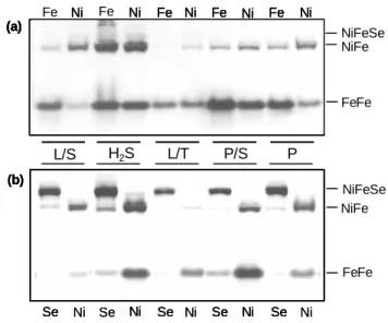
with only Fe and with Fe+Ni are shown in Figure 1a. For cells grown in the presence of these metals 3
only the [FeFe] and [NiFe]1 Hases are observed, whereas the [NiFeSe] Hase is not detected. In Fe-only
4
media the [FeFe] Hase is predominant, except for growth with H2/S in which the [NiFe]1 Hase displays
5
a similar band intensity. The [FeFe] Hase band intensity is highest in P/S and H2/S, and lowest in L/T.
6
The inclusion of Ni in the medium leads to a strong reduction in the band intensity of the [FeFe] 7
Hase in L/S medium, and a smaller effect is also observed in P/S and pyruvate fermentative conditions. 8
The presence of Ni increases the intensity of the [NiFe]1 Hase for all conditions tested except in H2/S in
9
which the [FeFe] and [NiFe]1 Hases show similar band intensities in Fe-only or Fe+Ni media. For
10
Fe+Ni media the [NiFe]1 Hase shows highest band intensity in H2/S followed by L/S, whereas it is
11
almost not detected in L/T. 12
The activity stained gel comparing cells grown with Fe+Ni and with Fe+Ni+Se are shown in 13
Figure 1b. The Fe+Ni conditions are repeated from Figure 1a, as controls for the Fe+Ni+Se conditions, 14
since band intensity comparisons should only be performed for samples in the same gel. It is readily 15
apparent that inclusion of Se in the growth media has a profound effect on the pattern of the DvH 16
periplasmic Hases, leading to the appearance of a strong band of the [NiFeSe] Hase, which becomes 17
the major Hase detected, and a concomitant decrease in the band intensity of the other two Hases. This 18
effect is observed irrespective of the electron donor/acceptor present. For Fe+Ni+Se media the 19
[NiFeSe] Hase shows highest band intensity in H2/S and lowest in L/T. For two of the growth
20
conditions (L/S and P) we also tested whether the effect of the three elements on the Hase synthesis 21
was constant along the growth curve. Activity-stained gel analysis of cells collected at initial, middle 22
and late exponential phase and at stationary phase showed that the effect of Ni and Se was constant 23
along the growth curve (data not shown). 24
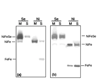
Previous work has shown that in DvH grown in the standard L/S conditions the [FeFe] Hase is a 1
soluble periplasmic protein, whereas the [NiFe]1 and [NiFeSe] are both membrane-associated proteins
2
(29, 36). However, a soluble form of the [NiFeSe] Hase was also detected. In order to check whether 3
the Hases are soluble or membrane-bound in the growth conditions tested here, we separated the 4
soluble and membrane extracts and analysed these with the activity-stained gels. Only the Ni+Fe versus 5
Ni+Fe+Se conditions were tested since the difference in Hase composition between the Fe and the 6
Fe+Ni crude extracts was less marked. For the L/S, L/T, P/S and P the same pattern was observed 7
where the [FeFe] Hase is only present in the soluble extract, and the [NiFe]1 and [NiFeSe] Hases are
8
found mainly in the membrane fraction, but are also detected with much smaller intensity in the soluble 9
fraction (Figure 2a, only L/S is shown). Interestingly, the cells grown in H2/S displayed a different
10
pattern where the band intensity of both the [NiFe]1 and [NiFeSe] Hases in the soluble fraction is
11
strongly increased relative to the other conditions. An [FeFe] Hase band in the membrane fraction of 12
Ni+Fe grown cells is also apparent (Figure 2b). This indicates that the increased synthesis of the three 13
Hases in the H2/S conditions is accompanied by changes in the cellular localization of the three Hases.
14 15
Analysis of H2-producing activity in DvH cell extracts
16
In the activity-stained gels the band intensity is related to the H2-uptake activity of the three
17
Hases. To complement these results we tested also the H2-production activity of the soluble and
18
membrane extracts of DvH cells grown in the different conditions, by GC chromatography (Figure 3). 19
These results cannot be directly compared to the previous ones because it is not possible to discriminate 20
between the three different Hases. Also, the relative ratio of the H2-uptake activities for the three Hases
21
is different from the ratio of their H2-production activities. Nevertheless, it is also of interest to analyse
22
the effect of the different growth conditions in the H2-producing activity. A fairly rough analysis of
23
each Hase contribution can be attempted based on the fact that the soluble extract contains most of the 24
[FeFe] Hase and only a small amount of the other two Hases, whereas the membrane extract contains 1
the [NiFe]1 and [NiFeSe] Hases (except in the H2/S conditions). Overall, for both the soluble and
2
membrane extracts, the H2-production activity is highest in H2/S and P/S conditions, and lowest in L/T.
3
The soluble extracts of P/S and P grown cells display similar activities in the three element conditions 4
(Figure 3a). The soluble extracts of L/S and L/T display higher activities in Fe-only medium probably 5
due to the increased expression of the [FeFe] Hase in these conditions. The soluble extracts of H2/S
6
grown cells have twice the activity level in Ni+Fe+Se medium relative to the other two metal 7
conditions, which is likely due to the high activity of the [NiFeSe] Hase that under H2/S conditions is
8
almost equally distributed between the soluble and membrane extracts, as shown above. The H2
-9
production activity of membrane extracts for all growth conditions tested displays a sharp increase in 10
the Ni+Fe+Se media (Figure 3b), reflecting the increase in the membrane-bound form of the [NiFeSe] 11
Hase under these growth conditions, which has a much higher H2-production activity than the soluble
12
form (36). 13
14
Analysis of periplasmic DvH hydrogenases by Western blot 15
The results presented above show striking differences in the Hase pattern of DvH under 16
different growth conditions, particularly in response to Ni and Se. However, they may be fallible for 17
assessing the quantity of these proteins since they are based on the enzymatic activity. The activity of 18
each of the three proteins is different, and is particularly sensitive to their activation state, which in turn 19
is affected by any possible contact with oxygen. Furthermore, the effects observed could be due to an 20
increase/decrease in activity and not to an actual change in protein content of the extracts. In order to 21
have a more clear picture of the Hase pattern in DvH we analysed these proteins in cell extracts by 22
Western blots. For this we obtained polyclonal antibodies against the [FeFe], [NiFe]1 and [NiFeSe]
Hases purified from DvH. These antibodies proved to be very selective, detecting only the respective 1
Hase, and showing no cross-reactivity with the other two Hases when tested with the pure proteins. 2
Crude cell extracts of DvH cells grown in the conditions mentioned were analysed by Western 3
blot (Figure 4). The pattern of the three Hases under the different growth conditions was broadly in 4
agreement to the results observed by activity-stained gel (Figure 1). Densitometric analysis of the 5
immunoblot bands permits a more quantitative view of the results that is easier to analyse (Figure 5). In 6
Fe-only media the [FeFe] Hase shows highest synthesis with P/S, followed by H2/S, L/S, P and is
7
lowest in L/T (Figure 5A). As observed in the activity-stained gels, the addition of Ni to the media 8
induces no pronounced effect on the [FeFe] Hase for the H2/S and L/T media, whereas its formation is
9
reduced in P and P/S conditions and even more markedly in L/S. In all Fe+Ni+Se media the synthesis 10
of the [FeFe] Hase decreases relative to Fe+Ni conditions, but is then constant irrespective of the 11
electron/donor acceptor. 12
The synthesis of the [NiFe]1 Hase (figure 5B) increases upon addition of Ni to the growth
13
media, except in H2/S conditions where its synthesis is surprisingly high even in Fe-only medium, and
14
is not affected by the further inclusion of Ni, as observed before. In Fe+Ni media the formation of the 15
[NiFe]1 Hase is highest with H2/S, followed by P/S, L/S and P and is lowest in L/T. As observed in the
16
activity-stained gels, the addition of Se to the media causes a sharp decrease in the [NiFe]1 Hase
17
amount for all conditions tested. 18
The [NiFeSe] Hase is only detected in Fe+Ni+Se media (Figure 5C), and its synthesis is not 19
markedly affected by the electron donor/acceptor, showing a small increase in H2/S and a small
20
decrease in L/T. Thus, the Western blot analyses confirms the effect of the elements Ni and Se on the 21
levels of the DvH Hases, particularly on the [NiFe]1 and [NiFeSe] Hases.
22 23 24
Detection of [FeFe], [NiFe]1 and [NiFeSe] Hase transcripts by RT-PCR
1
The results described above indicate that Ni, and even more strongly Se, are involved in 2
regulation of the periplasmic DvH Hase expression. To address the question of whether this regulation 3
occurs at transcriptional or post-transcriptional level we analysed the mRNA levels of the three Hases 4
in L/S medium. For this, primers were designed for RT-PCR amplification of DNA fragments from 5
hydA (coding for the [FeFe] Hase large subunit), hynA-1 (coding for the [NiFe] Hase large subunit) and 6
hysA (coding for the [NiFeSe] Hase large subunit). 7
The results shown in Figure 6 indicate that addition of Ni to the L/S medium does result in a 8
decrease of the [FeFe] Hase large subunit transcript, and a similar level is observed in the presence of 9
Fe+Ni+Se, in agreement with the immunoblot analysis. The mRNA levels of the [NiFe]1 Hase large
10
subunit show a very weak increase in Ni+Fe L/S medium compared to Fe-only medium. Further 11
addition of Se results in a decrease of the [NiFe]1 Hase large subunit transcript, confirming the previous
12
observations. The level of the [NiFeSe] Hase large subunit transcript is very low in Fe-only L/S 13
medium as predicted, but this level is quite high in Fe+Ni conditions and only slightly lower than the 14
level observed in Fe+Ni+Se conditions, which contrasts to the results obtained by activity-stained gel 15
and Western blot for which the [NiFeSe] Hase was only detected in cells grown in medium containing 16 selenium. 17 18 DISCUSSION 19
In this work we investigated the expression pattern of the DvH [FeFe], [NiFe]1 and [NiFeSe]
20
Hases in response to different electron donors and acceptors, and also to the elements Ni and Se that are 21
constituents of the active site of the two latter Hases. The [FeFe], [NiFe]1 and [NiFeSe] are the major
22
Hases detected in DvH and previous work has indicated a similar physiological role of H2 oxidation for
the three enzymes, suggesting that the difference between them could lie in different expression 1
conditions. 2
An analysis of each Hase individually shows that the synthesis of the [FeFe] Hase in Fe-only 3
medium was highest in P/S, followed by H2/S. Inclusion of Ni in the medium led to a significant
4
reduction of this Hase only in L/S medium, but Se further reduced its amount in all conditions tested. 5
The [NiFe]1 Hase showed highest formation upon growth with H2/S, even if Ni was not added to the
6
growth medium. For the other conditions, its synthesis increased in Fe+Ni media compared to Fe-only 7
media. Inclusion of Se in the growth media led to a significant decrease of this Hase for all conditions 8
tested. The [NiFeSe] Hase was only detected when Se was included in the media, and in these 9
conditions it did not show strong variations in response to the electron donors/acceptors tested, albeit 10
showing increased synthesis upon growth with hydrogen and lower when thiosulfate was used as 11
electron acceptor. 12
The most striking result obtained in this study was that Se is involved in the negative regulation 13
of the [FeFe] and [NiFe]1 Hases and the positive regulation of the [NiFeSe] Hase, irrespective of the
14
growth medium used. This Hase is not detected in cells grown in the absence of selenium, as would be 15
expected since selenocysteine is part of its polypeptide chain. However, when cells are grown in the 16
presence of Se the [FeFe] and [NiFe]1 Hases are strongly (but not completely) downregulated and the
17
[NiFeSe] Hase becomes the dominant Hase present. Downregulation of the [NiFe]1 Hase in the
18
presence of Se was somewhat expected. This Hase and the [NiFeSe] one are homologous, and 19
structurally very similar, and their genes are adjacently located hinting at coordinated regulation, in a 20
situation analogous to that found for the Hases of M. voltae. In contrast, the downregulation of the 21
[FeFe] Hase in response to Se is unexpected as this Hase belongs to a completely different family from 22
the [NiFe] Hases. These results are in agreement with the [NiFeSe] Hase having a similar physiological 23
role to the [FeFe] and [NiFe]1 Hases and so being capable of replacing them. Its preferred synthesis is
probably linked to its higher catalytic efficiency and possibly also to its resistance to oxygen 1
inactivation (36). The repression of the [FeFe] and [NiFe]1 Hases in the presence of Se occurs at a
2
transcriptional level, as revealed by RT-PCR. 3
The situation found in DvH is similar, but not identical, to that in M. voltae and M. maripaludis, 4
since in these organisms the selenium-free Hases are completely repressed when Se is present in the 5
growth medium and are only detected in its absence (4, 18, 30), whereas in DvH the [NiFe]1 and [FeFe]
6
Hases are still present, albeit at a lower level, in Se-grown cells. In M. maripaludis a mutant of the selB 7
gene encoding an archaeal translation factor specialised for selenocysteine insertion did not show 8
repression of the Se-independent enzymes in the presence of Se, revealing that it is not selenium itself 9
that is involved in regulation, but a molecule derived from it such as a selenoprotein or a selenocysteyl-10
tRNA (30). 11
Ni also affects synthesis of the [FeFe] and [NiFe]1 Hases, although in a less clear cut way than
12
Se. Growth in the presence of Ni led to a significantly reduced amount of the [FeFe] Hase only for L/S 13
and P/S media, whereas for other media the effect was not significant. The regulation of the [FeFe] 14
Hase by Ni in L/S medium occurs at the level of transcription. A nickel-dependent regulator, NikR, has 15
been identified in the genomes of DvH and Desulfovibrio desulfuricans G20, and putative binding sites 16
for this regulator were identified upstream of genes encoding Ni-transport systems (28). Interestingly, 17
in D. desulfuricans (but not DvH) a similar binding site is present upstream of the [FeFe] Hase genes 18
supporting regulation of these genes by Ni (28). 19
Formation of the [NiFe]1 Hase is increased in the presence of Ni for all growth conditions,
20
except in growth with hydrogen/sulfate. For this growth medium the amount of the [NiFe]1 Hase is
21
very high even in the Fe-only conditions, when Ni is not added. This indicates that H2 acts as a very
22
strong activating signal for expression of this Hase, and that there is enough Ni in the basal medium C 23
to allow for maturation of a large amount of this Hase, even though the Ni concentration in this 24
medium was below the detection limit (0.01 mg/l) as determined by atomic absorption spectrometry. 1
With lactate as electron donor (L/S and L/T) there is a strong increase in [NiFe]1 Hase synthesis when 1
2
μM Ni is included in the medium, whereas with pyruvate as electron donor (P/S and P) there is a less 3
pronounced increase. The RT-PCR studies of the Hase transcripts in L/S medium indicate that Ni does 4
not regulate transcription of the [NiFe]1 Hase, since similar mRNA levels are observed in growth in Fe
5
and Fe+Ni conditions. In these conditions it is likely that the regulation by Ni is due to modulation of 6
the post-translational processing, since insertion of Ni is one of the important steps in maturation of 7
Hases (7, 38). The different behavior of the [NiFe]1 Hase synthesis between the H2/S medium and the
8
other conditions suggests that hydrogen may induce a high-affinity uptake system for Ni, which is able 9
to scavenge the low levels of Ni in the Fe-only medium in order to support a high expression level of 10
this Hase, and that this system is not present in the other conditions. In E. coli, it was recently shown 11
that transcription of the nickel transport system NikABCDE is regulated in order to match the Hase 12
expression level of the cell (32). Regarding the effect of Ni on the [NiFeSe] Hase, this protein is not 13
detected, by activity stained gel or Western blot, in cells grown in media containing Fe+Ni. However, 14
its large subunit transcript is present in these conditions at an almost similar level to that found after 15
growth in Fe+Ni+Se, indicating that the gene is transcribed even in the absence of Se, but this absence 16
obviously interferes with translation as selenocysteine is part of the C-terminus of the polypeptide 17
chain. 18
In terms of the effect of the electron donors/acceptors available for growth on the synthesis of 19
the Hases, these had less impact than the two elements tested, with the three Hases being formed in all 20
the conditions tested. Growth with thiosulfate as electron acceptor led to the lowest levels of the three 21
Hases. On the other hand, hydrogen was undoubtedly the strongest activator for synthesis of the three 22
Hases, which agrees with a function of H2 uptake for all three proteins. Nevertheless, this effect was
23
more pronounced for the [NiFe]1 Hase than for the other two. A striking observation is that upon
growth with H2, the [NiFeSe] and the [NiFe]1 Hases, which are mainly membrane-associated in all
1
other growth conditions, are almost equally distributed between the soluble and the membrane fraction. 2
This is also reflected in the fact that the soluble fraction of cells grown in H2/S in the presence of Se
3
have a very high level of H2-production activity when compared, for example, to the soluble fraction of
4
P/S grown cells with Se, which have an almost similar level of the [NiFeSe] Hase that remains 5
membrane-associated in this case. The mode of association of the [NiFeSe] and [NiFe]1 Hases to the
6
membrane has not been completely established but is presumably due to a lipidic group attached to the 7
N-terminal (36). An operon containing Hase maturation genes, just downstream of the [NiFeSe] and 8
[NiFe]1 Hase genes in DvH and D. desulfuricans genomes, includes a lipase, which is unprecedented as
9
a Hase maturation protein. This lipase may be involved in the release of the lipid anchor from the two 10
Hases resulting in soluble forms of the proteins, as observed after growth in hydrogen. A soluble form 11
of the [NiFeSe] Hase, apparently lacking the lipidic group, has been detected in DvH (36). It can be 12
speculated that H2 uptake might be more efficient by having a higher amount of the [NiFe]1 and
13
[NiFeSe] Hases present in both soluble and membrane-bound forms. 14
In conclusion, this work revealed that inclusion of Se in the growth medium of DvH leads to a 15
strong downregulation of both [FeFe] and [NiFe]1 Hases and a strong increase in the level of the
16
[NiFeSe] Hase. This Hase is not detected when Se is not present during growth, but when Se is added it 17
becomes the major Hase present replacing the other two. Ni has a less clear effect, possibly causing 18
repression of the [FeFe] Hase in L/S medium, but not in other conditions. Ni leads to increase of the 19
[NiFe]1 Hase in all media, except with H2 as electron donor where its amount is highest even without
20
Ni added. Three observations made in this work support the proposal that the [FeFe], [NiFe]1 and
21
[NiFeSe] Hases have a similar physiological role of H2 oxidation in DvH metabolism: i) all three
22
hydrogenases are synthesised to highest levels upon growth with hydrogen; ii) the [NiFeSe] Hase 23
mostly replaces the other two when Se is added; iii) synthesis of the three DvH Hases responds in a 24
similar way to the different electron donors and acceptors. However, the fact that the [FeFe] and 1
[NiFe]1 Hases are still present even after growth with Se may indicate that not all functions of these
2
Hases are shared. The flexibility of Hase expression in response to the elements available surely 3
contributes to DvH adaptation and survival in changing environmental conditions. Further work should 4
be carried out to clarify the mechanisms of Hase regulation by Se and Ni in DvH. 5
6
ACKNOWLEDGMENTS
7
This work was supported by research grants no POCTI/ESP/44782/02 (to I.A.C.P) and 8
POCTI/BME/37406/2002 (to L.M.S.) funded by Fundação Ciência Tecnologia (FCT, MCTES, 9
Portugal) and FEDER program. F.M.A.V. is supported by FCT PhD grant nº SFRH/BD/9187/2002. 10
11 12
Abbreviations: DvH: Desulfovibrio vulgaris Hildenborough; Hase: Hydrogenase; L/S: lactate/sulfate; 13
L/T: lactate/thiosulfate; H2/S: hydrogen/sulfate; P/S: pyruvate/sulfate; P: Pyruvate.
14 15
Figure 1 1 2 3 4 5 6 7 8 9 10 11 12 13 14 15 16 17 18 19 20 21 Ni Se Ni Ni L/S H2S Ni Ni Se Se Se Se (b) Ni Ni Fe Fe Fe Ni Fe Ni Fe Ni (a) Ni Se Ni Ni Ni Ni Se Se Se Se (b) Ni Ni Se Se Se Se (b) Ni Ni Fe Ni Fe Ni Fe Ni (a) Ni Ni Fe Ni Fe Ni Fe Ni (a) L/T P/S P FeFe NiFeSe NiFe FeFe NiFeSe NiFe
Figure 2 1 2 3 4 5 6 7 8 9 10 11 12 13 14 15 16 M S M S Se Ni M S M S Se Ni (a) M S M S Se Ni M S M S Se Ni M S M S Se Ni (a) M S M S Se Ni (a) (b) FeFe NiFeSe NiFe FeFe NiFeSe NiFe M S M S Se Ni M S M S Se Ni (a) M S M S Se Ni M S M S Se Ni M S M S Se Ni (a) M S M S Se Ni (a) (b) FeFe NiFeSe NiFe FeFe NiFeSe NiFe
1 Figure 3 2 3 4 5 6 7 8 9 10 11 12 13 14 15 16 17 18 19 0 5 10 15 L/S H2/S L/T P/S P s pec if ic ac ti vi ty (U /m g ) Sol (Fe) Sol (Fe+Ni) Sol (Fe+Ni+Se) (a) 0 5 10 15 20 25 30 L/S H2/S L/T P/S P s pec if ic ac ti vi ty (U /m g ) Memb (Fe) (b) Memb (Fe+Ni) Memb (Fe+Ni+Se) 0 5 10 15 L/S H2/S L/T P/S P s pec if ic ac ti vi ty (U /m g ) Sol (Fe) Sol (Fe+Ni) Sol (Fe+Ni+Se) (a) 0 5 10 15 L/S H2/S L/T P/S P s pec if ic ac ti vi ty (U /m g ) Sol (Fe) Sol (Fe+Ni) Sol (Fe+Ni+Se) Sol (Fe) Sol (Fe+Ni) Sol (Fe+Ni+Se) (a) 0 5 10 15 20 25 30 L/S H2/S L/T P/S P s pec if ic ac ti vi ty (U /m g ) Memb (Fe) (b) Memb (Fe+Ni) Memb (Fe+Ni+Se) 0 5 10 15 20 25 30 L/S H2/S L/T P/S P s pec if ic ac ti vi ty (U /m g ) Memb (Fe) (b) Memb (Fe+Ni) Memb (Fe+Ni+Se)
1 2 Figure 4 3 4 5 6 7 8 9 10 11 12 13 14 15 16 17 18 19 20 21 22 23 24 25 26 27 28 Ni Se Ni Se Ni Se Ni Se Ni Se Fe Ni Fe Ni Fe Ni Fe Ni Fe Ni Ni Se Ni Se Ni Se Ni Se Ni Se Fe Ni Fe Ni Fe Ni Fe Ni Fe Ni Ni Se Ni Se Ni Se Ni Se Ni Se Fe Ni Fe Ni Fe Ni Fe Ni Fe Ni Ni Se Ni Se Ni Se Ni Se Ni Se Ni Se Ni Se Ni Se Ni Se Ni Se Fe Ni Fe Ni Fe Ni Fe Ni Fe Ni Fe Ni Fe Ni Fe Ni Fe Ni Fe Ni Ni Se Ni Se Ni Se Ni Se Ni Se Ni Se Ni Se Ni Se Ni Se Ni Se Fe Ni Fe Ni Fe Ni Fe Ni Fe Ni Fe Ni Fe Ni Fe Ni Fe Ni Fe Ni Ni Se Ni Ni Ni Se Ni Se Fe Ni Fe Ni Fe Ni Fe Ni Fe Ni Ni Se Ni Ni Ni Se Ni Se Fe Ni Fe Ni Fe Ni Fe Ni Fe Ni Fe Ni Fe Ni Fe Ni Fe Ni Fe Ni A B C L/S H2/S L/T P/S P Ni Se Ni Se Ni Se Ni Se Ni Se Fe Ni Fe Ni Fe Ni Fe Ni Fe Ni Ni Se Ni Se Ni Se Ni Se Ni Se Fe Ni Fe Ni Fe Ni Fe Ni Fe Ni Ni Se Ni Se Ni Se Ni Se Ni Se Fe Ni Fe Ni Fe Ni Fe Ni Fe Ni Ni Se Ni Se Ni Se Ni Se Ni Se Ni Se Ni Se Ni Se Ni Se Ni Se Fe Ni Fe Ni Fe Ni Fe Ni Fe Ni Fe Ni Fe Ni Fe Ni Fe Ni Fe Ni Ni Se Ni Se Ni Se Ni Se Ni Se Ni Se Ni Se Ni Se Ni Se Ni Se Fe Ni Fe Ni Fe Ni Fe Ni Fe Ni Fe Ni Fe Ni Fe Ni Fe Ni Fe Ni Ni Se Ni Ni Ni Se Ni Se Fe Ni Fe Ni Fe Ni Fe Ni Fe Ni Ni Se Ni Ni Ni Se Ni Se Fe Ni Fe Ni Fe Ni Fe Ni Fe Ni Fe Ni Fe Ni Fe Ni Fe Ni Fe Ni A B C L/S H2/S L/T P/S P
1 Figure 5 2 3 4 5 6 7 8 9 10 11 12 13 14 15 16 (B) 0,0 0,5 1,0 1,5 L/S H2/S L/T P/S P (C) 0,0 0,5 1,0 1,5 L/S H2/S L/T P/S P (A) 0,0 0,5 1,0 1,5 L/S H2/S L/T P/S P B a n d i n te n s ity (a rb itr a ry u n it s ) (B) 0,0 0,5 1,0 1,5 L/S H2/S L/T P/S P (B) 0,0 0,5 1,0 1,5 L/S H2/S L/T P/S P (C) 0,0 0,5 1,0 1,5 L/S H2/S L/T P/S P (C) 0,0 0,5 1,0 1,5 L/S H2/S L/T P/S P (A) 0,0 0,5 1,0 1,5 L/S H2/S L/T P/S P 0,0 0,5 1,0 1,5 L/S H2/S L/T P/S P B a n d i n te n s ity (a rb itr a ry u n it s )
1 2 Figure 6 3 4 5 6 7 8 9 10 11 12 13 14 15 16 17 18 19 20 Fe Ni Se M M Fe Ni Se M Fe Ni Se A B C Fe Ni Se M M Fe Ni Se M Fe Ni Se A B C
References
1 2
1. Afting, C., E. Kremmer, C. Brucker, A. Hochheimer, and R. K. Thauer. 2000. Regulation
3
of the synthesis of H-2-forming methylenetetrahydromethanopterin dehydrogenase (Hmd) and 4
of HmdII and HmdIII in Methanothermobacter marburgensis. Arch. Microbiol. 174:225-232. 5
2. Andrews, S. C., A. K. Robinson, and F. Rodriguez-Quinones. 2003. Bacterial iron
6
homeostasis. FEMS Microbiol. Rev. 27:215-237. 7
3. Axley, M. J., A. Böck, and T. C. Stadtman. 1991. Catalytic Properties of an Escherichia coli
8
Formate Dehydrogenase Mutant in Which Sulfur Replaces Selenium. Proc. Nat. Acad. Scienc. 9
88:8450-8454.
10
4. Berghofer, Y., K. Agha-Amiri, and A. Klein. 1994. Selenium is involved in the negative
11
regulation of the expression of selenium-free [NiFe] hydrogenases in Methanococcus voltae. 12
Mol. Gen. Genet. 242:369-73. 13
5. Birringer, M., S. Pilawa, and L. Flohé. 2002. Trends in selenium biochemistry. Nat. Prod.
14
Rep. 19:693-718. 15
6. Böck, A. 2000. Biosynthesis of selenoproteins--an overview. Biofactors 11:77-8.
16
7. Casalot, L., and M. Rousset. 2001. Maturation of the [NiFe] hydrogenases. Trends Microbiol
17
9:228-37.
18
8. Chuang, S. E., D. L. Daniels, and F. R. Blattner. 1993. Global regulation of gene expression
19
in Escherichia coli. J Bacteriol 175:2026-36. 20
9. Fauque, G., H. D. Peck, Jr., J. J. Moura, B. H. Huynh, Y. Berlier, D. V. DerVartanian, M.
21
Teixeira, A. E. Przybyla, P. A. Lespinat, I. Moura, and J. LeGall. 1988. The three classes of
22
hydrogenases from sulfate-reducing bacteria of the genus Desulfovibrio. FEMS Microbiol. Rev. 23
4:299-344.
10. Fournier, M., Z. Dermoun, M. C. Durand, and A. Dolla. 2004. A new function of the
1
Desulfovibrio vulgaris Hildenborough [Fe] hydrogenase in the protection against oxidative 2
stress. J. Biol. Chem. 279:1787-1793. 3
11. Garcin, E., X. Vernede, E. C. Hatchikian, A. Volbeda, M. Frey, and J. C.
Fontecilla-4
Camps. 1999. The crystal structure of a reduced [NiFeSe] hydrogenase provides an image of
5
the activated catalytic center. Structure 7:557-566. 6
12. Hantke, K. 2001. Iron and metal regulation in bacteria. Curr.Op. Microbiol. 4:172-177.
7
13. Heidelberg, J. F., R. Seshadri, S. A. Haveman, C. L. Hemme, I. T. Paulsen, J. F. Kolonay,
8
J. A. Eisen, N. Ward, B. Methe, L. M. Brinkac, S. C. Daugherty, R. T. Deboy, R. J.
9
Dodson, A. S. Durkin, R. Madupu, W. C. Nelson, S. A. Sullivan, D. Fouts, D. H. Haft, J.
10
Selengut, J. D. Peterson, T. M. Davidsen, N. Zafar, L. W. Zhou, D. Radune, G. Dimitrov,
11
M. Hance, K. Tran, H. Khouri, J. Gill, T. R. Utterback, T. V. Feldblyum, J. D. Wall, G.
12
Voordouw, and C. M. Fraser. 2004. The genome sequence of the anaerobic, sulfate-reducing
13
bacterium Desulfovibrio vulgaris Hildenborough. Nature Biotechnology 22:554-559. 14
14. Huynh, B. H., M. H. Czechowski, H. J. Kruger, D. V. DerVartanian, H. D. Peck, Jr., and
15
J. LeGall. 1984. Desulfovibrio vulgaris hydrogenase: a nonheme iron enzyme lacking nickel
16
that exhibits anomalous EPR and Mossbauer spectra. Proc. Natl. Acad. Sci. U S A 81:3728-32. 17
15. Kryukov, G. V., and V. N. Gladyshev. 2004. The prokaryotic selenoproteome. EMBO Rep.
18
5:538-43.
19
16. Lee, S. R., S. Bar-Noy, J. Kwon, R. L. Levine, T. C. Stadtman, and S. G. Rhee. 2000.
20
Mammalian thioredoxin reductase: oxidation of the C-terminal cysteine/selenocysteine active 21
site forms a thioselenide, and replacement of selenium with sulfur markedly reduces catalytic 22
activity. Proc. Natl. Acad. Sci. U S A 97:2521-6. 23
17. Matias, P. M., I. A. C. Pereira, C. M. Soares, and M. A. Carrondo. 2005. Sulphate
1
respiration from hydrogen in Desulfovibrio bacteria: a structural biology overview. Prog. 2
Biophys. Mol. Biol. 89:292-329. 3
18. Muller, S., and A. Klein. 2001. Coordinate positive regulation of genes encoding [NiFe]
4
hydrogenases in Methanococcus voltae. Molecular Genetics and Genomics 265:1069-1075. 5
19. Mulrooney, S. B., and R. P. Hausinger. 2003. Nickel uptake and utilization by
6
microorganisms. FEMS Microbiol. Rev. 27:239-261. 7
20. Noll, I., S. Muller, and A. Klein. 1999. Transcriptional regulation of genes encoding the
8
selenium-free [NiFe]-hydrogenases in the archaeon Methanococcus voltae involves positive and 9
negative control elements. Genetics 152:1335-41. 10
21. Odom, J. M., and H. D. Peck Jr. 1981. Hydrogen cycling as a general mechanism for energy
11
coupling in the sulfate-reducing bacteria, Desulfovibrio sp. FEMS Microbiol. Lett. 12:47-50. 12
22. Olson, J. W., C. L. Fu, and R. J. Maier. 1997. The HypB protein from Bradyrhizobium
13
japonicum can store nickel and is required for the nickel-dependent transcriptional regulation of 14
hydrogenase. Mol. Microbiol. 24:119-128. 15
23. Peck, H. D., Jr., and H. Gest. 1956. A new procedure for assay of bacterial hydrogenases. J
16
Bacteriol 71:70-80. 17
24. Pereira, A. S., P. Tavares, I. Moura, J. J. G. Moura, and B. H. Huynh. 2001. Mossbauer
18
characterization of the iron-sulfur clusters in Desulfovibrio vulgaris hydrogenase. J. Am. Chem. 19
Soc. 123:2771-2782. 20
25. Pereira, I. A. C., C. V. Romão, A. V. Xavier, J. LeGall, and M. Teixeira. 1998. Electron
21
transfer between hydrogenases and mono and multiheme cytochromes in Desulfovibrio spp. J. 22
Biol. Inorg. Chem. 3:494-498. 23
26. Pohorelic, B. K., J. K. Voordouw, E. Lojou, A. Dolla, J. Harder, and G. Voordouw. 2002.
1
Effects of deletion of genes encoding Fe-only hydrogenase of Desulfovibrio vulgaris 2
Hildenborough on hydrogen and lactate metabolism. J. Bacteriol. 184:679-86. 3
27. Postgate, J. R. 1984. The sulphate-reducing bacteria. University Press, Cambridge, Cambridge.
4
28. Rodionov, D. A., I. Dubchak, A. Arkin, E. Alm, and M. S. Gelfand. 2004. Reconstruction of
5
regulatory and metabolic pathways in metal-reducing delta-proteobacteria. Gen. Biol. 5:R90. 6
29. Romao, C. V., I. A. Pereira, A. V. Xavier, J. LeGall, and M. Teixeira. 1997.
7
Characterization of the [NiFe] hydrogenase from the sulfate reducer Desulfovibrio vulgaris 8
Hildenborough. Biochem. Biophys. Res. Commun. 240:75-9. 9
30. Rother, M., I. Mathes, F. Lottspeich, and A. Böck. 2003. Inactivation of the selB gene in
10
Methanococcus maripaludis: effect on synthesis of selenoproteins and their sulfur-containing 11
homologs. J. Bacteriol. 185:107-14. 12
31. Rother, M., A. Resch, R. Wilting, and A. Böck. 2001. Selenoprotein synthesis in archaea.
13
Biofactors 14:75-83. 14
32. Rowe, J. L., G. L. Starnes, and P. T. Chivers. 2005. Complex transcriptional control links
15
NikABCDE-dependent nickel transport with hydrogenase expression in Escherichia coli. J. of 16
Bacteriol. 187:6317-6323. 17
33. Sorgenfrei, O., S. Muller, M. Pfeiffer, I. Sniezko, and A. Klein. 1997. The [NiFe]
18
hydrogenases of Methanococcus voltae: Genes, enzymes and regulation. Arch. Microbiol. 19
167:189-195.
20
34. Stojanowic, A., G. J. Mander, E. C. Duin, and R. Hedderich. 2003. Physiological role of the
21
F-420-non-reducing hydrogenase (Mvh) from Methanothermobacter marburgensis. Arch. 22
Microbiol. 180:194-203. 23
35. Sun, J., and A. Klein. 2004. A lysR-type regulator is involved in the negative regulation of
1
genes encoding selenium-free hydrogenases in the archaeon Methanococcus voltae. Mol. 2
Microbiol. 52:563-71. 3
36. Valente, F. M. A., A. S. F. Oliveira, N. Gnadt, I. Pacheco, A. V. Coelho, A. V. Xavier, M.
4
Teixeira, C. M. Soares, and I. A. C. Pereira. 2005. Hydrogenases in Desulfovibrio vulgaris
5
Hildenborough: structural and physiologic characterisation of the membrane-bound [NiFeSe] 6
hydrogenase. J. Biol. Inorg. Chem. 10:667-682. 7
37. van Haaster, D. J., P. L. Hagedoorn, J. A. Jongejan, and W. R. Hagen. 2005. On the
8
relationship between affinity for molecular hydrogen and the physiological directionality of 9
hydrogenases. Biochem. Soc. Trans. 33:12-4. 10
38. Vignais, P. M., and A. Colbeau. 2004. Molecular biology of microbial hydrogenases. Curr.
11
Issues Mol. Biol. 6:159-88. 12
39. Voordouw, G., V. Niviere, F. G. Ferris, P. M. Fedorak, and D. W. S. Westlake. 1990.
13
Distribution of hydrogenase genes in Desulfovibrio spp. and their use in identification of 14
species from the oil field environment. Appl. Environ. Microbiol 56:3748-3754. 15 16 17 18 19 20 21 22 23 24 25 26 27
Figure legends
1 2 3
Figure 1 – Activity-stained gels of crude cell extracts (50µg) from DvH grown cells in: lactate/sulfate
4
(L/S), hydrogen/sulfate (H2/S), lactate/thiosulfate (L/T) pyruvate/sulfate (P/S), and pyruvate (P). Fe –
5
medium containing only Fe; Ni – medium containing Fe+Ni; Se - medium containing Fe+Ni+Se. 6
Results were reproduced in triplicate experiments. FeFe- [FeFe Hase; NiFe- [NiFe]1 Hase; NiFeSe-
7
[NiFeSe] Hase. a) gel comparing crude cell extracts of cells grown in Fe and Fe+Ni; b) gel comparing 8
crude cell extracts of cells grown in Fe+Ni and Fe+Ni+Se. The Fe+Ni extracts are repeated in both gels 9
because comparison of band intensities should only be performed within the same gel. 10
11
Figure 2 – Activity-stained gels of membrane (M) and soluble (S) extracts (50µg) from DvH cells
12
grown in lactate/sulfate (a) and hydrogen/sulfate (b). Se - medium containing Fe+Ni+Se; Ni –medium 13
containing Fe+Ni. 14
15
Figure 3 –Hydrogen production activity (U/mg) of soluble (a) and membrane (b) extracts of DvH cells
16
grown in lactate/sulfate (L/S); hydrogen/sulfate (H2/S); lactate/thiosulfate (L/T); pyruvate/sulfate (P/S)
17
and pyruvate (P), in the presence of Fe (grey bars), Fe+Ni (black bars) or Fe+Ni+Se (stripe bars). 18
19
Figure 4 - Western blots of DvH crude extracts from cells grown in lactate/sulfate (L/S),
20
hydrogen/sulfate (H2/S), lactate/thiosulfate (L/T), pyruvate/sulfate (P/S), pyruvate (P), in medium
21
containing only iron (Fe), iron and nickel (Ni), or iron and nickel and selenium (Se), using antibodies 22
against the DvH [FeFe] (panel A), [NiFe]1 (panel B) and [NiFeSe] Hases (panel C). The amounts used
23
for imunnodetection were 20 µg of crude extracts for [FeFe] Hase antibodies, and 5µg for [NiFe]1 and
24
[NiFeSe] Hase antibodies. 25
Figure 5 – Densitometric analysis of imunoblots in Figure 4 using antibodies against [FeFe] Hase (A),
1
[NiFe]1 Hase (B) and [NiFeSe] Hase (C). Media containing only iron (grey bars), iron plus nickel
2
(black bars) or iron plus nickel plus selenium (stripe bars). Values are normalized to the L/S medium in 3
Fe-only conditions in (A), Fe+Ni conditions in (B) and Fe+Ni+Se in (C), and are averages of two 4
experiments. 5
6
Figure 6 – RT-PCR analysis of the transcription of the genes encoding the DvH [FeFe], [NiFe]1
7
and [NiFeSe] Hases on L/S media: Fe – medium containing Fe; Ni – medium containing Fe+Ni; Se – 8
medium containing Fe+Ni+Se; M – 100bp DNA ladder. Panel A – hydA ([FeFe] Hase large subunit) 9
transcription; Panel B – hynA-1 ([NiFe] Hase large subunit) transcription; Panel C - hysA ([NiFeSe] 10
Hase large subunit) transcription. RT-PCR reactions were done with 100ng of total RNA. 11
12 13