Universidade Nova de Lisboa
Instituto de Higiene e Medicina Tropical
Macrophage direct activation
by Leishmania spp.
Madalena Bento Duarte
Dissertação para a obtenção do grau de mestre em Saúde Tropical Especialidade de Patologia Tropical
Agosto 2019
Universidade Nova de Lisboa
Instituto de Higiene e Medicina Tropical
Títulos do Candidato
Author : Madalena Bento Duarte
O
Macrophage direct activation
by Leishmania spp.
Dissertação apresentada para cumprimento dos requisitos necessários à obtenção do grau de Mestre em Saúde Tropical
Supervisor: Professora Doutora Gabriela Santos-Gomes Co-supervisor: Investigadora Doutora Ana Maria Armada
Agradecimentos
Começar por agradecer à minha grande orientadora, a Professor Doutora Gabriela Santos-Gomes, pela enorme paciência que teve comigo e pela amabilidade de estar sempre disponível para me ajudar ou para dar uma palava amiga. Não foi fácil ter-me como aluna, especialmente nos últimos tempos, por situações pessoais e profissionais que me impossibilitaram de avançar com este projeto. Agradeço imenso, novamente, por me ter aceite como sua aluna, e por todas as qualidades que lhe reconheço é, e será sempre, uma excelente orientadora, professora e mulher. Um grande obrigado por tudo. Aos meus colegas de bancada, Geraldina Majante, Pedro Ruas, Maria Pereira e Aurea Gabriel que me acolheram e ajudaram nos primeiros dias no laboratório. Estiveram sempre disponíveis, mesmo que fosse para um incentivo nos momentos mais difíceis. À Professora Doutora Rosa Teodósio pela enorme preocupação que teve para comigo desde o início da dissertação. Agradeço imenso por toda a ajuda disponibilizada.
A todos os meus queridos amigos e colegas de trabalho que se disponibilizaram e foram ao meu encontro para contribuírem para este projeto, um muito obrigado.
Um enorme agradecimento aos meus pais, amigos, namorado e restante família pela paciência e pela oportunidade que me deram ao fazer parte deste projeto. Por estarem sempre ao meu lado, por me incentivarem a continuar e nunca desistir de lutar pelos meus sonhos e ambições, porque estiveram sempre lá, e irão continuar, nos bons e nos maus momentos, um grande obrigado, sem vocês não seria possível.
Por último quero agradecer às minhas grandes amigas e colegas de turma, à Maria do Rosário Oliveira, à Iolanda Alves e à Maria Gabriela Lima, um muito obrigada por toda a ajuda e por todos os conselhos.
A
u
t
o
r
T
í
t
u
l
o
Abstract
Leishmanias is a group of diseases with different clinical manifestations and is the leading cause of high morbidity and mortality worldwide. The clinical manifestations of Leishmanias in humans depend on complex interactions between the virulence characteristics of infecting Leishmania spp. and the immune responses of its hosts. The disease can manifest in a number of forms, ranging from simple cutaneous ulcers to the involvement of visceral organs, causing highly fatal visceral disease or kala-azar. Drug resistance is a very evident problem in some of endemic countries within the increasing demand for effective alternatives. The urgent demand for a vaccine capable of halting the spread of the disease has been exhausting. One of the most important aspects of Leishmania spp. infection is the ability of these parasites to evade and sabotage the host immune responses, which allow the parasite to persist and established chronic infection. The aim of the present dissertation is to investigate Leishmania spp. (L. shawi, L. amazonensis, L. guyanensis, and L. infantum) ability in modulating MØ activation, by accessing cell phenotype through the expression of two surface biomarkers, CD68 and CD163that are associated with phagocytosis and oxidative burst. Results showed that Leishmania promastigotes induced a change in MØ phenotype that point towards active phagocytosis and inhibition of the oxidative burst. Modulating the MØ activity to increase phagocytosis and decrease oxidative burst allows the parasite to establish host infection and ensures their own survival and the accomplishment of the life cycle, at least by the parasites that cause cutaneous leishmaniasis.
Keywords: Macrophage phenotype; Leishmania spp., Surface biomarkers;
Resumo
Leishmanioses é um grupo de doenças causadas por parasitas do género Leishmania que apresenta elevada morbidade e de mortalidade. As manifestações clínicas dependem de interações complexas que se estabelecem entre Leishmania spp. e a resposta imunitária do hospedeiro. A doença pode manifestrar-se de maneiras diferentes, desde uma simples úlcera cutânea até ao envolvimento dos órgãos profundos que pode originar doença visceral fatal ou kala-azar. A resistência a drogas terapêuticas é um problema muito evidente em países endémicos, existindo uma crescente procura por alternativas eficazes. A pesquisa de vacina(s) capaz de deter a propagação da doença tem sido exaustiva. Um dos aspectos mais importante de Leishmania spp. é a capacidade destes parasitas conseguirem, por um lado, evitar e, por outro, alterar a resposta imunitária do hospedeito, que permita a sobrevivência do parasita e do hospedeiro, originando infeção crónica. O objectivo desta dissertação é investigar a capacidade de Leishmania spp. (L.shawi, L.amazonensis, L.guyanensis e L.infantum) modular a activação de MØ humanos, avaliando o fenótipo de MØ infetados através da expressão de dois biomarcadores de superfície, o CD68 e o CD163, associados à fagocitose e á actividade oxidativa. Os resultados mostraram que promastigotas de Leishmania induziram a mudança no fenótipo dos MØ que aponta para a existência de fagocitose intensa e inibação da actividade oxidativa. O incremento da fagocitose associado á diminuição da actividade oxidativa dos MØ infetados facilita o estabelecimento da infeção no hospedeito humano, garante a sobrevivência do parasita e assegura a conclusão do seu ciclo de vida, sobretudo nos parasitas causadores de leishmaniose cutânea
Palavras-chave: Fenótipo de macrófagos; Leishmania spp., Biomarcadores de
Índice
Abstract ... 5
Resumo ... 6
Index of Figures ... 9
List of Abbreviations ... 10
1 - Introduction ... 12
1.1.1 - Leishmania: zoonotic and anthroponotic disease ... 12
1.1.2 - Taxonomic position ... 14
1.1.3 - Epidimiology and Global Dispersion ... 15
1.1.4 - Parasite Morphology and Life Cycle ... 17
1.1.5 - Clinical presentation – Leishmaniasis, the disease ... 19
1.1.6 - L. (L) amazonensis ... 22
1.1.7 - L.(L.) infantum ... 22
1.1.8 - L. (V) shawi ... 22
1.1.9 - L. (V) guyanensis ... 23
2 – Immune System: defense mechanisms ... 23
2.1.1 - Innate Immunity ... 24
2.1.2 - Adaptive Immunity ... 24
2.1.3 - Monocytes and Macrophages ... 25
2.1.4 - Leishmania Virulence Factors ... 27
2.1.5 - Diagnostic and Treatment ... 27
2.1.6 - Prevention ... 29
3 - Objectives ... 30
3.1.1 – Immunophenotype macrophage population after being exposed to promastigotes of Leishmania spp... 30
3.1.2 – Analyze the surface expression level of CD68 and CD163 in macrophages exposed to
Leishmania spp. promastigotes ... 30
4 - Experimental Procedure ... 31
4.1.1 - Sample ... 32
4.1.3 - Isolation of peripheral blood mononuclear cells ... 33
4.1.4 - Monocyte culture and MØ differentiation ... 34
4.1.5 - Leishmania cultures ... 34
4.1.6 – Production of Leishmania antigen ... 35
4.1.7 - Leishmania count ... 36
4.1.9 - Flow cytometry ... 38
4.2.1 - Nitric Oxide Test ... 40
4.3 – Statistical analysis ... 40
5 – Results... 41
5.1 – Blood donors were negative for anti-leishmanial antibodies ... 41
5.2.Cutaneous species and the visceral species of Leishmania were uptake by macrophages ... 41
5.3. Leishmania spp. promote activation of macrophage phagocytosis ... 41
5.4. – Leishmania spp. promote the differentiation of M2-macrophages... 43
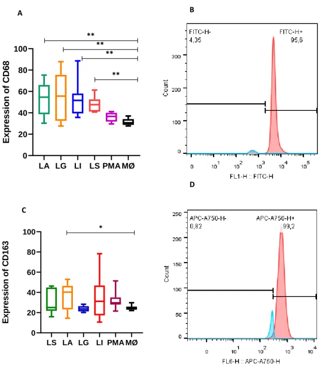
5.5. – Leishmania spp. enhance the density of surface CD68 molecules and L. amazonensis direct macrophage to highly express CD163... 46
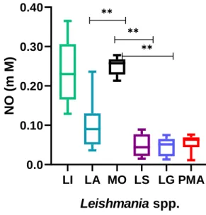
5.6. – Cutaneous species of Leishmania modulate macrophage oxidative burst ... 47
6 - Discussion ... 48
Index of Figures
Fig.1.: Representative scheme of Leishmanaia spp. Life
cycle……….18 Fig.2.: Table of species of Leishmania of Old and New World, geographic
distribution, the respective vectores and the clinical presentation………...20 Fig.3.: Schematique representation of the experimental design used in the present study………32 Fig.4.: Representation of the well plate………...37 Fig.5.: Components of a flow cytometry………..38 Fig.6.: Example of flow cytometry data representes by histogram and dot-plot….39 Fig.7.: Representation of the plate used in the flow cytometry apparatus………...39 Fig.8.. Macrophages infected with Leishmania spp………41 Fig.9.: CD68 macrophage subset after being infected with Leishmania spp………42 Fig.10.: CD163 macrophage subset after being infected with Leishmania spp……44 Fig.11.: Surface expression of CD68 and CD163 in Leishmania-infected
MØ………..46 Fig.12.: Nitric oxide production by macrophage exposed to Leishmania spp……..47 Fig.13.: Graphical representation of the ideal moment of infection by cultured promastigotes of Leishmania spp……….48
List of Abbreviations
APC – Antigrn Presenting Cells CL – Cutanuous Leishmaniasis
CPD-1 -Citratephosphate Dextrose Adenine DAT - Developed Agglutination Test DL – Disseminated Leishmaniasis
ELISA -Enzyme-Linked Immunosorbent Assay
FBS - Fetal Bovine Serum
FITC -Fluorescein Isothiocyanate GIPL - Glicoinositolphospholipids GP63 - Cysteine Protease
iNOS - NO synthase
KMP-11- 11 kDa Kinetoplastid Membrane Protein LA – Leishmania amazonensis LG – Lleishmania guyanensis LI – Leishmania infantum LS- Leishmania shawi LPG - Lipophosphoglycan ML – Mucocutaneous Leishmaniasis MMP 9 - Matrix Metalloprotease 9 Mn - Monocytes MØ – Macrophage
MP - Metalloproteinase NK – Natural Killer OPB - Oligopeptidase B
PBS - Phosphate Buffered Saline PPG - Proteophosphoglycan PCR - Polymerase Chain Reaction
RPMI - Roswell Park Memorial Institute Medium SI – Immune System
SP – Serine Proteins
12
1 - Introduction
1.1.1 - Leishmania: zoonotic and anthroponotic disease
Leishmania is an intracellular parasite member of Trypanosomatidae family and it is the etiological agent of leishmaniasis, a worldwide endemic disease of tropical and subtropical areas, including the Mediterranean region. This disease is considered by WHO as one of the most dangerous diseases. Regarding the prevalence and geographic distribution and counting at present with about 98 scountries from different continents with reported cases (González et al., 2015). Leishmaniasis is a neglected and poorly reported disease with an underestimated burden in most countries. Despite of being known as a dog disease, and others silvatic and domestic animals be infected by Leishmania, such as rodents (Akhounndi et al., 2016), there is a higher rate of human infection and in this case leishmaniasis behaves as zoonosis. In other cases the transmission is anthroponotic, the infection can be accidental and the parasitic cycle is completed in the human body instead in the animal body, and it is more common in the Indian subcontinent and during the epidemic spread in East Africa. Leishmaniasis is a major health problem that is affecting more than 300 million people (Gurung et al., 2015), and can be fatal when is not treated appropriately. Overall the incidence is underestimated because in the majority of countries are not reported. In some parts of both the Old and the New World the transmission can be domestic (Vries et al., 2015).
Leishmania is a heterogeneous parasite that has the ability of infecting two different hosts, vertebrate and invertebrate. The parasite natural habitat is the intracellular microenvironment of mammal’s reticuloendothelial system, including liver, spleen, bone marrow, lymphatic nodes and the intestinal tract of the insect vector (Palatnik-de-Sousa et al., 2011; Akhounndi et al., 2016).
The main vector responsible for parasite transmission is the blood sucking female of the flebotomine sand fly. There are about of 800 sand fly species described and characterized in the New and Old World (Akhounndi et al., 2016). In the New World the predominant vector is of Lutzomyia genus and in the Old World is the Phlebotomus, and due to their small size and physical appearance, they are sometimes mistakenly for mosquitoes (Branco et al., 2010). The female sandflies usually bite when night falls and they have differences according to the species (González et al., 2015).
13 The dog is the main domestic and a peridomestic reservoir of L. infantum in countries of Southern Europe, the Middle East, Asia, North Africa, Central and South America. It is considered a good reservoir because in addition to cohabiting with human beings, it maintains the infection for a long time before developing chronic disease, allowing several parasite transmission cycles. Despite canine leishmaniasis be characterized by a viscera cutaneous involvement, the presence of parasites in the skin will facilitates it’s transmission to the vector (Guimarães e Silva, et al., 2017).
Depending on the parasite species, leishmaniasis can cause different clinical forms such has cutaneous leishmaniasis, visceral leishmaniasis, mucocutaeous leishmaniasis and kala-azar form. However, several factors influence resistance or susceptibility to infection and disease, including genetic variation of the host, genetic variation of the parasites between species and strains, age of the host, nutrition, efficacy of the immuneresponse and other factors such as the inoculum size and number of infective bites received by the host.
Although the most documented form of transmission is the one that is transmitted by the vector, there are other forms of transmission like blood transfusion. Congenitally it is another way of transmission, but more rare than the others.
The disease is characterized by epidemiological and profiles of the several species of Leishmania, capable of expressing different clinical manifestations, transmitted to several vectors and be kept in mammalian reservoirs. Despite the high number of infected human cases, new strategies are needed for a more effective intervention for better prevention of leishmaniasis spread, not only in endemic but also in non-endemic areas (Guimarães e Silva, et al., 2017).
14
1.1.2 - Taxonomic position
Around 53 species of Leishmania were described and approximately 20 of them infect human beings (Akhounndi et al., 2016). Leishmania was classified in several stages with years of difference and by different biologists and is currently classified in the Protist Kingdom (by Haeckel, 1866), Class Kinetoplastea (by Honigberg, 1963 emend. Vickerman,1976), Subclass Metakinetoplastina (by Vickerman, 2004), Order Trypanosomatida (by Kent, 1880), Family Trypanosomatidae (by Döflein, 1901), Subfamily Leishmaniinae (Maslov and Lukeš, 2012) and Genus Leishmania (by Ross, 1903). Leishmania is a heterogeneous parasite and has the ability to survive within mammalian macrophages (MØ) and in the gut of sandflies (Akhoundi et al., 2016). The genus Leishmania is divided into two subgenera depending on its development in the vector: subgenus Leishmania with a development in the stomach (suprapylaric); Vianna subgenus with an initial development in the hindgut and migration to the vector proboscis (peripylaric) (Saridomichelakis, 2009; Akhoundi et al., 2016).
The first attempts of Leishmania classification were based only on extrinsic characteristics, such as geographic distribution, clinical manifestations and development in the intestine of sand fly, but the official classification appeared in the early twentieth century by Nicolle, who separated L.infantum from L.donovani, which has hampered Leishmania taxonomy (Fraga et al., 2010). In the late 1970s, Vickerman proposed, dividing into four complexes of species: the donovani complex, the tropic complex, the Mexican complex and the Braziliensis complex (Fraga et al., 2010).
Later, Leishmania parasites were separeated by sections according to the intravectorial development: the first one in peripylaria (hindgut and pylorus); and the second in suprapylaria (pre pylorus) (Fraga et al., 2010). This division into sections allowed Lainson and Shaw to differentiate Leishmania into two subgenera: Viannia to periplyaria and Leishmania to suprapylaria (Fraga et al., 2010).
More recently, using new techniques of differentiation and comparison with the data obtained previously, the parasite is classified into two phylogenetic lines: Euleishmania that included the subgenus Leismania, Viannia, Sauroleishmania and L. enrietti; and in Paraleishmania that includes L.hertigi, L. deanei, L. herreni, L. equatorensis, L. colombiensis and the genus Endrotrypanum (Fraga et al., 2010).
15
1.1.3 - Epidimiology and Global Dispersion
According by WHO, there were reported in 2014 more than 90% of new cases in countries like Brazil, Somalia and Sudan. But this statistic varies according to the leishmaniasis clinical form, for example the kala-azar form has been less reported, due to the elimination program implemented.
The cutaneous leishmaniasis (CL) of the Old World found in North Africa, Median Mediterranean, Northeast of India and Central Asia is frequently caused by L. (L) major, L. (L) tropica and L. (L) aethiopica. The clinical features vary between and within regions, reflecting different species, immunological status and responses of the patients. More recently, in 2017, 94% of new cases were reported in the same countries, and there were also an increase of CL in cases in Afghanistan and Syria. As already have been said the lack of notification strongly influences the final numbers. At present, Turkey, in Europe, is the most affected country by CL, and Turkmenistan and Uzbekistan, in Asia, account for almost 80% of total cases reported. Anthroponotic CL, which is caused by L. tropica, can be found in Azerbaijan, Israel, Turkey and Uzbekistan. The disease is predominantly reported in densely populated communities, where person-to-person transmission is maintained by P. sergenti. Cases of zoonotic CL due to L. major are mainly registered in South Caucasus and Kazakhstan in central Asia, in Israel at Middle East and in Turkey in Europe. Furthermore, L. major infection is prone to epidemics (Akhoundi, 2016).
In the New World (American continent) CL is caused by parasites belonging to the L. mexicana complex or to Viannia subgenus. The number of new cases of CL (ACL) reported comes from countries such as Colombia and Peru. This is a very complex disease, which can be caused by several different species of Leishmania, that have diverse reservoir hosts and vectors, and can give origin to different symptons (Akhoundi, 2016).
Visceral leishmaniasis (VL) is caused by L. donovani and is mainly found in the Indian subcontinent and East Africa and by L. infantum in the Middle East, Mediterranean bay and Central and South America (González et al., 2015), and more recently were discovered in Australia (Akhounndi et al., 2016). VL may be endemic, sporadic or epidemic, with different clinical features in each situation. In areas endemic
16 for VL the disease tends to be relatively chronic, and children are the most affected. In the Old World the main species are L.(L) donovani, L. infantum and in the New World L.(L) infantum, L. Mexicana and L. (V) braziliensis.
The number of reported cases has been increasing, especially in non endemic areas, due to a long term immigration and tourism in risk areas of contracting the parasite (Silva-Almeida et al., 2012). VL cases of due to L. infantum, are reported in countries of Western Europe, the Balkan region, central Asia, South Caucasus and Turkey, with an overwhelming majority (nearly 75%) found in Albania, Georgia, and a minimum percentage in Italy and Spain. The mainly reservoir hosts are domestic dogs, but foxes also can be sylvatic reservoirs. The main suspected vectors are thought to be P. chinensis, P. ariasi and P. pernicious (Akhoundi, 2016).
Despite its wide global distribution, leishmaniasis epidemiology depends on: the intrinsic characteristics of the parasite species, the local ecological factors, the current and past exposure of the human population to the parasite and the human behavior. Immunocompetence and lack of nutrition are others factors that will increase the risk of VL and mucocutaneous leishmaniasis (MCL). This clinical manifestation causes lesions in the mucous membranes, leading to disfigurations, and is most frequently caused by Leishmania parasites of the subgenus Viannia, mainly by L. braziliensis (Scorza et al., 2017).
The lack of iron, vitamin A and zinc seems to make people more sensitive to the spread of disease when in contact with the parasite. Although recent studies suggested that the increasing of zinc and iron in nutrition do not cause prevent parasite visceralization (Akhoundi, 2016). Other factors that influence the affected population are the age and the level of exposure. In endemic foci where the causative parasite is L. infantum, patients mean age tends to be under 5 years and in endemic foci of L. donovani predominates ages between 13–23 years (Scorza et al., 2017).
Because the CL clinical manifestations are very similar and generalized, the identity of the infecting species should be known as soon as possible. Although in Europe the risk of contracting leishmaniasis in humans is low, and when it happens only focuses on two groups: immune suppressed by medication and refugees from endemic
17 countries (anthroponotic). In dogs, however, remains highly due to the growing number of abandoned dogs or because the owners/tutors do not apply the available anti-Leishmania protection measures to their dogs.
About 1 to 2 million people are infected for the first time each year, with cutaneous Leishmania, being the most reported the emerging case of public health in the Middle East, such as in Syria, where a high incidence has been reported, and although there are national programs, vector control and treatment programs implemented for infected patients, their spread is worrying, especially in recent years with the increase in mass migration (Hijjawi et al., 2016).
The development of new molecular technologies for detection and identification has been an important allied in epidemiological research and management of clinical cases, particularly during outbreaks (for example for species determination).
1.1.4 - Parasite Morphology and Life Cycle
The parasite has two different morphological phases in its life cycle: the promastigote and the amastigote forms. Promastigote parasites are characterized by the presence of a flagellum and live inside the gut of their sand fly vector, and amastigote parasites, which do not have an external flagellum, can be found inside MØ and represents the mammal infective stage.
During the sand fly blood meal, metacyclic promastigotes, the parasite infective form, is inoculated in the host skin. The promastigote forms are deposited on the skin and at the dermo-epidermal junction they bind to some molecules in order to camouflage their entry into the body, as is the case of collagen (Fatoux-Ardore et al., 2014). Once metacyclic promastigotes come into contact with a new microenvironment have to face different obstacles, such has a high temperature and the innate defense mechanisms of the mammal immune system, which includes phagocytes and soluble serum factors, such as components of the complement system.
After phagocytosis, promastigotes differentiate into amastigotes and multiply by longitudinal binary fission inside MØ phagolysosome. Due to amastigotes saturation, MØ end up undergoing cell lysis, releasing the parasites in the extracellular space where found other MØ, being internalized again (Fig.1). By repeating the same cycle
18
A
3
2
5
6
7
8
1
B
4
facilitates parasite propagation in the organism. When the female sand fly gets another blood meal from a parasitized host the ingested amastigotes become procyclic promastigotes and multiply by mitosis in the sand fly gut (Solano-Gallego et al., 2011; Silva-Almeida et al., 2012)
As already mentioned, Leishmania presents a dymorphic life cycle, characterized by two morphological forms. These forms have a nucleus, a large mitochondria and a kinetoplast corresponding to mitochondrial deoxyribonucleic acid (DNA) highly condensed in a single region near the basal body.
At the local of sand fly bite it is evident an erythema, pointing out the local where the parasite was deposited. When the time goes by it can evolve to a CL characteristic wound. Within a two week to six month window, the infection progression trend is different according to the infecting parasite species. In VL case infected MØ home to organs rich in phagocyte cells (such as the spleen) whereas in CL become limited to the site of inoculation or are disseminated through the lymphatic system to other sites of the dermis, such as cartilage or mucosa (Solano-Gallego et al., 2011; Silva-Almeida et al., 2012).
Fig 1.: Representative scheme of Leishmania spp. life cycle. The letter A corresponds to the phlebotomine stage
and the letter B in the vertebrate stage. 1. metacyclic promastigotes are introduced into the dermis, in this case the human being by female sand flies during their meal; 2. promastigotes are phagocytosed by neutrophils, which are the first cells to be activated in the event of an invasion; 3. Parasites differentiated into anastigotes; 4. amastigotes replicate within the phagocytosoe of the MØ, causin cell rupture and are again phagocytosed by other MØ; 5. Female
19
sand flies ingest MØ infected with amastigotes; 6. During blood digestion amastigotes are released in the extracellular environment; 7. in the sand fly gut the amastigotes differentiate promastigotes; 8. promastigotes replicate by mitosis and migrate to proboscis, being able to infect another mammalian host during the next sand fly meal (adapted from CDC, 2018, https://www.cdc.gov/parasites/leishmaniasis/biology.html)
1.1.5 - Clinical presentation – Leishmaniasis, the disease
As already mentioned, 21 species of Leishmania are pathogenic to humans. According to their characteristics, signs, symptoms, endemic area, age and nutritional state of the host, the immunological status and the efficiency of the immune response are classified as Cutaneous Leishmaniasis (CL), Anergic Diffuse Cutaneous Leishmaniasis (ADCL), Mucocutaneous Leishmaniasis (MCL), and Zoonotic Visceral Leishmaniasis (ZVL) and Anthropoietic (AVL) Visceral Leishmaniasis (Akhounndi et al., 2016; Scorza et al., 2017).
In the Old World the genus Phlebotomus is involved in the transmission of different species of Leishmania, and in the New World only the vector Lutzomyia is responsible for the transmission of Leishmania (Fig.2). As already mentioned above, the development of the parasite in the vector is divided into suprapylaric and peripylaric.
20 Fig.2.: Table of species of Leishmania of Old and New World, geographic distributon, the respective vectores and the clinical presentation
In the Old World, CL begins as a papule at the site of inoculation that over time develops a crust, leading to a scar with altered pigmentation. When left untreated, the disease is destructive and disfiguring. The low number of amastigotes in the lesion may lead to late or incorrect diagnosis. In the Old World is caused by L. major, L. tropica and L. aethiopica and in the New World is caused by L. mexicana, L. amazonensis, L. venezuelensis, L. braziliensis, L. guyanensis, L. panamensis, L. lainsoni, L. naiffi, L.(V) shawi and L. peruviana. (Palatnik-de-Sousa et al., 2011; Scorza et al.,2017).
MCL is characterized by the presence of lesions in the nasal and buccal mucosa and respiratory tract and is mainly caused by L. braziliensis and L. panamensis .This disease is rarely seem in Old World, and is more present in aging people (or people with immune suppression). Laryngeal lesions may become chronic and may be mistaken for cancer (Palatnik-de-Sousa et al., 2011; Scorza et al., 2017).
ACDL is a serious clinical form of CL, presenting multiple, disseminated and nodular lesions. It can be a chronic and systemic infection with a fatal outcome if left
21 untreated. It is caused by L. amazonensis and L. aethiopica (Palatnik-de-Sousa et al., 2011; Scorza et al., 2017).
ACDL is a serious cinical form of CL, presenting multiple, disseminated and nodular lesions. It can be a chronic and systemic infection with a fatal outcome if left untreated. It is caused L. amazonensis and L. aethiopica (Scorza et al., 2017).
ACDL clinical forms, which presents lesions all around the body and commonly involves mucosal membranes, can be cause by L. guyanensis and L. amazonensis in the New World and by L. aethiopica in the Old World (Scorza et al., 2017).
CL recurrences are rare, but may occur after primary infections by species, such as L. major or L. braziliensis, This rare form is expressed for varying periods of time. The lesions appear around the scars caused by previous lesion, do not cause pain and do not form ulcers, unlike the other forms of the disease (Scorza et al., 2017).
ZVL is caused by L.infantum and its mains reservoir is the dog, with other animals such as foxes, such as sylvatic reservoirs. It is most common in rural areas with a varied geographic dispersion (Mediterranean basin, Middle East, Nortwest China and Africa). AVL is caused by L. donovani and found more in the Middle East and India (Scorza et. al., 2017). About 95% of cases VL occur in six countries: Bangladesh, Brazil, Ethiopia, India, South Sudan and Sudan (WHO, 2014). This clinical form affects the lymph nodes, spleen, liver and bone marrow. Symptoms are predicted by persistent fever, weight loss, hepatosplenomegaly, anemia and lymphdenopathy. Being progressive can be fatal.
Pos-Kalazar Dermal Leishmaniasis (PKDL), is the most severe form of the disease, and is a complication of VL. It is characterized by a macular, maculopapular and nodular eruption around the buccal mucosa, and can spread throughout the rest of the body. It occurs more frequently in Sudan and India, and its caused by L. donovani (Scorza et. al., 2017).
22
1.1.6 - L. (L) amazonensis
L. amazonensis is reported has highly virulent. In Latin America, L. amazonensis causes several clinical manifestations such as cutaneous, diffuse cutaneous leishmaniasis (although rare and long-lasting, it is caused by an anergic immune response, presenting progressive primary lesions and multiple metastatic lesions), and mucocutaneous. Is unique among other species because promotes chronic infection due to activation of extracellular signal regulated kinases (ERK), having the ability to manipulate the host immune response (Tschoeke et al., 2013; Pratti et al., 2016; Martinez et al., 2015).
1.1.7 - L.(L.) infantum
L. infantum is the etiological agent of VL, in humans and dogs of the Old and New World. The species was described in 1908, by Nicolle in the Old and, by Chagas and Cunha in 1937 in South America where it was identified as L. chagasi. Later on, molecular researches proved that L. infantum and L. chagasi are the same species. There are a few cases reported of CL in the Mediterranean basin caused by this species (Gouzelou et. al., 2013). In CL, the lesions may be single or multiple of chronic evolution. In Portugal the first CL case caused by L. infantum zimoderme MON-1 has been reportedin 2005 (Campino et. al., 2005).
1.1.8 - L. (V) shawi
Belonging to the subgenus Viannia, L. shawi has some epidemiologic relevance in the New World. Although not greatly studied, recent data were able to demonstrated that L. shawi antigens are immunogenic and have beneficial abilities (Jennings et al., 2014). However, this species causes large mucosal lesions with a severe inflammatory process that can progress to necrosis of the affected areas (Jennings et al., 2014).
Passero and collaborators stated that resistance to L. shawi infection can occur through the activation of CD4 + and CD8 + T lymphocytes, and the antigens capable of inducing this immunological profile may be good candidates for the development of vaccines against ACL (Passero et al., 2012). Despite being a monophyletic species, have antigens homologous to other species of Leishmania, which is a positive aspect for the development of an effective vaccine able to protect for Leishmania cross infections (Passero et al., 2012; Jennings et al., 2014)
23
1.1.9 - L. (V) guyanensis
L. guyanensis first described in 1954 occurs in South America and is responsible for ACL (Freitas, et. al., 2015; Coughlan et. al., 2018). More and more cases have been reported by this species, even in extensive areas separated by hydraulic barriers (Scarpassa, et al., 2012).
2 – Immune System: defense mechanisms
After infection, there are factors related to the immune system of the host, as well as to the parasite species that influence parasite spread, persistence and latency and even infection reactivation (Fernandes et al., 2016). The constant warfare of the host immune system against the parasite determines the infection outcome, and subsequently the disease evolution (Gupta et al., 2013).
The Immune System (IS) is composed of tissues, cells and molecules that interact to protect the organism against pathogens and structural alterations. The IS functionality is based on two types of defense: innate and adaptive immunity. Innate immunity represents the first line of defense consisting of anatomical barriers (skin and tissues), cells with phagocytic power (such as monocytes), Natural Killer (NK) cells, inflammatory mediators and complement system. The adaptive immunity generates specific defense strategies for each infectious agent, capable of selectively recognizing and eliminating various microorganisms and at the same time memorizing the previous contact with pathogens. It consists of B and T lymphocytes, antibodies and soluble mediators.
The constituents of innate and adaptive immunity are recruited to the sites when injury or infection occurs to eliminate the pathogens and to resolve the injury. This process however, may be beneficial for some situations, but can also give origin to not so beneficial responses, like high and lasting fever, hepatosplenomegaly, hemorrhages and icterus.
Leishmania parasites have mechanisms to subvert both innate and adaptive immunity (Isnard et al., 2012), escaping to IS deleterious effects and assuring its own survival and completion of its life cycle.
24
2.1.1 - Innate Immunity
The first cells to reach the site of infection are neutrophils, which in turn, destroy pathogens and leave the site “quickly” (they enter into apoptosis), while MØ remain in the same place for a longer period of time. As soon as the promastigotes are deposited in the host's dermis, immediate activation of the complement system occurs. The complement factors C3a and C5a are chemotactic, attracting neutrophils and MØ to the infection site. However, the parasite is able to inactivate the complement through the surface glycoprotein of 63kDa (GP63), which converts C3b into C3i and therefore avoid the assembly of membrane attack complex, preventing parasite lysis, contributing to parasite phagocytosis by MØ, parasite survival and spread, and infection establishment (Gupta et al., 2013).
2.1.2 - Adaptive Immunity
When the innate immune response fails to control and / or eradicate the infection, adaptive immunity takes effect. Activation is slower, however the response generated is specific to the pathogen and produces immune memory, which in the case of future infections (by the same pathogen) acts faster and is more effective. It is a specific antigen response mediated by lymphocytes, such as B cells (derived from bone marrow, involved in the humoral immune response) and T cells (thymus derived, cytokine producing cells). T cells regulate the activity of other immune cells, such as the BB cells and in particular the anti-parasitic activity of MØ (Gupta et al., 2013).
B cells only begin to differentiate after exposure to an antigen, while T cells require presentation of antigen through antigen presentation cells, as is the case of MØ. Although T cells help in eliminating the parasite, they are not able to recognize its surface. Thus, these cells have surface receptors able to recognize antigens from invading pathogens when complexed with molecules of major histocompatibility complex (MHC). Receptors present on the surface of T cells recognize Class I (MHCI) and Class II (MHCII) molecules of major histocompability complex. T cells can be divided into CD4+ (helper Tcells) and CD8- (cytotoxic cells). CD4+ T cells present receptors able to recognize antigen-MHCII complexes and the receptors of CD8-T cells bind to antigen-MHCI complexes. Following antigen recognition process, the selected cells differentiate and a clonal proliferation takes place. This process of specific
25 recognition distinguishes self antigens from non-self or invading antigens (Gupta et al., 2013).
CD4+ cells have several roles in the regulation of immune response. By producing anti-innflammatory and pro-inflammatory cytokines, these cells can promote MØ functionality, stimulate production of B-cell antibodies, stimulate the cytotoxic actibity and controls the intensity and duration of immune response. When CD4+ T cells lose these functions, the immune response does not have the desired effect and the host becomes more susceptible to disorders, such as parasitic invasion. After antigen recognition, the selected CD8+ cells differentiate and proliferate. These cytotoxic cells release granules (that contain perforin and granymes), which promote the lysis of the target cell. They are also producers of interferon (IFN)-γ, essential for the control of infection by inducing MØ together with tumor necrosis factor (TNF) - to produce nitric oxide (Gupta et al., 2013).
B cells represent only 15% of total leukocytes and support antibody production. Antibodies are immunoglobulins, that in mammals belong to five classes; IgA, IgM, IgG, IgE and IgD (Gupta et al., 2013).
2.1.3 - Monocytes and Macrophages
Monocytes (Mn) are heterogeneous cells differentiate in bone marrow by the CD34+ cells, a myeloid precursor. One of monocytes main characteristics is the antigen presentation, as these cells can express high levels of MHCII. Mn establishes a bridge between innate and adaptive immune response, have an active role in homeostasis and in the inflammatory process, being crucial in the body’s defense against microorganisms invasion. Three days after being in circulation, monocytes penetrate different tissues and differentiated into MØ, highly versatile cells, which have an active role when facing apoptotic cells and pathogens. Whenever inflammatory processes take place, monocytes production increase in the bone marrow and consequently, a hight number of these cells rises in the bloodstream. When monocytes progress to MØ, there is an increase in phagocytic capacity and the accumulation of hydrolytic enzymes (lysosomes). Macrophages are large cells, rich in lysosomes and have a direct involvement in host defense. As professional antigen presenting cells (APC), MØ has a high affinity with T and B lymphocytes (Raggi et al., 2017).
26 Macrophages, regarded as primary host cells of this parasite, are polarized cells and based on the type of stimulation received, they can be activated in two ways: M1 (pro-inflammatory MØ) or M2 (anti-(pro-inflammatory MØ) type. When activated in M1, MØ are responding to microbial factors (such as LPS) and to proinflammatory cytokines (such as IFN-γ, TNF-α and IL-1β). On the other hand, these cells also can be activated in M2 by exposure to various stimuli, such as anti-inflammatory interleukins IL-4 or IL-13 (M2a), by immune complexes combined with IL-1β or LPS (M2b), by regulatory anti- cytokine IL-10 (M2c) or by IL-6 (M2d) (Fernandes et al., 2016; Raggi et al., 2017).
In M1, the cells show proinflammatory immunostimulatory properties representing an important source of reactive oxygen and nitrogen and pro-inflammatory cytokines, mediating antimicrobial defense. M2 cells are oriented towards to resolution of any inflammation, tissue remodeling, scarring resolution and angiogenesis. MØ still have a degree of functional plasticity capable of reversibly changing their activation state, on the other hand any polarization disequilibrium may be associated with disorders such as, for example, an autoimmune disease or infection in the state chronic (Raggi et al., 2017).
Although MØ are at the same time phagocytic cells, they are also, in some ways, markers of infection, since they are present for a long period of time. Because they remain longer it became an important milestone that would allow us to understand more about the parasite-MØ interaction.
Toll-like receptors (TLRs), expressed by cells that participates in the innate response as is the case of MØ, are essential for recognition of the pathogen, its molecular patterns, and determinants of infection outcome [(TRL2 recognizes amastigote antigen and the promastigote lipophophaglycan ( LPG)]. Another of the TRLs involved is TRL3 which is activated by the most virulent strains of Leishmania guyanensis and by Leishmania of subgenus Vianna, increasing the expression of proinflammatory mediators (Gupta et al., 2013).
27
2.1.4 - Leishmania Virulence Factors
Leishmania parasite utilizes various surface proteins to enter, to survive inside the human organism, to replicate and to disseminate. Glicoinositolphospholipids (GIPL’s), lipophosphoglycan (LPG), proteophosphoglycan (PPG), 11 kDa kinetoplastid membrane protein (KMP-11) and glycoprotein of 63 kDa (GP63) are indicated as key molecules that ensure parasite infectivity (Isnard et al., 2012; Silva-Almeida et al., 2012). GP63 is described as the major surface antigen expressed in promastigotes of various species Leishmania and is synthesized in the endoplasmic reticulum (ER) (Isnard et al., 2012).
Activation of the complement cascade culminates in C3-C3b cleavage. C3b opsonizes pathogens, which become being killed by lysis (crucial in innate immunity). However Leishmania GP63 as described above, has the ability to cleave C3, converting C3b into the inactive form iC3b reducing terminal complement fixation to the parasite, thus allowing its resistance to lysis (Isnard et al., 2012).
2.1.5 - Diagnostic and Treatment
Over the years diagnostic methods have been improved to fast and accurately detect the parasite responsible for causing leishmaniasis. Unfortunately, it is not always possible to achieve and obtain a reliable result due to the available infrastructural conditions, so sometimes the diagnostic method will have to be adapted. However, there is a standard method, as in all diagnoses for the vast areas, which is direct microscopy of the parasite (Vries et al., 2015).
In serology, many methods have already been developed such as indirect fluorescent antibody test (IFAT), Enzyme-linked Immunosorbent Assay (ELISA), Western Blot and Direct Agglutination Test. However, these methods are not ideal for the detection of cutaneous leishmaniasis due to a decrease in the humoral response caused by the infection, which results in the low sensitivity of the test. An innovative test (CL DetectTM Rapid Test, not currently available on the market) is based on a qualitative membrane immunoassay for the detection of all cutaneous Leishmania species (Vries et al., 2015). ). The technique of isoenzyme electrophoresis continues to be a very useful tool for identifying intrinsic characteristics, like immunological, biochemical and molecular markers (Fraga et al., 2010).
28 A variety of rapid tests (quick and easy to perform) are available for VL diagnosis, which facilitates the determination of the disease as well as a more effective performance, such as IT-LEISH® (DiaMed AG, Switzerland - now Biorad, France), Kalazar Detect® (InBios International, USA), Onsite Leishmania Ab Fast Test (CTK Biotech, USA). Fluorescence-based serological tests are time consuming, expensive, and require more sophisticated laboratory infrastructure (Boelaert et. al., 2014).
Polymerase chain reaction (PCR) allows amplifying small amounts of parasite DNA. Is a fast technique, of high specificity and sensitivity, allowing detecting the presence of Leishmania DNA even in case of asymptomatic individuals. Real-time PCR allows assessment of parasite burden and helps to monitor therapeutic responses.
As already mentioned, depending on the infrastructures there may have to be an adaptation of the diagnostic method and in many low-yield laboratories the "detection" of CL is based on the patient's clinical history and on the physical examination, without recourse to any type of laboratory confirmation test. The risk factors to be taken into account are: age, the profession they perform (whether related to outsider activities such as agriculture or military activities, among others), whether they live, if lives in rural or suburban areas, whether use mosquito nets at home or at work, whether have domestic animals. In terms of observable clinical manifestations, ulcerative lesions should be taken into consideration for final diagnosis (Vries et al., 2015).
In most cases, CL eventually leads the infected host to a "self-cure", however, the total recovery may go for months to years, and in this period the patient has a very weak immune system that results in "exposure" to other potential secondary infections. In places where identification of the species its not possible, treatment is based only on local therapeutic drug options (Vries et al., 2015).
There are several drugs for leishmaniasis therapy, but pentavalent antimonials remain the first choice in most countries, such as meglumine antimoniate (Glucantime®) and sodium stibogluconate (Pentostam®), which allow the blockade of glutamic pyruvic transaminase formation (TGP) and adenosine triphosphate (ATP), inhibiting phosphofructokinase and pyruvate dehydrogenase, destroying the parasite. Despite being the first-line there are already many cases of reported resistance as well as
29 toxicity and side effects. There are other alternative drug therapies such as miltefosine, amphotericin B and paromomycin. In addition to etiologic therapy, conjugation with symptomatic therapies such as anti-inflammatories, broad-spectrum antibiotic therapy, antifungal agents, fluid therapy, blood transfusion, hypoprotein diets and cryotherapy may be appropriate (Vries et al., 2015).
2.1.6 - Prevention
Although the geographical distribution of leishmaniasis is restricted to certain regions of the world, the public health concern caused by the infectious parasite is noteworthy. Prevention is always the best weapon of combat. It can be initiated by identifying risk groups as well as risk factors, like infected vectors.
Thus, a way to prevent parasite infection, controlling the increased number of reported cases is by reducing vector bites. Sand fly is of small in size, even smaller that Anopheles mosquito, the malaria vector. To prevent sand fly bites proper insecticide pulverization is a good option. Another way is diminishing the number of infected reservoirs, especially in notified areas, therefore reducing parasite transmission. The obligation to vaccinate dogs, the reservoir of L. infantum, as a prophylaxis method has already proven a reduction in ZVL cases (Vries et al., 2015).
Prophylaxis vaccination would by far be the best method to combat this disease, however, has been a difficult task to get an efficient prophylactic vaccine.
Individuals infected with Leishmania spp. recovered from infection and become resistant to re-infections, being protected, indicating that prophylactic vaccines can be developed. Investigations and field research have been increasing in the hope of developing the first anti-Leishmania vaccine for more than one species, however the current knowledge is still based on animal models that cannot be easily extrapolated to humans (Srivastava et al., 2016).
Deveoping vaccines are divided into: live attenuated Leishmania vaccines; dead parasite vaccine consisting of the entire parasite or parasite fractions; vaccine constituted of recombinant proteins and DNA vaccines (Srivastava et al., 2016). The major difficulty of developing an effective vaccine is realated to the pathogenesis of the
30 parasite ant the host immune responses, since Leishmania has the ability to modulate the host immune system.
Current preventive measures are based on surveillance, development of technologies to improve and expedite diagnosis, development of new drug therapies (taking into account the resistance already encountered), vector control, and consciousness of populations in endemic areas.
3 - Objectives
This study investigated the MØ phenotype after being exposed to the following Leishmania species: L. guanensis; L. shawi; L. amazonensis and L. infantum. Whenever a cell is exposed to a pathogen there is a whole process that begins with the recognition, followed by signaling and cell activation, that can cause pathogen elimination. There are cases in which the pathogen manages to become "hide" and manipulate the cell in its favor, prolonging its lifetime with consequences for the host. Therefore, the present study has three main goals:
3.1.1 – Immunophenotype macrophage population after being
exposed to promastigotes of Leishmania spp.
Using CD68 (a glycoprotein of 110 kDa highly expressed in cells of monocyte/MØ linage is related to cell activation and phagocytosis) and CD163 (a glycoprotein of 130 kDa restrict to monocyte and MØ is associated to the M2 activation) monoclonal antibodies as cell-surface-marker, MØ populations exposed to visceral and cutaneous Leishmania parasites were evaluated by flow cytometry.
3.1.2 – Analyze the surface expression level of CD68 and CD163 in
macrophages exposed to Leishmania spp. promastigotes
The intensity of CD68 and CD163 in MØ exposed to visceral and cutaneous Leishmania parasites were evaluated by flow cytometry.
31
3.1.3 – Assessed macrophage leishmanicide activity
Metabolization of MØ, arginine and nitric oxide (NO) production, which is a powerful leismanicide molecule that mediates killing, was analyzed by Griess reaction.
4 - Experimental Procedure
Blood was collected from healthy volunteers at the Institute of Hygiene and Tropical Medicine. After collection with anticoagulant the blood was placed in tubes with Histopaque solution. The blood collected along with the Histopaque solution was centrifuged, resulting in layering, and the rings were removed and placed in Falcon tubes with PBS solution. A wash was done and centrifuged again. The resulting supernatant is discarded and the pellet is placed in Falcon tubes and resuspended with RPMI with 10% FBS and 20% CFS. The final solution is placed in wells and incubated for 72h which is the time that the monocytes reach their maximum differentiation in macrophages. At the end of incubation, macrophages are labeled with CD68 and CD163 and infected with the four Leishmania species: L. guyanensis; L.infantum; L.shawi; L.amazonensis. The samples were analyzed by flow cytometry, and at the end compared and the results discussed.
32 Fig. 3.: Schematique representation of the experimental design used in the present study. Blood was
collected from healthy volunteers who were subsequently placed in tubes with Histopaque solution. Total blood was layered through centrifugation, and the rings were removed and placed in Falcon tubes with PBS solution and washed. The supernatant is discarded and the pellet is placed in Falcon tubes and resuspended with RPMI with 10% FBS and 20% CFS. The pellet is plated in wells and incubated for 72 h, which is the time that monocytes reach their maximum differentiation in macrophages. At the end of the incubation the macrophages are labeled with CD68 and CD163 and infected with the species of Leishmania: L. guyanensis; L.infantum; L.shawi; L.amazonensis. Final samples were treated and analyzed by flow cytometry, and MHCI and MHCII results were compared.
4.1.1 - Sample
In this study were used samples of fresh human blood, collected from a group of healthy people with age between 23 and 35 years. The collections were all done at the Instituto de Higiene e de Medicina Tropical and the volunteers were informed about the nature of the study and signed an informed consent.
4.1.2- DAT Protcol
To confirm that the volunteers have no contact with the parasite, the direct agglutination test (DAT) was used to detect antileishmanial antbodies. When become positive, this assay shows a light blue plate over the well of the positive sample, and if it is negative the sample is deposited in the bottom of the well and forms a stain of dark
Peripheral Blood Colection from Healthy Volunteers
Isolation of Mononuclear Cells
Monocytes -- Macrophages L. infantum L. guyanensis L.amazonensis L. shawi CD68 and CD163 Surface expression
33 blue color. An incubation is performed using V-bottom microplates. A 50 µl diluent solution is used in each well [composed of 0.15 M HCL, 0.2% gelatin (Sigma-Aldrich) in distilled water and 0.2 M 2 -mercaptoethanol (Sigma-Aldrich)]. For negative control in the wells only the diluent solution was placed and for the positive control serum samples of patients with zoonotic visceral leishmaniasis. Then, undiluted serum samples of healthy volunteers (50 L) were placed in a 96-well plate, 50 L of L. infantum antigen was added, and at the microplates were incubated overnight at room temperature.The plates were incubated overnight at room temperature. The results were visually evaluated and are expressed as the maximum dilution at which agglutination is observed.
The positive samples were kindly provided by Prof. Carlos Henrique Nery Costa, from the Federal University of Piauí (Teresina, Brazil), and the antigen was generously given by Prof. Saul Semião Santos, from Tiradentes University (Aracaju, Brazil).
4.1.3 - Isolation of peripheral blood mononuclear cells
The blood samples were collected into tubes, with the anticoagulant citratephosphate dextrose adenine (CPD-1) (Polymed) to prevent clot formation, which, if it happens, could make the collected sample unfeasible.
A volume of 3 mL of Hystopaque® (density 1.077 g.mL-1, Sigma-Aldrich), a solution of polysucrose and sodium diatrizoate, that is used to create a density gradient, were placed in Falcon tubes, and 3 mL of blood was carefully laid on. Tubes were centrifuged at 2000g (RCF) at 25 ºC (room temperature) for 20 min. After centrifugation, was possible to observe layers (rings) of different densities. These rings act as a barrier between the cells of higher density (polimorfonuclear and erythrocytes), which is at the bottom of the tube, and the layer which contains mononuclear cells (lymphocytes and monocytes). With the help of a Pasteurs pipette, the layer of mononuclear cells was carefully removed, avoiding blood dragging and placed in a 50 mL Falcon tube. Tubes were fulfilled with phosphate saline (PBS) solution and three washes were made by centrifugation at 1200g, during 7 min at room temperature. Between the washes the supernatant was removed and more PBS solution was added to
34 the total volume of 50 mL was pre-filled, cleaning the cells that will set down at the bottom of the tube.
4.1.4 - Monocyte culture and MØ differentiation
After washing, the final pellet was resuspended in Roswell Park Memorial Institute Medium (RPMI) 1640 (Lonza) at pH 7.2 supplemented with 2 mM L-glutamine (Merck), 100 μg.mL-1 of streptomycin (Sigma-Aldrich) (complete RPMI 1640 medium), 10% (v/v) of heat-inactivated fetal bovine serum (FBS) (BioWhittaker) and 20% (v/v) of colony stimulating factor (CSF – hybridoma secreted glycoproteins that bind to monocyte surface receptors, inducing monocyte proliferation and MØ differentiation). Complete RPMI 1640 medium supplemented with 10 % FBS is an enriching medium and a source of nutrition for MØ.
After the washes it is necessary to know the concentration of viable cells, as the centrifugation can cause the lysis of some cells, thus increasing the number of non-viable cells. A trypan blue dye was used to stain the non-non-viable cells. The concentration of viable cells was estimated in a Neubauer counting chamber under optical microscope (OM).
Since one of the objectives of the study was to obtain the largest number of monocytes, after washing and determination of viable cells, the next step was to maximize the number of MØ. Cell suspension were placed in a six wells plate (3 mL per well) and incubated for 72 h at 37 º C ± 1 ºC, in a humidified atmosphere with 5 % CO2 until achieved complete differentiation on the 3th day of culture.
Every 24 h, cell growth was observed to verify if there is any contamination that could make the sample non viable for the study and to replace the medium by fresh medium heated in a water bath at 37 ºC ± 1 ºC, in order to avoid a thermal shock that could alter cell growth and differentiation and to prevent cell malnutrition.
4.1.5 - Leishmania cultures
As it was mentioned before, four species of Leishmania were used in this study. The species of L. infantum (MCAN/2012/IHMT0003SG) was isolated from a case of canine leishmaniasis from Seixal municipality (Portugal) and maintained in BALB/c mice by successive passages. L. amazonensis (MHOM/BR/1973/M2269) was isolated
35
N = n 50 DF 10
ᶟ
from a patient with ACDL, in Pará (Brazil), L. shawi (MHOM/BR/96/M15789) was obtained from a patient with CL from Buriticupu (Maranhão, Brazil), and L. guyanensis (MHOM/BR/2001/M19663) was isolated from a patient with CL in Santarém (Pará,Brazil). The identification of cutaneous species was based on monoclonal antibodies and multilocus enzyme electrophoresis at Institute Evandro Chagas (Passero et al., 2012). The cutaneous species of Leishmania kindly provided by Dr. F. Passero were maintained criopreserved.
Parasites were cultured in Schneider's Insect Medium (SCHN, Sigma-Aldrich) supplemented with 10% FBS (v/v) and penicillin-streptomycin (Biochrom, Germany) at 100 U.mL-1 and 100 μg.mL-1 respectively (complete SCHN medium ), and incubated at 24 °C ± 1 °C in a refrigerated incubator (Lovibond, Germany). Then, promastigotes concentration (promastigotes.mL-1) was determined in a Neubauer chamber under optical microscope, using a Neubauer chamber and the following equation:
N - Promastigotes. mL-1
n - Total number of promastigotes counted
50 - Factor associated with the number of squares counted DF - Dilution factor
10
ᶟ
- Volume correction factor4.1.6 – Production of Leishmania antigen
To produce Leishmania soluble antigens of the species to be used for MØ stimulation, cultures with a concentration of 1 108 promastigotes.mL-1. Were centrifuged at 1800 g for 15 min at room temperature. The pellet was resuspended in PBS and again centrifuged at 1800 g for 15 min. Then, the resuspended pellet was subjected to six cycles of freezing / thawing (with temperatures varying between 4 and 24 ° C) to provide cell lysis. Finally, the protein concentration was determined in a
36 Nanodrop 1000 (Spectrophotometer, EUA), and adjusted to 40 mg.mL-1 in PBS medium. Antigens were stored at -20 °C until further use.
4.1.7 - Leishmania count
To have a ratio of 1 MØ for 3 promastigotes it was necessary to estimate the promastigote concentration of each Leishmania species involved in the current study. Cultured promastigotes (1 mL) of was placed in a tube with a glycerol- RPMI solution and a 1:5 dilution was made in an Eppendorf tube. Mobile promastigotes were counted in the four4 of the inner square of a Neubaeur chamber (2 upper and 2 lower) and the final concentration was estimated, using the following formula.
1. C [ ] =
cells. mL
-1C [ ] – concentration
x, y, z,w = number of promastigotes in the quadrants of the Neubaeur chamber
4.1.8 – Macrophage infection by Leishmania spp.
After the end of the incubation time (of the differentiation) of the macrophages, it is necessary to make a cell count in Neubaeur's chambers (to evaluate and to count the number of viable macrophages to make the infection). At the end the cell concentration should be adjusted to. At least three counts should be made at different "plate sites" and three fields per "location" using the following formula:
x, y,w - number of cells per field
After counting the macrophages by fields, calculate the number of cells per well by the following formula:
37 Finally multiply the number of cells per well by 3 to obtain the amount of Leishmania spp. add to each well.
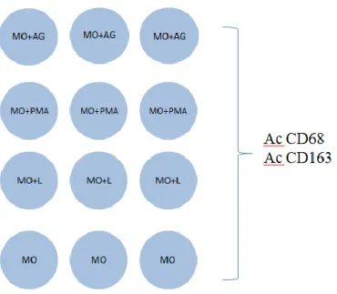
After knowing the amount of Leishmanias spp. add the four species per well. Each species has a different incubation time: L.infantum and L.amazonensis have 5 h of incubation; L.shawi 18 h incubation; L. guyanensis 24 h incubation. At the end of each incubation the medium should be removed and a 0.90% cold NaCl solution added (or use PBS). Place on a plate with ice for 30 min and centrifuge at 300 ×g for 8 min. Discard supernatant and resuspend 100 ul with buffer solution and re-incubate for 30 min on ice and protected from light. Wash with PBS (at least 1 time) at 300 ° C for 8 min. At the end of the centrifugation fix with 500 μl of 2% paraformaldehyde and incubate again for 20 min on ice and remove from the light. Do one last centrifugation at 400 × g (as the cells in the test are losing weight, it is necessary to change the rotation) for 10 min. At the end resuspended the pellet in 500 ul in PBS solution and store at - 4 ° C until use. In the plates, antigen, anticorpos and the four species of Leishmania were placed in each well, along with the positive and negative controls to be analyzed by flow cytometry (Fig 4).
Fig 4.: Representation of the well plates: Blood monocyte differentiated MØ were infected with L. infantum, L. amazonensis, L. shawi and L. guyanensis promastigotes (L). In parallel unprimed MØ (MØ) was used as negative
38 infantum, L. amazonensis, L. shawi and L. guyanensis soluble antigen (AG) Cells were marked with human monoclonal antibody anti CD68 and anti CD163.
.
4.1.9 - Flow cytometry
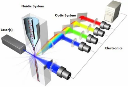
Flow cytometry is a widely used technique for analyzing multiple particle and cell characteristics through a liquid medium such as size (which is represented by light scattering), internal complexity (represented by the dispersion of the angle), cell cycle production and also immunophenotyping by evaluating the expression of surface and intracellular markers, using monoclonal antibodies associated with fluorescent molecules, called fluorochromes. This technique is based on the measurement of fluorescence, as well as its variation, measured between events (Fig.5). It is a technique increasingly used in several areas due to its ability to analyze individual cell with high sensitivity.
Fig 5: Components of a flow cytometry (
https://www.labome.com/method/Flow-Cytometry-A-Survey-and-the-Basics.html).
This method allows distinguishing two (or more) populations in the same sample, based on the change in in size and complexity and also fluorescence. As all cells emit an autofluorescence it is necessary to separate the living cells from the dead (Brown and Wittwer, 2000; Houston et al., 2010).
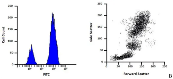
The results are presented by graphs charters, such as histograms in which the y-axis shows the number of events and the x-y-axis indicates size, complexity or the
39 fluorescence intensity and dot plot which is a graphical representation of two parameters in which each point corresponds to a single cell. The scales used in the axes, linear or logarithmic, depend on the intensity of the fluorescence of the amount of cells. The difference between the linear and the logarithm is that the first is used in cases of low intensity variation and the second in cases of greater oscillation of the fluorescence,
A B
Fig. 6.: Example of flow cytometry data represented by histogram and dot-plot. The histogram measuring the
intensity of the FITC signal vs the number of events (A); a scatter plot, used to define the size forward scatter and the complexity side scatter of the cells (B).
Prior to use, a cytometer should be rinsed, cleaning the impurities that may remain.
Fig.7 :. Representation of the plate used in the flow cytometry apparatus. Microplate wells used for initial
cleaning of the flow cytometer.
In the current study at first reading, no marked resting-MØs were used. In the following readings FITC labeled (fluorescein isothiocyanate) corresponding to CD68, and MØ marked with APC, corresponding to CD163, were used.