SYMPATHETIC RENAL DENERVATION
FOR RESISTANT HYPERTENSION
IMPACT ON BLOOD PRESSURE AND ON SURROGATE
MARKERS OF TARGET ORGAN DAMAGE.
MANUEL DE SOUSA ALMEIDA
Tese para obtenção do grau de Doutor em Medicina na Especialidade em Medicina Clínica (Cardiologia) na NOVA Medical School | Faculdade de Ciências Médicas
SYMPATHETIC RENAL DENERVATION
FOR RESISTANT HYPERTENSION
IMPACT ON BLOOD PRESSURE AND ON SURROGATE
MARKERS OF TARGET ORGAN DAMAGE.
Nome do autor: Manuel de Sousa Almeida Orientadores: Ana Maria Aleixo, Professora Associada Agregada Voluntária
Nuno Neuparth, Professor Associado Agregado Pedro de Araujo Gonçalves, Professor Auxiliar Convidado
Tese para obtenção do grau de Doutor em Medicina na Especialidade em Medicina Clínica (Cardiologia)
SYMPATHETIC RENAL DENERVATION
FOR RESISTANT HYPERTENSION
IMPACT ON BLOOD PRESSURE AND ON SURROGATE
MARKERS OF TARGET ORGAN DAMAGE.
Author’s Name: Manuel de Sousa Almeida Supervisors: Ana Maria Aleixo, Professora Associada Agregada Voluntária
Nuno Neuparth, Professor Associado Agregado Pedro de Araujo Gonçalves, Professor Auxiliar Convidado
Thesis to obtain the degree of Doctor of Medicine, in the speciality of Clinical Medicine (Cardiologia)
A
bstrActSYMPATHETIC RENAL DENERVATION FOR RESISTANT HYPERTENSION. IMPACT ON BLOOD PRESSURE AND ON SURROGATE MARKERS OF TAR-GET ORGAN DAMAGE.
Catheter‑based sympathetic renal denervation (RDN) is a new treatment option for resistant hypertension (rHTN) and its clinical impact is yet to be fully understood.
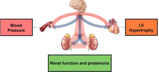
The aim of this study was to evaluate the impact of RDN in blood pressure (BP) and in 2 recognized surrogate markers of target organ damage (TOD): left ventricle (LV) hypertrophy (assessed by echocardiogram) and proteinuria (evaluated by the uri‑ nary albumin to creatinine ratio (ACR), at 1 year follow up.
All patients with rHTN under maximal tolerated antihypertensive drug therapy sub‑ mitted to RDN since July‑2011 were included in a prospective single centre registry. All clinical variables, medication, laboratory values, 24‑hour ambulatory BP measure‑ ments (ABPM) and echocardiogram results were recorded in an electronic database at baseline and at 1‑year follow‑up. The following objectives were addressed: changes on office and ABPM BP, on LV mass and structure, on ACR and renal function, as well as procedure safety.
Since 2011, 318 patients with rHTN were referred for RDN, of which 65 were considered to have true rHTN refractory to drug therapy and accepted for RDN. From those, 31 had a complete 1‑year follow‑up data at the time of the present analysis and are reported here. At 12 months there was a significant decrease in either office and ABPM systolic and diastolic BP, with 84% of patients considered responders to RDN regarding SBP and 71% for DBP. There was also a significant decrease in LV mass, from 152.3g/m2 to 135.7g/m2 (p<0.001) and in ACR, from 25.9mg/g to 14.8mg/g (p=0.007), independent of diabetes status, with no significant changes in renal func‑ tion. No clear linear correlations were found, between changes in BP and either LV mass or ACR, both surrogates of HTN related TOD. There were no major complications related with RDN. These results suggest benefits of RDN in recognized markers of HTN organ damage, which if confirmed are expected to translate to an improvement in clinical endpoints beyond BP control.
For all my masters
THESIS TO OBTAIN THE DEGREE OF DOCTOR OF MEDICINE
t
Able ofc
ontentsList of Figures . . . XIII
List of Tables . . . XV
Abbreviations and Acronyms . . . XVII
Aknowledgements . . . XIX
INTRODUCTION . . . . 1
Summary . . . 2
Hypertension . . . 2
Sympathetic nerve system . . . 3
Sympathetic control of blood pressure . . . 6
Role of sympathetic nerve system on renal function . . . 7
Human sympathetic nerve system assessment . . . 8
Sympathetic nerve system over activity . . . 9
Sympathetic nerve system and essential hypertension . . . 10
Resistant hypertension . . . 11
Renal denervation . . . 12
Clinical studies on catheter based sympathetic renal denervation . . . 13
New devices for renal denervation . . . 17
future indications for sympathetic renal denervation . . . 20
Conclusion . . . 21
Bibliography. . . . 23
CHAPTER 1 – BACKGROUND . . . . 31
Summary . . . 32
Limitations of previous therapeutic strategies on blood pressure control . . . . 33
Limitations of previous studies on renal denervation . . . 33
Target organ lesion – Beyond blood pressure control . . . 35
Redefinition of therapeutic success of hypertension . . . 36
Conclusion. . . . 39
Bibliography. . . . 40
CHAPTER 2 – METHODS . . . . 45
Summary . . . 46
Aims of present study . . . 46
Specific objectives of present study . . . 47
Study team . . . 48
Study design and patients selection . . . 48
Statistics . . . 56
Definitions of other used variables . . . 57
Ethical approvals . . . 57
Bibliography. . . . 58
CHAPTER 3 – RESULTS . . . . 61
Summary . . . 62
Study population . . . 62
Renal denervation procedure . . . 65
Impact on blood pressure . . . 66
Impact on left ventricle structure and function . . . 69
Impact on albuminuria and renal function . . . 76
Relationships between ABPM blood pressure measurements, ACR and LV mass index after renal denervation . . . 81
Safety of renal denervation . . . 84
CHAPTER 4 – DISCUSSION . . . . 87
Summary . . . 88
Impact on blood pressure . . . 89
Impact on left ventricular structure & function . . . 92
Impact on renal function . . . 93
Relationships between changes on blood pressure and on target organ damage . . . 955
Procedure safety . . . 96
Study limitations . . . 97
Conclusions . . . 99
Bibliography. . . . 101
CHAPTER 5 – FUTURE RESEARCH OPPORTUNITIES . . . 107
Summary . . . 108
New changes in hypertension management . . . 108
New side effects . . . 110
New indications and future clinical challenges . . . 111
Assessment of sympathetic nerve system activity . . . 113
Final remarks . . . 114 Bibliography. . . . 115 ATTACHMENTS . . . . 117 List of attachments . . . 117 Attachment A . . . 119 Attachment B . . . 133 Attachment C . . . 143
THESIS TO OBTAIN THE DEGREE OF DOCTOR OF MEDICINE
l
ist off
iguresFigure 1: Different approaches to assess HTN treatment efficacy. . . . 37 Figure 2: Impact of RDN on blood pressure and on target organ damage
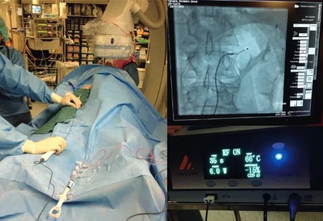
– left ventricle mass and renal function/proteinuria. . . . 47 Figure 3: RDN – Team, involving the Cardiology, Nephrology and Anesthesiology
Departments. . . . 48 Figure 4: Evaluation by CT angiography scan of right (A) and left (B) renal artery,
and angiography, displaying the ablation catheter (E) in the right renal artery (C)
with its ablation tip (D) positioned in the lower artery wall near the ostium. . . . 51 Figure 5: The most frequently used devices used in RDN along the study. . . . 52
Figure 6: An overview of the cathlab setup during an RDN procedure. . . . 53 Figure 7. Flowchart of patient selection. From the total number of patients
evaluated in the outpatient HTN clinic (n=318), 31 patients with ABPM monitoring, transthoracic echocardiography (TTE) and complete data
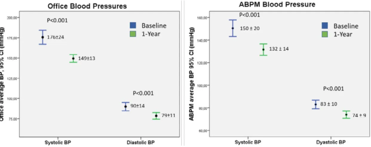
at 12‑months follow‑up, were select for the analysis. . . . 63 Figure 8. BP results one year after RDN. There was a statistically significant
decrease in both systolic and diastolic BP, in office and ABPM measurements. . . . . 67
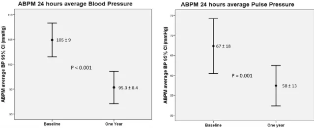
Figure 9. A statistically significant decrease on 24‑hours average pulse pressure
and mean BP, measured by ABPM. . . . 68 Figure 10: Statistically significant changes on LV end‑diastolic volume and function,
one year after RDN. . . . 71 Figure 11: Statistically significant changes on LV mass index, one year after RDN. . . . 71
Figure 12. Comparison of LV mass changes at baseline and one‑year follow‑up,
according to BP responders (n=19) and non‑responders (n=5) to RDN. . . . 72 Figure 13. RDN results one year after RDN, on BP (both office and ABPM)
and on LV mass index, with significant reductions in all parameters. . . . 72 Figure 14. Cross analysis relationship between LV mass index
and ABPM systolic BP changes at one‑year follow‑up. . . . 73 Figure 15. Left ventricle mass changes (g/m2) at baseline and one after RDN,
Figure 16. Comparison of different LV geometric patterns at baseline and one year
after RDN. . . . 75 Figure 17. Analysis of LV diastolic function at baseline and one year after RDN.
The percentage of patients in each diastolic function group
(Normal, Impaired relaxation, pseudo normal and restrictive) is depicted. . . . 76 Figure 18. Decrease in the median ACR after RDN. . . . 77 Figure 19: ACR changes after RDN, according to different ACR subgroups.
A decrease in the percentage of patients with an ACR >300mg/g and an increase
in patients with normal urinary albumin excretion one year after RDN. . . . 78 Figure 20: Results of ACR one year after RDN, according to ABPM systolic BP
responder subgroups. A significant reduction in the median values of ACR
on BP‑responder’s subgroup, and a numerically decrease also in non‑responders. . . . . 79 Figure 21: Results of ACR at 1 year after renal denervation according to ABPM
dipper status at baseline. There was a significant reduction in the median values
of ACR in the dippers subgroup, and a numerically decrease in non‑dippers. . . . 79 Figure 22: Results of ACR one year after RDN, according to diabetic status.
A significant reduction in the median ACR in patients with diabetes,
and a numerically decrease in the smaller subgroup of patients without diabetes. . . . 80 Figure 23. Crosse analysis relationships between ACR and ABPM systolic BP
changes at one‑year follow‑up. . . . 81 Figure 24: Cross analysis of correlation between changes in average systolic
BP on ABPM, ACR and LV mass one year after RDN. . . . 82 Figure 25: Cross analysis of responders one year after RDN,
to ABPM systolic BP, LV Mass and ACR. . . . 82 Figure 26: Cross analysis of responders one year after RDN,
excluding ACR extreme out layers (those with ACR >1500). . . . 83 Figure 27: Rate of of responders to RDN at one year, regarding the studied
endpoints, according to predefined cutoffs: >2mmHg decrease in average ABPM
systolic BP, > 5% decrease in LV mass and any decrease in ACR value. . . . 83 Figure 28: Changes in the median values of eGFR,
THESIS TO OBTAIN THE DEGREE OF DOCTOR OF MEDICINE
l
ist oft
AblesTable 1: Types and functions of adrenergic receptors. . . . 5
Table 2: Effects related to increased sympathetic nerve activity . . . 9
Table. 3: Factors leading to resistant hypertension. . . . 11
Table 4: Main studies and trials about catheter based renal denervation. . . . 14
Table 5: Catheter based sympathetic renal denervation devices. . . . 18
Table 6: Inclusion criteria for catheter based renal denervation. . . . 50
Table 7: Possible complications of catheter based renal denervation. . . . 54
Table 8: Patient’s demographic and clinical characteristics at baseline. . . . 64
Table 9. Antihypertensive medication at baseline. . . . 65
Table 10: Procedure characteristics of catheter based renal denervation. . . . 66
Table 11. Results of blood pressure and heart rate measurements at baseline and one‑year follow‑up. . . . 67
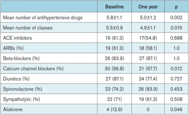
Table 12. Antihypertensive medication at baseline and at one‑year follow‑up. . . . 69
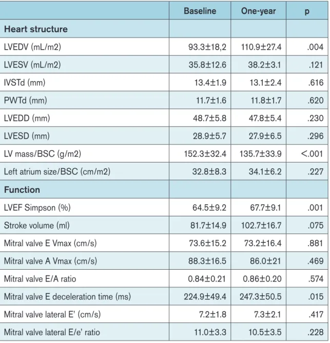
Table 13. Transthoracic echocardiographic parameters at baseline and at one‑year follow‑up. . . . 70
THESIS TO OBTAIN THE DEGREE OF DOCTOR OF MEDICINE
A
bbreviAtions And AcronymsABPM – 24 hours ambulatory blood pressure measurements ACE – Angiotensin converting enzyme
ACR – Urinary albumin to creatinine ratio ARB – Angiotensin receptors blockers BMI – Body mass index
BP – Blood pressure
DBP – Diastolic blood pressure
EDV – Left ventricle end‑diastolic volume eGFR – Estimated glomerular filtration rate ESV – Left ventricle end‑systolic volume EP – Epinephrine
GFR – Glomerular filtration rate HTN – Arterial Hypertension
IVS – Interventricular septum diameter in diastole LV – Left ventricle
LVEDD – Left ventricle end‑diastolic diameter LVEDV - Left ventricle end‑diastolic volume LVEF – Left ventricle ejection fraction
LVESD – Left ventricle end‑systolic diameter LVESV - Left ventricle end‑systolic volume LVM – Left ventricle mass index
NE – Norepinephrine
PWT – Left ventricle posterior wall thickness in diastole RAAS – Renin – Angiotensin ‑Aldosterone system RBF – Renal blood flow
RDN – Sympathetic nerve system denervation RWT – Left ventricle relative wall thickness SBP – Systolic blood pressure
SNS – Sympathetic Nerve System
THESIS TO OBTAIN THE DEGREE OF DOCTOR OF MEDICINE
A
knowledgementsThe author wishes to express its most sincere appreciation to Professors Ana Aleixo, Nuno Neuparth and Pedro Gonçalves for their assistance and continued support in the preparation of this manuscript.
Thanks for all the hard work, from all the fellows at the time, who participated, in all research projects, Helder Dores, Maria Salomé Carvalho, João Mesquita.
My profound appreciation for the partnership only made possible by the excel‑ lence of Dra. Augusta Gaspar and Patricia Branco, the nephrologists of my choice, who made possible the success of this project.
A special thanks to Professor Ricardo Seabra‑Gomes, Francisco Machado, and Pedro Gonçalves, for providing me an example of mastership in excellence in all we do, also for their friendship and belief.
In addition, my profound gratitude to all my colleagues and friends, who all share with me their passion for Santa Cruz, a school of cardiology, especially Rui Teles.
My most profound acknowledgement and gratitude for “Professor” Pedro Gon‑ çalves, a pupil who became a my Master. Thanks for the perseverance and belief.
Thanks also to the members of the Nova Medical School council for their valuable input.
Finally to my family, for their continuous support, trust and most of all, for their precious and most appreciated patience.
Adapted from a review article entitled. “Renal Denervation for Resistant Hy-pertension”, published in Revista Portuguesa de Cardiologia, with permis‑ sion from the editor.
M de Sousa Almeida, P de Araújo Gonçalves, E Infante de Oliveira, et al. Rev Port Cardiol. 2015; 34 (2):125‑135.
s
ections:
■ Summary ■ HypertenSion
■ SympatHeticnerveSyStem
■ SympatHeticcontrolofbloodpreSSure
■ roleofSympatHetic nerveSyStemon renalfunction ■ HumanSympatHetic nerveSyStemaSSeSSment
■ SympatHeticnerveSyStemoveractivity
■ SympatHetic nerveSyStemandeSSential HypertenSion ■ reSiStantHypertenSion
■ renal denervation
■ clinicalStudieS oncatHeter baSedSympatHeticrenal denervation ■ newdeviceSfor renaldenervation:
■ futureindicationS forSympatHetic renaldenervation: ■ concluSion
■ bibliograpHy
s
ummAryHypertension is a leading cause of death in developed countries, and although there have been large investments in drugs aiming its control, there is still a stag-gering contrast between its high prevalence and the low rates of adequate con-trol. A subset of patients with suboptimal blood pressure control have drug-resistant hypertension, in the pathophysiology of which chronic sympathetic hyperactivation is significantly involved. Sympathetic renal denervation has recently emerged as a device-based treatment for resistant hypertension. In this review, the pathophysiolog-ical mechanisms linking the sympathetic nervous system and cardiovascular disease are reviewed, focusing on resistant hypertension and the role of sympathetic renal denervation. An update on experimental and clinical results is provided, along with potential future indications for this device-based technique in other cardiovascular diseases.
H
ypertensionHypertension (HTN) is the leading global risk factor for cardiovascular mortality, with a prevalence worldwide projected to be approximately 3.5 billion in 2015, with 7.8 million related deaths each year1. Its strong association with myocardial infarction, heart failure, stroke, end‑stage renal disease and cardiovascular death, is well established, with 54% of stroke and 47% of ischemic heart disease attributable to high blood pres‑ sure (BP) worldwide,2 and it seems to have a continuous relationship between BP and cardiovascular risk from values as low as 115/75 mmHg, doubling the cardiovascular risk for every 20/10mmHg in pressure.1, 3
Meanwhile, effective blood pressure lowering has consistently been shown to reduce overall cardiovascular risk 4, but the rates of adequate BP control remains sub‑ optimal, despite the large amount of available antihypertensive drugs, from different classes and strong scientific evidence supporting their use, with only 37% of treated hypertensive patients, achieving recommended BP values, in European countries. 5
The blame for such very low rates of BP control cannot be attributable only to poor treatment management. The complex pathophysiology underlying human BP con‑ trol, with multiple interconnected systems, has certainly a relevant role for the failure in
INTRODUCTION
There are at least two main systems responsible for BP control: renal‑based and vascular‑based. The renin‑angiotensin‑aldosterone system (RAAS), plays a major role in renal control of salt and water homeostasis, but also in peripheral vascular resis‑ tance, acting directly through angiotensin but also by activating the sympathetic nerve system (SNS). Other mediators are also important, although their role is not so well understood.
For the last decades, RAAS system has been the central focus of HTN treatment and management. Availability of secure, efficacious and evidence proved drugs block‑ ing this system, as led to neglect the contribution of other systems, namely the autono‑ mous nervous system, for raising and maintaining high BP values.
The SNS and its possible role in the pathogenesis of HTN, has been receiving increasing attention. A more complete understanding of how the SNS could help to control the long‑term level of BP has developed recently and supports a complete new approach to treat resistant HTN.
s
ympAtHetic nerve systemMore than one hundred years have passed since Gaskell and Langley first elu‑ cidated us on the structure and function of the autonomous system, the sympathetic and parasympathetic system, and could show, that they are both distributed to the same body structures, their effects are antagonistic, and they subservice the functions of organic life, and are not under the control of the will.6 Later, Cannon has extended this, by pointing out the critical role of the SNS in preparing the body to struggle and to increase its powers of defence.7 Under SNS stimulation, the pupil dilates to increase perception of light, the heart beats faster and forcibly to supply the muscles with blood, the visceral blood vessels area constrict, raising the blood pressure and driving the flow from the digestive area, whose functions are inhibited, to the muscles, the heart, the lungs and the brain.
Sympathetic nerve system control. Under physiological conditions, the
autonomic nervous system, adjusts circulation in keeping with behaviour, environ‑ ment and emotions, via rapid changes in the cardiac output and regional arteriolar resistance. The neural control of the circulation operates via parasympathetic neu‑ rons that innervate the heart, and via three main classes of sympathetic efferents, the barosensitive, the thermosensitive and the glucosensitive, that do innervate the blood vessels, the heart, the kidneys and the adrenal medullae. Their control cen‑ ters are all located in the central nerve system: the rostral ventrolateral medulla, the spinal cord, the hypothalamus and the nucleus of solitary tract. The rostral ventro‑ lateral medullae, the nodal point for most, if not all, sympathetic reflexes that involve
thalamic centres, influencing sodium and water balance, but also, to the cortex, specially the limbic cortex, responsible for rapid behaviour‑related adjustments of sympathetic tone.8
Sympathetic nerve system effectors. The thermosensitive group of cardio‑ vascular efferents is primarily cutaneous vasoconstrictors activated by hypothermia, emotions and hyperventilation. The glucosensitive are activated by hypoglycemia and physical exercise and controls the release of epinephrine (EP) by the adrenal medulla. These two types of efferents presumably have a secondary role in short and long‑term regulation of BP.10, 11
The large group of barosensitive sympathetic efferents, under the input influence of arterial baroreceptors, have a dominant role on short and long‑term regulation of BP. Regardless of the organ or tissue that they innervate, these neurons have an ongoing activity at rest. They control the heart, the release of norepinephrine (NE) from the adrenal medullae, vasoconstriction of resistance arterioles and play a major influence in kidney function, where they control renin secretion, sodium reabsorption and renal blood flow.12
The SNS has also a special relation with adrenal medullae. Preganglionic sympa‑ thetic nerve fibres pass directly from medio‑lateral horn cells of the spinal cord into the two adrenal medullae, ending directly on modified neuronal cells that secrete EP and NE directly into blood.13 Anatomically, those sympathetic adrenal nerve fibres can go along with the sympathetic postganglionic renal fibres and in this way may be affected by renal denervation procedures.
Sympathetic nerve system neurotransmitters. The sympathetic fibres secrete one of two synaptic transmitter substances, acetylcholine or NE. All pregan‑ glionic neurons are cholinergic, it means, they secrete acetylcholine as neurotransmit‑ ter that excites the postganglionic neurons. Most of those postganglionic neurons are adrenergic, meaning that they secrete NE as a neurotransmitter, with the exception of postganglionic sympathetic fibres to the sweat glands, which are cholinergic.13
The NE secreted by terminal nerve endings is removed from site by reuptake into the adrenergic nerve endings through an active transport mechanism, account‑ ing for 50 to 80% of secreted neurotransmitter. The remaining neurotransmitter may diffuse away to the surrounding tissue and blood being destroyed later, by cate‑ chol‑O‑methyl transferase in the liver. Small amounts are also destroyed by local tis‑ sue monoamine oxidase enzyme. Although NE secret directly to tissue as a lifespan of a few seconds, once in blood, remains active from one to several minutes, until destroyed in the liver.13
Epinephrine and NE stimulates an effector organ through a membrane receptor, that either changes cell membrane permeability to ions (sodium, potassium or calcium) or altering an intracellular second messenger enzyme, the adenylcyclase, causing the
INTRODUCTION
There are at least two known types of adrenergic receptors, alfa and beta receptors and many subtypes of them. For a more comprehensive review, the receptors and their function are listed in Table.1.
Table 1: Types and functions of adrenergic receptors.
Alpha Receptors Beta Receptors
α1 postsynaptic α2 presynaptic β1 postsynaptic β2 postsynaptic Gq protein coupled Activation phospholi‑ pase C Gi protein coupled Inhibits Adenyl Cyclase
Gs protein coupled Activates Adenyl Cyclase
Vasoconstriction: • Skin
• Gut • Kidney • Brain
Smooth muscle cells contraction: • Ureter • Vas deferens • Urethral spinchter • Uterus • Cilliary body Glucose metabolism: • Gluconeogenesis • Glucolysis Glucose metabolism: • Inhibits insulin release • Stimulates glucagon release • Contraction of anal sphincter • Inhibits release of EP Heart: • Chronotropic + • Dromotropic + • Inotropic + • ↑ LVEF Renine release ↑ by juxtaglomerular cells Hunger ↑ • ↑ ghrelin release by stomach SMC relaxation: • Bronchus Bronchioles • Detrusor muscle • Uterine muscle Urethral sphincter Contraction: Renin release by justa‑
glomerular cells Glucose metabolism: • Inhibits insulin re‑
lease • Stimulate glucolysis and gluconeogene‑ sis Lipolysis Vasoconstriction of: • Coronary arteries • Veins
The beautifulness of this system, and its complexity, drives from the fact that using just two different kinds of neurotransmitters, NE and EP, it can initiate many different actions, depending on the target tissue, type of receptor and the relative affinity of the neurotransmitter for the receptor.
This mechanism must have been developed during a vast period, through innu‑ merable intermediate stages. Therefore, we are now in possession of a complex mech‑ anisms which discharge energy under strictly controlled fashion and adequate stimu‑ lation.
s
ympAtHetic control of blood pressureBlood pressure is primarily a function of peripheral arterial resistance, but also of cardiac output, which in turn, is dependent of heart rate, myocardial contractility and venous blood return, the later, also dependent on smooth muscle venous tonus. All under SNS control.
The neural control of BP and overall circulation uses the vast complex SNS and the more localized parasympathetic neurons that do innervate the heart and lungs.
As explained, the background activity of the barosensitive group of SNS effer‑ ents, is presumably the most crucial for long‑term physiological regulation of BP and it seems, that in hypertensive humans, the rise in the activity of barosensitive sympathetic efferents is not restricted to renal nerves but is generalized,8, 12, 14‑16 partly explaining the cluster relationship held by HTN and other metabolic disturbances. Regardless of the organs or tissues they innervate, they show continues ongoing activity at rest (the sympathetic tone) and they discharge in burst synchronized with arterial pulse and respiration, being responsible for short term fluctuation of BP.10, 17, 18 They control the heart, the release of NE from adrenal gland, constricting resistance arterioles, the kidneys, increasing renin secretion, tubular sodium reabsorption and renal blood flow, most likely, exerting a long‑term control on BP.12, 19
Atrial stretch or volume expansion has a strong inhibition effect on the renal SNS efferents. It appears that the selective control of renal SNS efferent by volume recep‑ tors, might be the most important of these differential regulations mechanisms.12, 19
The afferent limb of this loop reflex mechanism involves mechanoreceptors acti‑ vated by distension of the arterial wall. An increase in BP activates those receptors, inhibiting cardiac, renal and vasomotor sympathetic efferents, restoring BP to previ‑ ous values, and so helping to damp short‑term BP fluctuations.8, 9 This system may also be reset, through neural and humoral mechanisms, still largely unexplored, allow‑ ing higher BP values without reduction in reflex sensitivity, for example, circulating and brain derived angiotensin II can reset these reflex mechanism.8, 9 External, chronic
INTRODUCTION
monal indicators of sympathetic activity, namely NE and angiotensin II and with a pre‑ sumed increase in survival,7, 20 but apparently had little influence on long‑term control of BP.8, 21
Besides its nearness in the carotid bifurcation, the “road” into the brain, there is a close interaction between baroreceptors and chemoreceptors in the control of sym‑ pathetic activity, whereby the baroreflex activation is inhibitory and the chemoreceptor reflex is excitatory. Besides this contra regulatory action, there is a facilitator effect between them, whereby a reduced baroreceptor activity will enhance the chemore‑ ceptor response.7, 18 These reciprocal sensory modulation, exerted by baroreceptors and chemoreceptors, are beneficial in states of circulatory collapse and shock. When severe hypotension and hypoxia coexists, they mutually enhanced sympathetic drive and ventilation helping to overcome the crisis.
r
ole of sympAtHetic nerve system on renAl functionthe role of kidneys in long‑term regulation of BP, is mandatory in any discus‑ sion addressing this subject. The pressure‑natriuresis relationship described several years ago by Guyton,22 established that any increase in sodium retention produced an initial blood volume expansion, increasing cardiac output and therefore BP. The resulting peripheral tissues overperfusion leads to an increase in peripheral resis‑ tance, returning cardiac output towards normal. Accordingly, a reset of the pressure natriuresis relationship, establishing a new BP homeostatic set‑point, inevitably lead do HTN, regardless of the cause of the resetting. But we need to address the fact, not known at the time, that volume expansion also promotes atrial stretch and activation of baroreceptors located there, and subsequently to a selective inhibition of renal SNS activity.12, 19
Renal sympathetic nerve terminals innervate the three major renal neuroeffectors, directly influencing renal tubular function, glomerular flow rate (GFR) and renal blood flow, with a clear impact in all major components of renal function. Renal sympathetic efferents are in direct contact with the peritubular basement membrane, of all renal tubu‑ lar segments (α1A‑drenoreceptors), as well as the juxtaglomerular granular cells affect‑ ing renin secretion (β1‑adrenoreceptors) and renal arteries (α1A‑drenoreceptors). This control is frequency dependent, with increases in renin secretion rate without changes in urinary sodium excretion, renal blood flow and GFR at low frequencies. At slightly higher frequencies, the increase in renin secretion is associated with an increase in tubular sodium reabsorption, still without changes in renal blood flow and GFR.12 Much higher frequencies will decrease GFR and renal blood flow, maximizing renal ability to reabsorb sodium and water. Besides, additional neurophysiological renal studies
neuroeffector, making the overall system more powerful and precise, in controlling the body sodium contends and thus BP.12
We can assume that the coupling of regulation of total body fluid volume to arterial blood pressure, depends on kidney’s ability to excrete sodium in such a way as to achieve sodium internal balance in face of varying sodium intake, through the Guyton’s pressure natriuresis mechanism.22 Any defect on the kidney’s ability to main‑ tain this balance results in an increase in arterial pressure. The observation in various animal experiments,12 that renal denervation prevents or delays the onset of HTN, implies that an increase in renal sympathetic activity, may be a final common pathway required for a defective renal sodium excretion, leading to development and mainte‑ nance of HTN.
H
umAn sympAtHetic nerve system AssessmentUntil the early 70s, the most commonly used method to assess SNS activity, was blood measurements and urine excretion rates of NE and its derivate, a gross esti‑ mation of whole‑body sympathetic activity at best.23 Meanwhile, new methodologies emerged for measuring sympathetic nerve firing rates in subcutaneous nerves and for assaying the concentration of sympathetic transmitters in plasma.
Microneurography, a technique reported first by Hagbarth24, provided a tool to study sympathetic nerve firing in subcutaneous tissue and skeletal muscle vessels, through tungsten electrodes inserted in the skin. It records burst of nerve activity, syn‑ chronous to heart‑beat, generated by sympathetic efferent nerves. Highly reproducible and closely related to sympathetic traffic directed to other structures, it can be repeated over time, although painful, it allows the assess of interventions effects and direct quan‑ tification of sympathetic nerve traffic and vasomotor tone.
The spillover technique, a measurements of sympathetic NE transmitter release, first applied by Esler et al25, this isotope dilution method calculates the clear‑ ance and spillover of NE, using an infusion of tritium labelled NE administered intra‑ venously. The close relationship between the sympathetic nerve fibres firing rate of an organ and the rate of NE spillover into the venous effluent of that organ provides the rational for using measures of regional NE release as a surrogate of sympathetic tone in individual organs,26 allowing the assessment of regional SNS function in humans.
This technique was on the forefront in proving the concept that heart failure patients, had sympathetic overactivity rather than sympathetic denervation, as initially thought, and opened the way to the routine use of betablockers in heart failure.27
INTRODUCTION
s
ympAtHetic nerve system over ActivityBesides its central role in cardiovascular homeostasis, controlling the vascular tone through vasoconstriction of small resistance arteries, the SNS also interferes and regulates numerous other physiological processes (Table 2).
Table 2: Effects related to increased sympathetic nerve activity
Vascular
Smooth muscle cell hypertrophy and proliferation Endothelial dysfunction and damage
Arterial stiffness
Posture blood pressure control impairment and syncope Hypertension
Atherosclerosis Cardiac
Myocyte hypertrophy Left ventricle hypertrophy Arrhythmia
Psychogenic heart disease Renal Effects
Renal artery vasoconstriction Sodium and fluid retention Microalbuminuria
RAAS activation Metabolic Effects
Insulin resistance Dyslipidemia
There is growing evidence that sustained chronic changes in SNS activity are involved in the pathogenesis of many diseases states, ranging from metabolism to psychological disorders, including ischemic heart disease28, chronic heart failure,29, 30
tive sleep apnoea36, depression to inflammatory bowel disease.37, 38 A chronically over‑ active SNS is well known to worsen prognosis in patients with heart failure and end‑ stage renal disease39, 40.
Supported by accumulating scientific evidence41 is the fact that deleterious effects on vessels and myocardium, by an overactive SNS is independent of BP. Chronic SNS activation can cause hypertrophy and proliferation of vascular smooth muscles cells, as well as a direct trophic effect on cardiac myocytes with an increase on left ventricular mass (LVM) and wall thickness, without an increase in BP.41 The combined structural changes on the myocardium and the direct effect of an overactive SNS, is a major con‑ tribution to the high incidence of arrhythmias commonly seen in this patients.42
Virtually all cardiovascular conditions and diseases, in which an increased adren‑ ergic drive is involved are also characterized by endothelial dysfunction.43 Nitric Oxide, one of the most important endothelial function mediators, is also an important neu‑ rotransmitter cooperating in the autonomic regulation of cardiovascular function, act‑ ing as a sympathetic‑inhibitory substance within the central nerve system44. Acute and chronic increases in SNS activity, through endothelial dysfunction and endothelial cell damage, have been proven to contribute to subsequent development of atherosclero‑ sis. 37, 41, 43
SNS overactivity has also been linked to the development of metabolic distur‑ bances, such as insulin resistance and dyslipidemia.45 Not only, an increased SNS activity can itself led to insulin resistance, particularly in hypertensive patients,45 but also elevations in circulating insulin levels, from insulin resistance in obese patients, can precipitate an increase in SNS activity leading to HTN.41, 45, 46.
s
ympAtHetic nerve system And essentiAl HypertensionFrom the 1930s, surgical sympathectomy appeared to be very efficacious in low‑ ering high BP and improving the clinical outcome in patients with severe HTN,47, 48 but its poorly tolerated side effects and high surgical risk led to his abandon, especially after the appearance of ganglion blockers, the first efficacious antihypertensive drug class.49
The critical role of SNS in the pathogenesis of HTN, other cardiovascular diseases and disturbances, is unquestionable, but the existing complex and clinically impractical methodology, to assess the activity of SNS in humans, makes it difficult to adequately establish a relationship between SNS activity and HTN in a particular patient, a major reason why the SNS was so neglected until now.
A well‑known consequence of an overactive SNS is an increase in BP. Renal sympathetic nerve activity is pivotal in the pathogenesis of essential HTN, through influ‑
INTRODUCTION
diac contraction and venous capacitance.37 In previous studies, increased sympathetic outflow to kidneys were related to the magnitude of essential HTN.31, 33, 37, 50 Renal spill‑ over measurement, of sympathetic outflow to kidneys, in patients with HTN, revealed that more than 50% of them, had significant sympathetic hyperactivity.51
r
esistAnt HypertensionTreatment‑resistant HTN is defined as a BP persistently above 140/90 mm Hg and 130/80 mm Hg for patients with diabetes and renal disease, despite the prescrip‑ tion of three different antihypertensive drug classes, of complementary mechanism of action, at appropriate doses, preferably including a diuretic.52 Patients in this category might have pseudo‑resistance, that should be identified and differentiated from true resistance.
Pseudo‑resistance, comprising around 50% of patients claimed as having resis‑ tant HTN, includes those with inadequate treatment regimens, suboptimal adherence to medication and inadequate clinical BP measurements.53
Several factors can lead to true resistant HTN, and must be addressed appropri‑ ately (Table 3).54 Inappropriate levels of aldosterone are seen in up to 20% of patients with resistant HTN, whether or not, an adenoma is present. On such patients, aldoste‑ rone antagonists may provide relevant additional BP reduction.54
Table. 3: Factors leading to resistant hypertension. • Obesity • Male gender • Older age • African‑American • Insulin resistance • Volume overload • Renal dysfunction • Exogenous drugs
The prevalence of resistant HTN has been reported to be between 5 and 30%. This prevalence differs accordingly to the hypertensive population being studied and the methods used in BP assessment, with higher percentage of patients with resistant HTN in cohorts from centres specialized in the treatment of HTN as compared to gen‑ eral community based cohorts.55‑57 Recent evaluations on the prevalence of resistant HTN, especially if 24‑hours ambulatory blood pressure measurement (ABPM) is used, points to rates less than 10% of all hypertensive patients.58
In Portugal, data from the PHYSA study involving 3720 adults patients, the prev‑ alence of HTN was 42%, of which 76.6% were aware of the fact, 74,9% were treated and only 42,5 had their BP values below the recommended thresholds.59
Patients with resistant HTN, namely true resistant HTN exhibit a worst prognosis with a higher risk for cardiovascular events, when compared to hypertensive patients without resistant HTN.60, 61
In face of the available clinical studies, patients considered to be good candi‑ dates for RDN should have severe true resistant HTN, defined as office systolic BP of at least 160mmHg BP of at least 160 systolic (150mmHg in type 2 diabetes). 62‑64
r
enAl denervAtionThe finding that sympathetic renal activity was increased in spontaneous hypertensive rats, the animal model most used in essential HTN research, was of paramount for the role of renal SNS on HTN pathogenesis.65 In an experimen‑ tal model of HTN and obesity in dogs submitted to a high fat contend diet, RDN, not only prevented the appearance of HTN but also increased in 50% the urinary sodium excretion.66 In another animal model with chronic renal failure, sympathec‑ tomy prevented HTN and was associated to a decrease in the activity of the central adrenergic nucleus.67
Renal lesions induced by phenol injection, without changing the GFR, was linked to a sustained rise in HTN and NE release by the hypothalamus. RDN of those animals prevented the rise on BP.68 In different animal models, the effect of RDN has consistently showed the important role of renal SNS in the pathophysiol‑ ogy of HTN.
In humans, surgical sympathectomy exhibited a high efficacy in lowering high BP and improving the cardiovascular prognosis of patients with severe HTN.47‑49, 69‑71 Its poorly tolerated side effects, such as: severe orthostatic hypotension, anhi‑ drosis, bowel disturbances and sexual dysfunction, led to its abandon. Neverthe‑ less, it has reinforced the important link between the SNS and the mechanisms controlling BP.
INTRODUCTION
c
linicAl studies on cAtHeter bAsed sympAtHetic renAldenervAtion
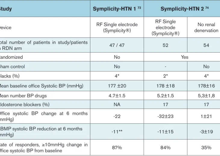
Since 2009, several clinical studies were published describing the effect of cath‑ eter based RDN for the treatment of resistant HTN (Table 4). The Symplicity HTN‑172, a proof‑of‑principle study, was the first to evaluate the effects of RDN on patients with severe resistant HTN. One year after the procedure, the office BP decreased on aver‑ age 27 mmHg for the systolic and 17 mmHg for the diastolic, and this was sustained until 24 months of follow‑up, with 13% of non‑responders to RDN therapy73. A sub‑ group analysis targeting the renal and systemic sympathetic activity, revealed an aver‑ age reduction of 42% in renal NE spillover on these patients.73
The Symplicity HTN‑2 study,74 a multicenter clinical trial, randomized 106 patients with severe resistant HTN under medication to optimal antihypertensive medical ther‑ apy alone or in association with RDN, with the primary endpoint of changes on office BP at 6‑month follow‑up. The results revealed a significant decreased on office BP, in patients submitted to RDN, 32 mmHg on systolic BP and 12 mmHg on the diastolic (p<0.01), compared to an increase in 1 mmHg on systolic BP and a no change on diastolic BP (p=NS), observed in patients under optimal medical therapy alone. A subgroup analysis regarding ABPM measurements, revealed a similar behavior, a drop of 11mmHg on systolic BP and 7mmHg on diastolic BP, in the RDN group (p<0.001) compared to a drop of 3mmHg and 1mmHg respectively, in the medical therapy alone group (p=NS). The magnitude of the decrease in BP, between the RDN treated group and the optimal medical therapy alone group was maintained at 12 months follow‑up.73
Alongside its efficacy, RDN was a very safe procedure. On both studies, only minor vascular complications occurred, mainly at the puncture site, haematomas, pseudoaneurysms and one renal artery dissection during the diagnostic procedure, successfully treated with a stent, all without sequalae. Regarding renal function, no significant changes on GFR occurred during follow‑up. No major complications were identified during the follow‑up.62, 75
Using a different RDN device, the EnlightHTN I study64 also revealed a significant decrease in BP measured by office and by ABPM, at 6 months, with a remarkable safety profile.
The available published scientific data supports an excellent safety profile on short term, although the risk of renal artery stenosis on long term is still lacking.
The results from the simplicity HTN 1‑2 trials and EnlighHTN I trial were very prom‑ ising, nevertheless, their open control design made impossible to properly address the important bias made by the placebo effect on BP measurement, in either study groups. Some of those limitations were addressed in the Symplicity HTN3 Trial.76
In the Symplicity HTN 3 clinical trial,77 a larger randomized study, to evaluate RDN for treatment of resistant HTN, the design included for the first time a sham con‑
trol‑group. On the primary endpoint, the change on office systolic BP from baseline to 6‑month follow‑up, between the RDN (n=353) and the sham control arm (n=171), the obtained average difference of ‑2.39mmHg between the 2 groups on office systolic BP, didn’t achieved statistical significance (95% CI: ‑6.89 to 2.12, p=0.26), with an average reduction of 14.1 mmHg on office systolic BP reduction in the RDN arm vs 11.7 mmHg reduction in the sham control arm.
In the secondary endpoint, the change on the mean systolic BP from baseline to 6‑month follow‑up, measured by ABPM, between RDN and the sham control arm, a statistically non‑significant difference of 1.96 mmHg (95% CI: ‑1.06 to 4.97, p=0.98) was achieved, with a systolic BP reduction of 6.8 mmHg in the RDN arm vs 4.8 mmHg reduction in the control arm, at 6‑month follow‑up.
The rate of major adverse events at 6 months, was very low, 1.4% in the RDN arm vs 0.6% in the sham control arm, much less than the 9.8% prespecified as the target for major safety events incidence. So, the primary safety endpoint was met, for a differ‑
Table 4: Main studies and trials about catheter based renal denervation.
Study Symplicity-HTN 1 72 Symplicity-HTN 2 74 Symplicity-HTN 3 77 EnligHTN-1 64 RAPID 101 REDUCE HTN
FIM102
Device RF Single electrode(Symplicity®) RF Single electrode
(Symplicity®)
No renal
denervation RF Single electrode(Symplicity®) denervation (sham)No renal RF multielectrode (EnligHTN®) (OneShot®)RF balloon RF balloon (Vessix®) Total number of patients in study/patients
in RDN arm 47 / 47 52 54 564 171 46 50 41
Randomized No Yes Yes No No No
Sham control No ‑ No ‑ Yes No No No
Blacks (%) 4* 2* 4* 2,2* NA 7.3
Mean baseline office Systolic BP (mmHg) 177 ±20 178 ±18 178±16 179±16 180±17 176 181.6 ± 20.8 183±18.1
Mean number BP drugs 4.7±1.5 5.2±1.5 5,3±1,8 5.1±1.4 5.2±1.4 4.1±0.6 4.9 5.1±1.7
Aldosterone blockers (%) NA 17 17 22.5 28.7 13 22 26.8
Office systolic BP change at 6 months
(mmHg) ‑22 ‑32±23 1±21 ‑14.1±23.9 ‑11.7±25.9 ‑26 ‑20 ‑27.6
ABMP systolic BP reduction at 6 months
(mmHg) ‑11** ‑11±15 ‑3±19 ‑6.8±15.1 ‑4.8±17.2 ‑10 ‑11 ‑8.5
Rate of responders, ≥10mmHg change in
office systolic BP from baseline 87% 84% 35% 58.3% 48.5% 80% 62 85%
INTRODUCTION
These conflicting results between the Symplicity HTN 377 and the previous Symplictity studies, HTN‑172 and Symplicity HTN‑274, were intensely analysed and discussed. Many factors were identified has being potentially related to the unex‑ pected results. Among others, differences in the selected population with the inclu‑ sion for the first time of a large group of African American patients (24.8% of blacks on RDN arm and 29.2% on control arm), a population well known to be resistant to RAAS system blockers that could have a negative impact on RDN efficacy, indeed a subgroup analysis revealed a statistical significant difference favouring RDN arm, in non‑African‑American patients.
A likely technical procedural variability, due to the high number of recruiting centres in HTN‑3,77 with a low case load per operator, three procedures on average (most of them did their first and only case of RDN) were also implied. It may help to explain. the puzzling fact in HTN 377, of a smaller decrease in office systolic BP from baseline to 6 months in the RDN arm, about half of that observed in the RDN
Table 4: Main studies and trials about catheter based renal denervation.
Study Symplicity-HTN 1 72 Symplicity-HTN 2 74 Symplicity-HTN 3 77 EnligHTN-1 64 RAPID 101 REDUCE HTN
FIM102
Device RF Single electrode(Symplicity®) RF Single electrode
(Symplicity®)
No renal
denervation RF Single electrode(Symplicity®) denervation (sham)No renal RF multielectrode (EnligHTN®) (OneShot®)RF balloon RF balloon (Vessix®) Total number of patients in study/patients
in RDN arm 47 / 47 52 54 564 171 46 50 41
Randomized No Yes Yes No No No
Sham control No ‑ No ‑ Yes No No No
Blacks (%) 4* 2* 4* 2,2* NA 7.3
Mean baseline office Systolic BP (mmHg) 177 ±20 178 ±18 178±16 179±16 180±17 176 181.6 ± 20.8 183±18.1
Mean number BP drugs 4.7±1.5 5.2±1.5 5,3±1,8 5.1±1.4 5.2±1.4 4.1±0.6 4.9 5.1±1.7
Aldosterone blockers (%) NA 17 17 22.5 28.7 13 22 26.8
Office systolic BP change at 6 months
(mmHg) ‑22 ‑32±23 1±21 ‑14.1±23.9 ‑11.7±25.9 ‑26 ‑20 ‑27.6
ABMP systolic BP reduction at 6 months
(mmHg) ‑11** ‑11±15 ‑3±19 ‑6.8±15.1 ‑4.8±17.2 ‑10 ‑11 ‑8.5
Rate of responders, ≥10mmHg change in
office systolic BP from baseline 87% 84% 35% 58.3% 48.5% 80% 62 85%
discrepancy fired doubts, if the radiofrequency shots were properly delivered, in the renal arteries.
A more aggressive antihypertensive therapy and the requirement that no changes could be made in the first 6 months after the procedure. Overall, the antihypertensive medication was more intensive than in previous studies, probably reflecting the more severe hypertensive patients included.
The presence for the first time of a sham procedure, in the control arm (a renal angiography was performed in all patients, before randomization) could eventually had increased the placebo effect.
The regression to the mean effect78, a more aggressive antihypertensive ther‑ apy allied to a bigger placebo effect, by the sham procedure may well explain the larger decrease in BP observed in the HTN 377 sham control‑group, compared with the much smaller decrease in the HTN 274 control group, and subsequently the lower than expected difference between the RDN‑treated and sham control groups, in HTN 377.
Altogether, these facts may have played a major role on HTN 377 final results. The inability to assess if renal SNS was in fact denervated by the procedure, because there is no test available able to do it, is a major limitation to this and to almost all the clinical studies performed until now.
Even though Symplicity HTN 377 follow‑up will continue as planed out to 5 years, the fact that patients were allowed to cross‑over from the sham‑control to the RDN arm at 6 month follow‑up, will make more difficult to drive significant conclusion on the long‑ term clinical results of RDN therapy, if not impossible, even to evaluate the long term impact of the sham placebo effect.
The Symplicity HTN 377 trial was a landmark study in the development of RDN treatment, driving the development of a new set of industry sponsored proof of concept trials, like the Spyral HTN ON‑MED and OFF‑MED trials.79 Such trials were designed to demonstrate the ability of RDN to influence BP in uncontrolled HTN, addressing some of the confounding factors identified from HTN‑377, such as drug changes, the patient adherence to drug therapy; the heterogeneity in studied population and the procedural variability. The recently published Spyral HTN OFF‑MED Trial80 addressed some of those non‑resolved issues from previous trials and proved the biological effect of RDN without the confounding factor of medication.
The Spyral HTN OFF‑MED Trial80 was an international, multicentre, randomized, blinded, sham controlled trial on RDN for the treatment of patients with HTN, in the absence of any antihypertensive medications. It was aimed to confirm the basic hypoth‑ esis that RDN therapy lowers BP in patients with HTN, without drug treatment, exclud‑ ing in such way the most important confounding variable in previous trials ‑ drug therapy. Prior to randomization, patients had to undergo an antihypertensive medication washout period of three to four weeks. The trial was intended to isolate the effect of
INTRODUCTION
the medical community. Key eligibility criteria included patient either on no antihyperten‑ sive medications or allowing discontinuation of drug therapy, or office systolic BP ≥ 150 and < 180 mm Hg, or office diastolic BP ≥ 90 mm Hg or ABPM mean systolic BP ≥ 140 and < 170 mm Hg. As on previous trials, patients with an ineligible renal artery anatomy, eGFR < 45 mL/min/1.73m2, type 1 diabetes mellitus or type 2 diabetes mellitus with HbA1C > 8.0%, and secondary causes of HTN were excluded.
The device used was the Spyral Catheter© (Medtronic™, Santa Monica), a flexi‑ ble multi‑electrode catheter with a quadrantic vessel contact for simultaneous ablation in up to 4 electrodes, 90º apart, with a 60‑second simultaneous energy delivery, allow‑ ing renal branch treatment. The trial randomized 80 patients, 42 in the RDN group and 38 in the sham control group. The primary efficacy endpoint was the BP reduction based on ABPM measurements, from baseline to 3‑month follow‑up, between RDN arm (n=38) and the sham control arm (n=42). At follow‑up, a reduction of 5.5mmHg on systolic BP (95% CI: ‑9.1 to ‑2.0, p=0.003) and of 4.8 in diastolic BP (95% CI: ‑7.0 to ‑2.6, p=0,<0.0001) occured in the RDN group against a reduction of 0.5mmHg on systolic BP (95% CI: ‑3.9 to ‑2.9, p=0.76) and of 0.4 in diastolic BP (95% CI: ‑2.2 to ‑1.4, p=0,65), in the sham control group. The blinding index was 0,65 at discharge and 0,59 at 3‑months, indicating a proper blinding.
Once again, the RDN safety profile was outstanding, with no major procedural or clinical safety events observed throughout the 3‑months follow‑up: no deaths, no myo‑ cardial infarctions, no stroke, no major bleeding, no serum creatinine elevation greater than 40%, no embolic events, no vascular complications, no renal artery re‑interven‑ tions or hypertensive crisis.
The Spyral HTN OFF‑MED study80 allowed the biologic proof of principle for the efficacy of RDN, in mild to moderate hypertensive patients, in the absence of anti‑hy‑ pertensive medications, with a Clinically meaningful reduction of BP at 3 months, com‑ pared to the sham control group. As in previous trials, no major safety events occurred, despite a more complete denervation procedure that extended into the branches of renal arteries. Nevertheless, two major limitation need to be pointed out, the Spyral HTN OFF‑MED80 was a proof of concept trial, not powered for statistical significance; in addition, as in previous trials, there were no direct assessment of SNS activity before or after the procedure to verify the extend of nerve trafficking damages.
n
ew devices for renAl denervAtionA high expectations and enthusiasm was created in the medical community, driv‑ ing many device companies to develop new and improved technical solutions for RDN (Table 5).
From predicable improvements of the original procedure to out of the box ideas, many innovations are being integrated in the new designs: a) alternative mechanisms of action, like ultrasound catheters and balloons with microinjection systems to deliver neurotoxins; b) simultaneous activation of multi‑electrodes, able to shorten significantly the procedural time and to increases reproducibility, guaranteeing that all quadrants are adequately denervated; c) radial artery access to reduce vascular complications, making the procedure less demanding, as manipulating renal catheters from a cra‑ neo‑caudal approach is generally easier and safer; d) radiofrequency catheters with
Table 5: Catheter based sympathetic renal denervation devices.
Company Medtronic Medtronic St. Jude Maya/Covidien ReCor Medical Boston Scientific Terumo Cordis
Product Symplicity Flex™ Symplicity Spyral™ EnligHTN™ OneShot™ Paradise™ Vessix V2™ Iberis™ Renlane™
Catheter Size 6 F 6F 8F 9F 6F 8F 6F 6F
Energy Type Radiofrequency Radiofrequency Radiofrequency Radiofrequency Ultrasound Radiofrequency Radiofrequency Radiofrequency
Catheter design Single electrodeMonopolar Multielectrode (4)Monopolar MultielectrodeMonopolar MultielectrodeMonopolar Irrigated Ultrasound Balloon with cooling Multielecrode Bipolar non‑compliant balloon Single Electrode
Monopolar Multielectrode Monopolar
Over Wire No yes no yes yes yes No no
Energy delivery time 2 min 1 min 1 min 2 min 5 min 30 sec 2 min 30 sec
Total treatment time 16‑24min 2 min 4 min 4 min Unknown 2 min 16‑24 min Unknown
Vessel Obstructtion No no no yes yes yes No no
Trials Symplicity HTN1
72
Symplicity HTN 273
Symplicity TN377
FIM103 EnligHTN‑164 RHAS101 PARADISE
104
REALISE105 Reduce HTN
102 ‑ RENABLATE I106
ABMP systolic BP reduction at 6 months
(mmHg) ‑11** ‑11±15 ‑3±19 ‑6.8±15.1 ‑4.8±17.2 ‑10 ‑11 ‑8.5
Rate of responders, ≥10mmHg change in
INTRODUCTION
activation, they produces less heat and tissue burning, creating adequate nerve dam‑ age and much less pain and discomfort.
The absence of a method allowing intraprocedural assessment, of the degree of renal SNS damage and subsequent decrease in its activity, is a major limitation, of all devices under development. In such scenario, procedural success is difficult to determine and correlate with BP changes or HTN related target organ damages (TOD) response. Many efforts are being made in the search for a biomarker or physiologic test allowing a precise control over RDN procedure and renal sympathetic trafficking modulation.
Table 5: Catheter based sympathetic renal denervation devices.
Company Medtronic Medtronic St. Jude Maya/Covidien ReCor Medical Boston Scientific Terumo Cordis
Product Symplicity Flex™ Symplicity Spyral™ EnligHTN™ OneShot™ Paradise™ Vessix V2™ Iberis™ Renlane™
Catheter Size 6 F 6F 8F 9F 6F 8F 6F 6F
Energy Type Radiofrequency Radiofrequency Radiofrequency Radiofrequency Ultrasound Radiofrequency Radiofrequency Radiofrequency
Catheter design Single electrodeMonopolar Multielectrode (4)Monopolar MultielectrodeMonopolar MultielectrodeMonopolar Irrigated Ultrasound Balloon with cooling Multielecrode Bipolar non‑compliant balloon Single Electrode
Monopolar Multielectrode Monopolar
Over Wire No yes no yes yes yes No no
Energy delivery time 2 min 1 min 1 min 2 min 5 min 30 sec 2 min 30 sec
Total treatment time 16‑24min 2 min 4 min 4 min Unknown 2 min 16‑24 min Unknown
Vessel Obstructtion No no no yes yes yes No no
Trials Symplicity HTN1
72
Symplicity HTN 273
Symplicity TN377
FIM103 EnligHTN‑164 RHAS101 PARADISE
104
REALISE105 Reduce HTN
102 ‑ RENABLATE I106
ABMP systolic BP reduction at 6 months
(mmHg) ‑11** ‑11±15 ‑3±19 ‑6.8±15.1 ‑4.8±17.2 ‑10 ‑11 ‑8.5
Rate of responders, ≥10mmHg change in
f
uture indicAtions for sympAtHetic renAl denervAtionThe probable decrease on the overall SNS activity after RDN, may therefore make RDN, a valid alternative in clinical scenarios characterized by an increased SNS activity, other than resistant HTN. Some of these alternative applications, have already been explored with promising results.
The association between heart failure and an increased sympathetic drive is well known. Interestingly, cardiac and renal spillover of NE are more closely related with car‑ diovascular mortality than circulating catecholamine concentrations, although both are related to worst outcomes.27, 81 This provides evidence that reducing NE spillover from the kidney could have a beneficial symptomatic and prognostic effects.82, 83
In animal models, RDN after myocardial infarction showed an improvement on sodium excretion84, increased cardiac output, improved renal blood flow85 and a down‑regulation of angiotensin AT1 receptors mediating maladaptive responses.86 In a multicentre study involving patients with resistant HTN treated by RDN, a subgroup with left ventricle (LV) dysfunction, with their anatomic and functional myocardial parameters assessed by magnetic resonance, had their ejection fraction and circumferential strain significantly increased, after RDN.87
In heart failure, the REACH pilot study provided evidence that RDN was able to improve the 6 min walk test results without affecting BP (average 120 mmHg at base‑ line).88 Ongoing clinical trials will provide further evidence on the potential of RDN, to influence the course and outcome of heart failure.
Type two diabetes and insulin resistance are other diseases with a strong asso‑ ciation with resistant HTN. About 50% of resistant HTN patients are considered to be insulin resistant, an increased risk for type II diabetes and since insulin resistance is dependent on sympathetic activity, it appears likely that it could also be a target for RDN.89, 90 In a pilot study, along with BP reductions, RDN improved fasting glucose, insulin, and C‑peptide concentrations as well as insulin sensitivity indices, in patients with resistant HTN and metabolic disease, suggesting that RDN might improve diabetic status on those patients.91 Witkowski et al. showed a decline in glycated hemoglobin concentrations after RDN.92
Association between obstructive sleep apnea and resistant HTN is well known.92 In 2011 Witkowski et al, published a pilot study on the effect of RDN in 10 patients with resistant HTN and obstructive sleep apnea. At 6 month after RDN, there was an improvement on apnea‑hypopnea indexes.92 In an experimental model, it has been shown that RDN reduces the post‑apneic BP rise, the renal hypoperfusion during apnea and activation of the RAAS system in the kidney.93, 94 The value of these findings is still controversial and confirmatory studies are needed.
INTRODUCTION
providing a better rate control.96 In another pilot trial, patients with resistant HTN and symptomatic paroxysmal or persistent atrial fibrillation, refractory to ≥2 antiarrhythmic drugs, were randomized to pulmonary vein isolation only or associated with RDN. At 12‑month follow‑up, 69% of patients treated with RDN were free of atrial fibrillation in comparison with 29% of patients treated with pulmonary vein isolation only.97 These experimental findings support the potential usefulness of RDN on atrial fibrillation treat‑ ment.
The scientific evidence supporting the pivotal role of SNS activity on the patho‑ physiology of ventricular arrhythmias is overwhelming. In an animal model of isch‑ emia‑reperfusion induced arrhythmias, RDN decreased the occurrence of ventricular arrhythmias and attenuated the rise in LV end diastolic pressure during LV myocardial ischemia, with no influence on infarct size, on ventricular contractility, on BP or on reperfusion arrhythmias.98 In small case series, of patients with dilated cardiomyopathy and electrical ventricular storm, RDN was able to reduce discharges from the implant‑ able cardioverter defibrillators and ventricular ectopies.99 Hoffmann et al.100 reported that RDN can be performed safely and effectively, as an adjunct to cardiac catheter ablation, in a hemodynamically unstable patient with ventricular storm after ST elevation myocardial infarction.
Even though preliminary findings, the biological plausibility underneath them and the promising results, will certainly rise the interest for RDN on those new clinical sce‑ narios.
c
onclusionIt seems now evident and well accepted, that an overactive sympathetic nerve system has a pivotal role in the pathophysiology of several diseases, besides essen‑ tial hypertension. All those clustered conditions like depression, mental stress, hyper‑ tension, diabetes, obesity, sleep apnea, metabolic syndrome, ischemic heart disease, heart failure and chronic kidney failure, have all a common “missing link”, the forgotten hyperactive sympathetic system. In this new era, with newer tools to control and treat effectively the sympathetic hyperactivity, it seems that this system will have finally, the long‑deserved attention.
The inability to effectively treat hypertension is due in part to a lack of understand‑ ing over the fundamental mechanism involved in blood pressure control. A complex mix‑ ture of hormonal, neural and intrinsic factors, all acting together, in different time scales and different feedbacks control pathways, and it seems unlikely, that any of the current treatment approaches is targeting altogether, the main factors that lead to hypertension. In patients with hypertension, catheter based renal denervation is a truly innovative
pressure, as well as sympathetic nerve activity and norepinephrine spill over, with high safety standards. Those achievements are well documented on several international multicentre trials.
Alongside with its proven efficacy in blood pressure reduction, its plausible ability to improve insulin resistance, diabetes, left ventricular mass and proteinuria, a cluster of known risk factors acting altogether in the pathophysiology of atherosclerosis, may act as an added value to be consider at any time a strategy is chosen to treat hypertension.
Nevertheless, there are still, important limitations that need to be properly addressed in the future, like the impossibility to determine if the denervation procedure was effective, or which patients have a suitable phenotype to renal denervation, or what are the proper endpoints to define as successful a treatment strategy for hypertension, just blood pressure reduction or it should include other endpoints such as improvement in target organ lesions and other known risk factors.
Certainly, much has to be done and will be done, in the next years, but for sure a new window has been open not only to address hypertension but most of all, to address sympathetic system dysfunction.