Effects of acute autonomic modulation on atrial conduction delay and local
electrograms duration in paroxysmal atrial
fibrillation
Mário Oliveira
a,⁎
, Nogueira da Silva
a, Pedro Cunha
a, Ruben Ramos
a, Fernando Marques
a, So
fia Santos
a,
Isabel Rocha
b, Luis Silva-Carvalho
b, Rui Ferreira
aa
Cardiology Department, Santa Marta Hospital, Lisbon, Portugal
b
Institute of Physiology, Faculty of Medicine, Lisbon, Portugal
a b s t r a c t
a r t i c l e i n f o
Article history: Received 30 July 2009
Received in revised form 2 December 2009 Accepted 4 February 2010
Available online 17 March 2010
Keywords:
Paroxysmal atrialfibrillation Atrial conduction Electrograms
Autonomic nervous system
Slowed atrial conduction may contribute to reentry circuits and vulnerability for atrialfibrillation (AF). The autonomic nervous system (ANS) has modulating effects on electrophysiological properties. However, complex interactions of the ANS with the arrhythmogenic substrate make it difficult to understand the mechanisms underlying induction and maintenance of AF.
Aim: To determine the effect of acute ANS modulation in atrial activation times in patients (P) with paroxysmal AF (PAF).
Methods and results: 16P (9 men; 59 ± 14 years) with PAF, who underwent electrophysiological study before AF ablation, and 15P (7 men; 58 ± 11 years) with atrioventricular nodal reentry tachycardia, without documentation or induction of AF (control group). Each group included 7P with arterial hypertension but without underlying structural heart disease. The study was performed while off drugs. Multipolar catheters were placed at the high right atrium (HRA), right atrial appendage (RAA), coronary sinus (CS) and His bundle area (His). At baseline and with HRA pacing (600 ms, shortest propagated S2) we measured: i) intra-atrial conduction time (IACT, between RAA and intra-atrial deflection in the distal His), ii) inter-intra-atrial conduction time (interACT, between RAA and distal CS), iii) left atrial activation time (LAAT, between atrial deflection in the distal His and distal CS), iv) bipolar electrogram duration at four atrial sites (RAA, His, proximal and distal CS). In the PAF group, measurements were also determined during handgrip and carotid sinus massage (CSM), and after pharmacological blockade of the ANS (ANSB). AF was induced by HRA programmed stimulation in 56% (self-limited— 6; sustained — 3), 68.8% limited — 6; sustained — 5), and 50% (self-limited— 5; sustained — 3) of the P, in basal, during ANS maneuvers, and after ANSB, respectively (p=NS). IACT, interACT and LAAT significantly lengthened during HRA pacing in both groups (600 ms, S2). P with PAF have longer IACT (pb0.05), a higher increase in both IACT, interACT (pb0.01) and electrograms duration (pb0.05) with S2, and more fragmented activity, compared with the control group. Atrial conduction times and electrograms duration were not significantly changed during ANS stimulation. Nevertheless, ANS maneuvers increased heterogeneity of the local electrograms duration. Also, P with sustained AF showed longer interACT and LAAT during CSM.
Conclusion: Atrial conduction times, electrograms duration and fractionated activity are increased in PAF, suggesting a role for conduction delays in the arrhythmogenic substrate. Acute vagal stimulation is associated with prolonged interACT and LAAT in P with inducible sustained AF and ANS modulation may influence the heterogeneity of atrial electrograms duration.
© 2010 Elsevier Ireland Ltd. All rights reserved.
1. Introduction
Atrial
fibrillation (AF) is the most common cardiac arrhythmia in
clinical practice. It has been recognized as a growing problem, with a
prevalence ranging from 1% in the general population to more than 5%
over the age of 65
[1,2]
. The complex pathophysiology of AF has not been
clearly elucidated, due to limitations in studying the mechanisms that
lead to the initiation and maintenance of this arrhythmia. Clinical and
experimental works have provided new insights into a better
understanding of AF, suggesting an important contribution of multiple
depolarization wavelets, single dominant reentry circuits, focal sources
of electrical activity, and different forms of atrial remodeling to the
creation of electrophysiologic substrate for both the recurrence and
progression to sustained AF
[3
–5]
. Patients with established AF have
regions of slowed conduction facilitating the functional substrate for the
occurrence of reentry circuits within the atria
[6]
.
⁎ Corresponding author. Cardiology Department, Santa Marta Hospital, Rua Santa Marta, 1169-024 Lisbon, Portugal. Tel.: + 351 213594311; fax: + 351 213560368.
E-mail address:[email protected](M. Oliveira).
0167-5273/$– see front matter © 2010 Elsevier Ireland Ltd. All rights reserved. doi:10.1016/j.ijcard.2010.02.006
Contents lists available at
ScienceDirect
International Journal of Cardiology
j o u r n a l h o m e p a g e : w w w. e l s e v i e r. c o m / l o c a t e / i j c a r d
Furthermore, atrial remodeling causes changes in atrial
refracto-riness and atrial conduction that may promote AF
[7]
. Autonomic
nervous system (ANS) activity is believed to play an important role
in AF pathogenesis
[8,9]
. The onset of AF is often preceded by
fluctuations in autonomic balance that are recognized as modulators
in mediating AF
[10,11]
. Also, a number of electrophysiological
properties related with vulnerability for AF may change as a result
of vagal or sympathetic activation
[12,13]
. Vagal stimulation reduces
velocity of the conduction in the atrial tissue and shortens the atrial
effective refractory periods (ERP) heterogeneously, whereas
sympa-thetic stimulation can increase atrial conduction velocity, favor trigger
activity and uniformly reduce atrial refractoriness. However, complex
interactions of the ANS with the arrhythmogenic substrate make it
dif
ficult to understand its influence in the mechanisms underlying
induction and maintenance of AF. The present study was performed to
assess the effects of acute ANS modulation in atrial conduction times
and the duration of atrial local electrograms in patients with paroxysmal
AF (PAF).
2. Methods
2.1. Patient groups
The study included a group of 16 patients (9 men and 7 women with a mean age of 59 ± 14 years) with≥1 year duration of clinical history of PAF, documented with electrocardiograms and/or Holter recordings, despite antiarrhythmic therapy, referred to our institution for AF ablation, and a control group of 15 patients (7 men and 8 women with a mean age of 58 ± 11 years), with clinically documented supraven-tricular tachycardia (all with electrophysiological diagnosis of atriovensupraven-tricular nodal reentry tachycardia). None of these patients had a history of AF or induction of AF during the electrophysiological study (EPS) performed before ablation. Each group included 7 patients with arterial hypertension, but without underlying structural heart disease assessed with transthoracic echocardiography.
Patients with previous myocardial infarction or angina, heart failure, evidence of sick sinus syndrome, failure to remain in stable sinus rhythm while in-hospital monitoring before the EPS, permanent pacemaker implanted, bronchopulmonary disease, sleep apnea, and pregnancy or thyroid dysfunction were excluded. Prior to the EPS, all antiarrhythmic drugs were withdrawn for at least 5 half-life times. Patients under amiodarone stopped treatment 2 months before the EPS. The study protocol was approved by the local ethics and performed according to the ethical guidelines of the Declaration of Helsinki. All subjects were required to give written informed consent.
2.2. Electrophysiological protocol
All patients underwent EPS in a non-sedated post-absorptive state. No serum electrolyte disturbances were found. Atrial electrical stimulation and recording of electrograms were performed by using 6F bipolar catheter electrodes inserted percutaneously into the femoral and internal jugular veins. Quadripolar electrode catheters (2-mm-spaced; DaigCo) were positioned in the high anterior wall of the right atrium (HRA), right atrial appendage (RAA), His bundle area (HBE), and a decapolar catheter with 2 mm interelectrode distance and 5 mm space between each electrode pair was advanced into the coronary sinus (CS) as distal as possible. Stability of the electrode catheters was maintained byfluoroscopic monitoring. Surface ECG leads I, II, V1, and V5 and four intracardiac electrograms (RAA, HBE, CS proximal and CS distal) were displayed on an oscilloscope and a multichannel electrophysiological recorder (Bard Lab System) with a frequency response of 50–500 Hz used onto optical disks for later analysis.
Intra-atrial conduction time (IACT), the interval from the RAA to the atrial electrogram at the HBE, interatrial conduction time (interACT), the interval from the RAA to the atrial electrogram at the distal part of the CS, left atrial activation time (LAAT), the interval from the atrial electrogram at the HBE to the atrial electrogram at distal CS, and local wave duration from different atrial sites (RAA, HBE, proximal and distal CS) were obtained during sinus rhythm, at baseline drive-train stimulation (S1–S1, cycle length of 600 ms) and at the earliest propagated extra-stimulus (S2) during S1 pacing at the HRA. The maximal prolongation of the atrial electrograms during S2 was represented by the % of increase compared to baseline at each recording site.
Stimulation was performed with impulses of 2 ms duration at twice the diastolic threshold. All atrial electrograms were recorded at afixed gain setting (accompanied by a 0.2 mV = 0.3 mm calibration signal) and remained almost consistent and reproduc-ible at each recording site in each patient. The duration of the local electrograms was measured from the beginning of the earliest electrical activity that deviated from the stable baseline value to the last point of the atrial electrogram at which the baseline value was crossed[14]. Fragmented activity was defined as a disorganised atrial electrogram, with multiple deflections, resulting in a prolonged duration of the activation complex greater than or equal to 150% of the electrogram duration of basic beats[15].
In the PAF group, measurements were made also during ANS stimulation maneuvers and after pharmacological ANS blockade (atropine 0.04 mg/kg + propran-olol 0.15 mg/kg). Sympathetic stimulation was achieved by 3 min of static, intermittent handgrip (HG) of submaximal intensity until fatigue set in, and vagal activity was induced by right carotid sinus massage (CSM), with pressure applied at the point of strongest pulse at the level of the cricoid cartilage (for 10 s at 10-s intervals in 3-min periods). In the absence of a response, CSM was repeated on the left side. Continuous ECG and blood pressure monitoring, together with spectral analysis of RR intervals in the frequency domain (Task Force Monitor 3040; CNSystems), were used to confirm ANS stimulation or blockade. The frequency spectrum was divided into three com-ponents: very low frequency (VLF) (0–0.04 Hz), low frequency (LF) (0.04–0.15 Hz) and high frequency (HF) (0.15–0.4 Hz). HF values, attributed to vagal modulation, are affected by mechanical stimulation of the carotid sinus, while LF values mainly reflect sympathetic activity and increase during HG. Intravenous administration of propran-olol and atropine resulted in total suppression of HF and LF activity, thus enabling assessment of the intrinsic electrophysiological properties[16].
All patients underwent programmed bipolar stimulation (drive-train cycle length of 600 ms using S2–S3 extra-stimuli delivered after eight paced beats) and incremental pacing protocols (short-term of burst pacing range from 600 to 300 ms) during sinus rhythm, by pacing from the distal electrode pair positioned at the HRA. AF was defined as a rapid atrial rhythm (rateN350 beats/min) characterized by variability of the beat-to-beat cycle length, polarity, configuration and amplitude of the recorded atrial electrograms and lasting more than 5 cycles[17]. AF was considered not inducible; inducible, self-limited (b 60 s) or inducible, sustained, terminated by therapeutic intervention[18].
2.3. Statistical analysis
Categorical variables are expressed as frequencies and percentages. Continuous variables were expressed as means ± standard. Student's t test was used to compare all paired data in the same group. Comparisons between groups were made using the unpaired Student's t test, repeated ANOVA for continuous variables (overall comparison) or Mann–Whitney's test as appropriate. The chi-square test with Yates correction was used for categorical variables. A value of pb0.05 was considered statistically significant. Data were analyzed using GraphPAD Instruments version 3.05 (GraphPad Software, Inc., California, USA).
3. Results
Table 1shows the clinical characteristics and the left atrial size (evaluated by M-mode echocardiography) of the patients with and without PAF. There were no significant differences between the groups.
Table 1
Baseline patient clinical characteristics and left atrial size.
Characteristic PAF group (n = 16) Control group (n = 15) Age, years 59 ± 14 58 ± 11
Male gender 56% 47%
Body mass index 27 ± 5 28 ± 6 History of hypertension 44% 47% Heart rate, bpm 65 ± 9 63 ± 8 Systolic blood pressure, mm Hg 132 ± 20 126 ± 21 Diastolic blood pressure, mm Hg 85 ± 11 81 ± 12 LA M-mode, mm 43 ± 3 40 ± 3 History of palpitations; years 2.5 ± 2.0 3.0 ± 2.0
PAF = paroxysmal atrialfibrillation; LA = left atrium (M-mode measurements in parasternal view).
None of the variables differed significantly between the groups.
Table 2
Comparison of the atrial conduction times between groups.
SR 600 ms S2 IACT (ms) PAF group ψ34±15 52 ± 19* §102 ± 45** Control group 24 ± 13 50 ± 22* 63 ± 19* InterACT (ms) PAF group 82 ± 19 118 ± 22** §176 ± 52** Control group 73 ± 20 115 ± 20** 126 ± 22** LAAT (ms) PAF group 53 ± 15 68 ± 17* 77 ± 58* Control group 49 ± 18 58 ± 19* 70 ± 18* SR = sinus rhythm; 600 ms = drive-train stimulation with a cycle length of 600 ms; S2 = the earliest propagated extra-stimulus; IACT = intra-atrial conduction time; interACT = interatrial conduction time; LAAT = left atrial activation time; PAF = paroxysmal atrialfibrillation; control = no clinical history of atrial fibrillation nor induction of atrial fibrillation. *pb0.05 (vs. measurement in SR); **pb0.01 (vs. measurement in SR);ψpb0.05 (between groups);§
AF was induced during programmed stimulation in 56% (self-limited— 6; sustained— 3), 68.8% (self-limited — 6; sustained — 5), and 50% (self-limited — 5; sustained— 3) of the PAF group in basal, during ANS maneuvers, and after ANS blockade, respectively (p = NS).
In baseline, mean P-wave duration was 108 ± 14 ms in patients with PAF and 96 ± 20 ms in patients without AF (p = 0.05). Baseline IACT, during sinus rhythm, was longer in the PAF group, compared to the control group, without significant differences in interACT and LAAT between groups (Table 2). IACT, interACT and LAAT significantly lengthened in both groups during HRA pacing (drive-train with a cycle length of 600 ms and during premature stimulation). Patients with PAF showed a greater prolongation in both IACT and interACT with the earliest propagated extra-stimulus (pb0.01) (Table 2).
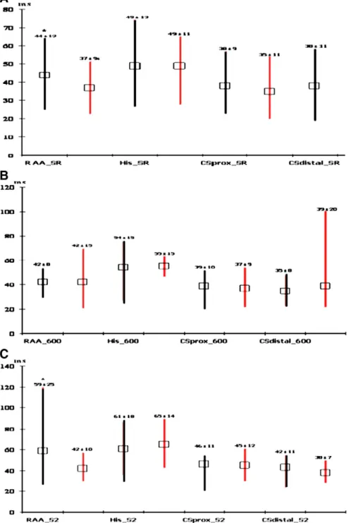
Electrogram duration showed significant differences at the RAA, when compar-ing both groups durcompar-ing baseline sinus rhythm and with S2 (Fig. 1). Also, patients with
PAF had greater prolongation of electrogram wave duration measured at the RAA and distal CS during the earliest propagated S2 (73 ± 35% vs. 11 ± 8% at the RAA and 13 ± 3% vs. 1 ± 6% ms at the distal CS, for PAF patients and control group, respectively; pb0.05). Fragmented atrial activity was identified in 43.8% of the PAF group and in 6.7% of the control group (p = 0.03).
3.1. Conduction parameters during acute autonomic modulation
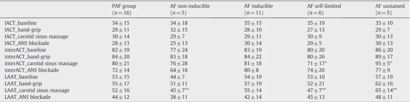
Table 3summarizes the results of the atrial conduction intervals during HG and CSM maneuvers, and after ANS blockade among patients with PAF. Atrial conduction times were not significantly changed during ANS stimulation. However, patients with inducible sustained AF had longer interACT and LAAT during CSM.
Despite longer electrograms in RAA and His during CSM when compared to baseline recordings, there were no significant differences in the mean duration of the
Fig. 1. Electrogram duration measured in sinus rhythm (SR) and during high right atrium pacing with a drive-train stimulation (600 ms) and with the earliest propagated extra-stimulus (S2). Comparison between the group with paroxysmal atrialfibrillation (black line) and the group without atrial fibrillation (red line). A: Baseline values during SR (*pb0.05, at RAA site). B: Values during 600 ms cycle length pacing (p=NS). C: Values during the earliest propagated S2 (*pb0.05, at RAA site). Values expressed in milliseconds, mean ± standard deviation. The lines represent mean, maximum and minimum values. RAA = right atrial appendage; His = His position; CSprox = proximal coronary sinus; CSdistal = distal coronary sinus.
measured electrograms during autonomic stimulation or after autonomic blockade (Fig. 2). Nevertheless, we observed an increased heterogeneity of the atrial wave duration, with significant differences between the recording sites, appearing during ANS maneuvers, and abolished after ANS blockade (Fig. 2). Representative intracardiac electrograms, obtained from the RAA during sinus rhythm, with S2, HG and CSM, and after ANSB are shown inFig. 3.
4. Discussion
Although the triggers for AF initiation appear to be located in the
pulmonary veins, established AF has been associated with conduction
disturbances and heterogeneous reduction of ERP, that facilitate the
Table 3
Atrial conduction times during autonomic modulation in paroxysmal atrialfibrillation patients.
PAF group AF non-inducible AF inducible AF self-limited AF sustained
(n = 16) (n = 5) (n = 11) (n = 6) (n = 5)
IACT_baseline 34 ± 15 34 ± 18 35 ± 15 35 ± 19 35 ± 10
IACT_hand-grip 29 ± 11 32 ± 15 28 ± 10 27 ± 13 29 ± 7
IACT_carotid sinus massage 30 ± 14 29 ± 7 29 ± 11 30 ± 9 30 ± 13
IACT_ANS blockade 28 ± 13 25 ± 13 30 ± 14 29 ± 5 30 ± 13
interACT_baseline 82 ± 19 77 ± 24 83 ± 19 80 ± 20 86 ± 20
interACT_hand-grip 84 ± 20 83 ± 18 84 ± 22 80 ± 26 89 ± 17
interACT_carotid sinus massage 80 ± 21 76 ± 28 81 ± 18 71 ± 17* 95 ± 5*
interACT_ANS blockade 72 ± 14 64 ± 16 80 ± 8 74 ± 20 77 ± 9
LAAT_baseline 53 ± 15 44 ± 7 54 ± 19 53 ± 10 57 ± 19
LAAT_hand-grip 55 ± 17 51 ± 11 57 ± 19 52 ± 21 62 ± 16
LAAT_carotid sinus massage 52 ± 16 45 ± 7** 55 ± 14 47 ± 7** 65 ± 14**
LAAT_ANS blockade 44 ± 12 38 ± 11 42 ± 14 45 ± 13 48 ± 11
PAF = paroxysmal atrialfibrillation; AF = atrial fibrillation; IACT = intra-atrial conduction time; interACT = interatrial conduction time; LAAT = left atrial activation time; ANS = autonomic nervous system. Values expressed in milliseconds, mean ± standard deviation. *pb0.05; **pb0.01.
Fig. 2. Electrogram duration measured during sinus rhythm in baseline, with handgrip, carotid sinus massage (CSM) and after pharmacological blockade of autonomic activity (ANSB). *pb0.05 compared to other sites. Values expressed in milliseconds, mean±standard deviation. The lines represent mean, maximum and minimum values. RAA = right atrial appendage; His = His position; CSprox = proximal coronary sinus; CSdistal = distal coronary sinus.
occurrence of multiple reentry circuits within the atria, probably
contributing to the electrophysiological substrate required for the
presence of AF
[3,6,18]
.
While prolongation of atrial conduction is a frequent
finding in
patients with AF
[19,20]
, the in
fluence of autonomic activity in the
atrial conduction intervals and local wave duration is incompletely
explored. The present study characterized the IACT, the interACT and
electrogram duration measured in different atrial recording sites in
response to an extra-stimulus with a short coupling interval and
during acute modulation of the ANS. There were no differences
between the baseline characteristics of PAF patients and controls.
However, the group with history of PAF showed longer IACT and RAA
electrograms in baseline, compared with control patients.
Further-more, they showed signi
ficant atrial conduction delays and greater
prolongation of atrial wave duration during early premature impulses
delivered at the HRA. Also, fragmented atrial activity was identi
fied in
more patients with PAF than in control patients. These
findings are
consistent with previous studies who demonstrated greater delays in
intra-atrial or inter-atrial conduction, and a higher incidence of atrial
fragmentation in patients with PAF
[6,20
–22]
. In fact, the presence of
marked conduction delay during an atrial premature beat with a short
coupling interval, combined with longer and fractionated
electro-grams is an important electrophysiological
finding, compatible with
the necessary conditions for the occurrence and maintenance of local
reentry circuits. Focal repetitive activity, most frequently originated
from pulmonary veins, plays an important role in the initiation of AF,
particularly when combined with abnormal atrial impulse
conduc-tion, which appears to be pre-requisite for the maintenance of AF
[23,24]
. Heterogeneity of atrial conduction delay and the presence of
local fragmented potentials have long been associated with the
substrate for AF
[25
–27]
. The greater prolongation of atrial activation
times and of local wave duration with the earliest propagated
extra-stimulus, showed in our results, might contribute to explain why the
mean coupling interval was signi
ficantly shorter for pulmonary veins
discharges initiating AF than for discharges that did not in a recent
study by Arentz et al.
[28]
. Therefore, a combination of atrial
premature complexes with short coupling intervals and delayed
activation of the atria may act as one component of the
arrhythmo-genic substrate for the vulnerability to PAF.
4.1. Autonomic modulation of conduction parameters in PAF patients
The supporting evidence of the impact of ANS activity in the
electrophysiological properties of the atria and its role in the initiation
and maintenance of AF has been mostly studied in experimental
preparations. Little is known about the effects of acute stimulation or
blockade of the ANS in atrial conduction and electrogram duration
during electrophysiological evaluation of patients with PAF. In our
data, obtained from patients with clinical history of PAF, the interACT
and LAAT were signi
ficantly prolonged during CSM in the group with
inducible sustained AF, supporting the notion that the substrate of AF
is associated with conduction abnormalities of the atria, which can be
more pronounced during vagal stimulation and contribute to the
maintenance of AF. Although conduction abnormalities in PAF have
been associated with increased age, atrial dilation and stretch,
fibrosis,
changes in the expression levels of connexins and electrophysiological
remodeling
[2,3,5
–7,24]
, acute autonomic modulation seems to
in
fluence atrial conduction properties in patients with PAF.
It has been known that vagal stimulation shortens the atrial ERP
and increases dispersion of atrial refractoriness
[8,9,12,13]
. Although
both vagal and sympathetic stimulations could produce signi
ficant
reductions on ERP, vagal stimulation appears more arrhythmogenic in
promoting AF
[29]
. One reason could be related with the lengthening
of atrial conduction time during vagal activity, that results in a
pronounced wavelength shortening (ERP x conduction velocity),
which would promote AF maintenance.
In the present study, we evaluated atrial electrograms duration
during autonomic stimulation and after pharmacological autonomic
blockade. Electrograms duration increased slightly in RAA and His
during CSM, but no signi
ficant differences were obtained during
acute autonomic modulation. However, when compared to baseline
recordings, differences in atrial wave duration between the
recording sites became more pronounced during ANS maneuvers
and were abolished after ANS blockade. Although the impact of
autonomic stimulation appears to be modest in the induction of AF
(68.8% during ANS maneuvers vs. 50% after ANS blockade), it is
possible that the electrogram duration heterogeneity and local
conduction delay produced by autonomic modulation contribute to
the initiation and maintenance of AF. Vagal stimulation has been
found to result in a large regional heterogeneity of atrial
electro-grams, and there is evidence that the appearance of complex
fractionated atrial electrograms during activation of the intrinsic
cardiac autonomic neural elements re
flects a change in the local
electrophysiological properties
[30,31]
. In fact, differential areas of
conduction velocity and dispersion of electrogram characteristics
may provide a substrate for functional reentry, creating a suitable
environment for AF
[32,33]
.
There have been limited data on autonomic in
fluences in the
characteristics of electrograms during sinus rhythm. Guo et al., in a
canine model, found that vagal stimulation shortened the
electro-gram duration in ischemic myocardium zone in the right atrium,
whereas sympathetic stimulation did not alter electrogram
dura-tion
[34]
. In a previous analysis of atrial electrograms during sinus
rhythm in patients with PAF, electrograms with
≥4 deflections and
duration
≥40 ms were associated with a parasympathetic response
during AF ablation
[35]
. The explanation for this
finding was
related with local effects of acetylcholine in atrial tissue, causing
conduction block between adjacent
fiber bundles. Recently, in a
different study, complex fractionated atrial electrograms, representing
slow conduction areas or pivoting points in reentry circuits, were
induced by local application of varying concentrations of acetylcholine
or by injecting acetylcholine into the anterior right ganglionated plexi,
providing evidence that ANS activity may induce changes in local atrial
conduction
[36]
.
There is a great need for experimental and clinical studies to better
understand the relationship between the dynamic changes in atrial
electrogram morphology and autonomic innervation and its role in
the maintenance of AF.
Fig. 3. Electrograms obtained from the right atrial appendage in a 72 years old woman with paroxysmal atrialfibrillation. Sinus rhythm (A), with the earliest propagated extra-stimulus (B), during handgrip (C) and carotid sinus massage (D), and after pharmacological autonomic blockade (E).
5. Study limitations
Although it was possible to identify slight changes in atrial activation
times and wave duration during acute autonomic modulation, the study
included a small number of patients. However, all patients acted as their
own controls to enable comparison of the parameters during
stimula-tion and after blockade of the ANS. Although obtaining high density
recordings by using multipolar catheters with better spatial resolution
from several simultaneous right and left atrial sites could give more
precise results, allowing a better comprehension of the problem,
transseptal punctures for the use of left atrial catheters were not
justi
fiable in a preliminary investigative study in humans. Another
concern is that despite the con
firmation of HG and CSM effects based on
frequency domain spectral analysis, direct stimulation of sympathetic
and parasympathetic nerves would have improved the results.
6. Conclusions
The presented study demonstrated that atrial conduction times,
electrograms duration and fractionated activity are increased in
patients with PAF when compared with control patients, suggesting
that conduction abnormalities in the atria contribute to the
arrhythmogenic substrate for AF. Also, acute vagal stimulation
prolonged interACT and LAAT in patients with inducible sustained
AF and ANS modulation in
fluenced the heterogeneity of atrial
electrograms duration in the recording sites. These should be taken
into consideration in future studies in order to better understand the
dynamic phenomena involved in the onset and perpetuation of AF
episodes.
Acknowledgement
The authors of this manuscript have certi
fied that they comply
with the Principles of Ethical Publishing in the International Journal of
Cardiology
[37]
.
References
[1] Friberg J, Scharling H, Gadsbøll N, Jensen GB. Sex-specific increase in the prevalence of atrialfibrillation (The Copenhagen City Heart Study). Am J Cardiol Dec 15 2003;92(12):1419–23.
[2] Fuster V, Ryden LE, Canmon DS, et al. ACC/AHA/ESC 2006 guidelines for the management of patients with atrialfibrillation-executive summary. Eur Heart J 2006;27:1979–2030.
[3] Nattel S. Atrial electrophysiology and mechanisms of atrialfibrillation. J Cardiovas Pharmacol Ther 2003;8(1):S5–S11.
[4] Haissaguerre M, Jais P, Shah DC. Spontaneous initiation of atrialfibrillation by ectopic beats originating in the pulmonary veins. N Engl J Med 1998;339:659–66. [5] Nattel S, Burstein B, Dobrev D. Atrial remodeling and atrial fibrillation.
Mechanisms and implications. Circ Arrhythm Electrophysiol 2008;1:62–73. [6] Pytkowski M, Jankowska A, Maciag A, et al. Paroxysmal atrialfibrillation is
associated with increased intra-atrial conduction delay. Europace 2008;10: 1415–20.
[7] Nattel S, Shiroshita-Takeshita A, Cardin S, Pelletier P. Mechanisms of atrial remodeling and clinical relevance. Curr Opin Cardiol 2005;20:21–5.
[8] Olshansky B. Interrelationships between the autonomic nervous system and atrial fibrillation. Prog Cardiovasc Dis Jul–Aug 2005;48(1):1409–17.
[9] van den Berg MP, Hassink RJ, Baljé-Volkers C, Crijns HJGM. Role of the autonomic nervous system in vagal atrialfibrillation. Heart March 2003;89(3):333–5. [10] Bettoni M, Zimmermann M. Autonomic tone variations before the onset of
paroxysmal atrialfibrillation. Circulation June 11, 2002;105(23):2753–9.
[11] Amar D, Zhang H, Miodownik S, Kadish AH. Competing autonomic mechanisms precede the onset of postoperative atrialfibrillation. J Am Coll Cardiol October 2003;42(7):1262–8.
[12] Chen PS, Tan AY. Autonomic nerve activity and atrialfibrillation. Heart Rhythm March 2007;4(3 Suppl):S61–4.
[13] Chen J, Wasmund SL, Hamdan MH. Back to the future: the role of the autonomic nervous system in atrialfibrillation. Pacing Clin Electrophysiol 2006;29:413–21. [14] Miyamoto K, Nakao K, Seto S, et al. Abnormal right atrial electrograms predict the
transition to chronic atrialfibrillation in paced patients with sick sinus syndrome. PACE 2004;27:644–50.
[15] Cozma D, Mornos C, Pescariu S, Petrescu L, Lighezan D, Dragulescu S. Electrophysiological and echocardiographic parameters predisposing to atrial fibrillation in patients with a structurally normal heart. Kardiol Pol 2006;64: 143–50.
[16] Oliveira M, Silva N, Feliciano J, et al. Effects of stimulation and blockade of the autonomic nervous system on atrial refractoriness in patients with lone paroxysmal atrialfibrillation. Rev Port Cardiol 2009;28(6):1–16.
[17] Friedman HS, Sinha B, Tun A, et al. Zones of atrial vulnerability. Relationships to basic cycle length. Circulation 1996;94:1456–64.
[18] Oliveira M, da Silva MN, Timoteo AT, et al. Inducibility of atrialfibrillation during electrophysiologic evaluation is associated with increased dispersion of atrial refractoriness. Int J Cardiol August 2009;136(2):130–5.
[19] Platonov PG, Mitrofanova LB, Chireikin LV, Olsson SB. Morphology of inter-atrial conduction routes in patients with atrialfibrillation. Europace 2002;4:183–92. [20] O'Donnell D, Bourke JP, Furniss SS. Interatrial transseptal electrical conduction:
comparison of patients with atrialfibrillation and normal controls. J Cardiovasc Electrophysiol November 2002;13:1111–7.
[21] Cozma D, Kalifa J, Lighezan D, et al. Mechanism of atrialfibrillation: decremental conduction, fragmentation, and ectopic activity in patients with drug resistant paroxysmal atrial fibrillation and structurally normal heart. PACE 2005;28: S115–9.
[22] Tai CT, Chen SA, Tzeng JW, et al. Prolonged fractionation of paced right atrial electrograms in patients with atrialflutter and fibrillation. J Am Coll Cardiol 2001;37:1651–7.
[23] Rha S, Kim Y, Hong M, et al. Mechanisms responsible for the initiation and maintenance of atrialfibrillation assessed by non-contact mapping system. Int J Cardiol Feb 2008;124(2):218–26.
[24] Chaldoupi SM, Loh P, Hauer RNW, Bakker JMT, van Rijen HVM. The role of connexin40 in atrialfibrillation. Cardiovasc Res 2009,doi:10.1093/cvr/cvp203. [25] Papageorgiou P, Monahan K, Boyle NG, et al. Site-dependent intraatrial conduction
delay: relationship to initiation of atrialfibrillation. Circulation 1996;94:384–9. [26] Saksena S, Giorgberidze I, Mehra R, et al. Electrophysiology and endocardial
mapping of induced atrial fibrillation in patients with spontaneous atrial fibrillation. Am J Cardiol 1999;83:187–93.
[27] Ehrlich JR, Nattel S. Electrophysiological basis of atrialfibrillation. In: Schwartz-man D, Zenati MA, editors. Innovative Schwartz-management of atrialfibrillation. 1st ed. Blackwell Publishing; 2005. p. 3–18.
[28] Arentz T, Haegeli L, Sanders P, et al. High-density mapping of spontaneous pulmonary vein activity initiating atrial fibrillation in humans. J Cardiovasc Electrophysiol January 2007;18:31–8.
[29] Liu L, Nattel S. Differing sympathetic and vagal effects on atrialfibrillation in dogs: role of refractoriness heterogeneity. Am J Physiol 1997;42(2):805–16. [30] Vigmond E, Tsoi V, Kuo S, Yin Y, Trayanova N, Pagé P. Using atrial electrograms to
estimate vagal influence. Heart Rhythm May 2005;2(5):S179 Supplement. [31] Lin J, Scherlag B, Zhou J, et al. Autonomic mechanism to explain complex
fractionated atrial electrograms (CFAE). J Cardiovasc Electrophysiol Nov 2007;18: 1197–205.
[32] Liuba I, Walfridsson H. Focal atrial tachycardia: increased electrogram fraction-ation in the vicinity of the earliest activfraction-ation site. Europace July 2008,doi:10.1093/ europace/eun192.
[33] Makati K, Alsheikh-Ali A, Garlitski A, et al. Advances in mechanisms of atrial fibrillation: structural remodeling, high-frequency fractionated electrograms, and reentrant AF drivers. J Interv Card Electrophysiol 2008;23:45–9.
[34] Guo H, Euler D, Wang Z, Olshansky B. Autonomic influences in atrial ischemia: vagally mediated atrial conduction improvement. Int J Cardiol September 1997;61 (2):157–63.
[35] Lellouche N, Buch E, Celigoj A, et al. Functional characterization of atrial electrograms in sinus rhythm delineates sites of parasympathetic innervation in patients with paroxysmal atrialfibrillation. J Am Coll Cardiol 2007;50:1324–31. [36] Lin J, Scherlag BJ, Zhou J, et al. Autonomic mechanism to explain Complex
Fractionated Atrial Electrograms (CFAE). J Cardiovasc Electrophysiol Nov 2007;18: 1197–205.