Article
J. Braz. Chem. Soc., Vol. 24, No. 11, 1857-1863, 2013. Printed in Brazil - ©2013 Sociedade Brasileira de Química
0103 - 5053 $6.00+0.00
A
http://dx.doi.org/10.5935/0103-5053.20130231*e-mail: [email protected]
New Isoflavones from the Leaves of
Vatairea guianensis
Aublé
Ronilson Freitas de Souza,a Victor H. S. Marinho,a Geilson A. da Silva,a Livio M. Costa-Jr.,b Joyce Kelly R. da Silva,c Gilmara N. T. Bastos,d Alberto C. Arruda,a Milton N. da Silvaa and Mara Silvia P. Arruda*,a
aPrograma de Pós-Graduação em Química, Instituto de Ciências Exatas e Naturais,
Universidade Federal do Pará, Campus Universitário do Guamá, 66075-970 Belém-PA, Brazil
bCentro de Ciências Agrárias e Ambientais, Universidade Federal do Maranhão,
MA-230, km 04, s/n, Boa Vista, 65500-000 Chapadinha-MA, Brazil
cPrograma de Pós-Graduação em Biotecnologia and dPrograma de Pós-Graduação em Neurociências e
Biologia Celular, Instituto de Ciências Biológicas, Universidade Federal do Pará, Campus Universitário do Guamá, 66075-970 Belém-PA, Brazil
Das folhas de Vatairea guianensis Aublé foram isoladas quatro isoflavonas identificadas como, 5,3’,-diidroxi-4’-metoxi-2”,2”-dimetilpirano-(5”,6”:8,7)-isoflavona (1), 5,7-diidroxi-3’,4’-metilenodioxi-8-prenil-isoflavona (2) e 5,3’-diidroxi-4’-metoxi-7-O-β -glicopiranosídeo-8-prenil-isoflavona (3) e derrona (4), juntamente com cinco triterpenos identificados em mistura de lupeol,
α-amirina, β-amirina, germanicol e ácido betulínico. As substâncias 1-3 são novos produtos naturais, porém 1 e 2 já foram citados como produtos de síntese. No entanto, todas essas substâncias são relatadas pela primeira vez para essa espécie. Suas estruturas químicas foram elucidadas com base nos seus dados de ressonância magnética nuclear (RMN) 1D e 2D e por espectrometria de massas de alta resolução. O extrato etanólico das folhas e os compostos 1-3 foram avaliados quanto ao seu potencial sequestrador do radical DPPH• (2,2-difenil-1-picril-hidrazila) e os resultados mostram que o extrato apresentou alta atividade (CI50 = 6,2 ± 0,4 µg mL
-1), enquanto as substâncias testadas apresentaram baixo poder antioxidante (CI50≥ 29,5 ± 2,5 µg mL
-1) quando comparadas com TROLOX (CI50 = 4,5 ± 0,4 µg mL-1).
Four isoflavones were isolated from Vatairea guianensis Aublé leaves and identified as 5,3’-dihydroxy-4’-methoxy-2”,2”-dimethylpyrano-(5”,6”:8,7)-isoflavone (1), 5,7-dihydroxy-3’,4’-methylenedioxy-8-prenyl-isoflavone (2), 5,3’-dihydroxy-4’-methoxy-7-O-β -glucopyranoside-8-prenyl-isoflavone (3) and derrone (4) together with five triterpenes identified in mixture, lupeol,
α-amyrin, β-amyrin, germanicol and betulinic acid. Substances 1-3 are novel natural products, although 1 and 2 have been cited as synthetic products. However, all these compounds are first reported from this species. Their chemical structures were elucidated based on their 1D and 2D nuclear magnetic resonance (NMR) data and high resolution mass spectrometry. The ethanol extract from the leaves and 1-3 were evaluated for their potential in scavenging DPPH• (2,2-diphenyl-1-picrylhydrazylradical) and the results showed that the extract presented high activity (IC50 = 6.2 ± 0.4 µg mL-1), while the isolated compounds showed low antioxidant power (IC
50 ≥ 29.5 ± 2.5 µg mL-1) when compared to Trolox (IC
50 = 4.5 ± 0.4 µg mL -1).
Keywords:Vatairea guianensis, Fabaceae, isoflavones, antioxidant activity, DPPH•
Introduction
The genus Vatairea (Fabaceae) includes seven species
distributed in Brazil, the Guianas and the Atlantic coastal
regions of Central America and Mexico.1 The species
Vatairea guianensis Aublé is a plant native to the Amazon,
known as “fava bolacha” or “fava de impingem”.2 Its fruits,
bark from the stems, roots and leaves are commonly used for
treating surface micoses.3,4 Few scientific references citing
biological activities for this species have been found in the literature. Fruits are the only part of the plant that has been
New Isoflavones from the Leaves of Vatairea guianensis Aublé J. Braz. Chem. Soc.
1858
action against Leishmania amazonensis,5 antimicrobial6 and
topical healing activity.7 Among the components reported
for the plant are antraquinones chrysophanol, physcion and emodin, antrones 9-anthronechrysophanol acid, 9-anthronephyscion and 10-anthronephyscion, triterpenes
oleanolic acid and dihydromacaerinic acid lactone.4,8 These
reports of phytochemical studies are restricted to fruits,
heartwood and bark of the stems. The leaves of V. guianensis
were chosen for this study in an effort to identify their chemical composition and assess their potential for
scavenging radical DPPH•. Four isoflavones were isolated
(1-4) (Figure 1) together with a mixture of known triterpenes
lupeol, α-amyrin, β-amyrin, germanicol and betulinic acid.
The ethanolic extract of the leaves and isoflavones 1-3 were
evaluated for their potential to scavenge DPPH•.
Experimental
General
UV spectra were obtained from a liquid chromatograph (LC) equipped with diode array detection (DAD) Prominence SPDM-20A (Shimadzu, Tokyo, Japan). Nuclear magnetic resonance (NMR) spectra including 1D and 2D experiments were recorded on a Varian Mercury-300 spectrometer,
operating at 300 MHz at 1H and 75 MHz at 13C, using
CDCl3 or CD3OD as solvent (0.6 mL). Mass spectral
analyses were performed on UltrOTOF-Q (Bruker Daltonics, Billerica, MA, USA) in the cationized ion region. The heated capillary and voltage were maintained
at 250 oC and 3 kV, respectively. A 20 V cone energy for
ion extraction and mass spectrometry data were acquired at positive and negative modes for all compounds. High performance liquid chromatography (HPLC) was carried out in a semi-preparative LC-8A Shimadzu system with SPD-10AV Shimadzu UV detector (Tokyo, Japan); using a Phenomenex Gemini C18 column (250 × 10 mm, 5 µm), isocratic system of 50% acetonitrile-water and 35%
acetonitrile-water, and a flow rate 4.7 mL min-1. Detection
was performed at 254 and 282 nm. All solvents were
filtered through a 0.45 mm nylon membrane filter prior to analysis. Open column chromatography was run using silica gel 60 (70-230 mesh, Maherey-Nagel). Thin layer chromatography (TLC) was performed on precoated silica gel aluminium sheets (Maherey-Nagel) by detection with a spraying reagent (vanillin/sulfuric acid/EtOH solution) followed by heating at 100 °C and with reagent NP-PEG (diphenylborinic acid aminoethylester-polyethylene glycol) for flavonoid detection.
Plant material
Leaves of Vatairea guianensis were collected in
November 2010 from Belém, State of Pará, Brazil. Identification was made by Manoel R. Cordeiro from Embrapa Amazônia Oriental, Pará, Brazil and a voucher specimen (IAN – 187050) has been deposited in the herbarium of Embrapa Amazônia Oriental.
Extraction and isolation
Dried and powdered leaves of Vatairea guianensis
(1.0 kg) were extracted with ethanol by maceration at room temperature. The solvent was removed under vacuum, furnishing a residue (180.0 g). The crude ethanol
residue (50.0 g) was dissolved in 500 mL MeOH-H2O
mixture (9:1), then partitioned three times with n-hexane
(3 × 500 mL), ethyl acetate (3 × 500 mL) and the remaining hydroalcoholic phase. The extracts obtained were dried
to provide three fractions: an n-hexane fraction (7.5 g),
an EtOAc fraction (22.0 g) and a remaining MeOH-H2O
fraction (20.0 g). The n-hexane fraction (7.0 g) was
subjected on silica gel column chromatography with
gradient elution of n-hexane-EtOAc (9:1, 8:2, 7:3, 5:5
and 0:10) and EtOAc-MeOH (5:5 and 0:10), to obtain seven fractions (Fr.1–Fr.7), respectively. In fraction Fr-1
(1.1 g) eluted with n-hexane/EtOAc (9:1), after analysis
by TLC and 1H and 13C NMR, it was possible to identify a
mixture of pentacyclic triterpenes (5-9). The fraction Fr-2
(0.5 g) eluted with n-hexane/EtOAc (8:2) was sonicated
in 4.8 mL of acetonitrile for 1 min. Next, 1.2 mL of H2O
was added and sonicated again for 1 min. The solution was subjected to solid phase extraction (SPE) in a C18 cartridge (Phenomenex, 1 g of stationary phase / 6 mL). After evaporation, the residue (about 175 mg) was submitted to semi-preparative reversed phase HPLC (250 × 10 mm Phenomenex Gemini C18, 50% acetonitrile-water, flow rate
4.7 mL min-1, 254 nm) to yield 1 (25.0 mg), 2 (16.0 mg) and
4 (15.0 mg), which showed chromatographic peak retention times of 35.0, 39.0 and 51.0 min, respectively. The fraction Fr-6 (0.6 g) eluted with EtOAc-MeOH (5:5), after
Souza et al. 1859 Vol. 24, No. 11, 2013
up (treated by ultrasonic bath using 4.8 mL of acetonitrile
for 1 min, after 1.2 mL of H2O was added and sonicated
again for 1 min, after which the solution was subjected to solid phase extraction (SPE) in a C18 cartridge). A residue (90.0 mg) was obtained that was submitted to semi-preparative reversed phase HPLC (250 × 10 mm Phenomenex Gemini C18, 35% acetonitrile-water, flow rate
4.7 mL min-1, 282 nm) to yield 3 (15.0 mg), which showed
chromatographic peak retention time of 8.3 min. These compounds were identified by NMR and mass spectrometry methods, and comparison with available reported data.
DPPH assays
A stock solution of DPPH• radical (0.5 mmol L-1) in
methanol was prepared. The solution was diluted in methanol
(60 µmol L-1 approx.) measuring an initial absorbance of
0.62 ± 0.02 in 517 nm at room temperature. The reaction
mixture was composed by 1950 µL of DPPH• solution
and 50 µL of the samples diluted in different methanol portions. The final concentrations were of 10.0, 7.5, 5.0
and 2.5 µg mL-1 for the crude extract, 75.0, 50.0, 25.0 and
12.5 µg mL-1 for substances 2 and 3 and 8.0, 6.0, 4.0 and
2.0 µg mL-1 for the Trolox standard. For each sample a
methanol blank was also measured. The absorbance was measured at 517 nm during 60 min. All experiments were conducted in triplicate and Trolox (6-hydroxy-2,5,7,8-tetramethylchroman-2-carboxylic acid) was used as the standard antioxidant. The radical scavenging activity of each
sample was calculated by the DPPH• inhibition percentage
(%IDPPH) according to equation 1, where A and B are the
blank and sample absorbance values in the end reaction.9
(1)
The concentration of antioxidant required for 50%
scavenging of DPPH• radicals (IC
50) was determined by
linear regression using Windows/Excel. All analyses were performed in triplicate. The data were expressed as means ± standard deviation (SD) and analyzed using GraphPad (version 5.0). One-way analysis of variance (ANOVA) and Tukey multiple comparisons were performed in order to test any significant differences between the means. Differences between means at the 5% level were considered significant.
5,3’-Dihydroxy-4’-methoxy-2”,2”-dimethylpyrano [5”,6”:8,7] isoflavone (1)
Amorphous pale green solid; UV λmax/nm
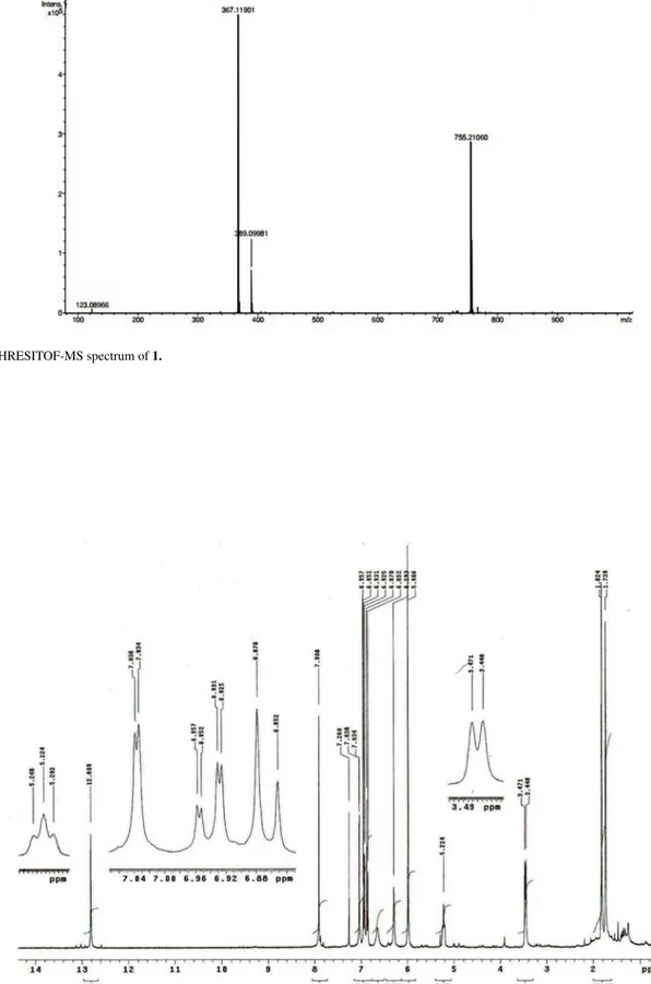
(acetonitrile-water): 268, 304 (sh); HRESITOF-MS m/z 367.11901
[M + H]+ (calc. for C
21H19O6+, 367,11816); 1H and 13C NMR
spectral data: see Tables 1 and 2.
5,7-Dihydroxy-3’,4’-methylenedioxy-8-prenyl-isoflavone(2)
Yellow amorphous powder; UV λmax/nm
(acetonitrile-water): 265, 292 (sh); HRESITOF-MS m/z 367.11723
[M + H]+ (calc. for C 21H19O6
+, 367.11816); 1H and 13C NMR
spectral data: see Tables 1 and 2.
5,3’-Dihydroxy-4’-methoxy-7-O-β
-glucopyranoside-8-prenyl-isoflavone (3)
Amorphous reddish solid; UV λmax/nm
(acetonitrile-water): 265, 290 (sh); HRESITOF-MS m/z 531.18615
[M + H]+(calc. for C 27H31O11
+, 531.18664); 1H and 13C NMR
spectral data: see Tables 1 and 2.
Results and Discussion
The hexane fraction from the ethanol extract of V.
guianensis leaves was fractionated as indicated in the Experimental section leading to the isolation of new isoflavones 1-3, together with known compounds derrone
(4), lupeol, α-amirin, β-amirin, germanicol and betulin
acid, which were identified by comparison of their spectral data with those reported in literature.10,11
Compound 1 was obtained as an amorphous pale
green solid, with the molecular formula C21H18O6, based
on the [M+H]+ peak at m/z 367.11901 (calc. for C
21H19O6 +,
367.11816) in the HRESITOF-MS, and confirmed by 1H
and 13C NMR experiments (Tables 1 and 2, respectively).
The 1H NMR signals at δ
H 7.87 (H-2) and 13C NMR
signal at δC 152.5 (C-2), 123.7 (C-3) and 180.8 (C-4),
were typical of isoflavones.12 Additionally, the 1H NMR
spectrum exhibited signals in the aromatic region at δH
6.91 (d, 1H, J 8.1 Hz), 7.03 (brd, 1H, J 8.1 Hz) and 7.06
(brs, 1H), which indicated an ABX spin system of a 1,3,4-trisubstituted phenyl group, as well as one singlet
at δH 6.28 assigned to a pentasubstituted benzene ring.
The signals observed at δH 5.58 and 6.67 (d, 1H each,
J 9.9 Hz) and 1.47 (s, 6H) revealed a 2,2-dimethylchromene
ring attached to an aromatic ring, whereas the singlets at
δH 12.95 and 3.91 indicated the presence of a hydroxyl
chelated to carbonyl and one OMe group connected to an aromatic ring, respectively. All couplings were confirmed
through analysis of the 1H-1H COSY spectrum. Besides the
signals related to C-ring carbons, the 13C NMR spectrum
of 1 exhibited another 17 signals attributed to eighteen
New Isoflavones from the Leaves of Vatairea guianensis Aublé J. Braz. Chem. Soc.
1860
A-ring at C-7 and C-8 was deduced by 3J
C,H correlations
from H-2 (δH 7.87) and H-4’’ (δH 6.67) to C-8a (δC 152.1)
observed in the HMBC spectrum (Table 2), as well as 2J
C,H
correlation between H-6 (δH 6.28) and OH-5 (δH 12.95) with
C-5 (δC 162.2). The location of the OMe and OH groups
at C-4’ and C-3’ of the aromatic B-ring, respectively, was
supported by the 3J
C,H correlations from H-6’ (δH 7.03)
and OMe-4’ (δH 3.91) to oxidized aromatic carbon C-4’
(δC 146.8) and confirmed by the NOE effect observed in
the NOE difference experiment, which revealed spatial
interactions between H-5’ (δH 6.91) and OMe-4’ (δH 3.91).
From the above features we can establish that compound 1 is 5,3’-dihydroxy-4’-methoxy-2”,2”-dimethylpyrano-(5”,6”:8,7)-isoflavone, here isolated for the first time from a natural source, though it has already been produced by
synthesis.13 Full spectrometric data for this compound are
presented.
Compound 2 was obtained as a yellow amorphous
powder and its molecular formula was defined as C21H18O6
on the basis of the quasi-molecular ion [M+1]+ observed
at m/z 367.11723 (calc. for C21H19O6+, 367.11816) in
HRESITOF mass spectrometry analysis. The 1H NMR
signal at δH 7.91 (H-2) and 13C NMR signal at δC 152.8 (C-2)
were characteristic of the isoflavone skeleton, similarly to
compound 1. Additionally, the 1H NMR spectrum (Table
1) exhibited signals at δH6.86 (d, J 7.8 Hz, H-5’), 6.94 (dd,
J 7.8 and 1.5 Hz, H-6’) and 7.03 (d, J 1.5 Hz, H-2’), which
indicated an AMX spin system of a 1,3,4-trisubstituted B-ring, as well as three singlets at δH 6.29 (1H), attributed
to a pentasubstituted A-ring, δH 5.99 (2H) assigned to a
methylenedioxy unit and δH 12.81 related to a hydroxyl
group bonded to C-5. The 13C NMR spectrum of 2 (Table
2) exhibited 21 signals attributed to twenty-one carbons with the aid of the HETCOR and HMBC experiments. The C-prenyl group [δH 3.46 (d, J 6.9 Hz, H-1’’), 5.22 (brt,
J 6.9 Hz, H-2’’), 1.74 (s, Me-trans) and 1.82 (s, Me-cis)]
attached at C-8 was deduced from the 3J
C,H correlations in
the HMBC experiments (Table 2) of the signals at δH 7.91
(H-2) and 3.46 (H-1’) with the signal at δC 155.0 (C-8a). On
the other hand, the HMBC 2,3J
C,H correlations from δH 7.02
(H-2’) and 6.86 (H-5’) to δC 147.8 and 147.7 (C-3’/C-4’)
revealed the connection of a methylenedioxy unit to C-3’ and C-4’. The structure of 2 was then identified as 5,7-dihydroxy-3’,4’-methylenedioxy-8-prenyl-isoflavone. This compound is a new natural product, although it has
been cited as a synthetic product.14 Itsspectrometric data
are presented for the first time.
Compound 3, an amorphous reddish solid, showed
molecular formula C27H30O11 that was established by the
quasi-molecular ion at m/z 531.18615 [M+H]+ in the
HRESITOF-MS (calc. for C27H31O11+, 531.18664), and
confirmed by 1H and 13C NMR experiments (Tables 1 and
2). Similarly to the compounds 1 and 2, 3 is an isoflavone
based on the 1H NMR signal at δ
H8.20 (H-2) and the 13C
NMR signal at δC 155.5 (C-2). Additionally, the
1H NMR
spectrum (Table 1) displayed signals in the aromatic region
at δH 7.06 (d, 1H, J 1.8 Hz,) and 6.78-6.99 (m, 2H), which
can be attributed, alike to compound 1, to the ABX spin system of a 1,3,4-trisubstituted B-ring. The pentasubstituted A-ring has been established as in compounds 1 and 2,
based on the singlet signal at δH 6.64 (s, 1H). In the
1H
NMR spectrum of 3 signals were also observed for a OMe group at δH 3.88 (s, 3H), a C-prenyl group at δH 3.60 (d, J
7.2 Hz, 2H-1’’), 5.22 (t, J 7.2 Hz, H-2’’), 1.82 (s, 3H-4’’) and 1.66 (s, 3H-5’’), and a glucose unit at δH 5.05 (d, J 7.2
Hz, H-1”) and 3.41-3.75 (m, H-2”- H-6”). The glucose residue was identified by comparison of NMR data with
the literature,15 where the anomeric proton signal at δ
H
5.05 with a large coupling constant of 7.2 Hz implies that
the glucose moiety must have a β-glucopyranose form.16
All the couplings were confirmed through analysis of the
1H-1H COSY spectrum. In addition to signals related to
the isoflavone skeleton containing a carbonyl (δC 182.7)
chelated to OH-5 group,17,18 the 13C NMR spectrum of 3
exhibited another 12 signals attributed with the aid of the
Table 1.1H NMR Chemical Shifts (dH / ppm) of Compounds 1, 2 (in
CDCl3) and 3 (in CD3OD)a
H 1 2 3
dH dH dH
2 7.87 (s) 7.91 (s) 8.20 (s)
6 6.28 (s) 6.29 (s) 6.64 (s)
2’ 7.06 (brs) 7.03 (d, 1.5) 7.06 (d, 1.8)
5’ 6.91 (d, 8.1) 6.86 (d, 7.8) 6.78-6.99 (m)
6’ 7.03 (brd, 8.1) 6.94 ( dd, 1.5 and 7.8) 6.78-6.99 (m)
1” 3.46 (d, 6.9) 3.60 (d, 7.2)
2” 5.22 (brt, 6.9) 5.22 ( brt,
7.2)
3” 5.58 (d, 9.9)
4” 6.67 (d, 9.9)
2Me-2” 1.47 (s)
4” (Me, cis) 1.82 (s) 1.82 (s)
5” (Me, trans) 1.74 (s) 1.66 (s)
OMe-4’ 3.91 (s) 3.88 (s)
OCH2O 5.99 (s)
OH-5 12.95 (s) 12.81 (s)
1”’ (Glc) 5.05 (d, 7.2)
2”’- 6”’ (Glc) 3.41-3.75 (m)
a1H NMR data were recorded at 300 MHz. Multiplicity and coupling
Souza et al. 1861 Vol. 24, No. 11, 2013
HETCOR and HMBC (Table 2) experiments to the carbons
of OMe and prenyl groups, as well as a glucose unit (δC
101.8, 78.3, 78.2, 74.9, 71.1, 62.3). The C-8 location of
the prenyl group was supported by the 3J
C,H correlations in
the HMBC spectrum from the signals at δH 8.20 (H-2) and
3.60 (H-1’’) to δC 156.0 (C-8a). Moreover, the cross-peaks
in the HMBC spectrum between H-6 (δH 6.64) and H-1’’’
(δH 5.05) of the glucose unit with C-7 (δC 162.1) indicated
that the glucose residue was attached to the 7-hydroxyl of the isoflavone moiety. The location of the OMe and OH groups at C-4’ and C-3’ of the B-ring, respectively, was sustained by combining the substitution pattern on
the aromatic B-ring (1,3,4-trisubstituted) displayed in the
1H NMR spectrum with the NOE effects observed in the
NOE difference spectra, which revealed spatial interactions between H-5’ and OMe-4’. Therefore, 3 was characterized
as 5,3’-dihydroxy-4’-methoxy-7-O-β
-glucopyranoside-8-prenyl-isoflavone, a new isoflavone glucoside.
The crude extract of leaves showed strong radical scavenging capacity, with inhibition percent of 77.2
at 22.0% at concentrations of 10.0 at 2.5 µg mL-1,
respectively, and IC50 value of 6.2 ± 0.4 µg mL
-1 (Trolox,
IC50 = 4.4 ± 0.1 µg mL-1). The results obtained from
isolated isoflavones 1-3 showed that all tested samples
Table 2. 13C NMR Chemical Shifts (dC / ppm) of Compounds 1, 2 (in CDCl
3) and 3 (in CD3OD)a
C 1 2 3
dC HMBCc dC HMBCc dC HMBCc
2 152.5 152.8 155.5
3 123.7b 2, 2’, 6’ 123.3 2 125.0b 2
4 180.8 2 181.1 2 182.7 2
4a 106.0 6, OH-5 105.4 6 107.8 6
5 162.2 6, OH-5 160.5 6 162.0 6
6 100.3 OH-5 99.6 99.7
7 159.5 6, 4’ 160.8 6, 1’ 162.1 6, 1”
8 101.1 6, 3’, 4’ 106.0 6, 1’ 110.4 6, 1’
8a 152.1 2, 4’ 155.0 2, 1’ 156.0 2, 1’
1’ 123.5b 2, 2’, 5’, 6’ 124.3 5’ 124.4b 2’, 5’, 6’
2’ 115.0 6’ 109.6 6’ 117.3 6’
3’ 145.6 2’, 5’ 147.8b 2’, 5’ 147.4
4’ 146.8 2’, 5’, 6’, OMe-4’ 147.7b 2’, 5’ 149.3 2’, OMe-4’
5’ 110.7 108.5 112.6
6’ 120.9 2’ 122.4 121.6 2’
1” 21.5 22.5
2” 78.0 3’, 4’, 2Me-2’ 121.1 1”, 4”, 5” 123.6 1”, 4”, 5”
3” 127.4 2Me-2’ 134.7 1”, 4”, 5” 132.6 1”, 4”, 5”
4” (Me, cis) 114.5 17.9 18.0 5”
5” (Me, trans) 25.8 25.9 4”
2Me-2’ 28.2
OMe-4’ 56.0 56.4
OCH2O 101.2
1’” (Glc) 101.8
2’” (Glc) 74.9
3’” (Glc) 78.2
4’” (Glc) 71.1
5’” (Glc) 78.3
6’” (Glc) 62.3
New Isoflavones from the Leaves of Vatairea guianensis Aublé J. Braz. Chem. Soc.
1862
were capable of scavenging free radical DPPH•, however,
compound 1 was weakly active with an inhibition
percentage below 50.0% at 100 µg mL-1. Compounds 2
and 3 presented greater inhibition effects, with inhibition percentage above 50.0% during 60 min of reaction at 25
and 50 µg mL-1 concentrations, respectively. However,
compounds 2 and 3 were about 6 and 14 times less active
than Trolox with IC50 values of 29.5 ± 2.5 µg mL
-1 and
64.3 ± 2.6 µg mL-1, respectively.
The free radical scavenging activity of flavonoids and other phenols is mostly due to their aromatic hydroxyl groups, which afford greater stability to the phenolic radical as soon as it is formed, after one hydrogen radical
donation to DPPH•.19 Although compounds 1-3 possess
two phenolic hydroxyl groups each, the isoflavonoid
2 was more effective in promoting DPPH• reduction,
when compared to 1 and 3. Early studies showed that the presence of a substituent (sugar residues) in position C-7 of A-ring in the flavonoids reduces the sequestering activity of the radicals, since the flavonoid structure loses its coplanarity due to the presence of voluminous
groups,20,21 and methylation in hydroxyl group in the
para-position decreased DPPH• scavenging activity.22 On
the other hand, the methylenedioxy function contributes
to stabilization of the phenoxy radical.23 The fact that
much higher values of IC50 were found for the isolated
compounds when compared to Trolox suggests that the significant anti-radical activity exhibited by the ethanolic
extract of V. guianensis leaves may be attributed to the
effect of synergism in the substances present in this extract.
Conclusions
Phytochemical investigation from the leaves of
Vatairea guianensis (Fabaceae) resulted in the isolation of compounds belonging to the isoflavone and triterpene classes, and it should be noted that the isoflavone class is being cited for the first time for this species. The evaluation of antioxidant activity, based on the method of sequestering
radical DPPH• demonstrated that the ethanolic extract of
V. guianensis leaves presents a high power for scavenging
radical DPPH•, similar to the TROLOX standard, while the
isoflavones tested present low anti-radical reaction, which suggests that the greatest antioxidant potential of the extract may be associated with synergism among its components.
Supplementary Information
Supplementary data are available free of charge at http:// jbcs.sbq.org.br, as PDF file.
Acknowledgments
The authors are grateful to Coordenação de Aperfeiçoamento de Pessoal de Nível Superior (CAPES) for scholarships and to Conselho Nacional de Desenvolvimento Científico e Tecnológico (CNPq) for financial support. We are also thankful to Dr. Norberto P. Lopes from the Faculdade de Ciências Farmacêuticas de Ribeirão Preto, USP for the HRESITOF-MS
References
1. Schongart, J.; Junk, W. J. J.; J. Hydrol. 2007, 335, 124. 2. Corrêa, M. P.; Dicionário de Plantas Úteis do Brasil e das
Exóticas Cultivadas, vol. 6; Imprensa Nacional: Rio de Janeiro, 1982.
3. Lima, H. C.; Arq.Jard. Bot. Rio de Janeiro1982, 26, 173. 4. Piedade, L. R; Wolter Filho, W.; Acta Amaz.1988, 18, 185. 5. Ottobelli, I.; Facundo, V. A.; Stabile, R. G.; Zuliane, J.; Frota,
D. B.; Badra, L. C.; Ottobelli, R. M.; Brasil, H. O. B.; Tomé, J. C. L.; Macedo, S. R. A.; Luz, C. C.; Silva-Jardim, I.; Rev. Patol. Trop.2009, 38, 1319.
6. Silva, C. T. L.; Mendonça, L. C.; Monteiro, M. C.; Carvalho, J. C. T.; Bol. Latinoam. Caribe Plant. Med. Aromat. 2011, 10, 462.
7. Silva, C. T. L.; Medeiros, B. J. S.; Santos, K. C.; Pereira Filho, R.; Albuquerque Jr., R. L. C.; Sousa, P. J. C.; Carvalho, J. C. T.; Int. J. Pharm. Sci. Rev. Res. 2011, 8, 1.
8. Ottobelli, I.; Facundo, V. A.; Zuliani, J.; Luz, C. C.; Brasil, H. O. B.; Militão, J. S. L. T.; Braz-Filho, R.; Acta Amaz.2011,
3, 393.
9. Silva, J. K. R.; Sousa, P. J. C.; Andrade, E. H. A.; Maia, J. G. S.;
J. Agric. Food. Chem. 2007, 55,9422.
10. Chibber, S. S.; Sharma, R. P.; Phytochemistry 1980, 19, 1857.
11. Mahato, S. B.; Kundu, A. P.; Phytochemistry1994, 37, 1517. 12. Bandeira, P. N.; de Farias, S. S.; Lemos, T. L. G.; Braz-Filho, R.;
Santos, H. S.; Albuquerque, M. R. J. R.; Costa, S. M. O.; J. Braz. Chem. Soc.2011, 22, 372.
13. Tsukayama, M.; Kawamura, Y.; Tahara, H.; Heterocycles1992,
3, 505.
14. Hastings, J. M.; Hadden, M. K.; Blagg, B. S. S.; J. Org. Chem.
2008, 73, 369.
15. Marin, P. D.; Grayer, R. J.; Grujic-Jovanovic, S.; Kite, G. C.; Veitch, N. C.; Phytochemistry 2004, 65,1247.
16. Ozden, S.; Durust, N.; Toki, K.; Saito, N.; Honda, T.;
Phytochemistry 1998, 49, 241.
17. Nara, K.; Nihei, K.; Ogasawara, Y.; Koga, H.; Kato, Y.; Food Chem.2011, 124, 710.
18. Xiaoli,L.; Naili, W.;Sau, W. M.; Chen, A. S. C.; Xinsheng, Y.;
Souza et al. 1863 Vol. 24, No. 11, 2013
19. Ribeiro, A. B.; Bolzani, V. S., Yoshida, M.; Santos, L. S.; Eberlin, M. N.; Silva, D. H. S.; J. Braz. Chem. Soc.2005, 16, 526.
20. Alves, C. Q.; David, J. M.; David, J. P.; Lima, L. da S.; Brandão, H. N.; Diálogos e Ciência 2007, 3, 7.
21. Khlebnikov, A. I.; Schepetkin, I. A.; Domina, N. G.; Kirpotina, L. N.; Quinn, M. T.; Bioorg. Med. Chem. 2007,5, 1770.
22. Lôbo, L. T.; Silva, G. A.; Ferreira, M.; Silva, M. N.; Santos, A. S.; Arruda, A. C.; Guilhon, G. M. S. P.; Santos, L. S.; Borges, R. S.; Arruda, M. S. P.; J. Braz. Chem. Soc.2009, 20, 1081. 23. Jinno, S.; Otsuka, N.; Okita, T.; Inouye, K.; Chem. Pharm. Bull.
1999, 47, 128.
Supplementary Information
S
I
J. Braz. Chem. Soc., Vol. 24, No. 11, S1-S14, 2013. Printed in Brazil - ©2013 Sociedade Brasileira de Química 0103 - 5053 $6.00+0.00
*e-mail: [email protected]
New Isoflavones from the Leaves of
Vatairea guianensis
Aublé
Ronilson Freitas de Souza,a Victor H. S. Marinho,a Geilson A. da Silva,a Livio M. Costa-Jr.,b Joyce Kelly R. da Silva,c Gilmara N. T. Bastos,d Alberto C. Arruda,a Milton N. da Silvaa and Mara Silvia P. Arruda*,a
aPrograma de Pós-Graduação em Química, Instituto de Ciências Exatas e Naturais,
Universidade Federal do Pará, Campus Universitário do Guamá, 66075-970 Belém-PA, Brazil
bCentro de Ciências Agrárias e Ambientais, Universidade Federal do Maranhão,
MA-230, km 04, s/n, Boa Vista, 65500-000 Chapadinha-MA, Brazil
cPrograma de Pós-Graduação em Biotecnologia and dPrograma de Pós-Graduação em Neurociências e
Biologia Celular, Instituto de Ciências Biológicas, Universidade Federal do Pará, Campus Universitário do Guamá, 66075-970 Belém-PA, Brazil
Figure S1. 1H NMR spectrum of 1 (CDCl
New Isoflavones from the Leaves of Vatairea guianensis Aublé J. Braz. Chem. Soc.
S2
Figure S2. 13C NMR spectrum of 1 (CDCl
3, 75 MHz).
Souza et al. S3 Vol. 24, No. 11, 2013
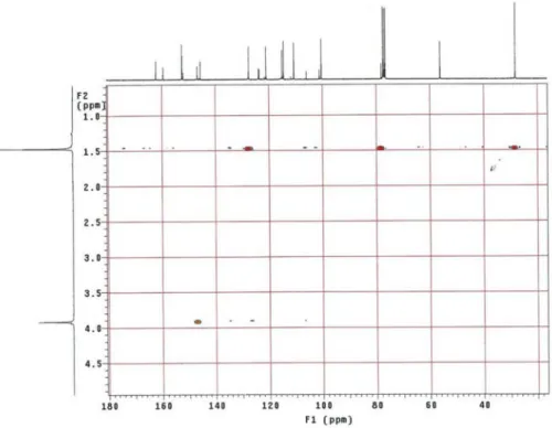
Figure S5. HMBC NMR spectrum of 1 (CDCl3, 300 x 75 MHz) (expansion 1).
New Isoflavones from the Leaves of Vatairea guianensis Aublé J. Braz. Chem. Soc.
S4
Figure S6. HMBC NMR spectrum of 1 (CDCl3, 300 x 75 MHz) (expansion 2).
Souza et al. S5 Vol. 24, No. 11, 2013
Figure S8. HRESITOF-MS spectrum of 1.
Figure S9. 1H NMR spectrum of 2 (CDCl
New Isoflavones from the Leaves of Vatairea guianensis Aublé J. Braz. Chem. Soc.
S6
Figure S10. 13C NMR spectrum of 2 (CDCl
3, 75 MHz).
Souza et al. S7 Vol. 24, No. 11, 2013
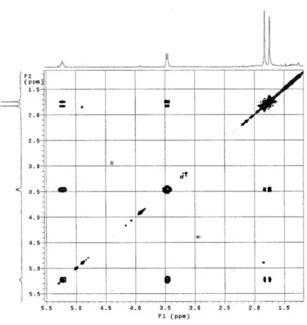
Figure S12. COSY NMR spectrum of 2 (CDCl3, 300 MHz).
New Isoflavones from the Leaves of Vatairea guianensis Aublé J. Braz. Chem. Soc.
S8
Figure S14. HMBC NMR spectrum of 2 (CDCl3, 300 x 75 MHz) (expansion 2).
Souza et al. S9 Vol. 24, No. 11, 2013
Figure S16. HRESITOF-MS spectrum of 2.
Figure S17. 1H NMR spectrum of 3 (CD
New Isoflavones from the Leaves of Vatairea guianensis Aublé J. Braz. Chem. Soc.
S10
Figure S19. HETCOR NMR spectrum of 3 (CD3OD, 300 x 75 MHz).
Figure S18. 13C NMR spectrum of 3 (CD
Souza et al. S11 Vol. 24, No. 11, 2013
Figure S21. HMBC NMR spectrum of 3 (CD3OD, 300 x 75 MHz).
New Isoflavones from the Leaves of Vatairea guianensis Aublé J. Braz. Chem. Soc.
S12
Figure S22. HMBC NMR spectrum of 3 (CD3OD, 300 x 75 MHz) (expansion 1).
Souza et al. S13 Vol. 24, No. 11, 2013
Figure S25. HRESITOF-MS spectrum of 3.
New Isoflavones from the Leaves of Vatairea guianensis Aublé J. Braz. Chem. Soc.
S14