(1) Instituto de Medicina Integral Professor
Fernando Figueira – IMIP, Recife, Pernambuco, Brasil .
(2) Universidade Federal de Pernambuco –
UFPE, Recife, Pernambuco, Brasil.
Conflict of interests: Nonexistent
Motor imagery and swallowing:
a systematic literature review
Ada Salvetti Cavalcanti Caldas(1)
Weldma Karlla Coelho(1)
Roberta Gomes Ferreira Ribeiro(1)
Daniele Andrade da Cunha(2)
Hilton Justino da Silva(2)
Received on: Semptember 17, 2017 Accepted on: March 21, 2018
Mailing Address:
Ada Salvetti Cavalcanti Caldas Pós-graduação em Neuropsiquiatria e Ciências do Comportamento -
Universidade Federal de Pernambuco – UFPE
Av. Prof. Moraes Rego, s/n Cidade Universitária
CEP: 50670-420 – Recife, Pernambuco,
Brasil
E-mail: adasc@hotmail.com
ABSTRACT
Objetive: to identify, in the literature, studies that address the use of motor imagery of swallowing.
Methods: a systematic review in SCOPUS databases, Science Direct and Medline, with
descriptors and free terms “Motor Imagery”; “Swallow”; “Feeding”; “Stomatognathic System”; “mastication”, “Chew”; “Deglutition”; “Deglutition Disorders”; and “Mental Practice”. Original articles using the motor imagery of swallowing were included, while reviews were excluded. For data analysis, at the first and second steps, the reading of titles and abstracts of the studies was carried out. In the third step, all studies that were not excluded were read in full.
Results: Four manuscripts were selected. The use of motor imagery in the rehabili
-tation of swallowing shows to be a recent proposal (2014-2015). The sample was reduced and comprised mainly healthy individuals. The EMG of the supra-hyoid mus
-cles was used in two manuscripts. The most used neuroimaging technique was the Near-Infrared Spectroscopy, demonstrating the occurrence of hemodynamic changes during motor imagery and motor execution of swallowing.
Conclusion: the motor imagery produces brain response in the motor area of the brain,
suggesting that mentalization of actions related to swallowing is effective. However, further studies are needed for the application of this approach in the swallowing rehabilitation.
INTRODUCTION
Swallowing is an essential function for the mainte-nance of life, fundamental in the intake and absorption of nutrients by the body. Thus, during swallowing smooth and safe passage of food and saliva from the oral cavity to the oropharynx occurs through a sequence of coordinated muscle contractions. And
thus swallowing can be classified as oral, pharyngeal
and esophageal depending on the location of the food bolus 1. This scheduled activity can be initiated
volun-tarily or synergistic awakened by movements triggered by sensory impulses from the posterior pharynx 2.
This whole process has underlying complex neuro-physiological mechanisms that require the activation of 55 muscles through six pairs of cranial nerves 3
and while it may initially be either conscious or
uncon-scious, the final phase of swallowing occurs through
blood pressure changes and contractions of contractile muscles of faringe1. These changes are part of a
pre-programmed sequence motor suffering specific
modulations that rely on sensory feedback arising from the oropharynx, considering the consistency and food size 4.
In the cortex, swallowing can be represented in the areas of the insula, the premotor cortex and the senso-rimotor cortex 5. This is a complex function, integrating
numerous systems: sensory (tactile, olfactory, gustatory and proprioceptive); motor (oral-pharyngeal motor coordination, integration between the compo-nents of mastication and breath; tone, range of motion, muscular strength) as well as cognitive components 6.
The inability or difficulty to swallow can be commonly
detected in patients with neurological disorders 4 and
requires motor trainings to favor functional recovery 6. In recent years have intensified research with
innovative techniques, and conventional approaches,
particularly in the field of neurological disorders 7,8,
highlighting the motor imagery (MI).
The MI can be conceptualized as a dynamic state
during which the representation of a specific motor
action is internally activated within a working memory without any motor response 9, in other words, the
individual imagines an action to be performed, describing the sequence of movements needed to perform this task without actually performing any movement. This technique, therefore, is a cognitive
strategy that can benefit the acquisition of motor habil
-The literature suggests that the MI can be an important therapeutic tool to facilitate individuals motor recovery after neurological injury 9,12. Since MI allows
you to activate the motor repertoire in all rehabili-tation levels, namely acute, sub-acute or chronic disease9 fase, can be used as a training strategy for
the relearning of Activities of Daily Living (ADL) 13 . This
approach is considered as a mental test of kinesthetic properties and / or visual movements 9 being directly
related to the activation of the motor area and areas of the somatosensory cortex 12.
Studies have shown that MI generates changes in motor performance 10,11 and can be used as a prepa-ratory approach, since it increases the efficiency of
subsequent physical training 9. The hypothesis of these
changes in functional performance may be related to the possibility of neural training establishing, in the
first phase of muscle training, the increase of muscle
strength, caused by adaptive changes in central processes and not by muscle hypertrophy, suggesting that the gains oberved after MI are due to neural changes in levels of programming and planning of motor system10.
From a neurological point of view, it is already well diffused in the literature that the primary motor area, primarily responsible for the motor, and the motor additional area involved in planning complex sequences are activated during movement execution 14.
Studies suggest that the pre-motor cortex and primary motor are essential during MI as well as in motor execution. Also propose that the primary motor cortex cause more changes in the motor areas during the motor execution (ME) than in the MI due to inter-action with supplementary motor area 15-17. In summary,
the results of different studies have shown that MI can increase the performance of motor tasks and that this increase may be associated with physiological and plastic changes of the Central Nervous System.
elevation suggest the possibility of using the motor imagery of the tongue protrusion movement as a means of practicing the motor imagery swallowing, revealing even that the input sensory language and swallowing are similar 6. In this way, the MI of the swallowing is
based on the principles of neuroplasticity, considering also the oral phase of swallowing, in which the position of the alimentary bolus in the tongue occurs and the ejection of the bolus into the pharynx voluntarily, in a coordinated sequence of muscular contractions 2.
Understanding the association between MI and feeding activity can enhance the swallowing rehabili-tation process of individuals after stroke, contributing to the clinical practice of professionals, and highlighting speech therapists involved in treating these individuals. In this context, this study aims to identify the literature studies that address the use of motor imagery function during the swallowing function.
METHODS
The systematic review of the literature was performed using the SCOPUS, Science and Medical Literature Direct Analysis and Retrieval System Online (Medline) databases, through the PUBMED platform. The data search was performed by three independent researchers, having occurred from September 2016 to
January 2017. A specific strategy has been drawn up
to the intersection of the descriptors (DeCS / MeSH) -
keywords for the scientific literature subjects recovery
and Free-Terms (FT) - terms not found in DeCS and MeSH, but of relevance to the search.
In all databases, a strategy of search was performed with the syntax: “Motor Imagery” (FT) AND “Swallow” (FT); “Motor Imagery” (FT) AND “Feeding” (DeCS); “Motor Imagery” (FT) AND “Stomatognathic System”(MeSH); “Motor Imagery” (FT) AND “masti-cation “(MeSH); “Motor Imagery” (FT) AND “Chew “(FT); “Motor Imagery” (FT) AND “Deglutition “(MeSH); “Motor imagery” (FT) AND “Deglutition Disorders “(MeSH). The crossings above were also performed with the free term “Mental Practice” by replacing the term “Motor Imagery” (FT).
The inclusion criteria for the studies found by searching the databases were: original articles; address the use of motor imagery rehabilitation of swallowing, the feeding activity and / or stomatognathic
system components related to this activity, without language and period restriction. Systematic reviews were excluded, as well as articles that addressed the attempted rehabilitation of other parts of the body, without focusing in the activity of swallowing, the feeding or the stomatognathic system components.
This review followed the methodology adopted
by Cochrane Brazil, and no filters were added during
the search of manuscripts, with no restriction of age, gender or year of publication. According to Cochrane criteria, a systematic review should have a broad liter-ature search in order to identify the greatest number of studies related to the issue, gathering similar data and evaluating it critically the methodology employed, promoting evidence-based practice 20.
Articles should meet the selection criteria, allowing, on the part of the evaluators, three answers: Yes, No and Maybe. The article that just got «yes» and / or «maybe» by two evaluators was included. On the other hand, the publication with reply «no» by two evaluators deleted the item analysis. The results of the two evalu-ators were compared and, in the case of disagreement over the article selection, it was requested that the article be assessed by a third expert (judge).
The selection of the articles was conducted in three
stages. In the first stage, reading the titles and excluded those who did not fit any of the selection criteria. In the
second stage, reading the abstracts of the studies and in the third stage, all the studies that were not excluded were read in full for the selection of those to be included in this review.
LITERATURE REVIEW
In the Medline database, via PubMed, crossing the keywords and free terms, 387 articles were found, of which, 57 were excluded by repetition. In the SCOPUS database, 476 articles were found (119 repeated articles). In Science Direct database, 77 articles were found, excluding 13 by repetition.
Considering the criteria of inclusion and exclusion, 4 articles were selected for this systematic review 6,21-23. The flowchart, which shows on detail, this process,
follows the model of the Preferred Reporting Items for Systematic Reviews and Meta-Analyzes (PRISMA) 24
Articles found in the databases of Scopus, Science Direct and Medline
/Pubmed (n = 940)
Duplicate studies removed (n = 189)
Selected studies (n = 751)
Studies excluded (n = 746) 728 articles excluded by title 18 articles excluded by abstract
Articles with full text to assess eligibility (n = 05)
Articles with full text excluded (n =01)
Article did not use motor imagery as a rehabilitation
strategy
Studies included in qualitative synthesis (n = 04)
Identification
Screening
Eligibility
In
cl
ud
ed
Figure 1. Flowchart and criteria for selection and inclusion of articles
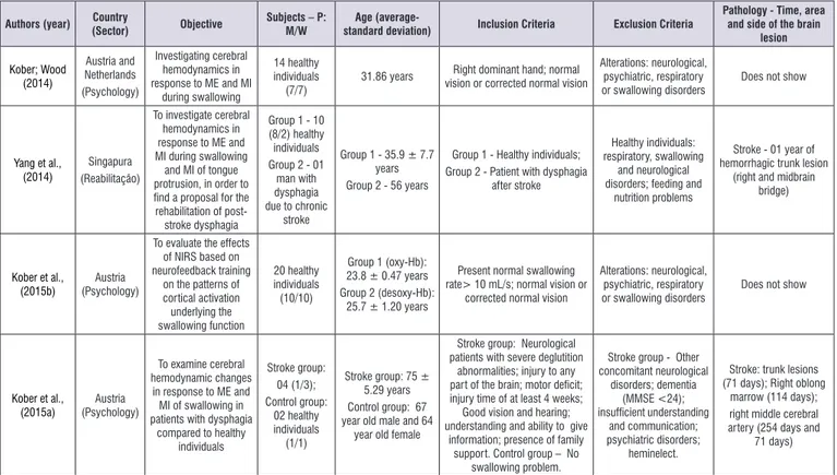
For a better presentation of the results, the following variables of the selected articles were considered: author (year); country (sector); objective; subject; age; Inclusion criteria; Exclusion criteria; pathology (time,
Authors (year) Country
(Sector) Objective
Subjects – P: M/W
Age
(average-standard deviation) Inclusion Criteria Exclusion Criteria
Pathology - Time, area and side of the brain
lesion Kober; Wood (2014) Austria and Netherlands (Psychology) Investigating cerebral hemodynamics in response to ME and MI
during swallowing
14 healthy
individuals
(7/7) 31.86 years
Right dominant hand; normal
vision or corrected normal vision
Alterations: neurological, psychiatric, respiratory
or swallowing disorders
Does not show
Yang et al., (2014)
Singapura
(Reabilitação)
To investigate cerebral hemodynamics in response to ME and
MI during swallowing and MI of tongue
protrusion, in order to find a proposal for the rehabilitation of
post-stroke dysphagia
Group 1 - 10 (8/2) healthy
individuals
Group 2 - 01 man with dysphagia due to chronic
stroke
Group 1 - 35.9 ± 7.7
years
Group 2 - 56 years
Group 1 - Healthy individuals; Group 2 - Patient with dysphagia
after stroke
Healthy individuals: respiratory, swallowing
and neurological
disorders; feeding and nutrition problems
Stroke - 01 year of hemorrhagic trunk lesion
(right and midbrain
bridge)
Kober et al.,
(2015b) (Psychology)Austria
To evaluate the effects of NIRS based on
neurofeedback training
on the patterns of
cortical activation underlying the swallowing function 20 healthy individuals (10/10)
Group 1 (oxy-Hb): 23.8 ± 0.47 years Group 2 (desoxy-Hb):
25.7 ± 1.20 years
Present normal swallowing
rate> 10 mL/s; normal vision or
corrected normal vision
Alterations: neurological, psychiatric, respiratory
or swallowing disorders
Does not show
Kober et al.,
(2015a) (Psychology)Austria
To examine cerebral hemodynamic changes
in response to ME and
MI of swallowing in
patients with dysphagia compared to healthy
individuals Stroke group: 04 (1/3); Control group: 02 healthy individuals (1/1)
Stroke group: 75 ± 5.29 years Control group: 67 year old male and 64
year old female
Stroke group: Neurological patients with severe deglutition
abnormalities; injury to any part of the brain; motor deficit; injury time of at least 4 weeks; Good vision and hearing;
understanding and ability to give
information; presence of family support. Control group – No
swallowing problem.
Stroke group - Other
concomitant neurological disorders; dementia (MMSE <24); insufficient understanding and communication; psychiatric disorders; heminelect.
Stroke: trunk lesions (71 days); Right oblong
marrow (114 days); right middle cerebral artery (254 days and
71 days)
Legend: ME: Motor Execution; MI: Motor Imagery; NIRS: Near-Infrared Spectroscopy Study; Oxy-Hb: Oxyhemoglobin; Desoxy-Hb: Deoxyhemoglobin; MMSE: Mini– Mental State Examination; P: study population; M: Men: W: Women; mL/s: Milliliter per second.
Figure 2. Characterization of the studies that performed the motor imagery of the deglutition activity
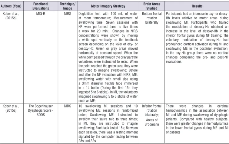
Authors (Year) Functional
Evaluations
Technique/
Image Motor Imagery Strategy
Brain Areas
Studied Results
Kober; Wood (2014)
Use of EMG in supra-hyoid muscles during
MI, ME and rest
NIRS MI/ME swallowing: Water use; Use of flexible tube (3mm diameter contained in a 1L bottle of water). ME of 15s; 15s IM; Rest from 28s to 32s. On the screen of a computer indication of action or rest.
Areas of Brodmann;
Topography: Change of oxy-Hb and desoxy-H during MI and ME. Signal in the lower frontal gyrus, including the Broca area;
Oxy-Hb higher during rest than during ME; - Oxy-Hb increased during MI and decreased during MI; Desoxy-Hb with higher concentration in tasks than at rest; Desoxy-Hb demonstrates positive correlation between MI and ME, mainly in the kinesthetic strategy of MI; Pre-motor; Supplemental motor area and subcentral area were involved in MI and ME; It involves the involvement of the insula during swallowing, not captured by the NIRS; Yang et al.,
(2014) and infrahyoid musclesEMG of the submental EEG 03 experimental sessions, each with two steps for healthy individuals: Sessions 1 and 2 with the MI of swallowing and rest; Session 3 with language MI and home; 02 experimental sessions, each with two stages for the patient with stroke: one of MI of swallowing versus rest and another of MI of tongue versus rest
Swallowing MI: Use of water, juice or food (noodles or large pill); MI of the tongue: Protrusion of the tongue as far as possible and by several times; Visual track through a screen and auditory indicating the beginning of the task to be imagined;
No specific area
In the search for data in the literature, the selected studies proposed searching the motor imagery of swallowing through different methodologies, and it was not possible to group data by means of statistical analysis. Given this heterogeneity, the results of this study are in the form of a systematic review, without meta-analysis.
The use of motor imagery rehabilitation of swallowing shows to be a recent proposal, since the four selected articles are between the years 2014 and 2015 6,21-23. The literature clearly indicates the efficacy
of MI as an adjuvant in motor rehabilitation of the upper limb in individuals with some neurological disorder 25-27.
But it is incipient the number of articles that suggest this technique correlate with tongue movements, and yet such research does not focus on rehabilitation of swallowing 28,29.
The countries which have published the research in question are different, however scholars of Austria
neuroimaging technique, checking changes in cerebral hemodynamics. In 201523, this same group deepens
the methodology with neurofeedback strategies, with training modulation of brain oxygen levels during MI swallowing. In the other hand, the authors of Singapore
6 proposed to investigate the detection of swallowing MI
as a proposal for rehabilitation through the MI of tongue movements (protrusion).
Certainlyi t is presumed that in South American countries, such as Brazil, where the rehabilitation of dysphagia is already consolidated 30,31, possibly was
not yet aroused the intention to enter the MI approach in clinical practice for the treatment of swallowing changes. Perhaps the limited number of studies will be a barrier to the expansion of this technique in Latin countries.
It is believed that the understanding of the thera-peutic approach of the MI by the speech therapist would enhance new perspectives in dysphagia
rehabili-Authors (Year) Functional
Evaluations
Technique/
Image Motor Imagery Strategy
Brain Areas
Studied Results
Kober et al.,
(2015b) MIQ-R NIRS Deglutition test with 100 mL of water at room temperature; Measurement of swallowing time; Seven sessions with NF were performed three to five times a week for 20 min; Changes in NIRS concentrations were shown by moving a white spot vertically on the feedback screen depending on the level of oxy- or desoxy-Hb; Green or gray areas moved horizontally at constant speed; When the white point passed through the gray part the volunteers were instructed to relax; When the point reached the green area, they were instructed to imagine swallowing; Before and after the NF evaluation with NIRS; ME: swallowing water with small sips using a 3mm diameter flexible tube immersed in a 1L bottle (During the first 15s they ingested 5 to 6 sticks); In MI, the volunteers imagined swallowing 5 to 6 sticks of water such as ME;
Bottom frontal rotation bilaterally
Participants had an increase in oxy- or deoxy-Hb levels relative to motor areas during swallowing MI. Participants who trained the modulation of desoxy-Hb obtained an increase in the level of desoxy-Hb in the inferior frontal gyrus during NF training; The voluntary modulation of desoxy-Hb had pronounced cortical activation during MI and swallowing ME in the posterior evaluation; In the oxy-Hb group there were no cortical changes comparing the pre- and post-NF evaluations.
Kober et al., (2015a)
The Bogenhauser Dysphagia Score -
BODS
NIRS 10 swallowing MI sessions and 10 swallowing ME sessions in randomized order; Swallowing ME: Instructed to swallow their saliva two to three times; In MI, they are instructed to imagine swallowing; Each task lasted 15s; Between each session, there was a resting moment signaled by the computer lasting between 28s and 32s
Inferior frontal rotation bilaterally;
Areas of Brodmann
There were changes in cerebral hemodynamics in the association between MI and ME during swallowing of dysphagic patients. Compared with healthy subjects, there were greater changes in hemodynamics in the lower frontal gyrus during ME and MI of patients
Legend: EMG: Surface electromyography; MI: Motor imagery; ME: Motor performance; NIRS: Near-Infrared Spectroscopy Study; mm: millimeters; L: Liter; s: seconds; Oxy-Hb: Oxyhemoglobin; Desoxy-Hb: Deoxyhemoglobin; EEG: Electroencephalogram; mL: milliliter; NF: Neurofeedback; min: minutes; MIQ-R: Motor Imagination Questionnaire in the revised version.
point to be reflected when looking at the departments in
wich the four studies of this review were subordinateds (rehabilitation and psychology).
This question is also anchored in the complexity of methodological approaches outlined in each manuscript, different from the selection of the sample to the choice of instruments and stages of MI execution.
In the sampling, the number of participants in the trials ranged from 6 to 20 individuals, composed mainly of healthy individuals aged up to 35 years. It was assumed that as a recent technique in swallowing rehabilitation, there was the interest of initially study a healthy population, in an attempt to better under-standing this therapeutic proposal.
Two studies involved neurological dysfunction 6,21,
wherein 05 patients with stroke, divided into brainstem lesion (03-60%) and the middle cerebral artery lesions (02 - 40%). The time after the event has been described with a range 71-254 days in the study series of cases22,
and, then considering a sample yet in subacute phase of the disease. Already in the manuscript with a single case of stroke 6, the patient had chronic injury a year
ago.
Lesions in the brainstem often brings changes in
swallowing, as well as the interruption of blood flow in
the middle cerebral artery can lead to various motor-sensory changes, affecting the face and the upper and lower members movements in the contralateral side of encephalic injury 32. In both studies the focus has
permeated to investigate the brain behavior of people with dysphagia during the MI and ME of swallowing, comparing them to individuals without neurological disorders.
In eligibility, for healthy individuals were ruled out any neurological and psychiatric were descarded, as
well as sensorial or motor deficit. Moreover, it wouldn’t
be possible the admission of people with swallowing disorders 6,21-23. In the other hand, in the participants
group with stroke, there was the requirement of dysphagia 6,22, however with no others neurological
and psychiatric disorders, with preserved capacity for understanding and communication 22.
To check the swallowing function, only one article used standardized assessment Bogenhausen Dysphagia Score (BODs) 22 to determine the dysphagia
degree of the 04 participants with stroke sequelae, classifying them into mild (score 4), moderate (score 5 6), moderate-severe (score 9) and severe (score 11).
The Bods is a German evaluation used by speech therapists in Europe, which assesses the ability to
swallow saliva and food intake33. The stratification of
the population in degree of dysphagia commitment, besides to identify the variability of the sample, raises the researcher concern in the risk analysis, referring the choice of safe intervention strategies to work swallowing rehabilitation. In the survey concerned the water was not used.
The surface electromyography (EMG) of the supra-hyoid muscles was used in two manuscripts during MI swallowing, bringing quantitative data on the electrical activity of these musculature 6,21. This information was
used as a control of active movement (motor) during MI in the swallowing oral phase.
In the analysis of the supra-hyoid muscles 21, the researchers identified that the electrical activity of
these muscles increased during the execution of the movement compared with the MI and rest. There was
also a numerical increase (not statistically significant)
in electrical activity during the MI when compared with rest. In discussion, it quotes the hypothesis that the reducing of the electrical signal magnitude during the MI has roots in cortical inhibition of movement (motor control) while the MI.
Studies show similarity in activation of brain areas during ME and MI, highlighting the Primary Motor (M1), Supplementary Motor Area (SMA), Pre-Motor and somatosensory cortex in the parietal lobe 15,16.
However, the suppression exerted by the SMA to the M1 during MI has also been reported in other studies
15,17. It is believed that this inhibitory effect is a
physio-logical mechanism to prevent movement during the MI.
To find out if the study population was under -standing the technique of motor imagery, a single article used the Motor Imagination Questionnaire in the revised version - MIQ-R 23. This questionnaire is
the most used to verify, through scores, the ability to imagine a movement through a visual or kinesthetic strategy, involving movements of the leg, arm and whole body 34.
The neuroimaging technique most widely used was the Near-Infrared Spectroscopy - NIRS (03-75%) 21-23 by
the researchers group from Austria and Netherlands. The infrared spectroscopy (NIRS) is one of the main techniques used to observe tissue oxygenation, most
specifically the oxyhemoglobin flow (Oxy-Hb) and
deoxyhemoglobin (Hb-Desoxy) in muscle and brain regions 35. The change in blood oxygenation response to a stimulus reflects in a higher level of brain activation,
that is, in the area where most cortical activation occurs
monitored, by NIRS, the concentrations of oxy- and deoxyhemoglobin. In this review, the authors proposed verify this change of cerebral hemodynamics in Brodmann areas, focusing on the inferior frontal gyrus, during the execution and imagination of the swallowing moviments.
With healthy individuals 21, the NIRS results
corrob-orate other neuroimaging studies about activation of premotor areas and SMA during the ME and MI 15,17.
There was an increase of oxy-Hb during ME (swallowing water) and Desoxy-Hb had greater concentration during the tasks than in the rest. During the motor execution
would be expected the increase of the oxy-Hb flow and
the inhibitory mechanism during MI, discussed above, may be a hypotesis for these hemodynamic changes. On the other hand, it was not possible on these results make inferences for the population with neurological alterations nor propose ways of treatment.
The oxygen flow modulation was aimed through
the use of NIRS and neurofeedback 23. The participants
were divided into two groups (oxy- and desosy-Hb) and instructed to observe a white dot on a computer screen with green and gray color stripes. The volunteer should imagine swallowing water depending on the color of the stripe in which the white point it was positioned.
For neurofeedback training, different values of oxy- or desoxy-Hb (respectively in each group) were achieved in the inferior frontal gyrus, being repre-sented by changes in the vertical direction of the white spot on the screen. The results showed that levels of oxy- and desoxy-Hb increases during both MI and ME. In the group of individuals who have trained the desoxy-Hb modulation, obtained an increase in the desoxy-Hb level in the inferior frontal gyrus. This study proposed intervention and the possibility of cortical reorganization, although it was carried out with healthy individuals.
In 2013, a pilot study used the NIRS with the neuro-feedback proposal in motor imagery of post-stroke
patients, confirming to be possible a cortical reorga -nization in neurological patients, with enhancement of functional recovery during rehabilitation. However, in this study the imagined movements were restricted to the upper member and has not been further analyzed the swallowing function 36.
The NIRS study with neurological patients 22 showed
the occurrence of hemodynamic changes in the MI and
through the MI. In the inferior frontal gyrus is located the
Broca’s area, largely responsible for the language, and
is also situated the control of orofacial sensory motor movements unrelated to speech, going on activation during swallowing 37.
The authors of Singapore 6 corroborate the idea of
rehabilitating dysphagia through swallowing of MI. In this study with electroencephalogram (EEG), the MI of tongue movements was used as a model for the detection of MI swallowing. The study has proposed that brain areas responsibless for swallowing are activated during the lifting movement of the tongue and quotes the cingulate cortex and the supplementary motor area.
The literature points out that the extent and distri-bution of brain activations may differ in MI and ME, however in both the MI and the execution activities, the neural networks involving central motor areas are activated. These areas participate in the planning, initiation and execution of motor commands 38. The
supplementary motor area along with pre motor cortex send neural impulses to the primary motor cortex and are constantly activated during MI and ME. In addition, the synchronization and coordination of the swallowing movements, is already evident in the literature the association of cortical areas such as the insula and pre motor cortex 5.
In consonance, study about the brain activation and connections that occurs during motor imagery indicates that the primary motor cortex, supplementary motor area and prefrontal cortex plays a crucial role during the MI and ME. The interaction between brain areas and effective network connectivity shows the importance of relationship of treatment as a mental practice and physical therapies during motor rehabili-tation 39.
In the event of neurological disorders, the MI associated with ME strengthens the occurrence of connections in the cortical motor area on the affected brain side and rearranges the connectivity of neural network of the contralateral cerebral side to the lesion
40, in the case of an effective strategy in dysphagia
rehabilitation, since that the cortical representation of normal swallowing is bilateral 19.
In this study 6, it is suggested that in clinical
the beginning of the pharyngeal phase and avoinding situations related to escape and aspiration.
However, it indicates the difficulty of accom -plishing the MI swallowing due to the complexity of the movement that involves sensory processing, coordination of chewing movements, breathing and attention. As the study realized with NIRS, it requires a larger number of participants to raise inference to the general population and facilitate the incorporation of the technique as an adjuvant of the conventional 22
swallowing rehabilitation.
CONCLUSION
The studies suggest that the motor imagery may cause some brain response in the motor area of the brain, suggesting that mentalization of actions related to swallowing may be effective to improve the engine
performance of this function and therefore, reflecting
also, in the feeding activity. However, new studies are needed with a larger number of participants allowing introducing this technique in the clinical intervention of professionals involved in swallowing rehabilitation.
REFERENCES
1. Matsuo K, Palmer JB. Anatomy and physiology of feeding and swallowing – normal and abnormal. Phys Med Rehabil Clin N Am. 2008;19(4):691-707. Doi: 10.1016/j.pmr.2008.06.001.
2. Yamada EK, Siqueira KO, Xerez D, Koch HA,
Costa MMB. A influência das fases oral e faríngea
na dinâmica da deglutição. Arq Gastroenterol. 2004;41(1):19-23.
3. Ramsey DJC, Smithard DG. Assessment
and management of dysphagia. Hosp Med. 2004;65(5):274-9.
4. Barritt AW, Smithard DG. Role of cerebral cortes plasticity in the recovery of swallowing function following dysphagic stroke. Dysphagia. 2009;24(1):83-90.
5. Sessle BJ, Yao D, Nishiura H, Yoshino K, Lee JC, Martin RE et al. Properties and plasticity of the primate somatosensory and motor córtex related to orofacial sensorimotor function. Clin Exp Pharmacol Physiol. 2005;32(1-2):109-14.
6. Yang H, Guan C, Chua KSG, Chok SS, Wang CC, Soon PK et al. Detection of motor imagery of swallow EEG signals based on the dual-tree complex wavelet transform and adaptive model selection. J. Neural Eng. 2014;11(3):1-13.
7. Duncan PW, Zorowitz R, Bates B, Choi JY, Glasberg JJ, Graham GD et al. Management of adult stroke rehabilitation care: a clinical practice guideline. Stroke. 2005;36(9):100-43.
8. Kleim JA, Jones A. Principles of experience-dependent neural plasticity: implications for rehabilitation after brain damage. J Speech Lang Hear Res. 2008;51(1):S225-39.
9. Sharma N, Pomeroy VM, Baron JC. Motor imagery: a backdoor to the motor system after stroke? Stroke. 2006;37(7):1941-52.
10. Jackson PL, Doyon J, Richards CL, Maloui F. The
efficacy of combined physical and mental practice
in the learning of a foot-sequence task after stroke: a case report. Neurorehabilitation Neural Repair. 2004;18(2):106-11.
11. Schuster C, Butler J, Andrews B, Kischka U, Ettlin T. Comparison of embedded and added motor imagery training in patients after stroke: study protocol of a randomised controlled pilot trial using a mixed methods approach. Trials. 2009;10(1):97.
12. Jackson PL, Lafleur MF, Malouin F, Richards C,
Doyon J. Potential role of mental practice using motor imagery in neurologic rehabilitation. Arch Phys Med Rehabil. 2001;82(8):1133-41.
13. Liu K, Chan C, Lee TM, Hui-chan CW. Mental imagery for promoting relearning for people after stroke: a randomizes controlled trial. Arch Phys Med Rehabil. 2004;85(9):1403-8.
14. Machado A. Neuroanatomia Funcional. Editora Atheneu, 2a ed. 2006.
15. Gao Q, Duan X, Chen H. Evaluation of effective connectivity of motor areas during motor imagery and execution using conditional Granger causality. Neuroimage. 2011;54(2):1280-8.
16. Guillot A, Collet C. Contribution from neurophysiological and psychological methods to the study of motor imagery. Brain Res Rev. 2005;50(2):387-97.
17. Kasess CH, Windischberger C, Cunnington R, Lanzenberger R, Pezawas L, Moser E. The
suppressive influence of SMA on M1 in motor
imagery revealed by fMRI and dynamic causal modeling. Neuroimage. 2008;40(2):828-37.
19. Hamdy S, Aziz Q, Rothwell JC. The cortical topography of humam swallowing musculature in health and disease. Nat Med. 1996;2(11):1217-24. 20. Ataliah AN, Castro AA. Revisão Sistemática da
literatura e Metanálise. In: Ataliah AN, Castro
AA (orgs). Medicina baseada em evidências: fundamentos da pesquisa clínica. São Paulo:
Lemos Editorial; 1998. p.20-8.
21. Kober SE, Wood G. Changes in hemodynamic signals accompanying motor imagery and motor execution of swallowing: A near-infrared spectroscopy study. Neuroimage. 2014;93(1):1-10. 22. Kober SE, Bauernfeind G, Woller C, Sampl M,
Grieshofer P, Neuper C et al. Hemodynamic signal changes accompanying execution and imagery of swallowing in patients with dysphagia: a multiple single-case near-infrared spectroscopy study. Front. Neurol. 2015;6:151. doi: 10.3389/ fneur.2015.00151
23. Kober SE, Gressenberger B, Kurzmann J, Neuper C, Wood G. Voluntary modulation of hemodynamic responses in swallowing related motor areas: A near-infrared spectroscopy-based neurofeedback study. Plos One. 2015;10(11):e0143314. doi:10.1371/journal.pone.0143314.
24. Moher D, Liberati A, Tetzlaff J, Altman DG., The PRISMA Group. Preferred reporting items for systematic reviews and meta-analyses: The
PRISMA Statement. Disponível em:
www.prisma-statement.org. Acesso em: 20 de maio de 2016. 25. Braun SM, Haastregt LCV, Beurskens AJ, Gielen AI,
Wade DT, Schols JM. Feasibility of a mental practice intervention in stroke patients in nursing homes; a process evaluation. Neurology. 2010;10(1):1-9. 26. Bastos AF, Carrapatoso BC, Orsini M, Leite MAA,
Silva JG, Souza GGL. Functional recovery of upper limb post-stroke: mental practice with motor and non-motor imagery. Am Medic J. 2012;3(1):50-5. 27. Page SJ, Dunning K, Hermann V, Leonard A, Levine
P. Longer versus shorter mental practice sessions for affected upper extremity movement after stroke: a randomized controlled trial. Clin Rehabil. 2011;25(7):627-37.
28. Morash V, Bai O, Furlani S, Lin P, Hallett M. Classifying EEG signals preceding right hand, left hand, tongue and right foot movements and motor imageries. Clin Neurophysiol. 2008;119(11):2570-8.
classification of different motor imagery tasks.
NeuroImage. 2006;31(1):153-9.
30. Itaquy RB, Favero SR, Ribeiro MDE C, Barea LM, Almeida ST, Mancopes R. Dysphagia and cerebrovascular accident: relationship between severity degree and level of neurological impairment. J Soc Bras Fonoaudiol. 2011;23(4):385-9.
31. Steimbergtu C, Frazãot YS, Furmm AM. Disfagia no Brasil : a construçao de uma pratica. Rev. CEFAC 2003;5(2):117-25.
32. Lundy-Ekman L. Neurociência: Fundamentos para
reabilitação. Editora Elsevier. 3a ed. 2008.
33. Schiele JT, Penner H, Schneider H, Quinzler R, Reich G, Wezler N et al. Swallowing tablets and capsules increases the risk of penetration and aspiration in patients with stroke-induced dysphagia. Dysphagia. 2015;30(5):571-82. http:// dx.doi.org/10.1007/s00455-015-9639-9
34. Gregg M, Hall C, Butler A. The MIQ-RS: A suitable option for examining movement imagery ability. Evid Based Complement Alternat Med. 2010;7(2):249-57.
35. Lima A, Bakker J. Espectroscopia no infravermelho próximo para a monitorização da perfusão tecidual. Rev Bras Ter Intensiva. 2011;23(3):341-51.
36. Mihara M, Hattori N, Hatakenaka M, Yagura H, Kawano T, Hino T. Near-infrared spectroscopy–
mediated neurofeedback enhances efficacy of
motor imagery–based training in poststroke victims a pilot study. Stroke. 2013;44(4):1091-8.
37. Hirsch J, Ruge MI, Kim KHS, Correa DD, Victor JD, Relkin NR et al. Integrated functional magnetic resonance imaging procedure for preoperative mapping of cortical areas associated with tactile, motor, language, and visual functions. Neurosurgery. 2000;47(3):711-22.
38. Bajaj S, Butler AJ, Drake D, Dhamala M. Oscilatory motor network activity during rest and movemen: an fNIRS study. Front. Syst. Neurosci. 2014;8(13): 1-12. http://dx.doi.org/10.3389/ fnys.2014.0001324550793.
39. Bajaj S, Butler AJ, Drake D, Dhamala M. Brain effective connectivity during motor-imagery and execution following stroke and rehabilitation. Neuroimage. 2015; 8(1):572-82.