UNIVERSIDADE DA BEIRA INTERIOR
Ciências da Saúde
Genetic susceptibility to thyroid cancer:
contributions of RET polymorphisms
Marina Silva dos Santos
Dissertação apresentada à Universidade da Beira Interior para obtenção do grau de Mestre em
Ciências Biomédicas
(2º ciclo de estudos)
Orientador: Prof. Doutor Manuel Carlos Loureiro de Lemos
iii
Agradecimentos
Mais do que um trabalho individual, esta dissertação é o resultado do contributo de várias pessoas que ao longo destes meses estiveram comigo estimulando-me intelectual e emocionalmente. Pelo que não poderia deixar de agradecer a todos os que de algum modo contribuíram para a sua concretização.
Em primeiro lugar, ao meu orientador, Professor Doutor Manuel Lemos pela disponibilidade, competência científica e oportunidade de pertencer ao seu grupo de trabalho.
Ao Director do Serviço de Endocrinologia do Instituto Português de Oncologia de Coimbra, Dr. Fernando Rodrigues, e restante equipa médica pela valiosa contribuição no recrutamento e caracterização clinica dos doentes.
Ao Dr. Mário Chin responsável pelo Centro Regional de Sangue de Coimbra por possibilitar o acesso às colectas de sangue dos dadores de sangue.
Ao grupo de trabalho de Endocrinologia que se tornou a família da qual me orgulho de fazer parte. Obrigada a todos vocês pelos momentos especiais, amizade, apoio e convivência diária. Sem vocês isto não teria graça nenhuma!
À Eduarda por seres uma verdadeira mãe para nós, por todo o tempo que perdeste comigo, pela paciência e pelo carinho. À Catarina por seres uma alegria constante e uma pessoa tão única, pelas conversas e pela companhia nas horas de almoço. À Inês, pelo teu sentido de humor que tanto gosto, pelas valiosas indicações, sugestões, críticas e correcções e acima de tudo pelas perguntas difíceis. À Ângela e à Susana meninas lindas a quem desejo tudo de bom. Ao Tito, meu companheiro desde início nesta jornada, custou mas conseguimos! À Gabi, a filha adoptiva, por toda a ajuda, força e apoio e por me ouvires sempre.
À minha família, aos meus irmãos e em especial aos meus pais, pelo exemplo de humildade, perseverança, simplicidade e determinação, por sempre se esforçarem tanto para me darem o melhor, por me incentivarem e me darem apoio para estudar mais do que eles puderam. E por fim, Ao Jorge, por seres o que és meu amor! Por acreditares em mim, por me ajudares e amparares sempre. E por me dares a força que às vezes me falta.
Este projecto foi financiado pela FCT através do programa COMPETE (projecto PEst-C/SAU/UI0709/2011).
v
Resumo alargado
A glândula da tiróide é o maior órgão do corpo humano exclusivamente endócrino. Localiza-se na parte anterior do pescoço, em frente à traqueia e abaixo da laringe, é um órgão bastante vascularizado e em forma de borboleta. Histologicamente a glândula da tiróide é constituída por folículos formados por epitélio simples de células foliculares produtoras das hormonas T3 e T4, nos interstícios dos folículos localizam-se as células parafoliculares produtoras da hormona calcitonina. As hormonas T3 e T4 produzidas por esta glândula regulam a taxa do metabolismo e afectam o aumento e a taxa funcional de muitos outros sistemas do organismo, já a hormona calcitonina possui um papel importante na homeostase do cálcio. Como qualquer outro órgão também a tiróide está sujeita a fenómenos de carcinogénese, o cancro da tiróide tem uma incidência anual de 212.000 casos por todo mundo, este número tem vindo a aumentar nos últimos anos devido, provavelmente, aos progressos nas técnicas de diagnóstico e ao efeito dos factores ambientais.
O cancro da tiróide tem uma etiologia multifactorial resultante da interacção de factores genéticos e ambientais. Dependendo do tipo de células a partir das quais o cancro se desenvolve, o cancro da tiróide pode ser dividido em dois grandes grupos, os carcinomas medulares da tiróide (MTC) e os carcinomas diferenciados da tiróide (DTC). A última categoria (DTC) abrange o carcinoma folicular da tiróide (FTC) e o carcinoma papilar da tiróide (PTC), ambos originam-se a partir das células foliculares e representam a maioria dos tumores desta glândula. Apesar da grande incidência, de um modo geral, estes subtipos apresentam bom prognóstico e boa taxa de sobrevivência. Os DTC podem evoluir para um estado indiferenciado com prognóstico muito mais reservado designado carcinoma anaplásico da tiróide (ATC). O MTC tem origem nas células parafoliculares da tiróide. Os mecanismos moleculares subjacentes ao desenvolvimento do cancro da tiróide têm sido amplamente estudados, e o proto-oncogene RET tem sido desde há muito associado a este cancro, desde rearranjos cromossómicos no caso do PTC a mutações pontuais a nível germinativo e somático no MTC. Este proto-oncogene localiza-se no braço longo do cromossoma 10 e é composto por 21 exões, codifica um receptor transmenbranar tirosina-cinase com o mesmo nome do gene. O receptor RET é expresso nas células precursoras da crista neural e do tacto urogenital, sendo necessário na maturação de várias linhagens celulares do sistema nervoso periférico bem como na morfogénese dos rins e na espermatogénese. Mutações que levam à perda de função deste receptor têm sido associadas à doença de Hirchsprung, doença poligénica associada à ausência de neurónios entéricos no tracto gastrointestinal. Por outro lado, mutações que levam ao ganho de função deste receptor estão associadas ao cancro, em particular ao cancro da tiróide.
vi
Um locus é considerado polimórfico quando um ou mais dos seus alelos raros têm uma frequência de pelo menos 1% numa população. Um conjunto de marcadores genéticos ou polimorfismos que se transmitam como uma unidade através de gerações é designado de haplótipo. Variantes polimórficas comuns têm vindo a ser associadas com o fenótipo de várias doenças complexas, modificando assim o risco de ocorrência das mesmas. Igualmente, nos últimos anos, os polimorfismos do proto-oncogene RET têm sido estudados de modo a determinar se estas variantes polimórficas podem representar alelos de baixa penetrância que predisponham para doenças associadas ao RET (como o PTC, o MTC familiar e esporádico e a doença de Hirschprung). No caso da tiróide, a maioria dos estudos existentes diz respeito ao MTC, de um modo geral os polimorfismos que têm mostrado uma maior prevalência nas populações de doentes relativamente à população geral são: o G691S, o L769L, o S836S e o S904S. Estes localizam-senos exões 11, 13, 14 e 15, respectivamente, e albergam os hot spots mutacionais para o MTC. Contudo, é clara a controvérsia entre os diversos estudos no que respeita à influência dos polimorfismos na susceptibilidade para o cancro da tiróide.
Com este trabalho, pretendeu-se investigar a associação dos polimorfismos G691S, L769L, S836S e S904S, isoladamente ou em conjunto, com o aumento do risco para o desenvolvimento do cancro da tiróide, em particular dos DTC na população portuguesa. Paralelamente pretendeu-se avaliar a associação destes polimorfismos com 4 parâmetros clínicos: o subtipo de cancro, o género, a idade de diagnóstico e o tamanho do tumor no diagnóstico. Para alcançar este objectivo, fez-se um estudo de associação caso-controlo. Para além de uma população controlo, foi necessária a recolha de uma população de doentes com PTC ou FTC e respectivos dados clínicos relevantes. A população de doentes consistiu em 282 indivíduos, portugueses e caucasianos, cedidos pelo Instituto Português de Oncologia de Coimbra. A população controlo foi formada por 245 voluntários, também portugueses e caucasianos, não relacionados, recrutados entre pessoal da Faculdade e dadores de sangue do Instituto Português de Sangue da Região Centro.
Após a obtenção do consentimento informado, foi recolhida uma amostra de sangue periférico de cada indivíduo. A genotipagem dos indivíduos do grupo de doentes e do grupo controlo iniciou-se pela extracção do DNA a partir de uma amostra de sangue periférico pelo método de salting-out adaptado, seguiu-se a quantificação desse mesmo DNA de forma a obter informação sobre a concentração e nível de pureza da amostra de DNA. Em seguida, fez-se a amplificação por PCR dos exões, onde se encontram os quatro polimorfismos estudados. Para conseguir distinguir os genótipos dos indivíduos para os vários polimorfismos fizeram-se incubações com enzimas de restrição, dado que que todos os polimorfismos criam ou eliminam um local de restrição para uma endonuclease específica. Para garantir a correcta genotipagem pelo método anterior, procedeu-se à sequenciação de DNA de 3 indivíduos representativos dos 3 genótipos para cada um dos polimorfismos estudados. Assim, foi então possível a formação de uma base de dados dos genótipos das duas populações, permitindo a
vii
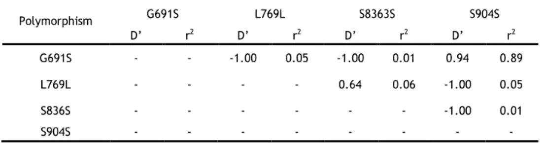
análise estatística das mesmas. Nesta análise, avaliou-se o equilíbrio de Hardy-Wenberg para a população controlo, bem como o desequilíbrio de ligação (LD) entre os vários polimorfismos, seguiu-se a comparação das distribuições alélicas, genotípicas e haplotípicas entre controlos e doentes. Para a avaliação dos vários parâmetros clínicos em estudo, fizeram-se subdivisões da população de doentes de acordo com cada um dos parâmetros e compararam-se as mesmas. Os resultados obtidos permitiram-nos observar que a população portuguesa, usada como controlo neste estudo, se assemelha à população europeia no que diz respeito à distribuição genotípica dos polimorfismos estudados, com excepção do polimorfismo L769L. Verificou-se um forte LD entre as variantes polimórficas G691S e S904S, sugerindo a sua co-segregação conjunta, tal como verificado em outros estudos populacionais. Pelas várias comparações realizadas, este estudo permitiu sugerir que o polimorfismo S836S parece estar associado com um risco aumentado de desenvolver DTC. Verifica-se também, uma possível associação entre os genótipos heterozigóticos dos polimorfismos G691S e S904S e a idade de desenvolvimento do DTC e, isoladamente, o genótipo heterozigótico do polimorfismo G691S parece ter associação com o tamanho do tumor. O haplótipo GGTC parece estar associado com uma maior susceptibilidade para o DTC em particular em idades mais avançadas, diagnóstico depois dos 45 anos. Contudo, com o ajuste para comparações múltiplas, muitos destes resultados não se mostraram estatisticamente significativos. Assim, estes resultados necessitam de ser confirmados em outros estudos com uma maior amostra para se poder reavaliar o papel destas variantes na susceptibilidade para o DTC.
Palavras-chave
Cancro da tiróide; carcinoma papilar da tiróide; carcinoma folicular da tiróide; gene RET; polimorfismos.
ix
Abstract
Thyroid cancer is the most common malignancy of the endocrine system, represents more than 1% of all malignancies and has an estimated annual incidence of 212,000 cases worldwide. The term differentiated thyroid carcinoma (DTC) comprises the subtypes papillary thyroid carcinoma (PTC) and follicular thyroid carcinomas (FTC), these subtypes represent the two most common subtypes of thyroid cancer (approximately 80% and 10% respectively). Despite its incidence DTCs have a good prognosis with relatively few metastases and deaths associated. The polymorphisms (variants in DNA sequence among individuals that have a frequency of at least 1% in a population) of RET proto-oncogene have been studied in different populations for association with susceptibility to thyroid cancer, but with inconsistent findings mainly in DTC. To clarify the contribution of single locus or haplotypes (polymorphisms that are transmitted through generations as a unit) of RET polymorphisms to genetic susceptibility to DTC among Portuguese patients, we conducted a case–control study by analyzing four well-characterized RET polymorphisms (G691S, L769L, S836S and S904S). To achieve this aim, the RET polymorphisms were genotyped and haplotype frequencies were estimated in a population of 282 individuals with DTC and in a control population of 254 individuals. Allele, genotype and haplotype distributions were compared among cases and controls. Patient population was subdivided according to several clinical parameters and allele, genotype and haplotype distributions were compared among the subgroups.
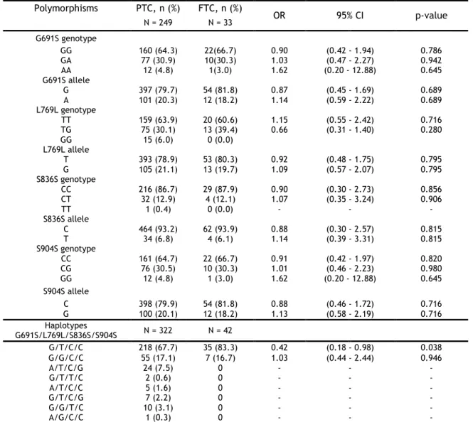
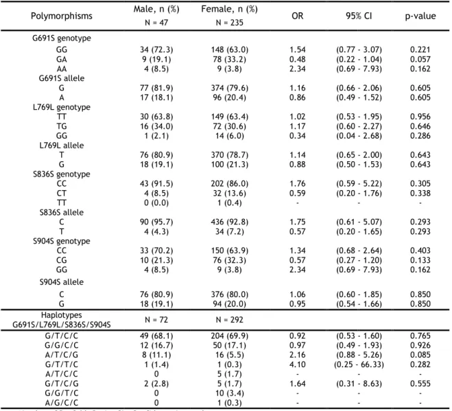
The single locus analysis showed an overrepresentation of the S836S polymorphism in patients when compared to controls. Also the heterozygous genotypes of the G691S/S904S polymorphisms were overrepresented in cases diagnosed after the age of 45 years and the heterozygous genotype of G691S polymorphism revealed an overrepresentation in patients with tumors larger then 10mm of diameter at diagnosis. The haplotype analysis showed an overrepresentation of GGTC haplotype in patients particularly in those diagnosed after the age of 45 years.
In conclusion, our data suggest that the S836S polymorphism may be associated with increased risk of DTC. Also the heterozygous genotype of the G691S/S904S polymorphisms seems to be associated with age of onset of DTC and additionally the heterozygous genotype of G691S polymorphism appeared to be in association with tumor size. Finally, one haplotype appears to be associated with increased risk of DTC particularly in those developed in later age (after the age of 45 years). These findings need to be confirmed by larger studies in order re-evaluate the role of these variants in the susceptibility to DTC.
x
Keywords
Thyroid cancer; Papillary Thyroid Carcinoma; Follicular Thyroid Carcinoma; RET gene; polymorphisms.
xii
Table of contents
Agradecimentos ... iii
Resumo alargado ... v
Abstract ... ix
Table of contents ... xii
List of figures ... xv
List of tables ... xvii
List of abbreviations ... xix
1. Introduction ... 1
1.1. Thyroid Gland ... 2
1.1.1. Anatomy and histology... 2
1.1.2. Thyroid hormones ... 3
1.2. Thyroid cancer ... 4
1.2.1. Follicular-cell-derived carcinoma ... 4
1.2.2. Medullary thyroid carcinoma ... 5
1.2.3. Molecular genetics of thyroid cancer ... 6
1.2.3.1. BRAF mutations ... 7
1.2.3.2. RAS mutations ... 8
1.2.3.3. PAX8/PPAR-γ rearrangement ... 8
1.2.3.4. TRK rearrangements ... 8
1.2.3.5. Mutations in carcinoma dedifferentiation ... 8
1.3. RET gene and RET protein ... 9
1.4. Genetic alterations in RET ... 12
1.4.1. RET and follicular-cell-derived carcinomas ... 12
1.4.2. RET and medullary thyroid carcinomas ... 13
1.5. RET Polymorphisms and Haplotypes in human diseases ... 13
1.5.1. RET Polymorphisms and Haplotypes in MTC ... 14
1.5.1.1. RET G691S and S904S Polymorphisms ... 15
1.5.1.2. RET L769L Polymorphism ... 16
1.5.1.3. RET S836S Polymorphism ... 16
1.5.2. RET Polymorphisms and Haplotypes in DTC ... 16
xiii
2. Materials and methods ... 18
2.1. Studied population ... 19
2.2. Genotyping ... 19
2.2.1. DNA Extraction ... 19
2.2.2. DNA quantification ... 20
2.2.3. Polymerase Chain Reaction ... 20
2.2.4. Enzymatic digestion ... 21 2.2.5. Electrophoresis ... 22 2.2.6. DNA sequencing ... 23 2.3. Statistical analysis ... 23 3. Results ... 25 3.1. Genotyping ... 26 3.1.1. RET G691S polymorphism ... 26 3.1.2. RET L769L polymorphism ... 27 3.1.3. RET S836S polymorphism ... 27 3.1.4. RET S904S polymorphism ... 28 3.2. Statistical analysis ... 29
3.2.1. Prevalence of the RET polymorphisms in a Portuguese population... 29
3.2.2. Linkage disequilibrium coefficients between the RET polymorphisms in a Portuguese population ... 30
3.2.3. Analysis of RET polymorphisms and haplotypes in thyroid cancer ... 31
3.2.3.1. Analysis of cancer subtypes ... 33
3.2.3.2. Analysis of gender ... 34
3.2.3.3. Analysis of age at diagnosis ... 34
3.2.3.4. Analysis of tumor size ... 35
4. Discussion and conclusion ... 37
xv
List of figures
Figure 1 – Frontal view of the thyroid gland. ... 2
Figure 2 - Histology of the thyroid gland. ... 3
Figure 3 - Schematic representation of the RET receptor tyrosine kinase. ... 10
Figure 4 - Different mechanisms of ligand-mediated RET activation. ... 11
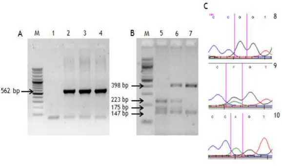
Figure 5 – Genotyping results for the G691S polymorphism. ... 26
Figure 6 - Genotyping results for the L769L polymorphism. ... 27
Figure 7 - Genotyping results for the S836S polymorphism. ... 28
Figure 8 – Genotyping results for the S904S polymorphism. ... 29
xvii
List of tables
Table 1 - Thyroid cancer types and correspondent mutational profiles. ... 7 Table 2 - Primers and PCR conditions for amplification of gene RET exons 11, 13, 14 and 15..
... 21
Table 3 - Gene RET polymorphisms, expected genotypes and the respective restriction
fragments. ... 22
Table 4- Comparison of the prevalence of RET polymorphisms between the studied
Portuguese population and a European population. ... 30
Table 5 - Pairwise linkage disequilibrium coefficients (D’ and r2) between the RET
polymorphisms in the control group. ... 31
Table 6 - Single locus analysis of RET polymorphisms and respective haplotypes between
patients and controls. ... 32
Table 7 - Single locus and haplotypes analysis of RET polymorphisms in PTC and FTC patients.
... 33
Table 8 - Single locus analysis and haplotype analysis of RET polymorphisms in male and
female patients. ... 34
Table 9 - Effect of RET polymorphisms and haplotypes on age at diagnosis of DTC. ... 35 Table 10 - Effect of RET polymorphisms and respective haplotypes on tumor size at age of
xix
List of abbreviations
AKAP9 A-kinase anchor protein 9
AKT Also known as Protein Kinase B (PKB)
AKT1 V-akt murine thymoma viral oncogene homolog 1 ARAF V-raf murine sarcoma 3611 viral oncogene homolog ARTN Artemin
ATC Anaplastic thyroid carcinoma ATP Adenosine triphosphate bp Base pairs
BRAF V-raf murine sarcoma viral oncogene homolog B1 BSA Bovine serum albumin
CCDC6 Coiled-coil domain containing 6
CRAF Raf-1 murine leukemia viral oncogene homolog 1 CTNNB1 Catenin (cadherin-associated protein) beta 1 DNA Deoxyribonucleic acid
dNTPs Deoxyribonucleotide triphosphates DTC differenciated thyroid carcinoma EDTA Ethylenediaminetetraacetic acid ERK Extracellular signal-regulated kinase FMTC Familial MTC
FTC Follicular thyroid carcinoma
GDNF Glial cell line-derived neurotrophic factor GFL GDNF family of ligands
GFRα GDNF receptor alpha Gly Glycine
H-RAS Harvey rat sarcoma viral oncogene homolog K-RAS Ras2 Kirsten rat sarcoma viral oncogene homolog LD Linkage disequilibrium
Leu Leucine
MAPK Mitogen-activated protein kinase MEK Mitogen-activated protein kinase kinase MEN-2 Multiple endocrine neoplasia type-2 MEN-2A Multiple endocrine neoplasia type-2A MEN-2B Multiple endocrine neoplasia type-2B MTC Medullary thyroid carcinoma
NCOA4 Nuclear receptor coactivator 4 NGF Nerve growth factor
xx
NIH 3T3 Mouse embryonic fibroblast cell line
N-RAS Neuroblastoma RAS viral (v-ras) oncogene homolog NRTN Neurturin
NTRK1 Neurotrophic receptor-tyrosine kinase OD Optical density
PAX8 Paired box gene 8
PCR Polymerase chain reaction PI3K Phosphatidyl-inositol-3'-kinase PIK3CA PI3K subunit alpha
PPARƴ Peroxisome proliferator-activated receptor gamma PSPN Persesephin
PTC Papillary thyroid carcinoma PTEN Phosphatase and tensin homolog RAS Rat sarcoma
RBC Red blood cells
RET Rearranged during transfection
RET43 RET isoform with 43 amino acids in carboxy - terminal tail RET51 RET isoform with 51 amino acids in carboxy - terminal tail RET9 RET isoform with 9 amino acids in carboxy - terminal tail RNA Ribonucleic acid
rpm Rotations per minute SD Standard deviation Ser Serine
SNP Single nucleotide polymorphism T3 Triiodothyronine T4 Thyroxine TK Tyrosine - kinase TRH Thyrotropin-releasing hormone TRK Tropomyosin-related kinase TSH Thyroid-stimulating hormone UV Ultraviolet
1
2
1.1. Thyroid Gland
1.1.1.
Anatomy and histology
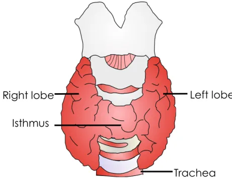
The thyroid gland is the largest organ, in the body, uniquely qualified to produce hormones. The thyroid gland weighs approximately 20 to 25 g, is located in the anterior neck at the level of the fifth cervical vertebra to the first thoracic vertebra, in front of the trachea, and immediately below the larynx. It is covered by the neck muscles and its fascia. It has a butterfly-shape and is constituted by two lobes joined together by an isthmus (narrow band of thyroid tissue). The thyroid is a very vascularized organ rich in blood and lymph capillaries and therefore presents a more reddish color than the surrounding tissues [1, 2].
Figure 1 – Frontal view of the thyroid gland. The thyroid gland is located in the anterior neckin front of the trachea, and immediately below the larynx, it has a butterfly-shape and is constituted by two lobes joined together by an isthmus.
Histologically the thyroid gland is composed by numerous thyroid follicles, which are spherical sacs filled with colloid rich in a protein called thyroglobulin to which thyroid hormones are bound. Thyroid follicles are lined by a single layer of thyroid epithelial cells, the follicular cells. In the soft connective tissue that separates and surrounds the thyroid follicles lie the parafollicular or C cells [3].
Trachea
Isthmus
Left lobe
Right lobe
3
Figure 2 - Histology of the thyroid gland. Histologically the thyroid gland consists in many spherical
thyroid follicles containing thyroglobulin. Parafollicular cells are in the tissue between the thyroid follicles. F- Thyroid follicle. Adapted from Toda et al., 2011 [4].
1.1.2.
Thyroid hormones
Briefly the function of the thyroid gland is to secrete an appropriate amount of the thyroid hormones. The thyroid gland produces two main types of hormones that are structurally related: thyroxine (named T4 since it carries 4 iodine atoms) and triiodothyronine (named T3 since it carries 3 iodine atoms). Under normal circumstances T4 constitutes approximately 90 percent of the hormones produced in the thyroid gland [5]. However, T3 is a much more active hormone, and the majority of the T4 produced by the thyroid is converted into T3 in the liver and kidneys [6].
T3 and T4 are produced and secreted by follicular cells and are stored in the thyroid follicles. The secretion of T3 and T4 is increased by the hypotalamic thyrotropin-releassing hormone (TRH), which stimulates release of thyroid-stimulating hormone (TSH), by the anterior pituitary, in response to low T3 and T4 levels, low metabolic rate, pregnancy, cold and high altitudes. High T3 and T4 levels inhibit TRH and TSH secretions; high iodine level suppresses the secretion of these hormones [7]. Almost all body cells have receptors for thyroid hormones, so T3 and T4 exert their effects throughout the body. The principal actions of T3 and T4 are to increase basal metabolic rate, increase use of glucose and fatty acids for ATP production, stimulate synthesis of proteins, enhance cholesterol excretion, accelerate body growth and contribute to development of the nervous system [7].
In addition to T3 and T4, the thyroid gland also produces calcitonin. Calcitonin is a polypeptide hormone produced and secreted by the parafollicular cells, which helps regulate
Parafollicular cells
4
calcium homeostasis by diminishing the concentration of Ca2+ in body fluids, when Ca2+ levels
are elevated. Taking into account this effect, calcitonin is a direct antagonist of the hormone produced by the parathyroid glands (parathyroid hormone). The target of calcitonin is the skeleton where it inhibits the action of osteoclasts (cells that break down bone extracellular matrix), so the level of Ca2+ in the blood decreases. Consequently, calcitonin promotes calcium deposition and bone formation by stimulating osteoblast activity [7]. Excessive blood levels of Ca2+ (approximately 20% above normal) act as a humoral stimulus for the release of
calcitonin in the blood, while the low levels of Ca2+ inhibit the secretory activity of the
parafollicular cells. Calcitonin appears to be important only in childhood, when the skeleton has an accentuated growth and the bones are changing in mass, size and shape [3].
1.2. Thyroid cancer
Thyroid cancer is the most common malignancy of the endocrine system [8]. Thyroid cancer represents more than 1% of all malignancies and has an estimated annual incidence of 212,000 cases worldwide. This number has increased quickly in recent years, probably due to the improvement of diagnostic techniques and to the effect of environmental factors [9]. Thyroid cancer has a multifactorial etiology resulting from the interaction of genetic and environmental factors in individuals at risk. Regarding the environmental level the major parameters to take into account are: the exposure to ionizing radiations, age at the time of the exposure, presence of a previous history of benign thyroid disease, role of the dietary iodine intake, role of the body mass index, and role of hormonal factors [10]. Thyroid cancer usually develops in thyroid nodules. Thyroid nodules are common, principally in adults with the increase of age, and most are benign. They can be detected by palpation and by imaging, the clinical challenge lies in the rapid and accurate identification of those nodules that hide cancer [11].
Thyroid cancer comprises two main sets of neoplasias, depending on the type of cell affected by malignant transformation. When the cancer develops from follicular cells it gives rise to follicular thyroid carcinoma (FTC) or papillary thyroid carcinoma (PTC), representing the majority of thyroid tumors, and the anaplastic thyroid carcinoma (ATC) is the undifferentiated type of carcinoma derived from follicular cells. Only a small part of cancers originates from parafollicular cells giving rise to medullary thyroid carcinoma (MTC) [9].
1.2.1. Follicular-cell-derived carcinoma
Follicular-cell-derived tumors include benign adenomas and well differentiated (papillary or follicular), poorly differentiated (insular), and undifferentiated (anaplastic) carcinomas.
5
Between them, PTC and FTC represent the two most common subtypes, representing 80-85% and 10-15% respectively [12, 13]. PTC and FTC are termed differentiated thyroid carcinoma (DTC)[14].
These subtypes, in general, display a good prognosis with relatively few metastasis and deaths[15], contrasting with the insular and anaplastic carcinomas, that though less frequent (approximately 5% of all thyroid carcinomas) show a poor prognosis. PTC is defined on the basis of the histological pattern and distinct nuclear features, that have ground glass appearance and longitudinal grooves with cytoplasm invaginations [16]. PTC may be sporadic (95%) or familial (5%) [17], its incidence is remarkably high in developed countries, and metastasizes to local lymph nodes [18]. PTC has a sex ratio female:male of around 3:1, predicting the influence of hormonal factors which are still unknown. Exposure to ionizing radiation is the only established environmental factor related to PTC [19].
There are numerous histopathologic variants of PTC according to a set of distinctive nuclear features (include nuclear enlargement and irregularity, overlapping, clearing, grooves, and pseudo-inclusions). The criteria used to subclassify PTC are not clearly defined, so classifying PTC is a major problem. The well-established subtypes include: classical variant, follicular variant, papillary microcarcinoma (micro-PTC), oncocytic variant (Hurthle cell), tall cell variant, clear cell variant, solid variant, cribriform-morular variant, columnar cell variant, diffuse sclerosing variant and macrofollicular variant [20, 21].
Emphasizing the micro-PTC, this subtype is defined as a papillary carcinoma that measures in maximum 1 cm that belongs to the low-risk well-differentiated PTC. Micro-PTCs are very common and is important distinguish between micro-PTC associated with clinically larger and significant PTC and an incidental micro-PTC found after thyroidectomy performed for other indications or during thyroid ultrasound. In the first case micro-PTC have been considered to represent dissemination of the larger tumor, on other hand incidental micro-PTC has an outstandingly good prognosis and there is nearly no risk of recurrence or death [20, 22-24]. What distinguishes the FTC is the absence of nuclear morphological features that define the PTC. The majority of the tumors is encapsulated, and may be composed of follicles or follicular cells arranged in follicular, solid or trabecular patterns. FTC compared to PTC tends to be more aggressive, produces distant metastases, and presents a more balanced sex ratio (about 2:1, female:male). FTC has been associated with a deficiency in iodine intake and is more frequent in developing countries [25].
1.2.2. Medullary thyroid carcinoma
Medullary thyroid carcinoma (MTC) originates from the parafollicular cells and represents about 3% to 5% of all thyroid cancers [13]. Around 75% of MTC occur sporadically and the
6
other 25% are hereditary. The hereditary MTC is part of a syndrome known as multiple endocrine neoplasia type-2 (MEN-2) that has an autosomal dominant model of inheritance. MEN-2 can be divided in three clinically distinct forms: MEN-2A, MEN-2B, and familial medullary thyroid carcinoma (FMTC).
MTC is histopathologically much more homogeneous than DTC. These carcinomas have a poorer prognosisas more than 50% of the patients develop local metastases to cervical and mediastinal nodal groups, and approximately 20% have distant metastases to the lung, liver, or bone at time of diagnosis [18].Patients with MTC have a 10 year rate mortality that varies between 13.5% and 38% [26].
1.2.3. Molecular genetics of thyroid cancer
Thyroid cancer initiation and progression includes a set of genetic (as somatic mutations) and epigenetic alterations (as aberrant gene methylation and micro RNA dysregulation) as occurs in other cancers. Somatic mutations represent most of the information obtained until now; these mutations have a role in the early carcinogenic process and are vital for the development of cancer.
The critical genes involved in thyroid cancer are usually mutated under two types of molecular mechanisms: i) point mutation, which is a result of single nucleotide change within the DNA chain; ii) chromosomal rearrangement, which is a genetic abnormality with breakage and fusion of parts of the same or different chromosomes, this mechanism encompass several different classes of events (deletions, duplications, inversions and translocations). These two molecular mechanisms have been associated with specific etiologic factors involved in thyroid cancer development [13].
The majority of the mutations in thyroid carcinomas involve the effectors of the mitogen-activated protein kinase (MAPK) pathway and the PI3K–AKT pathway. The MAPK pathway is a crucial intracellular cascade that regulates cell growth, differentiation, apoptosis and survival, and when aberrantly activated, tumorigenesis [27, 28]. These pathways are affected by mutations in genes encoding the transmembrane receptor tyrosine kinase (TK) RET and NTRK1 and intracellular signal transducers BRAF and RAS. In the case of FTC there is another quite common event in addition to RAS mutations, this event is PAX8/PPAR-γ rearrangement. Progression and dedifferentiation of thyroid carcinomas involves other mutations that affect the PI3K–AKT pathway and other cell signaling pathways [29, 30]. Table 1 compiles the mutational profiles for the major types of thyroid cancer. The genetic alterations of the RET gene are described in more detail in the section intended for this gene.
7
Table 1 - Thyroid cancer types and correspondent mutational profiles [13].
Features carcinoma Papillary carcinoma Follicular Anaplastic carcinoma carcinoma Medullary
Cell type Follicular Follicular Follicular Parafollicular Prevalence % 80 - 85 10 - 15 1 - 2 3 - 5 Frequency of familial forms % 5 5 0 15 – 30 10 years survival % 95 - 98 90 - 95 < 10 60 - 80 Common mutations (prevalence %) BRAF (40–45) RAS (10–20) RET/PTC (10–20) TRK (<5) RAS (40–50) PAX8/PPARγ (30– 35) PIK3CA (<10) PTEN (<10) TP53 (50–80) CTNNB1 (5–60) RAS (20–40) BRAF (20–40) PIK3CA (10–20) PTEN (5–15) AKT1 (5–10) Familial forms: RET (>95) Sporadic: RET (40–50) RAS (25)
1.2.3.1. BRAF mutations
The RAF family encodes serine/threonine kinases that are key signal transducers of diverse extracellular stimuli and MAPK signaling pathway [27, 28].
The RAF family is composed of three isoforms, ARAF, CRAF (RAF-1) and BRAF. BRAF (v-raf murine sarcoma viral oncogene homolog B1) protein is the most abundant and potent in the RAF family and is able to phosphorylate and activate MEK, which in turn phosphorylates and activates ERK (or MAPK). When activated by RAS, ERK phosphorylates and cytoplasmic proteins are translocated into the nucleus, where they regulate transcription of genes involved in cell differentiation, proliferation and survival [31, 32].
The vast majority of BRAF mutations found in thyroid cancer are a thymine to adenine transversion at nucleotide 1799 (T1799A) leading to a substitution of valine by glutamic acid at residue 600 of the protein (V600E). This point mutation leads to constitutive activation of BRAF kinase and chronic stimulation of the MAPK pathway, and is tumorigenic for thyroid cells. [33] This mutation is the most frequent in PTC (40–45%), also occurs in 20–40% of poorly differentiated thyroid carcinomas and 30–40% of anaplastic thyroid carcinomas [13].
Other significant mutations in BRAF are a substitution of lysine by glutamate in residue 601, some in-frame insertions and deletions nearly codon 600 and also the A-kinase anchor protein
8
9 (AKAP9)/BRAF rearrangement resulting from the fusion between the portion of the BRAF gene that encodes the protein kinase domain and the AKAP9 gene[13].
1.2.3.2. RAS mutations
RAt Sarcoma (RAS) genes code for multiple G proteins that are involved in intracellular signaling through the RAF-MEK-MAPK kinase pathway. Point mutations in the N-RAS, H-RAS, and K-RAS genes produce continuously activated signaling proteins leading to uncontrolled growth [34].
In thyroid carcinomas the most common activating mutations are in N-RAS codon 61 and
H-RAS codon 61. H-RAS mutations are found in all types of thyroid carcinomas derived from
follicular cells, with different prevalence (10–20% of papillary carcinomas, 40–50% of follicular carcinomas and 20–40% of poorly differentiated and anaplastic carcinomas) [13]. Regarding the prognosis, the role of RAS mutations is not well established. In certain cases, RAS mutations were associated with aggressive tumor phenotypes and poor prognosis [35], whereas in others this association was not observed [36]. Metastatic capability has been attributed to RAS genes activation, since cases with N-RAS mutations are consistently associated with the appearance of haematogenous metastases,particularly to bone [37].
1.2.3.3. PAX8/PPAR-γ rearrangement
This rearrangement results from the fusion of part of the DNA- binding segment of paired box gene 8 (PAX8) and the peroxisome proliferator-activated receptor gamma (PPAR-γ); PAX8 is a thyroid transcription factor and PPAR-γ is a transcription factor that stimulates cell differentiation and inhibits cell growth. Initially the presence of this fusion protein was useful to distinguish PTC from FTC, as this rearrangement is found in FTC (frequency of 30-35%) and not in PTC. However, later studies reported the presence of PAX8/PPAR-γ rearrangement in cases of follicular variant of PTC, although at a lower frequency [38, 39].
1.2.3.4. TRK rearrangements
The neurotrophic receptor-tyrosine kinase (NTRK1) gene is located on chromosome 1q22 and encodes the receptor for nerve growth factor. NTRK1 gene can be fused to three different partner genes situated in the same or other chromosome; these rearrangements are known as
TRK rearrangements and are found in PTC [40].
1.2.3.5. Mutations in carcinoma dedifferentiation
It is believed that mutations in BRAF and RAS genes are an early event in progression of thyroid cancer, as these mutations are consistently found in well differentiated thyroid
9
carcinomas and in less differentiated thyroid carcinomas [41]. However, poorly and undifferentiated thyroid carcinomas present with high frequency of other mutations inexistent in well differentiated thyroid carcinomas, suggesting that these genetic alterations lead to tumor dedifferentiation. These are late events in the carcinogenic process and occur in the TP53 and CTNNB1 genes and mutation in genes that encode effectors of the PI3K–AKT signaling pathway (as PIK3CA, PTEN and AKT1) [13, 41]. TP53 encodes the cell cycle regulator p53 and these mutations lead to inactivation of this tumor suppressor gene. CTNNB1 encodes a β‑catenin that is involved in cell adhesion and Wnt signaling [13].
In conclusion, the undifferentiated thyroid carcinomas reflect not only the changes related to the type of differentiated carcinoma from which it has evolved, but also new genetic alterations associated with its loss of differentiated thyroid cell function.
1.3. RET gene and RET protein
The RET proto-oncogene was identified by Takahashi et al. in 1985, who reported, during a classical experiment, a novel gene re-arrangement with transforming activity in NIH 3T3 cells transfected with human lymphoma DNA. Therefore the nomenclature of the gene as REarranged during Transfection (RET) [42]. The human RET gene is localized on the chromosome band 10q11.2 [43], comprises 21 exons and has a size of approximately 55,000 bp [44]. The RET proto-oncogene encodes a transmembrane receptor of the TK family of proteins, with the same name of the gene, that is expressed in precursor cells of the neural crest and urogenital tract. RET protein is required for maturation of several cell lineages of the peripheral nervous system, kidney morphogenesis, and spermatogenesis [45].
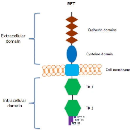
Like every membrane receptor, RET protein (figure 3) is composed of three domains: an extracellular domain, a transmembrane domain and an intracellular domain. The extracellular domain is constituted by four cadherin-like repeats,a calcium binding site and a large cysteine-rich region, responsible to transduction of extracellular signals of proliferation, growth, differentiation, migration, survival and cell apoptosis. The intracellular domain is divided into 2 TK subdomains (TK1 and TK2), separated by 28 amino acids. TK1 and TK2 include tyrosine residues that are phosphorylated during receptor activation, and are involved in the activation of the signaling intracellular pathways [46].
Three isoforms of RET are generated by alternative splicing of the 3’ region which differ in the number of amino acids in the carboxy - terminal tail, these isoforms are RET9, RET43 and RET51, consisting of 1072, 1106 and 1114 amino acids respectively [46]. The two main isoforms in vivo and best-characterized are RET9 and RET51, these isoforms are highly conserved over a broad range of species. Studies suggest that these isoforms have different
10
tissue-specific effects during embryogenesis. For example, mice with only the RET9 isoform are viable and normal, contrary to the monoisoformic RET51 mice that have kidney hypoplasia and lack enteric ganglia in the colon [47].
Figure 3 - Schematic representation of the RET receptor tyrosine kinase. The extracellular domain
includes four cadherin domains and a cysteine rich domain. A single transmembrane region spans the cell membrane. The intracellular domain contains two tyrosine kinase domains (TK1 and TK2). The three RET isoforms (RET9, RET43 and RET51) are indicated.
The main ligands of the RET receptor TK belong to the glial cell line-derived neurotrophic factor (GDNF) family of ligands (GFLs), namely GDNF, neurturin (NRTN), persesephin (PSPN) and artemin (ARTN). The GFLs bind to and activate the RET receptor when bound to the GDNF receptor-alpha (GFRα) family.
Four different GFRα co-receptors have been characterized (GFRα1 to GFRα4), these differ in their specificity for GFLs. The primary ligands for the co-receptors GFRα1, GFRα2, GFRα3, and GFRα4 are GDNF, NRTN, ARTN, and PSPN, respectively. However, signs of cross-talk between ligands and co-receptors have been observed in vitro [48]. Finally the complex GFL-GFRα binds to the extracellular domain of RET and this conjugation leads to auto-phosphorylation of the intracellular tyrosine residues [49].
11
Usually, GFRαs are bound to the plasma membrane; however they can also occur in a soluble form (non-membrane bound). Therefore, RET can be activated in two different forms: cis and
trans (figure 4). In the cis RET activation model the GFL binds to GFRα anchored on a lipid
platform, this complex will promote the dimerization of RET, allowing the phosphorylation of the intracellular tyrosine residues. In the trans RET activation model the GFL binds to the soluble form of GFRα stimulating the dimerization of RET outside the lipid platform, thus allowing tyrosine residues phosphorylation [26]. Independently of the form of activation, once activated, RET begins the different intracellular pathways involving the regulation of many processes such as differentiation, survival, proliferation, migration and cell chemotaxis [26].
Figure 4 - Different mechanisms of ligand-mediated RET activation. A: cis RET activationmodel, the GFL binds to GFRα anchored on a lipid platform, this complex will promote the dimerization of RET and allowing the phosphorylation of the intracellular tyrosine residues. B: trans RET activationmodel, the GFL binds to the soluble form of GFRα stimulating the dimerization of RET outside the lipid platform, thus allowing tyrosine residues phosphorylation
The RET receptor is mainly activated by GFLsand GFRα co-receptors, however other growth factors can activate this receptor. Studies have proved that, through an inter-receptor- kinase-signaling-mechanism independently of ligands or co-receptors, binding of nerve growth
12
factor (NGF) to its receptor TK (NTRK1) modulate the phosphorylation of isoform RET51. This mechanism results in increased growth, metabolism, and gene expression [50].
As NGF activates only RET51 phosphorylation and not the phosphorylation of the other isoforms, it is improbable that NGF would simply modulate the levels of GDNF protein. These results show how growth factors and their receptors engage in cross-talk to form a dynamic network of inter-related trophic signals that guide development. The molecular mechanism of the cross-talk between NTRK1 and RET appears to be indirect, however this mechanism remains unknown [51].
1.4. Genetic alterations in RET
Alterations of the RET proto-oncogene are associated with a variety of disorders, since RET is a proto-oncogene, a single activating mutation in one allele is sufficient to cause neoplastic changes [52]. Loss of function mutations of the RET proto-oncogene results in Hirchsprung disease, a genetic disorder characterized by congenital absence of enteric neurons in the gastrointestinal tract. In contrast, gain of function mutations leading to aberrant RET activation, are involved in a number of human cancers such as MTC [53].
1.4.1. RET and follicular-cell-derived carcinomas
The clinical relevance of RET in human diseases was first recognized in PTC. The chromosomal rearrangement RET/PTC is found in PTC (table 2) and results by fusion of the 3’ portion of
RET with one of several possible partner genes. The RET proto-oncogene is expressed at very
low levels in thyroid follicular cells, however when the TK domain of the RET protein is fused to genes with a promoter that is active in the follicular cells, this causes ligand-independent dimerization of the RET/PTC protein, which leads to chronic stimulation of MAPK signaling in thyroid follicular cells [54].
So far there are about 12 different fusion partner genes that vary according to the identity of the fusion partner of RET [55]. RET/PTC1 (60–70%) is the most common followed by RET/PTC3 (20–30%), which are fused to either CCDC6 or NCOA4, respectively. These prevalent rearrangements result from paracentric inversions on chromosome 10, as fusion partners reside on chromosome 10 [8].
An association between RET/PTC and exposure to external radiation was established, as RET/PTC rearrangements have been found in over 60% of PTCs emerged from the nuclear power plant disaster in Chernobyl on 26 April 1986 [56]. Also patients previously subjected to external irradiation for benign or malignant disease showed a high prevalence of RET/PTC
13
rearrangements [57]. More recent studies showed frequent RET/PTC rearrangements in sporadic PTC from children and adults that were not exposed to irradiation [58]. Regarding FTC, no associations with the RET proto-oncogene have been established.
1.4.2. RET and medullary thyroid carcinomas
Germline mutations of the RET proto-oncogene confer predisposition to hereditary MTC (Table 1). The mutations can be divided into two main groups: (a) Mutations affecting the extracellular domain: These primarily involve cysteine residues 609, 611, 618 and 620 (exon 10), and 634 (exon 11). (b) Mutations affecting the RET-TK domain: These involve codons 768, 790 and 791 (exon 13), 804 (exon 14), 883 and 891 (exon 15), and 918 (exon 16) [39].
About 98% of MEN-2A families have germline mutations of one of the five conserved cysteine residues in exon 10 (codons 609, 611, 618, and 620) or exon 11 (codon 634) in the RET proto-oncogene extracellular domain, this mutations convert a cysteine into another amino acid leading to RET constitutive activation. The mutations affecting codon 634 are the most common in MEN-2A, where the substitution of the cysteine for an arginine represents 50% of all cases. Rarer mutations that are associated with MEN-2A include those at codons 768, 790 and 791 (exon 13), 804 (exon 14), and 891 (exon 15) [39, 59].
In the case of FMTC the majority of the germline mutations are in codons 618 and 620 (exon 10) and a fewer in codons 630, 631 or 634 (exon 11). Recently, mutations in other exons have been associated to FMTC, like exon 13 (codons 768, 790 and 791), exon 14 (codon 804 and 844), and exon 15 (codon 891), which are located in the TK domain, thereby interfering with intracellular ATP binding. Approximately 95% of MEN-2B families have a single mutation that converts methionine to threonine at codon 918 (exon 16), other rare intracellular mutations associated with MEN-2B are in codon 882 (exon 15) [59].
In the case of sporadic MTC, no RET germline mutations are found. However a significant part of these MTCs has a somatic mutation in RET. In these cases the mutation is only present in the tumor and the codon 918 is the most affected, but rare mutations in codons 634 and 883 have also been associated with sporadic MTC [60].
1.5. RET Polymorphisms and Haplotypes in
human diseases
A genetic locus is considered polymorphic when one or more of the rarer alleles have a frequency of at least 1% in a population. Most polymorphisms are silent, that is, do not alter
14
the functional activity of the encoded protein; however there are some polymorphisms that are not neutral.
A set of closely linked genetic markers or polymorphisms that are transmitted through generations as a unit is called a haplotype. When there is correlation or association of a polymorphism or haplotype with a particular phenotype it is possible that the polymorphism or haplotype functions as a genetic modifier and is associated with a small to moderate increase in risk of developing a disease. In addition polymorphisms may also interact with other genetic variants modulating their action. Another important point for the study of polymorphisms is that these are relatively common, so that they can present a much greater risk in a population than rare mutations in genes with high susceptibility to cancer as the RET proto-oncogene [61].
The polymorphisms are divided into: minisatellite and microsatellite sequence polymorphisms and single-nucleotide polymorphisms (SNPs). SNPs represent approximately 90% of all the human genome variations and occur every 100 to 300 bases along the three-billion-base human genome and are the most commonly used genetic markers. A SNP which leads to modification of the amino acid sequence of the protein that is produced is called nonsynonymous SNP. When the SNP does not lead to a change in amino acid sequence, it is designed synonymous SNPs [62]. There are about 11 million SNPs described in the human genome, which are distributed uniformly and, so, these are important biomarkersin screening of complex diseases [63].
Carcinogenesis is a multistep process that occurs through an interaction between many genetic and environmental factors. Thus it is unlikely that the effect of a single polymorphism in this process is substantial. However, it has been shown that an approach based on the combined polymorphisms that interact in the same way can amplify the effect of individual variants and improve the predictive power of polymorphism analysis of complex diseases [64]. Over the last years, many groups have used SNPs of the RET proto-oncogene to determine whether polymorphic variants might represent low penetrance alleles predisposing to RET associated disorders. Several SNPs of this gene have been described in the general population as well as in patients with endocrine tumors, PTC, familial and sporadic MTC and Hirchsprung disease. An issue still unclear is how polymorphisms could have interacting, predisposing, or modifying roles in the pathogenesis of these diseases [65].
1.5.1. RET Polymorphisms and Haplotypes in MTC
Large improvements have been made in the study of the pathogenesis of MTC since the RET proto-oncogene was identified as the susceptibility gene for MTC. But there are many aspects about the MTC, principally about the sporadic form, that remain poorly understood. For
15
example, the heterogeneity at clinical level observed in individuals with the same mutation [26].
In the last few years various studies have been made to understand if the presence of certain
RET polymorphisms are associated or not with the susceptibility for the development or
progression of MTC. In general these studies have described an increased prevalence of the
RET polymorphisms G691S (exon 11, rs1799939), L769L (exon 13, rs1800861), S836S (exon 14,
rs1800862), and S904S (exon 15, rs1800863) in individuals with hereditary or sporadic MTC when compared to the general population.
Nevertheless these results are still controversial, as several studies indicate an involvement of
RET polymorphisms in the development of sporadic MTC, whereas several others failed to
demonstrate an association between these RET polymorphisms and MTC development or progression [26].
1.5.1.1. RET G691S and S904S Polymorphisms
The RET polymorphism G691S (Gly/GGT → Ser/AGT) is a non-synonymous variant present in exon 11 which leads to the alteration of a glycine to a serine in the protein. The RET polymorphism S904S (Ser/TCC → Ser/TCG) is a synonymous variant located in exon 15.
Two large studies, one in an Italian population and the other in a British population, demonstrated that the RET variant G691S is more frequent in MTC patients than in the general population. These two studies hypothesized, through a functional assessment of RET transcription and splicing, that G691S could be the functional variant, but the results were inconclusive [66, 67]. Additionally the Elisei et al. (2004) study described a positive significant co-segregation between G691S and S904S Polymorphisms [66].
The co-segregation between these two polymorphisms in MTC cases were confirmed in a Spanish population by Robledo et al. (2003), reporting a strong linkage disequilibrium. Other conclusions of this study were the higher prevalence of the haplotype G691S/S904S, in homozygosity, in patients with MEN 2A compared to the control group; the authors also observed that this haplotype could modify the age of onset of MTC patients [68]. In the Portuguese population, one study described an over-representation of the G691S polymorphism, particularly in females, in MTC patients with respect to controls, although not reaching the level of significance [69].
Although various authors have demonstrated an association between G691S/S904S polymorphisms and MTC, some other studies did not find a difference in the frequency of these polymorphisms between MTC patients and the general population. These negative results were found and replicated in studies with Polish, Indian and Brazilian populations [62, 70, 71].
16
1.5.1.2. RET L769L Polymorphism
The RET L769L polymorphism (Leu/CTT → Leu/CTG) is a synonymous variant present in exon 13. A study developed by Wiench et al. (2001) described that patients with MTC younger than 30 years presented a higher frequency of the RET L769L variant than those diagnosed between 31 and 45 years,but the absence of a control group decreased the relevance of this finding [72].
Baumgartner-Parzer et al. (2005) described an association between the presence of L769L polymorphism and F791Y mutation in FMTC patients. the authors deduced that the F791Y mutation and L769L polymorphism are located on the same allele and predicted whether the presence of this polymorphism could predispose the respective allele for the occurrence of a F791Y de novo mutation or would modulate the disease phenotype [65].
The RET L769L polymorphism was also associated with an increased risk for MTC whereas patients homozygous for the minor allele of this variant were younger at the MTC diagnosis [62]. On the other hand, several studies in distinct populations failed to demonstrate an association between this polymorphism and MTC [70, 71, 73].
1.5.1.3. RET S836S Polymorphism
The RET S836S polymorphism is a neutral variant and is located in exon 14. Gim et al. (1999) developed a study that reported a higher frequency of this variant minor allele in MTC patients comparing to a control group, Ruiz et al. (2001) obtained similar data in Spanish population [74, 75]. Another interesting finding on Gim et al. (1999) study was that eight of nine patients (89%) with the minor allele of the S836S polymorphism also had a somatic mutation in the M918T [75]. A large study in a Brazilian population associated the variant S836S with early onset of sporadic and hereditary MTC and also with the formation of lymph nodes and distant metastases [70].
In spite of this, other association studies failed to demonstrate any differences in the presence of this polymorphic allele between MTC patients and controls. For example the studies in Polish, French and Indian populations demonstrated a similar frequency in the MTC patients and in the controls groups [62, 71, 73].
1.5.2. RET Polymorphisms and Haplotypes in DTC
So far, only a few studies have evaluated the association between RET SNPs and DTC. As in MTC, in DTC, polymorphic variations in the DNA sequence of RET might modify expression of the RET protein and/or lead to activation of the gene, so it is expected that much still remains to be discovered [76].
17
Lesueur et al. (2002) conducted an association study with PTC cases and controls from four countries (France, Portugal, Italy and Australia) matched for sex, age, and population. The four RET SNPs studied were A45A in exon 2, L769L in exon 13, S836S in exon 14, and S904S in exon 15 and, also, a total of 10 haplotypes. The authors concluded that some RET polymorphic variants and some specific haplotypes could have low penetrant alleles for the PTC phenotype. The strongest association with PTC was found for A45A and L769L polymorphisms, moreover a specific haplotype, designed GGCC haplotype, might actas a low penetrance predisposing allele for PTC in the Italian and French populations [76].
Other studies demonstrated a weak association with PTC and the SNPs A432A, S836S, G691S, and S904S. Ho et al. (2005) described an association with DTC for A432A and S836S polymorphisms [14]. Stephens et al. (2005) investigated loss of heterozygosity (LOH) for three
RET SNPs (G691S, S904S, and L769L) in patients from Ukraine and Belarus that developed PTC
due to exposition to radioactive fallout following the Chernobyl nuclear accident and in a control group. The results showed an association with PTC for G691S and S904S [77].
Taking into account all these studies, the dimension of the effect among RET SNPs and/or haplotypes and PTC is only slightly significant. This association should be confirmed on larger samples.
1.6. Aims of the present study
Polymorphisms of the RET proto-oncogene have been studied in different populations for association with susceptibility to thyroid cancer, but with inconsistent findings mainly in thyroid carcinomas derived from follicular cells. There are multiple approaches to identify susceptibility genes and biological mechanisms in a complex disease. Association studies, one of the most used methods, seek to identify genetic variants that are associated with pathology, comparing affected individuals with a control population.
To clarify the contribution of RET polymorphisms to genetic susceptibility to DTC among Portuguese patients, we conducted an association case–control study by analyzing four well-characterized RET polymorphisms (G691S, L769L, S836S and S904S). This general goal can be divided into three points: i) to determine the prevalence of these RET polymorphisms in the Portuguese population; ii) to determine the association of these RET polymorphisms and haplotypes with DTC; iii) to define the association of these RET polymorphisms and haplotypes with clinical parameters (cancer subtype, gender, age at diagnosis and tumor size at diagnosis).
18
19
2.1. Studied population
The study group consisted of 282 Caucasian Portuguese patients with thyroid cancer (47 males and 234 females; mean age ± SD, 48.5 ± 14.8 years, median 49) from the Instituto Português
de Oncologia de Coimbra. The control group consisted of 245 (100 males and 145 females;
mean age ± SD, 32.0 ± 14.3 years, median 25) Caucasian Portuguese unrelated volunteers who were recruited among faculty staff or blood donors of the Instituto Português de Sangue da
Região Centro, with no history of thyroid cancer. This study was approved by local research
ethics committee.
2.2. Genotyping
2.2.1. DNA Extraction
A total of 10 mL of venous blood was collected, in EDTA tubes, from each individual. Genomic DNA was extracted from peripheral blood leucocytes collected and stored at 4ºC until manipulation. The DNA extraction procedure was based on the “salting-out” method, described by Miller [78].
The whole blood sample was transferred to a 50mL falcon tube and the final volume was completed until 45mL with Red Blood Cell (RBC) lysis buffer (155 mM NH4Cl; 20 mM KHCO3; 0.1 mM Na2EDTA; pH 7.4). The tubes were vortexed and incubated on ice for 15 minutes, with frequent inversions. After this incubation period, the tubes were centrifugated (AllegraTM X-22R Centrifuge, Beckman Coulter, USA) at 2500 rpm for 10 minutes at 4ºC, then, the supernatant was discarded, leaving the leucocytes intact.
Once the leucocytes were isolated, the pellet was completely resuspended and incubated with 5mL of Secundary Extraction buffer (75 mM NaCl; 25 mM Na2EDTA; pH 8.0), 12.5 μL proteinase K (20 mg/ml) and 500 μL SDS 10%. The digestion was left overnight at 55ºC. To precipitate the proteins 3 mL of saturated NaCl (6M)were added followed by a 10 minutes incubation at 55ºC, tubes were vortexed and centrifuged at 4000 rpm for 30 minutes at 15ºC. The supernatant was decanted to a new 50 mL falcon tube, avoiding foam. To perform the DNA precipitation, cold 100% ethanolwas added at about 2x the volume of the supernatant, tubes were inverted gently about 50 times observing the formation of the DNA precipitate and centrifuged at 4500 rpm for 5 minutes at 4ºC, the supernatant was decanted and discarded. The DNA pellet was washed with 10 ml of cold 70% ethanol and centrifuged at 4500 rpm for 5
20
minutes at 4ºC, finally was transferred into a 1.5 mL microfuge tube and allow to air dry for about 30 minutes. DNA was hydrated with 1mL of Tris-EDTA buffer and left in slow and constant stirring (Programmable Rotator Mixer, Star Lab) overnight at room temperature. When the blood sample was insufficient to implement the protocol described above, the DNA was extracted using the Puregene Blood Core Kit C (Qiagen), according to the manufacturer’s instructions.
2.2.2. DNA quantification
Accurate analysis of the DNA preparation may be impeded by the presence of impurities in the sample or if the amount of DNA is too little or too high, so it is necessary to quantify the samples.
DNA quantity and quality was assessed by spectrophotometric determination (NanophotometerTM, Implen, Germany). Analysis of UV absorption by the nucleotides provides
a simple and accurate estimation of the concentration of nucleic acids in a sample. Purines and pyrmidines in nucleic acid show absorption maxima around 260nm (dATP: 259nm; dCTP: 272nm; dTTP: 247nm) if the DNA sample is pure without significant contamination from proteins or other components. To assess the purity of the samples, the ratio of OD260/OD280
was determined. A ratio between 1.8-2.0 denotes that the absorption in the UV range is due to nucleic acids. A ratio lower than 1.8 indicates the presence of proteins and/or other UV absorbers. A ratio higher than 2.0 indicates that the samples may be contaminated by RNA. After DNA quantification all samples were diluted to the same concentration (100 ng/µL).
2.2.3. Polymerase Chain Reaction
The Polymerase Chain Reaction (PCR) was developed by Kary Mullis in 1985 and is used to exponentially amplify a specific DNA fragment, in vitro [79].
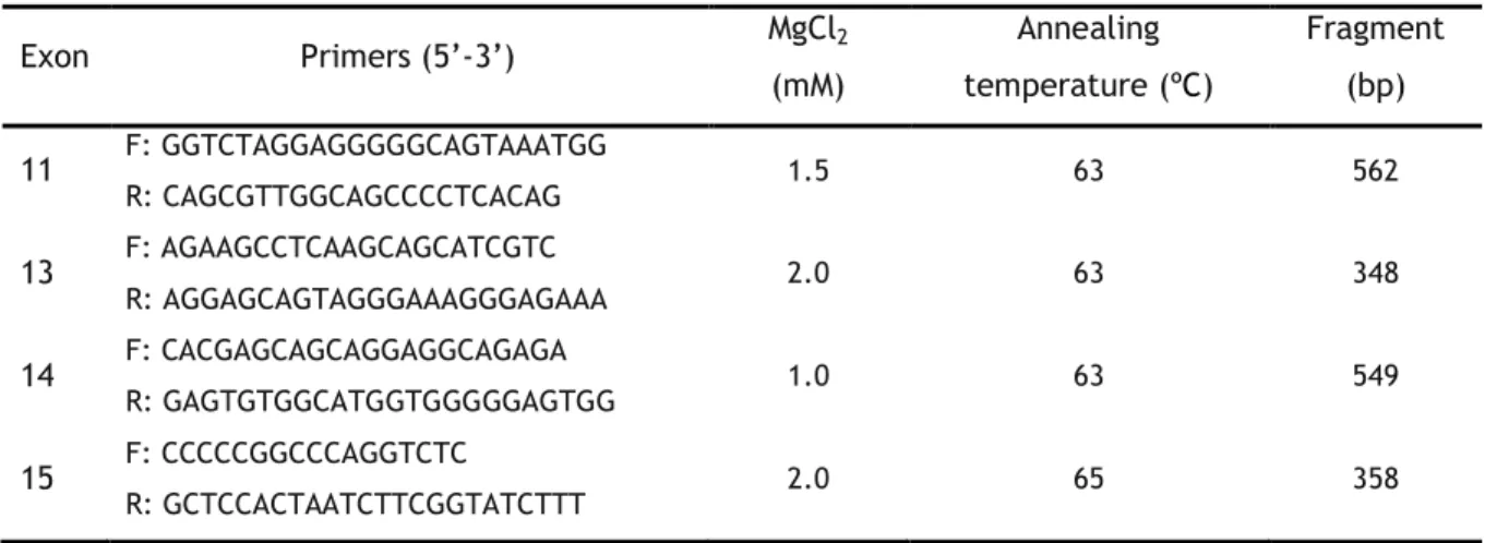
The amplification of RET exons 11, 13, 14 and 15 was conducted using specific primers and optimized PCR conditions (see table 2). The PCR reactions of RET exons 11, 13 and 14 occurred in a final volume of 25 µL: 1X buffer (20 mM Tris HCL; pH 7.5; 100 mM NaCl; 0.1 mM EDTA; 1mM dithiothreitol; 50% (v/v) glycerol), 0.2 mM of dNTPs, 0.2 µM of each primer (forward and reverse), optimized concentration of MgCl2 (Table 2), 1 U of NZYTaq (NZYtech
Lda., Lisbon, Portugal) and 100-300 ng of genomic DNA. The amplification of exon 15 was performed using Supreme NZYTaq 2X Green Master Mix with 2,5 U of Taq (NZYtech Lda., Lisbon, Portugal), optimized concentration of MgCl2 (table 2) and 100–300 ng of genomic DNA.
The PCR reaction occurred in a thermal cycler (T100TM Thermal Cycler, Bio Rad) and the amplification thermal cycling profile was: initial denaturation at 95ºC for 5 minutes, 35 cycles of denaturation of 30 sec at 95ºC, annealing for 30 sec at a particular temperature dependent
21
on the primer sequences (Table 2), and an extension for 30 sec at 72ºC. This was followed by a final extension step of 7 minutes at 72ºC. Negative and positive controls were included in each amplification analysis.
Table 2 - Primers and PCR conditions for amplification of gene RET exons 11, 13, 14 and 15.
Exon Primers (5’-3’) MgCl2 (mM) Annealing temperature (ºC) Fragment (bp) 11 F: GGTCTAGGAGGGGGCAGTAAATGG R: CAGCGTTGGCAGCCCCTCACAG 1.5 63 562 13 F: AGAAGCCTCAAGCAGCATCGTC R: AGGAGCAGTAGGGAAAGGGAGAAA 2.0 63 348 14 F: CACGAGCAGCAGGAGGCAGAGA R: GAGTGTGGCATGGTGGGGGAGTGG 1.0 63 549 15 F: CCCCCGGCCCAGGTCTC R: GCTCCACTAATCTTCGGTATCTTT 2.0 65 358
2.2.4. Enzymatic digestion
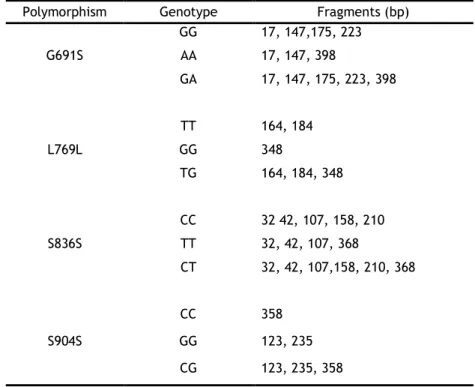
The amplified PCR products were subjected to enzymatic digestion because all the polymorphisms create or abolish a restriction site for a specific endonuclease. The BanI, TaqI,
AluI, and RsaI enzymes (New England Biolabs, Beverly, MA, USA) were used to study the
G691S, L769L, S836S, and S904S polymorphisms, respectively.
BanI was used to distinguish the RET exon 11 G691S polymorphism in which the homozygous
common GG genotype produced 4 fragments (17, 147, 175 and 223, bp). The homozygous AA genotype abolish one BanI restriction site, so 3 fragments (17, 147, 398 bp) were produced and the heterozygous GA genotype resulted in 5 fragments (17, 147, 175, 223, 398 bp) (table 3). Digestion of 10 µL of PCR product was performed in 15 µL reaction volume using 5 units of
BanI, 1X NEbuffer 4 (50 mM potassium acetate; 20 mM Tris-acetate; 10 mM magnesium
acetate; 1 mM dithiothreitol; pH 7.9) and 100 µg/mL of BSA supplied with the restriction enzyme at 37ºC overnight.
TaqI enzyme was used in order to identify the RET exon 13 L769L polymorphism. The
homozygous common TT genotype produced 2 fragments (164, 184 bp). The homozygous GG genotype abolishes the TaqI restriction site avoiding the digestion, remaining the fragment intact. Finally, the heterozygous TG genotype produced 3 fragments (164,184, and 348 bp) (table 3). Digestion of 10 µL PCR product was performed in 15 µL reaction volume using 5 units of TaqI, 1X NEbuffer 4 (50 mM potassium acetate; 20 mM Tris-acetate; 10 mM magnesium acetate; 1 mM dithiothreitol; pH 7.9) and 100 µg/mL of BSA supplied with the restriction enzyme, at 65ºC overnight.