.
CONSTITUTIVE OGG1 VARIANT TOGETHER
WITH BRCA MUTATIONS DISPLAY
ACCELERATED TELOMERE SHORTENING
Sofia Maria Morgadinho Ferreira
Dissertation for the Master Degree in Oncobiology
Thesis supervisors
Dr. Ana Osório
Dr. Ana-Teresa Maia (UAlg)
.
CONSTITUTIVE OGG1 VARIANT TOGETHER
WITH BRCA MUTATIONS DISPLAY
ACCELERATED TELOMERE SHORTENING
Sofia Maria Morgadinho Ferreira
Dissertation for the Master Degree in Oncobiology
Thesis supervisors
Dr. Ana Osório
Dr. Ana-Teresa Maia (UAlg)
Constitutive OGG1 variant together with BRCA mutations
display accelerated telomere shortening
Declaração de autoria de trabalho
Setembro de 2015
Declaro ser a autora deste trabalho, que é original e inédito. Autores e
trabalhos consultados estão devidamente citados no texto e constam da
listagem de referências incluídas
_______________________________________
Copyright
Setembro de 2015
Eu, Sofia Maria Morgadinho Ferreira, na qualidade de estudante (nº
42291) do mestrado Oncobiologia da Universidade do Algarve, declaro que
não autorizo a divulgação dos dados contidos na Tese intitulada
“Constitutive OGG1 variant together with BRCA mutations display
accelarated telomere shortening".
_________________________________________
This thesis, submitted for the degree of Master at the University of Algarve, has been elaborated in the Human Cancer Genetics laboratory at the Spanish National Cancer Research Center (CNIO). The student was supported by the Exchange Programme
Scholarship Erasmus +
Erasmus plus Exchange Programme Funding Human Genetics Group
Human Cancer Genetics Programme
To my dearest and beloved family
Para a minha querida e amada família
Page | vi
This thesis would have taken far longer without the contribution of many others. Here, I express my immensurable appreciation and deepest gratitude to the people who contributed somehow for that.
My foremost thanks and gratitude go to my supervisors, Dr. Ana Osório and
Dr. Ana-Teresa Maia for their extraordinary supervision, guidance since very early
stages of my stay in CNIO and for their input in this thesis. From them I received the most valuable comments, constructive critics and knowledge that I will bring with me to my future career. My particular acknowledgement to the extreme kindness and very useful advices of Dr. Ana Osório, and to Dr. Ana-Teresa Maia, who since the very beginning have been a source of inspiration to pursue a scientific, and always found a time in her very thigh schedule to give her assistance.
I gratefully acknowledge Dr. Javier Benítez, Director of the Human Cancer Genetics Programme, to have hosted me in his Programme to perform my Master Research Project and for his close supervision of my work outcomes.
Thanks to Carlos Benítez, who made a close but relaxed supervision, which easily contributed to a positive work environment.
Thanks to my Spanish laboratory mates, Ali, Javi, Bea, Nere and Carlos included for their contagious joviality and good disposition, that always contributed to a very light and pleasant environment in my daily work. A particular thank to Laura for her kindness, support and active interest in my welfare, to Oriol for making my work time even more pleasant with his very bright and tasteful musical suggestions, and to Maria whit whom I got precious recommendations.
My thanks to Irene, who helped me to deal with the undesirable bureaucracies always with a great smile and good disposition.
A very sincere thanks to my dear Ale, for being there in the most crucial times, for her immense support and without her days would have passed more slowly. To
Tere, for being the first and more compassionate one receiving in the laboratory, and for
all much needed lucid and mature advices. To both, for encouraging me to fight for more and better and for providing such rich source of conversation and amusement.
A very special thanks with all my heart to Sílvia, aquela míuda que curto tótil, for her sensibility and determination, for her advices and her sharing, for her care and concern, without whom the learning curve would be steeper. Also, for making my stay in Madrid far more pleasant and familiar
My sincere thanks to Martim, whom is a great classmate and always supported and encouraged me to go abroad. Without his very inspirational words I would never had this opportunity to do my Master thesis project in such a great institute.
A special thanks to my extraordinary friends Lilocas and Marta for their helpful “informatic” support and obviously to their friendship.
Page | vii
Words fail me to express my deepest appreciation to my boyfriend, who made everything worth it and always made me believe, being the greatest source of encouragement, wisdom and love.
And above all, I offer my utmost gratitude to all my Family, without whom this thesis would have been possible. I will be eternally indebted to them, for their immeasurable love and endless support.
Page | ix
Osorio, Milne et al. (2014) reported a SNP, rs2304277, in the OGG1 gene, with evidence of potential association with increased ovarian cancer risk in BRCA1 germline mutation carriers (p=4.8x10⁻³). The protein OGG1 is a main player in the DNA BER pathway, responsible for recognizing and excising oxidized guanines (8-oxoG).
In the literature, oxidative stress has been well characterized to have a natural site-specific preference for guanines, in which telomeric DNA is enriched due to several TTAGGG repeats. 8-oxoG is highly mutagenic and affects DNA replication, and at the telomere level it can impair the recruitment and affinity of the shelterin complex proteins. This complex caps and protects telomeres from aberrant chromosomal rearrangements. Not only, when not properly corrected, 8-oxoG can give rise to ssDNA breaks. Therefore, telomeres are more susceptible to this kind of damage, and disruption of the normal telomere function and length can result in carcinogenesis, as a consequence of genomic instability. Hence, given the role of OGG1 and telomeres composition, we aimed to explore whether the increase of cancer risk in the carriers of rs2304277 (together with a germline mutation in the BRCA gene), might be due to an altered OGG1 function, which could accelerate the telomere shortening, resulting from a weaker response upon oxidative stress harms.
For the functional characterization of the polymorphism, the experimental approach was based on the evaluation of the OGG1 mRNA expression levels by qPCR and measurement of telomeres length by High Throughput Q-FISH of the different
OGG1 genotypes and BRCA1/2 mutation status from peripheral blood samples. Very
preliminary results, suggest that the variant leads to a decrease in OGG1 mRNA levels which, and together with a mutation in BRCA gene might contribute to an accelerated telomere shortening. These results might explain, in part, the increase of an individual’s lifetime risk of developing cancer when harbouring both genetic conditions.
Page | xi
Recentemente, Osorio, Milne et al. (2014) realizaram uma análise compreensiva de dezoito genes que envolvem a via de reparação de DNA Base
Excision Repair (BER), numa ampla série de portadores de uma mutação na linha
germinal em um dos genes de elevada penetrância BRCA1 ou BRCA2 provenientes do consórcio CIMBA (Consortium of Investigators of Modifiers of BRCA1 and BRCA2). A escolhida via de reparação de ADN, BER, foi feita com base no conceito de letalidade sintética entre membros da via BER, como PARP1 (Poly ADP ribose polymerase), BRCA1 e BRCA2.
Desses 18 genes escolhidos, foram identificados onze SNPs (polimorfismos de base única), com evidências de associação com o aumento de risco para o desenvolvimento de cancro da mama e/ou dos ovários. Desses onze SNPs, cinco localizam-se em genes de ADN glicosilases, e aqueles com mais fortes evidências de associação ao risco de desenvolver os referidos cancros, localizam-se em dois genes de ADN glicosilases: rs1466785 no gene NEIL2 (endonuclease VIII-like2) (HR: 1.09, 95% CI (1.03–1.16), p= 2.761023) para o cancro da mama em portadores de uma mutação no BRCA2, e rs2304277 no gene OGG1 (8-guanine DNA glycosylase) (HR: 1.12 95%CI: 1.03–1.21, p= 4.861023) para o cancro dos ovários em portadores de uma mutação no gene BRCA1. Este estudo focou-se e contribuiu para a caracterização funcional do polimorfismo rs2304277 no gene OGG1, de forma a estabelecer uma possível explicação para a sua associação com o aumento de risco de desenvolver cancro, mais concretamente cancro do ovário em portadores de uma mutação na linha germinal no gene BRCA1.
8-oxoguanina DNA glicosilase, OGG1, é uma enzima com um papel fundamental nas etapas primárias de ação da via de reparação BER, sendo responsável por fazer o reconhecimento e excisão de bases quimicamente alteradas que perturbam minimamente a hélix do ADN, no caso concreto desta ADN glicosilase, as guaninas oxidadas: 8-oxodeoxyguanosina (8-oxoG). Na literatura, está muito bem descrita e caracterizada uma tendência natural do stress oxidativo sobre as guaninas, que torna o ADN telomérico mais suscetível e sensível a este tipo de agressão, uma
Page | xii
vez que são estruturas enriquecidas em guaninas, devido às suas várias repetições da sequência “TTAGGG”.
8-oxoG é o produto mais comumente gerado por stress oxidativo e é altamente mutagénico pois possuí uma grande propensão em emparelhar com adeninas, podendo conduzir a transversões GC para TA durante a replicação do ADN. Embora essa transversão possa ser não-sinónima, alterando o produto final da proteína e, num cenário mais drástico, a sua função, a substituição de guaninas por timinas diminui o recrutamento e afinidade das proteínas que cobrem e protegem fisicamente os telómeros. O conjunto destas proteínas forma um complexo denominado de shelterin.
As proteínas do complexo shelterin impedem o reconhecimento das extremidades cromossomais, conhecidas por telómeros, como quebras intra-cromossomais formando uma configuração fechada destas estruturas. Desta forma, os mecanismos de reparação de ADN danificado não são ativados e, consequentemente, os telómeros não são sujeitos a fusões cromossómicas aberrantes, o que levaria à instabilidade cromossómica. Na literatura, está muito bem descrito que telómeros criticamente curtos podem funcionar como um promotor da carcinogénese devido à sua subsequente instabilidade cromossómica.
No entanto, este não representa o único problema relativo às guaninas oxidadas. Algumas ADN polimerases, as de maior fidelidade, poderão encontrar dificuldades na replicação de ADN na presença de 8-oxoG, uma vez que guaninas oxidadas conduzem a uma ligeira modificação na cadeia de ADN. Comprometendo assim a replicação normal do ADN e possivelmente a dos telómeros, podendo contribuir ainda para um encurtamento dos telómeros mais acelerado. A disrupção do tamanho normal dos telómeros e da sua normal função pode resultar em carcinogénese, como consequência da instabilidade genómica advinda de telómeros curtos que acabam por perder a proteção do complexo shelterin. Telómeros criticamente curtos têm sido também descritos como promotores de tumores. As quebras de ADN, mais especificamente em apenas em uma das cadeias (ssDNA) podem também acontecer quando a intervenção da via BER não é corretamente executada ou concluída. Os sítios abásicos provocados pela intervenção BER
Page | xiii
fragilizam o ADN facilitando a sua quebra. Quebras de ADN de cadeia simples são assim as maiores responsáveis pelo encurtamento acelerado de telómeros, segundo von Zglinicki, Pilger et al. (2000).
ADN oxidado é a maior fonte de danos de ADN, e o stress oxidativo poderá vir tanto de fontes endógenas (espécies reactivas de oxigénio provindas do metabolismo celualr) como exógenas (elementos patogénicos ou radiação ionizante). O que propencia a tumourigénese é o desequilíbrio entre as espécies reativas de oxigénio (ROS) e as defesas antioxidantes, tal como OGG1. Resumidamente, os telómeros são estruturas mais suscetíveis à oxidação, devido ao seu conteúdo rico em guaninas que são entre as bases, as preferencialmente oxidadas. As consequências de 8-oxoG no ADN telomérico podem promover uma disrupção no tamanho e função dos telómeros conduzindo a um encurtamento acelerado e crítico dos telómeros, destabilizando a homeostase cromossómica levando à génese tumoral.
Portanto, dado o papel de OGG1 no reconhecimento e correção das 8-oxoG e a composição enriquecida dos telómeros em guaninas, procurámos explorar se o aumento de risco de desenvolver cancro nos indivíduos portadores do polimorfismo rs2304277 no gene OGG1 e uma mutação nos genes BRCA1/2 se deve a uma perturbação na eficácia da ação da enzima OGG1, que poderá favorecer um encurtamento de telómeros mais acelerado, como resultado de uma resposta alterada às consequências provocadas pelo stress oxidativo.
Para a caracterização funcional do polimorfismo, a abordagem experimental foi feita com base na avaliação dos níveis de expressão de mRNA de OGG1 por qPCR e medição do tamanho dos telómeros (TL) por High Troughput Q-FISH para os diferentes genótipos de OGG1 (wild type ou portadores da variante) e BRCA status de amostras de sangue periférico. Resultados muito perliminares, sugerem que a variante possa levar à diminuição dos níveis de mRNA de OGG1 que, juntamente com uma mutação no gene BRCA contribuí a um encurtamento dos telomeros mais acelarado, o que poderá explicar em parte, o aumento de risco de desenvolver cancro em pessoas que abrigam ambas as condições genéticas.
Page | xiv
Sendo o cancro uma doença poligénica, são então importantes estudos como este, feitos com base na descoberta e caracterização de variantes genéticas que possam contribuir para o desenvolvimento de cancro, contribuindo para uma melhor avaliação de risco da população saudável.
FIGURES
Figure 3.1 T-loop structure 9
Figure 3.2 Structure of human telomeres 10
Figure 3.3 Telomere length dynamics in germ, stem, somatic and tumour
cells 12
Figure 3.4 Critically short telomeres can undergo into aberrant fusions 12
Figure 3.5 Oxidative stress effects over telomeres 14
Figure 3.6 ROS effect in cells and its role in cancer development 14
Figure 3.7 Scheme of Base Excision Repair pathway action mode 15
Figure 3.8 Structural representation of hOGG1 17
Figure 3.9 Hallmarks of cancer 19
Figure 3.10 Estimate incidence and mortality rates of the different
cancers 20
Figure 3.11 Pie with the representative percentages of the different
stages of breast cancer at the time of diagnosis 20
Figure 3.12 Percentage of ovarian cancer diagnosed and five-year
relative survival by stage 21
Figure 3.13 Genealogic tree of Paulo Broca’s wife’s family (1788-1856) 25
Figure 3.14 Timeline of hunt for the genetic underpinnings of the familial
breast cancer 26
Figure 3.15 Pie with the representative percentages of familial breast
cancer mutations in low-, moderate-, high-penetrance genes, BRCAX and related syndromes
Figure 3.16 Some BRCA1 and BRCA2 HR-mediated functions at
different stages of cell division
29
Figure 3.17 Genome-wide association studies are effective in detecting
common alleles with low penetrance in a disease
31
Figure 6.18 Carriers of the rs2304277 minor allele (A) show a decreased OGG1 mRNA expression levels in all groups
46
Figure 6.19 Carriers of the rs2304277 minor allele (A) show lower
relative OGG1 mRNA expression compared with the non-carriers 46
Figure 6.20 Telomere shortening rate during lifetime in FBOC series and
Controls 48
Figure 6.21 Carriers of the rs2304277 minor allele (A) harbouring a
mutation in the BRCA1/2 gene show significant shorter telomeres 49
TABLES
Table 1 Risk factor and its relative risk for breast cancer in women 23
Table 2 Oligonucleotide primers used for PCR 38
Table 3 Oligonucleotide primers used for qPCR 40
Table 4 Number and relative frequencies of the different series within the
1. ABBREVIATIONS 1 2. GLOSSARY 5 3. INTRODUCTION 8 3.1. Telomeres 9 3.1.1. Shelterin Complex 10 3.1.2. Telomere shortening 10
3.1.2.1. Oxidative stress and ts contribute to telomere crisis 12
3.2. Base excision repair pathway 15
3.2.1. 8-oxoguanine DNA glycosylase, OGG1 16
3.3. Cancer 17
3.4. Breast and Ovarian cancer 19
3.4.1. Epidemiology 19
3.4.2. Risk factors 21
3.4.3. Familial breast and ovarian cancer (FBOC) syndrome 24
3.4.3.1. Breast cancer susceptibility genes 26
3.5. Breast Cancer Susceptibility genes, BRCA 28
3.5.1. Function as homologous recombination DNA repair members 28
3.5.2. Functional involvement in telomere maintenance 29
3.6. Genome-wide association studies, GWAS 30
3.6.1. TagSNP for association studies 30
3.6.2. SNPs in DNA glycosylases involved in the BER pathway 31
3.6.2.1. SNP in OGG1 associated with breast and ovarian cancer
risk 32
4. OBJECTIVES 34
5. MATERIAL & METHODS 36
5.1. Samples 37
5.2. Genotyping 37
5.2.1. DNA extraction 38
5.2.2. PCR – DNA amplification 38
5.3. RNA expression analysis 39
5.3.1. RNA extraction 39
5.3.2. Reverse transcription PCR (RT-PCR) 39
5.3.3. Quantitative PCR (qPCR) 39
5.4. Telomere Length measurement 40
5.4.1. High throughput Q-FISH 40
5.5. Statistical analysis 41
6. RESULTS 42
6.1. Different genotypes for rs2304277 in the population 43
6.2. Levels of OGG1 mRNA is reduced in minor alleles carriers of the
rs2304277 variant 43
6.3. Accelerated telomere shortening in individuals who carry the minor
allele and a BRCA1/2 mutation 46
7. DISCUSSION 50
7.1. OGG1 mRNA expression levels 51
7.2. Telomere Length 52
7.3. Advantages and disadvantages of using peripheral blood 53
8. CONCLUSIONS 55
9. BIBLIOGRAPHY 58
Page | 2
Abbreviations are displayed in alphabetic order
8-oxoG 8-oxodeoxyguanosine
53BP1 53 binding protein 1
A
A Adenine
AP Apurinic/apyrimidinic
APE Apurinic or apyrimidinic endonuclease
B
BER Base excision repair
BRCA1 Breast cancer susceptibility gene 1
BRCA2 Breast cancer susceptibility gene 2
BRCAX Breast cancer susceptibility gene X
C CIMBA
Consortium of investigators of modifiers of BRCA1 and BRCA2
D
DDR DNA damage response
DHPLC High-performance liquid chromatography
dsDNA Double-stranded DNA
F
F Forward
FBC Familial breast cancer
FBOC Familial breast and ovarian cancer
G
G Guanine
Page | 3
H HR Homologous recombination
L LD Linkage disequilibrium
M
miRNA MicroRNA; micro ribonucleic acid
MRN Mre11-Rad50-Nbs1 complex
mRNA Messenger ribonucleic acid
N
NEIL2 Endonuclease VIII-like2
NHEJ Non-homologous end joining
O OGG1 8-oxoguanine DNA glycosylase
P
PARP1 Poly ADP ribose polymerase
PCR Polymerase Chain Reaction
POT1 Protection of telomeres 1
Q
Q-FISH Quantitative Fluorescence in situ hybridization
qPCR Quantitative Polymerase Chain Reaction
R
R Reverse
RAP1 Repressor and activator protein 1
ROS Reactive oxygen species
RT-PCR Reverse Transcription Polymerase Chain Reaction
S
SNP Single-nucleotide polymorphism
Page | 4
T
TIN2 TRF1-interacting protein 2
TL Telomere length
TRF1 Telomeric repeat-binding factor 1
TRF2 Telomeric repeat-binding factor 2
TPP1 POT1- and TIN2 interacting protein
U UTR Untranslated region
Page | 6
Concepts are displayed in alphabetic order
C Causal SNP The SNP which is affecting a trait/phenotype/disease
F
Fine Mapping Pos-genotyping analysis with greater resolution that
searches for the causal SNP at a susceptibility locus
Five-year survival rate
Percentage of patients who are alive at least five years after the diagnose of their cancer
Five-year relative survival rate
A more accurate prognosis with a percentage of a group of patients with a certain type and stage of cancer, who are alive at least five years after the diagnose of their cancer
H
Haplotype A set of alleles in LD inherited in a dependent way, as a
unit
HapMap Project
Catalogue that aimed to map the haplotypes of the human genome, describing what there variants are and how they are distributed among populations
I Incidence Number of new cases of a specific disease occurring
during a certain period in a population
L Linkage
Disequilibrium
Correlation of neighbouring alleles at different loci, that are inherited dependently, reflected in haplotypes. In other words, individuals who carry a specific SNP are often predictably carrying specific nearby alleles that are in LD
M
Major Allele Represents the commonest allele in a given population Minor Allele Represents the least common allele in a given population
Page | 7
Polygenic Model Describes a trait/phenotype/disease which is influenced
not by a single gene but for more
Prevalence Total number of cases with a specific disease in a
population at a certain period
R Risk Factors
Anything that might affect the chance of an individual of getting the disease
S
Synthetic Lethality Combination of perturbations in at least two genes/proteins
leads to cellular death
SNP
DNA sequence variation occurring in at least 1% of the population. Polymorphic site which is diallelic, having a major allele and a minor allele
Susceptibility Likelihood to be influenced or harmed by a particular
condition
T TagSNP
Representative SNP in a determined region of the genome with high LD (r2 ≥ 0.8) with a group of other SNPs:
Page | 9
3.1. Telomeres
Derived from the Greek words telos and meros meaning “end” and “part”, respectively, telomeres are the structures that caps the chromosome ends representing a crucial role in the genome integrity maintenance (Aubert and Lansdorp 2008). Discovered and described for the first time in the early 1930’s by Herman Muller and Barbara McClintock (Mason and Perdigones 2013).
These eukaryotic specialized nucleoproteins structures prevent abnormal chromosomal fusion or rearrangements shielding the natural ends of linear chromosomes that resemble DNA breaks (Rhodes and Giraldo 1995, O'Sullivan and Karlseder 2010). Since linear DNA fragments are toxic to mammalian cells, enzymes that degrade or repair those fragments are activated, and inappropriate repair can lead to genomic instability and, eventually, carcinogenesis (O'Sullivan and Karlseder 2010). Hanahan and Weinberg (2011) include genome instability in the ten cancer hallmarks. Telomeres are therefore vital structures for maintenance of genomic integrity.
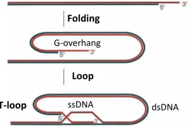
Mammalian telomeres are made of long stretches of double-stranded DNA TTAGGG repeats (9-15kb in humans) and at the 3’ end are 50-300 nucleotide single-stranded repeats, the so-called G-overhang (Figure 3.1 and Figure 3.2, left) (O'Sullivan and Karlseder 2010). Telomeric-loop, also referred as T-loop, is the end structure in telomeres that works as a protective cap masking the natural ends of the telomeres from the DNA damage response (DDR) machinery, through a closed configuration (O'Sullivan and Karlseder 2010).
Loop
ssDNA dsDNA
T-loop
G-overhang
Folding
Figure 3.1 T-loop structure. The G-overhang (ssDNA) bends into the double-stranded DNA (dsDNA) telomeric repeat array. Adapted from de Lange (2005)
Page | 10
3.1.1. SHELTERIN COMPLEX
Besides this T-loop configuration, the distinction of telomeres from intra-chromosomal breaks is also ensured by the recruitment of the shelterin complex protein to the TTAGGG telomeric repeats. The shelterin complex has a wide and vital role in the telomere length regulation and protection (Figure 3.2, right) (Liu, O'Connor et al. 2004, de Lange 2005, O'Sullivan and Karlseder 2010). These proteins cap the chromosome ends hiding them from the surveillance of the DDR, avoiding an inappropriate action from the DNA repair pathways, which would process telomeres ends as intra-chromosomal breaks and start fusing them (de Lange 2005). Shelterin proteins also have a part in the intracellular signalling for cell proliferation, DNA repair and recombination regulation (Xin, Liu et al. 2008).
This complex consists of six proteins: telomeric repeat-biding factor 1 and 2 (TRF1 and TRF2); repressor and activator protein 1 (RAP1); TRF1-interacting protein 2 (TIN2); protection of telomeres (POT1) and POT1- and TIN2 interacting protein (TPP1) (Figure 3.2, right) (Xin, Liu et al. 2008, O'Sullivan and Karlseder 2010). TRF1, TRF2 and POT1 recognize and bind to TTAGGG repeats and interrelate with the rest of the shelterin proteins: TIN2 TPP1 and RAP1, allowing cells to differentiate telomeres from DNA damaged sites (de Lange 2005).
3.1.2. TELOMERE SHORTENING
Losing around 100-200 bp in every division (Aubert and Lansdorp 2008), somatic cells can only undergo a limited number of divisions before telomeres become
Figure 3.2 Structure of human telomeres consists in many kilobases of TTAGGG repeats that extend in the 3’ direction, forming the G-overhang strand (left image). Shelterin complex (TRF1, TRF2, RAP1, TIN2, POT1 and TPP1) covers the double- and single-strand telomeric DNA, protecting these structures (right image). Adapted from O'Sullivan and Karlseder (2010).
Page | 11
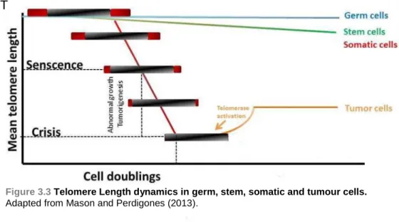
dysfunctional (< 3Kb) and establish replicative senescence (Figure 3.3). Telomeres are therefore vital to preserve genetic information and integrity in each cell division (Sun, Tan et al. 2015)
Telomerase is a ribonucleoprotein, which binds to the first few nucleotides in the 3’ end of telomeres, and adds the six-nucleotide repeating sequence: 5’-TTAGGG-3’, using RNA primers as template. Like that, it elongates telomeres reversing their shortening, a normal consequence of cell division (Weinrich, Pruzan et al. 1997, Cong, Wright et al. 2002).
During normal cell division, somatic cells do not express or have low expression of telomerase and therefore, have progressive telomere shortening (Hodes 1999), but is strongly active in germ, stem and most of the tumour cells, in order to prevent telomere attrition (Mason and Perdigones 2013).
Critically short telomeres can lose their shelterin complex bond domains, and as mentioned above, activate DDR mechanisms. To avoid recognition of critically short telomeric ends as double-stranded (dsDNA) breaks and inappropriate end-to-end DNA fusions, the cell gets into a terminal arrest, known as replicative senescence (Aubert and Lansdorp 2008, Munoz-Espin and Serrano 2014, Sun, Tan et al. 2015). This occurs when a cell reaches its limit of replicative lifespan being a major tumour suppressor mechanism together with apoptosis (Collado, Blasco et al. 2007, Munoz-Espin and Serrano 2014).
Cells in which this tumour suppressor mechanism, replicative senescence, does not work properly, might go through inappropriate DNA fusions and rearrangements, which can favour consequently, translocations, aneuploidy and amplifications/deletions (Figure 3.4) (Artandi and DePinho 2010). These phenomena of aberrant rearrangements lead to chromosomal instability, which is one of the two most important cancer hallmark (Hanahan and Weinberg 2011).
There are several retrospective case-control and longitudinal studies suggesting that short telomeres may predispose to cancer (Wu, Amos et al. 2003, Shao, Wood et al. 2007, Wentzensen, Mirabello et al. 2011, Zhang, Chen et al. 2015)
Page | 12
Figure 3.3 Telomere Length dynamics in germ, stem, somatic and tumour cells. Adapted from Mason and Perdigones (2013).
T
3.1.2.1. OXIDATIVE STRESS AND ITS CONTRIBUTE TO TELOMERE CRISIS
Besides the normal cell division and aging, oxidative stress is also a contributor to telomere shortening/crisis due to its natural site-specific preference to oxidize guanines, for which telomeric DNA is enriched (TTAGGG repeats) (Oikawa and Kawanishi 1999).
Oxidized guanines, also called 8-Oxodeoxyguanosines (8-oxoG), are the most common product generated by oxidative stress, and are highly mutagenic because of
Figure 3.4 Critically short telomeres can undergo into aberrant fusions, promoting chromosome breaks, translocations, aneuploidies, amplifications and deletions. Adapted from Mason and Perdigones (2013).
and/or translocations, aneuploidy, amplifications or deletions
Page | 13
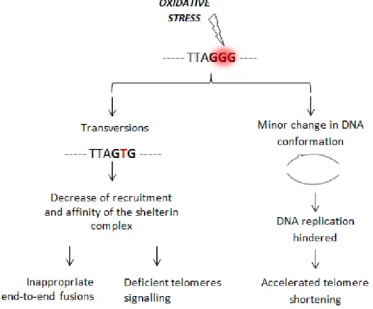
their propensity to mismatch with Adenine promoting GC-to-TA transversions during DNA replication (von Zglinicki 2002). Despite the fact that those transversions can be non-synonymous, modifying the final product of a protein and its function, the loss of guanines to thymines, at telomeres level, decreases the recruitment and affinity of the shelterin complex, possibly leading to aberrant and inappropriate end-to-end fusions, since the shelterin complex is no longer able to mask the telomeric ends from the DDR (Figure 5) (von Zglinicki 2002, Klaunig, Kamendulis et al. 2010, Wang, Rhee et al. 2010, Georgakilas 2012). Cellular processes such as apoptosis, ageing and chromosomal stability maintenance are affected by the loss of the shelterin complex, insofar as mediators of the telomeres signalling (Figure 3.5) (Oikawa and Kawanishi 1999, von Zglinicki 2002, Coluzzi, Colamartino et al. 2014).
Additionally, 8-oxoG can contribute to disturb telomere homeostasis also by affecting the DNA replication, as they can lead to change in the normal DNA molecule conformation, which can hinder the binding of the high-fidelity DNA polymerases during normal replication, and possibly disrupting telomere length and its function (Figure 3.5) (Oikawa and Kawanishi 1999, von Zglinicki 2002, Wang, Rhee et al. 2010).
Several studies have shown higher levels of 8-oxoG in various human cancers: Miyake, Hara et al. (2004), Weiss, Goode et al. (2005), Diakowska, Lewandowski et al. (2007) and Tanaka, Fujita et al. (2008).
Oxidative stress also gives rise to DNA breaks, mostly single-stranded DNA (ssDNA) breaks, either generated as a consequence of the disintegration of the oxidized sugar or the intervention of DNA Base Excision Repair (BER) pathway over the oxidized bases that create abasic sites (Oikawa and Kawanishi 1999, von Zglinicki, Pilger et al. 2000, von Zglinicki 2002, Klaunig, Kamendulis et al. 2010, Georgakilas 2012). A study (von Zglinicki, Pilger et al. 2000) has shown that the accumulation of ssDNA breaks is the major responsible for telomere shortening in human fibroblasts.
Oxidized DNA is the major source of DNA damage in living organisms, and both endogenous and exogenous sources of oxidative stress can contribute to carcinogenesis when the load of Reactive Oxygen Species (ROS) is not counteracted by the antioxidant defences (Figure 3.6) (Klaunig, Kamendulis et al. 2010). In
Page | 14
conclusion, since telomeric DNA is a highly protected structure its repair is not that efficient, making it more vulnerable to aggressions, e.g. oxidative stress, as the proteins that cover it hamper the DNA repair machinery access (von Zglinicki 2002, Coluzzi, Colamartino et al. 2014).
Figure 3.6 ROS effect in cells and its role in cancer development
Page | 15
3.2. Base Excision Repair pathway
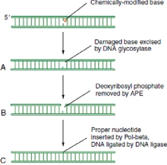
The BER pathway is the principal responsible for the correction of oxidative lesions in the DNA, being the unique process specialized in the single-base lesions reparation with only four core proteins: a DNA glycosylase, an apurinic or apyrimidinic endonuclease (APE), a DNA polymerase and a DNA ligase (Kow 1994, Kim and Wilson 2012). BER action mode is the following: the damaged base is excised by the DNA glycosylase creating an apurinic/apyrimidinic (AP) site; the AP endonuclease cleaves the AP site generating a 3’OH and 5’ deoxyribose phosphate terminus, which is forward filled by the DNA polymerase and sealed by the DNA ligase (Figure 3.7) (Kow 1994, Mendelsohn 2008, Kim and Wilson 2012). Additionally, BER pathway also corrects damaged bases by alkylation (Sedgwick, Bates et al. 2007) and deamination (Kavli, Otterlei et al. 2007). All BER repair processes restore the appropriate DNA base with high accuracy, maintaining the template function of the DNA and avoiding the decay of genetic information (Jacobs and Schar 2012, Kim and Wilson 2012).
Figure 3.7 Scheme of Base excision Repair pathway action mode. Small lesions in DNA are often recognized and corrected by BER mechanisms repair. At first the lesion is recognized and flipped out by DNA glycosylase(A), and then the phosphodiester backbone of the DNA is cleaved by APE (B). An error-free DNA polymerase (Pol-β) fills the gap with the appropriated nucleotide ending with DNA ligase sealing the base to the double-stranded DNA restoring the normal sequence(C). Adapted from (Mendelsohn 2008)
Page | 16
3.2.1. 8-oxoguanine DNA glycosylase, OGG1
OGG1, 8-oxoguanine DNA glycosylase, is an enzyme that acts in the early steps of the mentioned pathway. First found in yeast (Nash, Bruner et al. 1996) and later in humans (Radicella, Dherin et al. 1997), OGG1 is an enzyme which plays a key role in the repair of such lesions, making the recognition and excision of the chemically modified bases causing minor perturbations in the DNA helix, cleaving the glycosidic bond (Jacobs and Schar 2012, Kim and Wilson 2012). Single-base lesions are then flipped out generating an abasic- AP-site, being further processed by the subsequent enzymes, in order to restore the original DNA sequence before the DNA polymerase gets the opportunity to miss-insert an adenine, preventing a transversion mutation (Jacobs and Schar 2012).
Up till now, eleven different DNA glycosylases have been identified in mammals which can be divided into distinct superfamilies (Jacobs and Schar 2012). OGG1 is a helix-hairpin-helix glycosylase (Figure 3.8) whose respective motif is able to recognize both single-strand and double-strand DNA, by hydrogen bond-mediated interactions with the DNA-phosphate backbone (Shinmura and Yokota 2001). Such interactions are fundamental for DNA glycosylases function.
The OGG1 gene is localized in the human chromosome 3p25.3 and consists of 11 exons, covering about 38.275 bp of DNA. Even though with 18 different possible transcripts, its principal protein product has 424 amino acids residues with a theoretical molecular weight of 4.95kDa (Ensembl 2015).
Ablation of ogg1 in Saccharomyces cerevisiae leads to the accumulation of G->T transversion mutations (Thomas, Scot et al. 1997), and results in telomere base damage and length alteration (Lu and Liu 2010). Also, the Ogg1 null mice exhibit abnormal level of chromosomal 8-oxoG but are still viable (Klungland, Rosewell et al. 1999). In humans, there is evidence that non-synonymous polymorphisms in the OGG1 gene might associate with cancer (Zhou, Li et al. 2015), such as non-small cell lung cancer (Duan, Hua et al. 2012) and esophageal cancer (Wang, Gan et al. 2013). Moreover, Osorio, Milne et al. (2014) showed an association between a polymorphism
Page | 17
in the 3’-untranslated region (UTR) in the OGG1 gene and cancer risk in Breast Cancer Susceptibility Genes 1 and 2 (BRCA1 and BRCA2) germline mutation carriers.
3.3. Cancer
The denomination of cancer was initially given by the “Father of Medicine”, Hippocrates (460 – 370 BC) as karkinos and karkinoma to describe non-ulcer and ulcer forming tumours, respectively. This Greek designation for crab was most likely applied because of the finger-like projections, which remind the crab legs shape. Later the “cancer” domination was given by the roman physician, Celsus (28 – 50 BC), which translated the previous Greek words into Latin (ACS 2014).
Cancer is a common designation for many different diseases (Britannica 2015). Although, there are many kinds of cancer, they share this designation because different cancers start the same way: an abnormal cell growth out of control behaving like autonomous cells (Mendelsohn 2008, NCI 2015). All body cells can effectively become into tumour/cancer, but each type has its own characteristics (UK 2015).
Carcinogenesis is a multistage disease in which not one, but several mutations are required. At least three to six mutations seem to be the necessary to reach this development (Vogelstein and Kinzler 1993). These mutations have to be acquired by
Figure 3.8 Structural representation of hOGG1. The six helices are coloured in dark blue and the three DNA binding loops in yellow. Adapted from (Drohat, Kwon et al. 2002)
Page | 18
oncogenes and/or tumour suppressor genes, in a way that affects the net rate of cell division. According to Land, Parada et al. (1983), at least one mutation in two cooperating oncogenes are necessary for a tumourigenic transformation. Not only a multistage disease, cancer is also multifactorial, combining genetic and environmental factors (Hanahan and Weinberg 2011).
Before reaching the malignant stage, denominated cancer, tumour cells undergo through a series of stages, which in the majority of cases include: a hyperplasia (characterized by an increased number of cells in a tissue), dysplasia (where the tissue looks abnormal and disorganized but still not invasive, and cells look less differentiated). Cancer stage is achieved when tumour cells acquire invasive properties (Mendelsohn 2008).
Hanahan and Weinberg (2011) defined the necessary functional competences that a cell needs to contribute to genesis of malignancies: the hallmarks of cancer, which allow cancer cells to survive, proliferate and disseminate. Those hallmarks include: resistance to cell death; genome instability and mutation; sustenance of proliferative signalling; replicative immortality; escape from immune system surveillance; evasion of growth suppressors; deregulation of cellular energy; angiogenesis induction; inflammation promoted by tumour and activation of invasion and metastasis properties (Figure 3.9).
The acquisition of this set of malignant competences is mainly regulated by genomic instability and premalignant inflammation. The first one involves processes that are involved in major reprogramming processes of cell growth/proliferation and metabolism, the second allows cancer cells to escape from immune system surveillance and antagonistically operates for an enhancement of tumour progression through the immune system (Hanahan and Weinberg 2011).
Page | 19
3.4. Breast cancer and Ovarian Cancer
3.4.1. Epidemiology
Breast:
According to GLOBOCAN (2012), female breast cancer is by far the most frequent cancer, the most common cause of death (Figure 3.10) and the most frequently diagnosed cancer among women worldwide, accounting for 23% of all women’s malignant cancers. In 2012, 1,67 million new breast cancer cases were diagnosed representing 25% of all diagnosed cancers (GLOBOCAN, 2012).
The lifetime risk for breast cancer, in the United States, is approximately 1 in 8, and 12.3% of worldwide women will be diagnosed worldwide with breast cancer at some point during their lifetime (Ghoussaini, Pharoah et al. 2013).
With respect to prevalence, in 2011, 2,899,726 women were living with breast cancer. The 5-year survival rate is 89.2%. Such a high percentage is influenced by the stage at the time of the diagnosis, referring to the extent of cancer, where 61% is detected in the localized state (Figure 3.11) (NCI 2015).
Figure 3.9 Hallmarks of cancer that gives the necessary functional competences a cell need to contribute to carcinogenesis. Adapted from Hanahan and Weinberg (2011)
Page | 20
Ovary:
As to the incidence in 2012, around 239,000 new cases of ovarian cancer were reported, representing nearly 4% of the new cases of women cancer. Ovarian cancer is the seventh most common cancer affecting women worldwide, and the 18th overall (Ferlay, Soerjomataram et al. 2015).
Among all cancers of the female reproductive system, ovarian cancer is the one that causes more deaths, being the eighth most common cause of cancer death in women worldwide. 14 030 deaths were estimated for 2013. The five-year relative survival rate is 44%, considering all the stages of diagnosis (Ferlay, Soerjomataram et al. 2015). In 2012, 192,446 women were living in the United States with ovarian cancer, and approximately 1.3% of women will be diagnosed at some point of their lifetime (GLOBOCAN 2012).
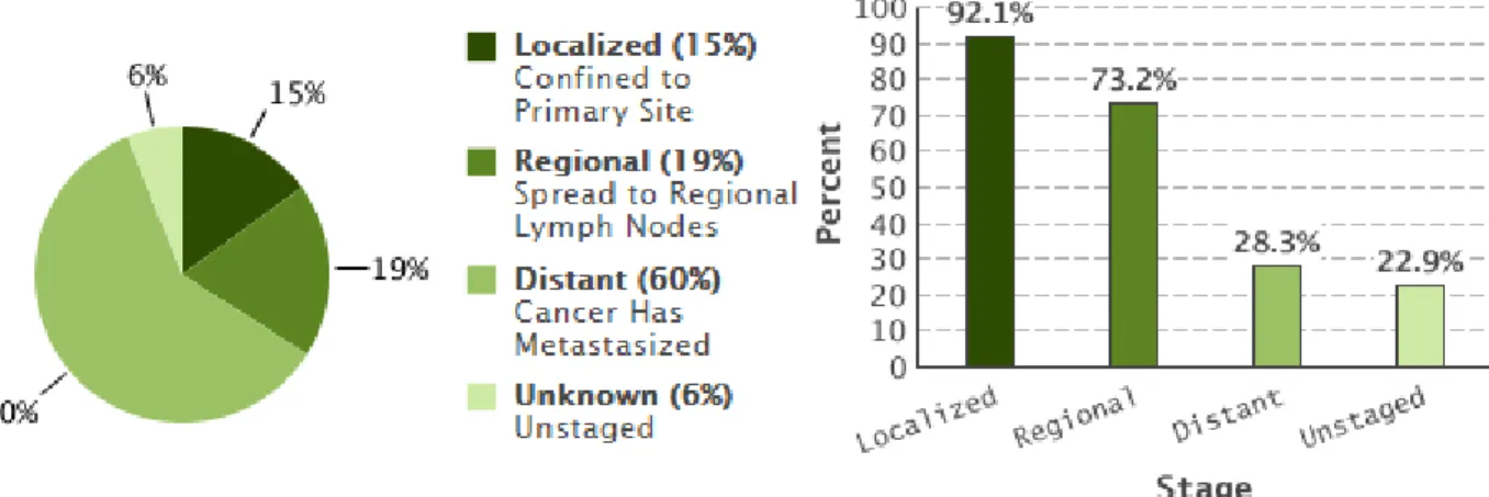
This type of cancer is usually fatal because most of the cases (60%) are diagnosed in an advanced stage, when the cancer is already metastasized. The five-year relative survival rate for this stage is very low, being 28.3% (Figure 3.12) (Cronin, Ries et al. 2014).
Figure 3.10 Estimate incidence and mortality rates of the different cancers per 100 000 women per year (ASR: age-standardised rate). Adapted from GLOBOCAN (2012) 61% 32% 5% 2% Localized - Confined to Primary site Regional - Spread to Regional Lymph Nodes Distant - Cancer has metastasized Unknown - Unstaged
Figure 3.11 Pie with the representative percentages of the different stages of breast cancer at the time of diagnosis. Adapted from GLOBOCAN (2012)
Page | 21
3.4.2. Risk factors
The risk factor concept involves anything that might affect the chance of an individual of getting the disease (WHO 2015). A risk factor can be environmental or even genetic. Evidently, different cancers have different risk factors and different load of the relative risk.
Breast:
Together with gender, i.e. being a woman, the highest risk factor for breast cancer is the age, with the incidence rate higher among older women, although, the risk increases across all ages until 80 years-old (UK 2015).
Another risk factor that substantially increases the risk of developing breast cancer is personal history of the disease (Figures 2013). The risk is highly correlated with the number of affected first-degree relatives: 1.8 times higher for women with one first-degree relative affected, 3 times higher with two, and nearly 4 times higher with 3 or more affected relatives (Epidemiology 2015). The younger the age of onset of the affected relative, the greater the risk for a woman to develop the disease (Figures 2013, Epidemiology 2015).
Up to date, have been identified high-, moderate- and low-susceptibility genes that modifies the risk for breast cancer. For instances, mutations in the BRCA1 (Hall,
Figure 3.12 Percentage of ovarian cancer diagnosed (left) and 5-year relative survival by stage (right). Adapted from Cronin, Ries et al. (2014)
Page | 22
Lee et al. 1990) and BRCA2 (Wooster, Neuhausen et al. 1994) high-susceptibility genes increase the risk for breast cancer, but also to ovarian, and other cancers. Deleterious mutations in the BRCA1 gene confer around 65% increase of cumulative risk for breast cancer, while deleterious mutations in the BRCA2 gene confers around 39% (Antoniou, Pharoah et al. 2003, Chen, Iversen et al. 2006, Chen and Parmigiani 2007, Milne, Osorio et al. 2008).
It is believed that differences in worldwide incidence rates are due to the variability in reproductive patterns, hormonal factors, lifestyle and environmental factors. All these factors are partially responsible for modulating the probability of breast cancer development throughout a woman’s lifetime. Table 1 briefly introduces some of the known risk factors with their relative risk values (Figures 2013, Epidemiology 2015, UK 2015).
Ovary:
Like breast cancer and most other cancers, the risk of developing ovarian cancer increases with age. Most of the ovarian cancers are developed after menopause. Women who had a first full-term pregnancy after 35 years-old or who never carried a pregnancy to term show higher risk. Also, breastfeeding may lower the risk (UK 2015).
Long-term use of hormonal contraceptives, on the contrary of breast cancer risk, lowers ovarian cancer risk, and the longer the use the lower the risk. Curiously, risk continues lowering for years after stoppage of use. Oestrogen/hormone therapies after menopause show an increased risk of developing ovarian cancer (UK 2015).
A strong family history of breast and/or ovarian cancer is a very important risk factor, where about 1 in 10 ovarian cancers, 10%, are caused by an inherited faulty gene, like BRCA1 and BRCA2. The risk increases the number of affected relatives. Familial history of other cancers such as breast cancer is also linked to an increased risk. Women who have a personal history of breast cancer have an increase risk, as well (UK 2015). A germline deleterious mutation in the BRCA1 gene confers approximately 39% increase of cumulative risk, while in the BRCA2 gene conferes
Page | 23
around 11% (Antoniou, Pharoah et al. 2003, Chen, Iversen et al. 2006, Chen and Parmigiani 2007, Milne, Osorio et al. 2008).
As to lifestyle, a low-fat diet for at least 4 years showed a reduced risk and being overweight during pre-menopause increases the risk. While smoking shows a slight increase, drinking alcoholic beverages does not seem to increase the risk of ovarian cancer overall (UK 2015).
Table 1Risk factor and its relative risk for breast cancer in women. Relative risk is a comparison of the absolute risk of disease among people with a particular risk factor with the risk among people without that risk factor. Relative risk higher than 1.0 means the risk is higher among people with the particular risk factor vs. the people without. Adapted from (Figures 2013).
Relative risk Risk factor
>4.0 Age (65 or older vs. <65 years-old)
Certain inherited genetic mutations for breast cancer (BRCA1 and/or
BRCA2)
Mammographically dense breasts
Personal history of early breast cancer onset (<40 years-old) Two or more first-degree relatives with early onset age 2.1-4.0 Personal history of breast cancer (40 or older years-old)
High endogenous estrogen or testosterone levels (postmenopausal) High-dose radiation to chest
One first-degree relative with breast cancer 1.1-2.0 Alcohol consumption
Ashkenazi Jewish heritage Early menarche (<12 years-old) Height (tall vs. short)
Page | 24
Late menopause (>55 years-old) Never breastfed
No full term pregnancies Obesity
Personal history of endometrium, ovary or colon cancer Recent oral contraceptive use
Recent and long-term use of menopausal hormonal therapy
3.4.3. Familial breast and ovarian cancer (FBOC) syndrome
Cancer is a sporadic disease that occurs due to the accumulation of genetic changes, but a small percentage of the cases are considered hereditary. Familial cancer is characterized by having a higher number of cancer cases within a family than statistically expected, which have a genetic component. While hereditary cancer shows a clear pattern of inheritance, clustering of early-onset age and multiple primary cancer cases in an individual (Berliner, Fay et al. 2007, Berliner, Fay et al. 2013).
The first detailed and significantly description about hereditary breast cancer was published in 1866 by Paul Broca (1824 – 1880) (van der Groep, van der Wall et al. 2011). Broca made a pedigree of his wife’s family who suffered from early onset of breast cancer, and he suggested that breast cancer could be inherited because four generations had breast cancer (Figure 3.13) (van der Groep, van der Wall et al. 2011).
Up to now high-, moderate- and low-susceptibility genes that increase the risk in Familial breast and ovarian cancer (FBOC) have been identified. FBOC syndrome is mostly caused by a germline mutation in high-susceptibility genes BRCA1 (chromosome 17q21) (Hall, Lee et al. 1990) and BRCA2 (chromosome 13q12-13) (Wooster, Neuhausen et al. 1994) (Figure 3.14). These mutations not only increase the risk for breast and ovarian cancer, but also prostate and pancreatic cancer (van der
Page | 25
Groep, van der Wall et al. 2011). The overall prevalence of these mutations is 1 in 400 for BRCA1 and 1 in 800 to BRCA2 (Petrucelli, Daly et al. 2010).
The penetrance is the most significant clinical aspect in FBOC syndrome, being breast and ovarian cancer the predominant phenotypes. The penetrance of these mutations vary within families therefore, there is no exact estimate risk for individuals harbouring germline BRCA1 and BRCA2 mutations (Petrucelli, Daly et al. 2010). Those differences can be due to the type and/or position of the mutation and other genetic and/or environment factors (Petrucelli, Daly et al. 2010).
Page | 26
3.4.3.1. Breast cancer susceptibility genes
Germline mutations in BRCA1 and BRCA2 genes, which predisposes to breast, ovarian and even other cancers, explain around 25% of familial breast cancers (FBC). There are considerable differences in the manifestation of the disease among all the carriers of an inherited mutation in BRCA. This differences, in addition to the fact that not all the individuals that inherited a BRCA mutation will develop cancer, suggest the existence of other genetic and/or environmental factors modifying the risk of developing cancer (Couch, Nathanson et al. 2014).
Approximately 5% of the FBC cases are due to mutations in high-susceptibility genes involved in other familial syndromes such as TP53, PTEN, STK11 and CDH11 (Borresen, Andersen et al. 1992, Chen, Lindblom et al. 1998, Pharoah, Guilford et al. 2001, Leggett, Young et al. 2003).
Figure 3.14 Timeline of hunt for the genetic underpinnings of the familial breast cancer. Adapted from Hurst (2014)
Page | 27
Moderate penetrance genes include the RAD51C, ATM and CHEK2 genes, involved in fanconi and non-fanconi anemias (e.g. (Meindl, Hellebrand et al. 2010)) (Meijers-Heijboer, van den Ouweland et al. 2002, Thompson, Duedal et al. 2005)), representing 5% of the FBC.
Recently, 41 low susceptibility polymorphisms were associated to FBC (Michailidou, Hall et al. 2013), which together with the 26 polymorphisms previously identified explain around 14% of the FBC cases (Ghoussaini, Pharoah et al. 2013)
Nevertheless, 51% of FBC cases do not have mutations or the alleles described above and are thus categorized as BRCAX families. These cases can be explained by a polygenic model or by some gene that remains to be identified (Melchor and Benitez 2013). Figure 3.15 shows the distribution of FBC patients according with their mutations/variants in low-, moderate-, high-susceptibility genes/loci.
Established in 2006, The Consortium of Investigators of Modifiers of BRCA1 and BRCA2 (CIMBA), provides currently the largest sample size for the study of common genetic modifiers of breast and ovarian cancer risk. So far, 94 single-nucleotide polymorphisms (SNPs) have been associated with low susceptibility to breast cancer by Genome-Wide Association Studies (GWAS). (Cox, Dunning et al. 2007, Easton, Pooley et al. 2007, Stacey, Manolescu et al. 2007, Ahmed, Thomas et al. 2009, Milne, Benitez et al. 2009, Thomas, Jacobs et al. 2009, Turnbull, Ahmed et al. 2010, Broeks, Schmidt et al. 2011, Figueroa, Garcia-Closas et al. 2011, Michailidou, Hall et al. 2013, Michailidou, Beesley et al. 2015).
14% 5%
25% 51%
5% Low Susceptibility Genes
Moderate Susceptibility Genes
High Susceptibility Genes
BRCAX families
Related-syndromes - High Susceptibility Genes
Figure 3.15 Pie with the representative percentages of familial breast cancer mutations in low-, moderate-, high-penetrance genes, BRCAX and related syndromesup to datedescribed that predispose to breast cancer. Adapted from (Melchor and Benitez 2013)
Page | 28
3.5. Breast Cancer Susceptibility Genes, BRCAs
3.5.1. Function as Homologous Recombination DNA repair members BRCA proteins play a very important role in genome and chromosome integrity and maintenance, during cell division. It is notable how BRCA-deficient cells accumulate espontaneously aberrations in chromosomes’ structure and number, due to impairment in Homologous DNA Recombination (HR) pathway, (Venkitaraman 2002, Venkitaraman 2014). Both BRCA1 and BRCA2 are individually crucial for an efficient HR, a mechanism of DNA repair error-free using as template an intact homologous sequence, such as sister chromatid (Venkitaraman 2014).
DsDNA breaks may be create during chromosome duplication thus, BRCA-deficiency is typically accompanied by dsDNA breaks or aberrant chromosomes structure and number since their absent or normal performance leads the cells to reroute the DNA reparation to an error-prone mechanism: nonhomologous end joining (NHEJ) (Venkitaraman 2014). BRCAs are therefore classified as tumour suppressor proteins, with fundamental functions in the genomic stability maintenance.
The BRCA genes follow the Knudson’s “two-hit” hypothesis (Knudson 1971) which postulates that mutation or loss in one of the alleles of a tumour suppressor gene is not enough to trigger cancer, being necessary the occurrence of a second “hit” to completely inactivate the protein. However, it is known that for BRCA genes just one impaired allele can actually affect BRCA normal performance due to haploinsufficiency (Cousineau and Belmaaza 2007, Konishi, Mohseni et al. 2011, Nisman, Kadouri et al. 2013). That phenomenon might increase the susceptibility of the carrier and accelerate the loss of the second allele.
BRCA1 and BRCA2 have many and distinct functions although, in a simplified perspective BRCA1 acts in the early steps of HR reparation, whereas BRCA2 stabilizes and supports the replication-associated lesions structures. BRCA2 not only plays as HR
Page | 29
member but also works in the surveillance of the mitotic spindle assembly checkpoint. In
Figure 3.16 is possible to see BRCA1 and BRCA2 HR-mediated functions.
Figure 3.16 Some BRCA1 and BRCA2 HR-mediated functions at different stages of
3.5.2. Functional involvement in telomere maintenance
It has been described that BRCA1/2 have a functional role in the telomere maintenance. For instance, previous reports (Xiong, Fan et al. 2003, Ballal, Saha et al. 2009) suggest that BRCA1 can regulate both telomere length and stability due to its interaction with Mre11-Rad50-Nbs1 (MRN) complex (complex that binds to shelterin proteins) and telomerase. Regarding BRCA2, Badie, Escandell et al. (2010) have shown BRCA2 is with telomeres during the S and G2 cell cycle phases, in order to facilitate the loading of RAD51 into telomeres. BRCA2 conditional deletion leads to shorter and fragmented telomeres representing telomere fragility (Badie, Escandell et al. 2010).
Figure 3.16 Some BRCA1 and BRCA2 HR-mediated functions at different stages of cell division. Both BRCA1 and BRCA2 participate in the G2 checkpoint: BRCA1 helps in the initiation of HR moving 53 Bindingprotein 1 (53BP1) and triggers the end resection (represented by the scissors). Thus, BRCA2 binds to the ssDNA and dsDNA junctions at the lesion, moving RPA away. During mitosis, BRCA1contributes to mitotic spindle assembly, and BRCA2 watches for mitotic checkpoint and participates in the abscission step of cytokinesis. Adapted from Venkitaraman (2014)
Page | 30
Together BRCA1 and BRCA2, despite mediating HR also contribute to telomere integrity and length.
3.6. Genome-wide association studies, GWAS
Genome-wide association studies (GWAS) look for genetic variation, such as SNPs, that are more frequently present in a group of people with the disease than controls. The search for variants/SNPs/polymorphisms can be made without a priori information about its genetic function or mechanism, or can be restricted to candidate genes. The HapMap project made possible the existence of comprehensive studies such as GWAS, where describes common patterns of human DNA sequence variation displayed as a haplotype map (International HapMap 2005). GWASs are a good approach to identify common alleles with low penetrance in the disease (Figure 3.17), of common multifactorial and polygenic diseases (Easton and Eeles 2008). GWAS are used to mapping genomic areas associated with a disease/phenotype (International HapMap 2005).
Systematic studies of common genetic variants are simplified by the linkage disequilibrium (LD) phenomenon, because it is possible to obtain a large amount of information on genomic variation without complete resequencing and to get an efficient selection of tag SNPs, optimizing the association analyses (International HapMap 2005).
GWAS are widely used in cancer research, particularly in the commonest cancer types such as breast, prostate, colorectal, lung and melanoma (Easton and Eeles 2008).
3.6.1. Tag SNPs for association studies
A “tag SNP” is a representative SNP in a determined region of the genome with high LD (r2 ≥ 0.8) with a group of other SNPs: haplotype. Like that, it is possible to search for an association with a phenotype based on the genotypes: a unique SNP or a
Page | 31
combination of various SNPs. LD phenomenon saves the entire genotyping of every SNP in a chromosomal region (International HapMap 2005).
The tag SNPs selection looks for maximum efficiency with the least loss of information. Hence, some methods need a single SNP serving as a proxy for others, while others use the combination of alleles, or haplotypes, to serve as proxies. The causal SNP for a disease is rarely genotyped but a good set of tag SNPs will provide association between a genotype and phenotype, which the pinpointed SNP might be in LD with the causal one. For the identification of the causal SNP, is necessary a greater resolution in the selection of haplotypes blocks as a Fine mapping approach.
3.6.2. SNPs in DNA glycosylases genes involved in the BER pathway
Given the synthetic lethality between members of BER pathway: Poly ADP ribose polymerase (PARP1) and BRCA1 and BRCA2, Osorio, Milne et al. (2014) performed a comprehensive analysis using a tagging SNP method of 18 genes involved in BER pathway in a large series of BRCA1 and BRCA2 mutation carriers from CIMBA consortium. 144 tagSNPs were analysed in a sample of 23,463 individuals.
Figure 3.17 Genome-wide association studies are effective in detecting common alleles with low penetrance in a disease. Adapted from Eeles, Goh et al. (2014)
Page | 32
Evidence of association with breast and/or ovarian cancer was obtained for eleven SNPs, of which, five located in DNA gycosylases genes. The strongest evidences of was for rs1466785 in the NEIL2 gene (endonuclease VIII-like2) (HR: 1.09, 95% CI (1.03–1.16), p = 2.761023): associated with breast cancer risk in BRCA2 mutation carriers, and for rs2304277 in the OGG1 gene (HR: 1.12 95%CI: 1.03–1.21, p = 4.861023), associated with ovarian cancer risk in BRCA1 mutation carriers (Osorio, Milne et al. 2014).
These loci should be extensively studied, considering these results.
3.6.2.1. SNP in OGG1 associated with ovarian cancer risk
An impaired Homologous Recombination DNA repair mechanism, due to a
BRCA mutation, makes cells critically dependent on other DNA repair machineries such
as BER pathway. As mentioned above, Osorio, Milne et al. (2014) reported the rs2304277 variant in the OGG1 gene with strong evidences of association with ovarian cancer in BRCA1 mutation carriers. Knowing, this SNP was identified by a tagging SNP approach, it is possible this is not the causal SNP, but only a SNP in LD with the causal one.
This SNP has two alleles, an Adenine (A) and a Guanine (G), whit the ancestral and minor allele being the A allele, present in 35% of the population, according to the 1000 Genomes project (dbSNP 2015). This polymorphism is located in a 3’UTR, a regulatory region that can in many aspects, influence the mRNA molecule in its stability or translation. At 3’URT regions, contain specific binding sites for both regulatory proteins and microRNAs. Hence, the stability of the mRNA molecule or its translation efficiency can be altered. A polymorphism at 3’UTR can, for instance, decrease or increase the binding affinity of those microRNA (miRNA) and regulatory proteins, thus changing the final product of mRNA and respective translated protein (Barrett, Fletcher et al. 2012, Pichon, Wilson et al. 2012).
OGG1 is a key player in BER pathway, involved in the repair of oxidized guanines, bases in which telomeres are enriched. Given the natural preference of oxidative stress over guanines, OGG1 role, and telomeres composition, this study
Page | 33
followed this line of reasoning to understand and answer whether the increased cancer risk is due to a less effective OGG1 response upon oxidative stress, leading to accelerate telomere shortening.
Page | 35
We hypothesized that the association with cancer risk in the carriers of the rs2304277 variant might be due to an altered OGG1 function, that could accelerate the telomere shortening, resulting from a weaker response upon oxidative stress consequences.
In order to answer the biological question the aims of this study were:
First aim:
- To investigate whether the rs2304277 variant, located in the 3’ UTR region of
OGG1 can modify the expression of its messenger ribonucleic acid (mRNA),
leading to an altered response upon oxidative stress.
Second aim:
- To investigate whether the presence of the rs2304277 variant in OGG1 alone, or in conjunction with the BRCA mutation can modify telomere length, given the role of OGG1 in the telomere homeostasis.
Page | 37
5.1. Samples
The samples mentioned below and used for this study were collected through individuals that attended the genetic counselling consultancy for familial cancer in the Fuenlabrada Hospital University – Madrid, Spain from 2011 to 2014. Informed consent was obtained from all individuals involved in this research project.
We have followed a cross-sectional approach in a cohort of familiar breast and ovarian cancer cases (FBOC) from 101 families, harbouring deleterious mutations in
BRCA1 and BRCA2 and BRCAX. With BRCAX it is mean cases with familiar history for hereditary breast cancer but that do not harbour mutations in any of the identified genes for FBC, as reported above in the “Breast Cancer Susceptibility Genes” section.
Individuals from these families met high risk criteria (Milne, Osorio et al. 2008) and had been previously screened for mutations in BRCA1 and BRCA2 by a combination of denaturing high-performance liquid chromatography (DHPLC) and direct sequencing as previously reported (Diez, Osorio et al. 2003)
Peripheral blood samples from 223 individuals were used, which 68 (31%) of the samples corresponded to controls: healthy women and men with no personal or familiar antecedent of cancer and age range from 18 to 84 years old. 45 (20%) and 49 (22%) individuals harboured a mutation in BRCA1 and BRCA2, respectively, while 61 samples (27%) a mutation in BRCAX. In the Table 4 it is possible to see the mentioned numbers and its relative frequencies within the sample.
5.2. Genotyping
The genotyping for the polymorphism rs2304277 present in OGG1 gene was made in order to know the alleles of which sample included in this study: homozygote for the common allele (G/G), heterozygous (GA) or homozygote for the minor allele (A/A).