Brief Communications
Blockade of Adenosine A
2A
Receptors Prevents
Protein Phosphorylation in the Striatum Induced by
Cortical Stimulation
Ce´sar Quiroz,
1Catarina Gomes,
2Arlene C. Pak,
1Joaquim A. Ribeiro,
2Steven R. Goldberg,
1Bruce T. Hope,
1and
Sergi Ferre´
11Behavioral Neuroscience Branch, National Institute on Drug Abuse, Intramural Research Program, National Institutes of Health, Department of Health and Human Services, Baltimore, Maryland 21224, and2Institute of Pharmacology and Neurosciences, Faculty of Medicine, Institute of Molecular Medicine, University of Lisbon, 1649-028 Lisbon, Portugal
Previous studies have shown that cortical stimulation selectively activates extracellular signal-regulated kinase 1/2 (ERK1/2)
phosphor-ylation and immediate early gene expression in striatal GABAergic enkephalinergic neurons. In the present study, we demonstrate that
blockade of adenosine A
2Areceptors with caffeine or a selective A
2Areceptor antagonist counteracts the striatal activation of cAMP–
protein kinase A cascade (phosphorylation of the Ser
845residue of the glutamate receptor 1 subunit of the AMPA receptor) and
mitogen-activated protein kinase (ERK1/2 phosphorylation) induced by the
in vivo stimulation of corticostriatal afferents. The results indicate
that A
2Areceptors strongly modulate the efficacy of glutamatergic synapses on striatal enkephalinergic neurons.
Key words: caffeine; adenosine A
2Areceptor; striatum; phosphorylation; ERK1/2; AMPA receptor
Introduction
The striatum is the main input and information processing
struc-ture of the basal ganglia. Cortico-limbic-thalamic glutamatergic
and mesencephalic dopaminergic systems converge in the
GABAergic medium-sized spiny neurons, which constitute
⬎90% of the striatal neuronal population (Gerfen, 2004). These
are the striatal efferent neurons, which can be classified into
GABAergic enkephalinergic and GABAergic dynorphinergic
neurons. Enkephalinergic neurons predominantly express
aden-osine and dopamine receptors of the A
2Aand D
2subtype,
respec-tively, whereas dynorphinergic neurons predominantly express
adenosine and dopamine receptors of the A
1and D
1subtype,
respectively (Ferre´ et al., 1997, 2005; Gerfen, 2004).
The dopamine D
1receptor is a G
s/olf-coupled receptor, whose
main signaling pathway is the cAMP–protein kinase A (PKA)
cascade. The D
1receptor modulates neuronal excitability and
glutamatergic neurotransmission in the GABAergic
dynorphin-ergic neuron by inducing PKA-mediated phosphorylation of
dif-ferent substrates, such as L-type voltage-dependent calcium
channels, DARPP-32 (the dopamine and cyclic adenosine 3
⬘,
5⬘-monophosphate-regulated phosphoprotein, 32 kDa), the NMDA
and AMPA glutamate receptors (GluRs), and the nuclear
consti-tutive transcription factor cAMP response element binding
pro-tein (for review, see Greengard et al., 1999). Furthermore, the D
1receptor can potentially activate mitogen-activated protein
ki-nase (MAPK) [extracellular signal-regulated kiki-nase 1/2 (ERK1/2)
phosphorylation] by PKA-dependent and -independent
mecha-nisms (Zanassi et al., 2001), which seem to require a concomitant
stimulation of NMDA receptors (Valjent et al., 2005).
The adenosine A
2Areceptor is functionally very similar to the
D
1receptor. It is also a G
s/olf-coupled receptor whose activation
stimulates the cAMP–PKA cascade. A
2Areceptor forms
hetero-mers with D
2receptor, which antagonistically modulates A
2Are-ceptor function by inhibiting adenylyl cyclase activation (Kull et
al., 1999; Hillion et al., 2002). Furthermore, stimulation of A
2Areceptor decreases the ability of dopamine to bind the D
2receptor
by means of an intramembrane A
2A–D
2receptor interaction
(Ferre´ et al., 1991). The same as with the D
1receptor, A
2Areceptor-mediated PKA activation can potentially phosphorylate
different substrates and induce MAPK activation. However,
un-der basal conditions, there is a strong tonic activation of striatal
D
2receptors, which impairs the ability of A
2Areceptors to signal
through the cAMP–PKA cascade (for review, see Ferre´ et al.,
2004, 2005).
Both enkephalinergic and dynorphinergic neurons receive
corticostriatal afferents and display excitatory postsynaptic
re-sponses to corticostriatal stimulation (Kawaguchi et al., 1990).
Nevertheless, corticostriatal stimulation selectively activates
MAPK and immediate early gene (IEG) expression in the
en-kephalinergic neurons without activation in dynorphinergic
neurons (Berretta et al., 1997; Gerfen et al., 2002). In fact, MAPK
activation is responsible for the induction of striatal IEG
expres-sion after corticostriatal stimulation (Sgambato et al., 1998). In
the present study, we demonstrate that blockade of A
2Areceptors
Received April 18, 2006; revised Sept. 11, 2006; accepted Sept. 11, 2006.This work was supported by the Intramural Research Program of the National Institutes of Health, National Institute on Drug Abuse, Department of Health and Human Services.
Correspondence should be addressed to Sergi Ferre´, Preclinical Pharmacology Section, National Institute on Drug Abuse, Intramural Research Program, National Institutes of Health, Department of Health and Human Services, 5500 Nathan Shock Drive, Baltimore, MD 21224. E-mail: sferre@intra.nida.nih.gov.
DOI:10.1523/JNEUROSCI.1661-06.2006
with caffeine or a selective A
2Areceptor antagonist counteracts
the striatal activation of cAMP–PKA cascade (phosphorylation of
the Ser
845residue of the GluR1 subunit of the AMPA receptor)
and MAPK (ERK1/2 phosphorylation) induced by the in vivo
stimulation of corticostriatal afferents. The results support our
hypothesis that strong cortico-limbic-thalamic input to the
GABAergic enkephalinergic neuron allows the A
2Areceptor to
override the inhibitory effects of the D
2receptor with significant
activation of the cAMP–PKA cascade and MAPK (Ferre´ et al.,
2004, 2005).
Materials and Methods
Electrical cortical stimulation. Male Sprague Dawley rats (250 –300 g;
Charles River Laboratories, Wilmington, MA) were used in all experi-ments. Animals were maintained in accordance with the guidelines of the Institutional Care and Use Committee of the Intramural Research Pro-gram, National Institute on Drug Abuse, National Institutes of Health. The animals were implanted unilaterally under Equithesin (National In-stitute on Drug Abuse Pharmacy, Baltimore, MD) anesthesia with bipo-lar stainless steel electrodes, 0.15 mm in diameter (Plastics One, Roanoke, VA), into the orofacial area of the lateral agranular motor cortex (3 mm anterior, 3 mm lateral, and 4.2 mm below bregma). The electrodes and a head holder (connected to a swivel during stimulation) were fixed on the skull with stainless steel screws and dental acrylic resin. Five days after surgery, rats were placed in individual bowl chambers, and the implanted electrodes were attached to an electrical stimulator (Grass S88K; Grass Instruments, West Warwick, RI). Ten minutes before corti-cal stimulation, the animals were given an intraperitoneal administration of saline, the A2Areceptor antagonist MSX-3 (3,7-dihydro-8-[(1
E)-2-(3- methoxyphenyl)ethenyl]-7-methyl-3-[3-(phosphonooxy)propyl-1-(2-propynyl)-1 H-purine-2,6-dione]disodium salt hydrate) (3 mg/kg), the A1receptor antagonist 8-cyclopentyl-1,3-dimethylxanthine (CPT) (4.8
mg/kg), or the nonselective adenosine antagonist caffeine (anhydrate base; 10 mg/kg). All drugs were purchased from Sigma (St. Louis, MO). All drugs were dissolved in sterile saline (with a few drops of 0.1N NaOH for MSX-3, final pH 7.4) and administered in a volume of 3 ml/kg of body weight. The doses of the adenosine antagonists were previously shown to provide motor activation by selectively antagonizing A1receptors (CPT),
A2Areceptors (MSX-3), or both A1and A2Areceptors (caffeine)
(Karcz-Kubicha et al., 2003). The parameters of stimulation were the same as those shown previously to induce selective phosphorylation of ERK1/2 in enkephalinergic cells of the projecting striatal area, the lateral caudate– putamen (Gerfen et al., 2002). After 10 min of habituation, biphasic current pulse trains (pulse of 0.1 ms; 150 –200A, 100 Hz, 160 ms trains repeating once per second) were delivered using two coupled constant current units (Grass PSIU6; Grass Instruments). The intensity was 150 A for most cases or it was increased up to 200 A, until small jaw movements were observed. The cases that failed to show visible somatic movements were excluded from additional analysis. In no case did ani-mals display evidence of seizure activity from the electrical stimulation. Stimulation was applied for 20 min, and the animals were killed imme-diately after the stimulation offset.
Immunohistochemistry. Immediately after the offset of cortical
stimu-lation, rats were deeply anesthetized with Equithesin and perfused tran-scardially with 0.1MPBS, followed by 4% formaldehyde in 0.1Msodium phosphate monobasic buffer, pH 7.4. Brains were postfixed in the same fixative for 2 h and immersed in 20% sucrose/0.1Msodium phosphate, pH 7.4, solution for 48 h at 4°C. Twenty-micrometer-thick coronal sec-tions were cut at the anteroposterior (AP) level of bregma 0.0⫾ 1.0 mm in a Leica (Nussloch, Germany) CM3050S cryostat at⫺20°C, collected in PBS, and stored in antifreeze-buffered solution (20% ethylene glycol, 10% glycerol, and 10% sucrose in PBS) at⫺80°C until processing. The slices were then rinsed with PBS, incubated with 100 mMsodium boro-hydride and 0.1% hydrogen peroxide in PBS for 10 min, rinsed, and incubated in blocking buffer (PBS/0.1% Triton X-100/5% bovine serum albumin) for 2 h before incubation with rabbit polyclonal anti-phospho-Thr202/Tyr204ERK1/2 (1:2000 dilution; Cell Signaling Technology,
Dan-vers, MA) in blocking buffer for 24 h at 4°C. Sections were washed and
incubated in 1:200 biotinylated goat anti-rabbit antibody (Vector Labo-ratories, Burlingame, CA) for 2 h, then washed and incubated for 2 h in ABC reagent (PK-6100; Vector Laboratories), washed again, treated with 0.33 mg/ml 3,3⬘-diaminobenzidine, 50 mMammonium nickel sulfate, and 0.003% peroxide until development (5–10 min), transferred to PBS, mounted onto chrom-alum gelatin-coated slides, air-dried, dehydrated through graded ethanols, cleared in xylene, and coverslipped with DPX (mixture of xylene and dibutyl phthalate; Fisher Scientific, Tustin, CA). Images were acquired using a digital camera (HRC; Zeiss, Gottingen, Germany) coupled to an Axioimager A1 microscope (Zeiss).
Western blotting. Immediately after the offset of cortical stimulation,
the animals were decapitated, and the brains were rapidly extracted, frozen in dry ice-cold isopentane, and stored at⫺80°C. Subsequently, unilateral tissue punches of the lateral striatum (16 gauge) at the AP level of bregma 0.0, corresponding to the area with maximal expression of phosphorylated ERK1/2 (Fig. 1), were obtained from ⬃1 mm-thick coronal sections cut in the cryostat at⫺20°C. The rostral side of the coronal sections was localized approximately between bregma 0.5 mm and bregma⫺0.5 mm. For Western blot assays, tissue punches were sonicated for 10 –15 s in sonication buffer (1% SDS in deionized sterile water). The volume of sonication buffer was 200l. The protein concen-trations of all samples were determined using a bicinchoninic acid assay kit (Pierce, Rockford, IL) and were further diluted with 1% SDS to equal-ize the protein concentrations in each sample. Loading buffer (16% glyc-erol, 20% mercaptoethanol, and 0.05% bromophenol blue) was added to each sample (3:1, sample to loading buffer ratio) before heating at 95°C for 10 min. The proteins in the samples were separated by SDS-PAGE (10% acrylamide/0.27% N, N⬘-methylenebisacryalamide resolving gel) Figure 1. Striatal ERK1/2 phosphorylation induced by cortical electrical stimulation. A, D, Immunohistochemical localization of phosphorylated ERK1/2 in coronal sections (AP level of bregma 0.0) through the striatum ipsilateral to the implanted electrode from stimulated (A) and sham nonstimulated (D) animals. B, C, E, F, Higher magnification of the respective fields indicated in A and D, which shows cells displaying immunoreactivity to phospho-ERK1/2 anti-body in the lateral caudate–putamen of the stimulated animals. Scale bars, 250m.
for 3– 4 h at 200 V. For each electrophoresis run, 20l of sample (con-taining 8 –10g of total protein depending on each particular run) were loaded in each well, and increasing amounts of protein pooled from the samples were also loaded and electrophoresed to produce a standard curve. Proteins were transferred electrophoretically to polyvinylidene fluoride Immobilon-P membranes (Millipore, Bedford, MA) at 0.3 A for 4 h. Membranes were washed four times for 15 min in blocking buffer: 2% polyvinylpyrrolidone in PBST (phosphate-buffered saline plus 0.05% Tween 20, pH 7.4) for phospho-ERK1/2 and phospho-GluR1 or 2% nonfat dry milk in PBST for total ERK1/2 and total GluR1. Mem-branes were then incubated overnight at 4°C with their primary antibody diluted in the respective blocking buffers plus 0.01% sodium azide. The antibodies used were rabbit polyclonal anti-GluR1 (1:2000 dilution; Chemicon, Temecula, CA), rabbit polyclonal anti-phospho-Ser845
GluR1 (1:2000 dilution; Upstate, Charlottesville, VA), rabbit polyclonal anti-ERK1/2 (1:2000 dilution; Cell Signaling Technology), and rabbit polyclonal anti-phospho-Thr202/Tyr204ERK1/2 (1:5000 dilution; Cell
Signaling Technology). After four washes for 15 min in blocking buffer, the blots were incubated for 2 h at room temperature with horseradish peroxidase-conjugated secondary goat anti-rabbit IgG antibody in blocking buffer (1:2000 dilution, PI-1000; Vector Laboratories). Finally, the blots were washed six times for 10 min in PBS and incubated during 60 s in the reagents of the enhanced chemiluminescence procedure of Amersham Biosciences (Piscataway, NJ). Luminescence from the blots was detected by exposing the membranes to Amersham Biosciences Hy-perfilm during 30 s to 5 min, followed by digital scanning of the devel-oped film in transparency mode. The scanned image of the membranes and band intensities were calibrated and quantified using NIH ImageJ software (version 1.34). The amount of the protein of interest in each sample was interpolated from the band intensities of the standard curves. The standard curves run with the Western blots in our assays indicate that the band intensities for each of the test samples quantified were within linear range of detection. For each animal, the values obtained from the experiments with phosphorylated and total ERK1/2 corre-sponded to the addition of the bands intensities of both ERK1 and ERK2 (also called p44 and p42 MAP kinases, in relation to their molecular weights, 44 and 42 kDa, respectively). For each animal, the values of phosphorylated ERK1/2 and phosphorylated GluR1 were normalized (as percentage of control) to total ERK1/2 and total GluR1 subunit of the AMPA receptor, respectively. In each Western blot, all values were nor-malized (as percentage of control) with respect to the average of the values from two vehicle-treated animals included in the same blot.
Results
As reported previously (Sgambato et al., 1998; Gerfen et al.,
2002), cortical stimulation in the orofacial area of the lateral
agranular motor cortex (demonstrated by the selective elicitation
of jaw movements) induced a predominant phosphorylation of
ERK1/2 in the lateral caudate–putamen at the AP level of bregma
0.0
⫾ 1.0 mm (Fig. 1). Also as reported previously (Sgambato et
al., 1998), ERK1/2 phosphorylation could also be observed in the
contralateral striatum (data not shown). Western blot assays
were therefore performed from punches of the lateral striatum
corresponding to the area with maximal ERK1/2
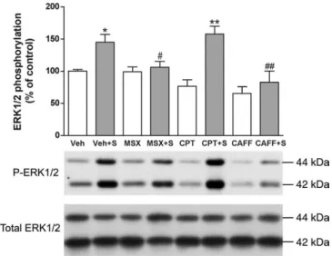
phosphoryla-tion. Cortical stimulation induced a significant 50% increase in
ERK1/2 phosphorylation in the ipsilateral striatum compared
with electrode-implanted nonstimulated controls (Fig. 2). This
effect was significantly counteracted by the nonselective
adeno-sine receptor antagonist caffeine and by the selective A
2Areceptor
antagonist MSX-3 but not by the A
1receptor antagonist CPT
(Fig. 2). None of the adenosine receptor antagonists produced
any significant effect in sham nonstimulated animals (Fig. 2).
Corticostriatal stimulation was also found to significantly
in-crease phosphorylation of the Ser
845residue of the GluR1 subunit
of the AMPA receptor in the ipsilateral striatum (⬃75% increase
compared with controls) (Fig. 3). Striatal AMPA receptor
phos-phorylation was also completely counteracted by caffeine or
MSX-3, whereas CPT did not significantly modify AMPA
recep-tor phosphorylation induced by cortical stimulation. Again, none
of the adenosine receptor antagonists produced any significant
effect in sham nonstimulated animals (Fig. 3). There were not
significant differences in the values of total ERK1/2 and GluR1
subunit of the AMPA receptors between the different sham and
stimulated groups.
Discussion
It can be assumed that there are two major distinct types of
stri-atal glutamatergic synapses, the D
1receptor-regulated and the D
2(and A
2A) receptor-regulated glutamatergic synapses, mostly
lo-calized at the heads of the dendritic spines of the GABAergic
dynorphinergic and GABAergic enkephalinergic neurons,
re-Figure 2. Striatal ERK1/2 phosphorylation induced by cortical electrical stimulation. Caf-feine and the A2Areceptor antagonist MSX-3, but not the A1receptor antagonist CPT, counteract
ERK1/2 phosphorylation induced by cortical electrical stimulation. Results are shown in means⫾ SEM (n ⫽ 6–10 per group) of representative Western blots. *p ⬍ 0.05 and **p ⬍ 0.01, significantly different compared with the vehicle (Veh)-treated group;#p⬍ 0.05 and ##p⬍ 0.01, significantly different compared with the stimulated vehicle-treated group
(Veh⫹S) (ANOVA with Tukey–Kramer tests).
Figure 3. Striatal GluR1 phosphorylation induced by cortical electrical stimulation. Caffeine and the A2Areceptor antagonist MSX-3, but not the A1receptor antagonist CPT, counteract
GluR1 phosphorylation induced by cortical electrical stimulation. Results are shown in means⫾ SEM (n⫽ 5–13 per group) and representative Western blots. *p ⬍ 0.05 and ***p ⬍ 0.001, significantly different compared with the vehicle (Veh)-treated group;###p⬍ 0.001,
signifi-cantly different compared with the stimulated vehicle-treated group (Veh⫹S) (ANOVA with Tukey–Kramer tests).
spectively (Ferre´ et al., 2005). Dopamine afferents make
prefer-ential synaptic contact with the neck of the dendritic spines
(Gerfen, 2004). This arrangement allows mesencephalic
dopami-nergic inputs to modulate cortico-limbic-thalamic glutamatergic
excitatory inputs. In the GABAergic dynorphinergic neuron,
do-pamine stimulates D
1receptors, which modulate neuronal
excit-ability and glutamatergic neurotransmission by inducing
PKA-mediated phosphorylation of different substrates, which include
NMDA and AMPA receptors. PKA-mediated phosphorylation of
NMDA receptors (in the C terminus of the NR1–1 subunit)
po-tentiates NMDA receptor-mediated currents (Greengard et al.,
1999), and there is also evidence for a tight crosstalk between the
second-messenger pathways of D
1and NMDA receptors
(Dud-man et al., 2003). These functional interactions depend on the
existence of physical interactions, heteromerization, between the
D
1receptor and specific subunits of the NMDA receptor (Lee et
al., 2002; Woods and Ferre´, 2005). Coactivation of D
1and
NMDA receptors induces MAPK activation (Valjent et al., 2005),
although a significant phosphorylation of ERK1/2 can be
ob-tained with selective stimulation of D
1receptors under
condi-tions of chronic dopamine depletion (Gerfen et al., 2002; Kim et
al., 2006). D
1receptor-mediated PKA activation can also induce
phosphorylation at Ser
845of the GluR1 subunit of the AMPA
receptor (Wolf et al., 2003), which, together with
phosphoryla-tion of ERK1/2, are critical initial steps in the establishment of
plastic changes in excitatory synapses, involving recruitment of
AMPA receptors to the postsynaptic density (Malinow and
Malenka, 2002; Lee et al., 2003; Thomas and Huganir, 2004). In
fact, D
1receptor-mediated AMPA receptor phosphorylation (at
Ser
845of the GluR1 subunit) has been shown to be involved in the
dopamine-mediated modulation of glutamate-dependent
stria-tal synaptic plasticity (Wolf et al., 2003).
The D
1receptor-mediated mechanisms by which dopamine
can potentiate the function and plasticity of glutamatergic
syn-apses in the GABAergic dynorphinergic neuron cannot operate
in the D
2–A
2Areceptor-regulated glutamatergic synapse of the
GABAergic enkephalinergic neurons, in which dopamine, by
acting on D
2receptors, inhibits the cAMP–PKA cascade and
MAPK (Ferre´ et al., 1997, 2004, 2005; Gerfen et al., 2002). In the
GABAergic enkephalinergic neurons, it is adenosine, by acting on
A
2Areceptors, that mimics the role of dopamine and D
1receptors
on the glutamatergic synapses of GABAergic dynorphinergic
neurons. A
2Areceptors are more concentrated in the striatum
than anywhere else in the brain, and they are preferentially
lo-cated both presynaptically and postsynaptically at glutamatergic
synapses, in a strategic position to modulate corticostriatal input
to the GABAergic enkephalinergic neurons (Hettinger et al.,
2001; Ciruela et al., 2006). At the postsynaptic site, A
2Areceptors
form heteromeric complexes with D
2and metabotropic
gluta-mate mGlu5 receptors localized in the perisynaptic ring outside
the synaptic cleft (Ferre´ et al., 2005). Costimulation of A
2Aand
mGlu5 receptors exerts a synergistic effect on their ability to
in-hibit dopamine binding to the D
2receptor and on MAPK
activa-tion (Popoli et al., 2001; Ferre´ et al., 2002; Nishi et al., 2003). At
the presynaptic site and inside the synaptic cleft, A
2Areceptors
form heteromeric complexes with adenosine A
1receptors, which
provide a switch mechanism by which low and high
concentra-tions of adenosine inhibit and stimulate, respectively, glutamate
release (Ciruela et al., 2006).
In the GABAergic enkephalinergic neurons, A
2Areceptor-mediated PKA activation can potentially phosphorylate the
dif-ferent substrates involved in synaptic plasticity. However, under
basal conditions, endogenous dopamine exerts a strong tonic
activation of D
2receptors in the striatum, which impairs the
ability of A
2Areceptors to signal through the cAMP–PKA
cas-cade. For instance, A
2Areceptor blockade does not produce a
decrease in the basal PKA-mediated phosphorylation of GluR1
(at Ser
845of the GluR1 subunit), although it counteracts GluR1
phosphorylation induced by D
2receptor blockade (Hakansson et
al., 2006). We have postulated that a strong
cortico-limbic-thalamic glutamatergic input produces a sufficient amount of
intrasynaptic glutamate and adenosine (most probably derived
from ATP coreleased with glutamate), with a significant
stimula-tion of presynaptic A
2Areceptors and postsynaptic perisynaptic
A
2Aand mGlu5 receptors. Then, under conditions of strong
glu-tamatergic input, adenosine and A
2Areceptors can provide a
mechanism of facilitation of plastic changes in the excitatory
syn-apses of the GABAergic enkephalinergic neurons (Ferre´ et al.,
2005). The present results strongly support this hypothesis,
be-cause phosphorylation of ERK1/2 and GluR1 at Ser
845induced by
stimulation of corticostriatal afferents was completely dependent
on A
2Areceptor function. The results also indicate that caffeine,
the most consumed psychoactive drug in the world, can
poten-tially alter this form of striatal plasticity, which suggests that
caf-feine may be deleterious for the normal development of striatal
function.
References
Berretta S, Parthasarathy HB, Graybiel AM (1997) Local release of GABAer-gic inhibition in the motor cortex induces immediate-early gene expres-sion in indirect pathway neurons of the striatum. J Neurosci 17:4752– 4763.
Ciruela F, Casado V, Rodrigues RJ, Lujan R, Burgueno J, Canals M, Borycz J, Rebola N, Goldberg SR, Mallol J, Cortes A, Canela EI, Lopez-Gimenez JF, Milligan G, Lluis C, Cunha RA, Ferre´ S, Franco R (2006) Presynaptic control of striatal glutamatergic neurotransmission by adenosine A1–A2A
receptor heteromers. J Neurosci 26:2080 –2087.
Dudman JT, Eaton ME, Rajadhyaksha A, Macias W, Taher M, Barczak A, Kameyama K, Huganir R, Konradi C (2003) Dopamine D1 receptors mediate CREB phosphorylation via phosphorylation of the NMDA re-ceptor at Ser897-NR1. J Neurochem 87:922–934.
Ferre´ S, von Euler G, Johansson B, Fredholm BB, Fuxe K (1991) Stimulation of high-affinity adenosine A2 receptors decreases the affinity of dopamine D2 receptors in rat striatal membranes. Proc Natl Acad Sci USA 88:7238 –7241.
Ferre´ S, Fredholm BB, Morelli M, Popoli P, Fuxe K (1997) Adenosine-dopamine receptor-receptor interactions as an integrative mechanism in the basal ganglia. Trends Neurosci 20:482– 487.
Ferre´ S, Karcz-Kubicha M, Hope BT, Popoli P, Burgueno J, Gutierrez MA, Casado V, Fuxe K, Goldberg SR, Lluis C, Franco R, Ciruela F (2002) Synergistic interaction between adenosine A2A and glutamate mGlu5 receptors: implications for striatal neuronal function. Proc Natl Acad Sci USA 99:11940 –11945.
Ferre´ S, Ciruela F, Canals M, Marcellino D, Burgueno J, Casado V, Hillion J, Torvinen M, Fanelli F, Benedetti Pd P, Goldberg SR, Bouvier M, Fuxe K, Agnati LF, Lluis C, Franco R, Woods A (2004) Adenosine A2A-dopamine D2 receptor-receptor heteromers. Targets for neuro-psychiatric disorders. Parkinsonism Relat Disord 10:265–271.
Ferre´ S, Borycz J, Goldberg SR, Hope BT, Morales M, Lluis C, Franco R, Ciruela F, Cunha R (2005) Role of adenosine in the control of homo-synaptic plasticity in striatal excitatory synapses. J Integr Neurosci 4:445– 464.
Gerfen CR (2004) Basal ganglia. In: The rat nervous system (Paxinos G, ed), pp 445–508. Amsterdam: Elsevier Academic.
Gerfen CR, Miyachi S, Paletzki R, Brown P (2002) D1dopamine receptor
supersensitivity in the dopamine-depleted striatum results from a switch in the regulation of ERK1/2/MAP kinase. J Neurosci 22:5042–5054. Greengard P, Allen PB, Nairn AC (1999) Beyond the dopamine receptor:
the DARPP-32/protein phosphatase-1 cascade. Neuron 23:435– 447. Hakansson K, Galdi S, Hendrick J, Snyder G, Greengard P, Fisone G (2006)
Regulation of phosphorylation of the GluR1 AMPA receptor by dopa-mine D2 receptors. J Neurochem 96:482– 488.
Hettinger BD, Lee A, Linden J, Rosin DL (2001) Ultrastructural localization of adenosine A2A receptors suggests multiple cellular sites for modulation of GABAergic neurons in rat striatum. J Comp Neurol 431:331–346. Hillion J, Canals M, Torvinen M, Casado V, Scott R, Terasmaa A, Hansson A,
Watson S, Olah ME, Mallol J, Canela EI, Zoli M, Agnati LF, Ibanez CF, Lluis C, Franco R, Ferre´ S, Fuxe K (2002) Coaggregation, cointernaliza-tion, and codesensitization of adenosine A2A receptors and dopamine D2 receptors. J Biol Chem 277:18091–18097.
Karcz-Kubicha M, Antoniou K, Terasmaa A, Quarta D, Solinas M, Justinova Z, Pezzola A, Reggio R, Muller CE, Fuxe K, Goldberg SR, Popoli P, Ferre´ S (2003) Involvement of adenosine A1 and A2A receptors in the motor effects of caffeine after its acute and chronic administration. Neuropsy-chopharmacology 28:1281–1291.
Kawaguchi Y, Wilson CJ, Emson PC (1990) Projection subtypes of rat neos-triatal matrix cells revealed by intracellular injection of biocytin. J Neu-rosci 10:3421–3438.
Kim DS, Palmiter RD, Cummins A, Gerfen CR (2006) Reversal of supersen-sitive striatal dopamine D1 receptor signaling and extracellular signal-regulated kinase activity in dopamine-deficient mice. Neuroscience 137:1381–1388.
Kull B, Ferre´ S, Arslan G, Svenningsson P, Fuxe K, Owman C, Fredholm BB (1999) Reciprocal interactions between adenosine A2A and dopamine D2 receptors in Chinese hamster ovary cells co-transfected with the two receptors. Biochem Pharmacol 15:1035–1045.
Lee FJ, Xue S, Pei L, Vukusic B, Chery N, Wang Y, Wang YT, Niznik HB, Yu XM, Liu F (2002) Dual regulation of NMDA receptor functions by di-rect protein-protein interactions with the dopamine D1 receptor. Cell 111:219 –230.
Lee HK, Takamiya K, Han JS, Man H, Kim CH, Rumbaugh G, Yu S, Ding L, He C, Petralia RS, Wenthold RJ, Gallagher M, Huganir RL (2003) Phos-phorylation of the AMPA receptor GluR1 subunit is required for synaptic plasticity and retention of spatial memory. Cell 112:631– 643.
Malinow R, Malenka RC (2002) AMPA receptor trafficking and synaptic plasticity. Annu Rev Neurosci 25:103–126.
Nishi A, Liu F, Matsuyama S, Hamada M, Higashi H, Nairn AC, Greengard P (2003) Metabotropic mGlu5 receptors regulate adenosine A2A receptor signaling. Proc Natl Acad Sci USA 100:1322–1327.
Popoli P, Pezzola A, Torvinen M, Reggio R, Pintor A, Scarchilli L, Fuxe K, Ferre´ S (2001) The selective mGlu5 receptor agonist CHPG inhibits
quinpirole-induced turning in 6-hydroxydopamine-lesioned rats and modulates the binding characteristics of dopamine D2receptors in the rat
striatum: interactions with adenosine A2areceptors.
Neuropsychophar-macology 25:505–513.
Sgambato V, Pages C, Rogard M, Besson MJ, Caboche J (1998) Extracellular signal-regulated kinase (ERK) controls immediate early gene induction on corticostriatal stimulation. J Neurosci 18:8814 – 8825.
Thomas GM, Huganir RL (2004) MAPK cascade signalling and synaptic plasticity. Nat Rev Neurosci 5:173–183.
Valjent E, Pascoli V, Svenningsson P, Paul S, Enslen H, Corvol JC, Stipanov-ich A, Caboche J, Lombroso PJ, Nairn AC, Greengard P, Herve D, Girault JA (2005) Regulation of a protein phosphatase cascade allows conver-gent dopamine and glutamate signals to activate ERK in the striatum. Proc Natl Acad Sci USA 102:491– 496.
Wolf ME, Mangiavacchi S, Sun X (2003) Mechanisms by which dopamine receptors may influence synaptic plasticity. Ann NY Acad Sci 1003:241–249.
Woods AS, Ferre´ S (2005) Amazing stability of the arginine-phosphate elec-trostatic interaction. J Proteome Res 4:1397–1402.
Zanassi P, Paolillo M, Feliciello A, Avvedimento EV, Gallo V, Schinelli S (2001) cAMP-dependent protein kinase induces cAMP-response element-binding protein phosphorylation via an intracellular calcium release/ERK-dependent pathway in striatal neurons. J Biol Chem 276: 11487–11495.