2016
UNIVERSIDADE DE LISBOA
FACULDADE DE CIÊNCIAS
DEPARTAMENTO DE BIOLOGIA VEGETAL
Aloe species in colorectal cancer therapy:
Friend or Foe?
Eduarda Pimenta Veríssimo
Mestrado em Biologia Molecular e Genética
Dissertação orientada por:
Prof. Dr. Ricardo Boavida Ferreira
Prof. Anabela Rosa Bernardes dos Santos Silva
~ III ~
Agradecimentos
Ao finalizar este trabalho, gostaria de expressar o meu sincero agradecimento pelo apoio, disponibilidade e colaboração concedida por diversas pessoas que, de forma direta ou indireta, contribuíram para a sua realização. A todas elas, os meus maiores agradecimentos:
À Faculdade de Ciências da Universidade de Lisboa, pelo contributo que deu para o desenvolvimento do meu conhecimento académico ao longo do mestrado.
Ao Instituto Superior de Agronomia, por me ter recebido e ter permito a realização desta dissertação de mestrado.
Ao Prof. Ricardo Boavida Ferreira, por todo o conhecimento transmitido ao longo de vários meses, por nunca perder a vontade de trabalhar mais e melhor e por me ter incutido também esse sentimento. Por todas as palavras de incentivo e por todo o reconhecimento concedido ao longo de todo este tempo.
À Dra. Ana Lima, por toda a formação que me deu no laboratório e fora dele, por todo o apoio e paciência, por toda a ajuda durante todos estes meses de trabalho. Obrigada por tudo!
À Prof. Anabela Silva, por todo o interesse demonstrado num tema algo diferente mas acima de tudo pela ajuda e compreensão que concedeu ao longo da dissertação.
A todos os colegas que ao longo do tempo partilharam tanto o laboratório como o gabinete comigo, em especial à Joana Guerreiro, ao Ricardo Chagas, Raimundo Diz, à Regina Freitas e à Ana Cristina Ribeiro que de uma forma ou de outra sempre me ajudaram e incentivaram.
À especial Ana Margarida Pinheiro e Filipe Rollo de quem sinto muita falta, obrigado por todos os momentos partilhados, bons e maus; obrigado pela ajuda e incentivo; obrigada por sempre me fazerem sentir bem onde estava e nunca me deixarem desistir!
Ao Lucas e ao João Fernandes pela ajuda que me deram a desenvolver uma última parte do trabalho que sem vocês nunca seria possível.
À Maria João pela disponibilidade em ajudar e contribuir para o bom funcionamento de todas as atividades e pelas palavras de apoio que sempre me dirigiu.
À Rita Francisco e à Mariana Santos, que sempre estiveram ao meu lado e com quem debati imensos assuntos e a quem sempre pude recorrer quando surgiram dúvidas e certezas.
À minha família que sempre me apoiou e a quem muitas horas foram roubadas para a realização deste trabalho. A eles dedico o meu empenho, dedicação e esforço na realização deste trabalho.
Ao meu namorado Pedro, muito obrigado por seres quem és, por todo o apoio, ajuda e incentivo. Nunca me deixaste desistir e mesmo nos momentos mais difíceis sempre soubeste o que dizer.
A todos, um enorme obrigado! Este período ficará para sempre no meu coração e memória, pois
Index
Agradecimentos ………. II Abstract ... VI Resumo...VII List of Figures ... X List of Tables... X List of Abbreviations... XI 1 - Introduction……… 11.1 The Aloe genus: century-old known medicinal plants... 1
1.1.1 Anatomy... 1
1.1.2 Reported bioactive components and properties ... 2
1.2 Aloe as a pharmacological tool for cancer treatment... 3
1.3 Matrix metalloproteinases-2 and -9 as targets for cancer therapy... 3
1.3.1 MMP inhibition aimed at cancer prevention / treatment... 4
1.3.2 Can Aloe compounds target gelatinases MMP-9 and MMP-2? ... 4
1.4 The Aloe vera paradigm: friend or foe? ... 5
1.5 Objectives... 5
2 – Materials and Methods ... 7
2.1 Species selected and collection of plant samples ... 7
2.2 Preparation of the leaf extracts... 7
2.3 Quantification of bioactive compounds... 7
2.3.1 Proteins... 7
2.3.2 Phenolic compounds ... 7
2.3.2.1 Anthranquinone... 7
2.3.3 Total polysaccharides... 8
2.4 In vitro colon cancer cell assays... 8
2.4.1 HT29 cell culture ... 8
2.4.2 Cell proliferation assay ... 8
2.4.3 Minimal inhibitory concentrations (MICs) ... 8
2.4.4 Cell migration assay... 8
2.5 MMP-9 and MMP-2 catalytic activities... 9
2.5.1 Gelatinolytic activity... 9
2.5.2 Sodium dodecyl sulfate-polyacrylamide gel electrophoresis... 9
~ V ~
2.6 Statistical analysis ... 10
3 – Results and Discussion……….11
3.1 Selected species: A. vera and A. arborescens... 11
3.2 Bioactive compounds in A. vera and A. arborescens... 11
3.2.1 The amount of bioactive compounds extracted from A. arborescens and A. vera is influenced by the species and by the extraction procedure... 12
3.2.2 The majority of the phytochemical components from A. vera are better extracted in 50% (v/v) methanol... 14
3.3 Effects of total Aloe extracts in colon cancer cells... 14
3.3.1 Aloe extracts reduce cancer cell growth... 15
3.3.2 Aloe extracts differently reduce cancer cell invasion... 16
3.4 Aloe extracts inhibit gelatinase activity... 17
4 – Conclusions ... 20
5 – References ... 20
Abstract
Aloe plants have been suggested to be an important natural source of medical therapy agents
(including for cancer) for several years. Throughout the world, pharmaceutical companies and institutions are struggling to isolate its most potent bioactive compounds as a primary means of using its activities. Regardless of the species used, the extraction procedure selected or the lack of specific compounds responsible for any given bioactivity, the fact is that we have been witnessing several debates, disputes and a lot of conflicting results arising on the claimed anticancer properties of Aloe species.
During the last decade, several reports demonstrated that a subgroup of matrix metalloproteinases (MMPs) called gelatinases (MMP-2 and especially MMP-9), are largely responsible for colorectal cancer progression/metastasis, suggesting that MMP inhibitors (MMPIs) may be a powerful tool to reduce cancer invasion. Although Aloe’s activities suggest they might inhibit MMPs, no studies have related their reported anticancer activity with MMPI activities. Also, although approximately 500 species have been identified so far within the Aloe genus, A. vera is the most widely studied albeit other species such as A. arborescens have also been reported to exhibit similar bioactivities. Hence, the goal of the present study was to compare the anticancer potential of two Aloe species, A. vera and
A. arborescens and to ascertain if they are specific MMP-9 and/or MMP-2 inhibitors.
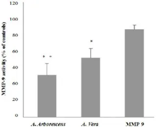
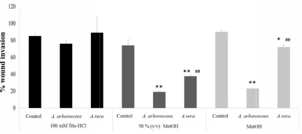
Different types of extraction were tested (100% (v/v) methanol, 50% (v/v) methanol and 100 mM Tris-HCl buffer pH 7) and specific bioactive compounds (proteins, total phenolic compounds, anthraquinones and total carbohydrates) were quantified and compared. Although there were a few variations between species and between extractions, 50% (v/v) methanol was selected as the best extraction procedure. Anticancer activities were measured in vitro using the wound healing model assay in the human colon cancer cells HT29, and A. vera showed significantly higher inhibitory potencial regarding wound closure. Furthermore, it was also evaluated their effects on gelatinase activities, measured by gelatin zymography and the DQ gelatinase assay. When assessing the total gelatinolytic activity, both species had a similar result of around 20% inhibition. Through the gelatin zimography we demonstrated that both MMP-9 pro-form and active form were inhibited, but only the MMP-2 pro form were inhibited, which corroborates the results obtained with the total gelatinolytic activity assay. The gelatinolytic activity of MMP-9 showed that although both species have the potencial to inhibit specifically this metalloproteinase, A. arborescens have a significantly higher inhibitory effectiveness.
Overall, our data provided clear indication that A. arborescens and A. vera have indeed potential as inhibitory agents in cancer therapy, being able to reduce colon cancer cell proliferation and invasion, possibly via specific MMP-9 inhibition. A. arborescens appears to be a more effective cancer cell invasion inhibitor than A. vera. Also, we observed that different types of extraction are important to obtain higher amounts of bioactive compounds and also higher levels of bioactivities. This might be due to a greater amount of extractable bioactive compounds such as anthraquinones. These results open novel perspectives on the mode of action of Aloe species in cancer invasion and may provide clues as to why there are so many conflicting results.
~ VII ~
Resumo
Desde a antiguidade que as espécies do género Aloe são consideradas como uma importante fonte natural de compostos terapêuticos. Contudo, nas últimas décadas tem-se verificado um crescimento rápido e exponencial do interesse popular nestas plantas. O Aloe barbadensis (mais vulgarmente conhecido como Aloe vera) é, entre as espécies deste género, a mais reconhecida e popularizada sendo-lhe atribuídas uma miríade de aplicações e indicações terapêuticas, muitas vezes não fundamentadas cientificamente. Consequentemente, a nível mundial, tanto empresas como instituições farmacêuticas procuram avidamente separar e identificar os compostos bioativos mais potentes entre espécies de Aloe, como forma de isolar potenciais bioatividades para aplicar em fármacos ou em produtos vendáveis. Contudo, tem surgido um grande debate entre a comunidade científica sobre o verdadeiro valor terapêutico desta espécie; enquanto alguns trabalhos científicos advogam o seu grande potencial, outros desacreditam totalmente os seus efeitos, alertando mesmo os seus eventuais perigos para a saúde. Existem várias razões possíveis para justificar estas inconsistências nos estudos publicados até à data. A. vera é a espécie mais utilizada e conhecida atualmente, sendo o elemento do seu género mais amplamente estudado, apesar de outras espécies tais como A. arborescens e A. ferox terem sido igualmente descritas como tendo bioatividades semelhantes ou mesmo superiores. Existe também grande inconsistência em relação ao tipo de solventes a utilizar nas extrações, o que pode influenciar a biodisponibilidade e quantidade de compostos ativos. Além disso, cada vez mais parece evidente que a ação terapêutica das plantas de Aloe poderá estar associada a uma sinergia entre os vários compostos bioativos presentes nas suas folhas e não apenas à ação de um único composto isolado. Por tudo isto, existe ainda muito trabalho a realizar e muitas questões que deveriam ser abordadas em relação a estas espécies.
Uma das aplicações mais conhecidas e procuradas do género Aloe é o seu potencial efeito anti-tumoral. Vários estudos têm comprovado que existe de facto um efeito preventivo e inibidor de vários tipos de tumores induzido pelos extratos e compostos de Aloe. Contudo, o mecanismo através do qual estes efeitos são exercidos ainda não está totalmente esclarecido.
Durante a última década, vários estudos revelaram que existe um subgrupo de metaloproteinases de matriz (MMPs), as gelatinases (MMP-2 e MMP-9), que estão envolvidas no processo de metastização. Estes trabalhos têm demonstrado que os seus inibidores (MMPIs) poderão ser uma ferramenta muito útil para reduzir a invasão tumoral. Estas enzimas (sobretudo a MMP-9) também estão fortemente envolvidas em processos de cicatrização e de inflamação. Deste modo, o facto de os efeitos das plantas de Aloe estarem associados simultaneamente a atividades cicatrizantes, inflamatórias e anti-tumorais sugere que poderão influenciar o mecanismo de ação da MMP-9 e da MMP-2. Contudo, até à data, ainda não existem estudos que associem o potencial anti-tumoral das espécies de Aloe com uma possível inibição das duas gelatinases Assim, neste contexto, no presente trabalho decidiu-se determinar e comparar a atividade anti-tumoral de duas espécies do género Aloe bastante conhecidas,
A. vera e A. arborescens, avaliando o seu impacto no crescimento e na migração de células de
adenocarcinoma do cólon e determinando se estes efeitos estão ou não associados a alguma influência na atividade das MMP-9 e/ou da MMP-2.
Foram primeiramente avaliadas as diferenças entre as duas espécies de Aloe, através da caraterização de diferentes compostos com potencial bioativo já descritas para este género, nomeadamente proteínas, compostos fenólicos, antraquinonas e hidratos de carbono totais. Foi também objetivo testar diferentes métodos de extração e tentar compreender qual o melhor processo de modo a obter o maior teor de compostos bioativos. Todos os compostos foram quantificados em três tipos de extracção: aquosa (em tampão Tris-HCl), hidro-metanólica (50% (v/v) metanol) e metanólica (100% (v/v) metanol).
Os resultados mostraram que, apesar de o teor em compostos bioativos não ter variado de forma marcada entre as duas espécies, A. vera apresentou um teor mais elevado em compostos fenólicos totais, enquanto A. arborescens apresentou valores superiores em antraquinonas. Os resultados comprovaram também que o método de extração não só influencia fortemente a quantidade de compostos bioativos isolados, como também difere entre as duas espécies. Por exemplo, enquanto A.
vera mostrou de um modo geral um maior teor em compostos extraídos a 50% (v/v) metanol, no A. arborescens estes valores variaram consoante os compostos determinados. Isto é de extrema
importância uma vez que muitos estudos utilizam apenas um tipo de extração, independentemente da espécie e do composto a isolar, sendo raros os artigos que utilizam a extração hidro-metanólica.
Em segundo lugar, foi determinado o potencial anti-tumoral dos melhores extratos de Aloe em células de adenocarcinoma do cólon humano (células HT29), utilizando os métodos padrão de proliferação celular e de invasão celular. O trabalho efetuado mostrou que as duas espécies estudadas apresentam de facto potencial para serem inibidoras do desenvolvimento tumoral, reduzindo tanto o crescimento celular como a invasão por células HT29. A inibição do crescimento das células tumorais foi dependente da dose e semelhante entre as duas espécies. Estes resultados permitiram ainda selecionar uma concentração EC50, que foi utilizada nos estudos subsequentes. Nos estudos de invasão tumoral, verificou-se que, apesar de as duas espécies terem tido atividades similares na proliferação celular, na invasão A. arborescens revelou ser a espécie com maior atividade, atingindo valores de inibição semelhantes ao conhecido inibidor de MMP-9, doxiciclina. Estes resultados sugerem que as pequenas diferenças de compostos bioativos observadas anteriormente, nomeadamente o maior teor em antraquinonas, poderão estar associados a um maior potencial de inibição por parte de
A. arborescens.
Uma vez que, segundo a literatura, uma maior inibição na invasão está normalmente associada a uma redução na atividade das gelatinases MMP-9 e MMP-2, foi subsequentemente avaliada a atividade destas duas enzimas no meio extracelular das células HT29, após exposição aos extratos de cada uma das espécies de Aloe. Para tal, foi utilizado o método da DQ gelatina que avalia a atividade gelatinolítica total e a zimografia de substrato, que separa as MMP-9 das MMP-2 num gel SDS-PAGE, permitindo a avaliação da atividade específica de cada uma. Os resultados das atividades das gelatinases corroboraram que os extratos das duas espécies possuem de facto capacidade de inibição das gelatinases, mas com maior especificidade para a MMP-9 do que para a MMP-2. De forma a testar se esta inibição ocorre por influência direta na enzima, foi ainda efetuado um ensaio de inibição enzimática com uma MMP-9 comercial na presença da DQ gelatina. Ambas as espécies mostraram inibir a gelatinase MMP-9. Contudo, o extrato de A. arborescens mostrou maior atividade inibitória que o de A. vera. Não obstante, a inibição da MMP-9 não foi tão elevada como a verificada nas zimografias do meio extracelular das células HT29. Deste modo, os resultados sugerem que poderão existir outros mecanismos de ação que não apenas a inibição direta da enzima.
Em conclusão, o trabalho efetuado mostrou que as duas espécies estudadas de Aloe apresentam de facto um potencial para serem inibidoras do desenvolvimento tumoral, inibindo tanto o crescimento celular como a invasão. Os resultados mostram também que esta inibição pode estar relacionada com uma redução específica da atividade da MMP-9 e da MMP-2.
Apesar de terem concentrações semelhantes das várias classes de compostos bioativos, A.
arborescens parece ser um inibidor da invasão tumoral mais eficaz do que A. vera. Isto pode ser
devido a uma maior quantidade de compostos bioativos individuais, como as antraquinonas presentes em A. arborescens, ou à presença de compostos específicos ainda não identificados nessa espécie. Os resultados aqui apresentados mostram igualmente que a utilização de espécies de Aloe, quer seja na prevenção ou na terapia tumoral, deve ter sempre em consideração o tipo de extração, devendo esta adaptar-se de acordo com a espécie e o objetivo em mente; de outro modo podem não ser obtidos os resultados mais realistas e reveladores do verdadeiro potencial da espécie.
~ IX ~
De um modo geral, A arborescens parece ser uma alternativa mais eficaz do que A. vera no caso da inibição e da invasão celulares, sugerindo um elevado potencial de utilização como coadjuvante em terapias tumorais, ou ainda na prevenção da formação de metástases em tumores já estabelecidos. Uma vez que a inibição direta das enzimas parece não ser o seu único mecanismo de ação, mais estudos deveriam ser efetuados para determinar a sua toxicidade e efeitos. Não obstante, os resultados obtidos neste trabalho abrem as portas para novos trabalhos sobre a inibição de MMPs por parte das espécies de Aloe, chamando a atenção para a necessidade de uma reavaliação do tipo de metodologias de extração utilizados, bem como as espécies utilizadas e respetivos alvos terapêuticos.
List of Figures
Figure 1.1 – Ancient Greek Dioscorides recorded his use of Aloe to treat war wounds as well as piles ………. 1 Figure 1.2 – Components of the typical leaf structure of an Aloe plant …….………. 2 Figure 1.3 – MMP-2 and MMP-9 structures ……….... 4 Figure 3.1 – Representative photos of the selected species A. arborescens and A. vera from the
herbarium collection at the Instituto Superior de Agronomia and during the flowering season …... 11
Figure 3.2 – Quantitative chemical characterization of A. vera and A. arborescens considering tha
main phytochemical groups defined by Hamman et al. (2008) ………... 13
Figure 3.3 – Cell proliferation assay ...………... 15 Figure 3.4 – A representative image of the wound healing assay on HT29 cells exposed to A. vera and
A. arborescens extracts prepared with 50% (v/v) methanol at time 0h and 48h of exposure
………..…….……… 17 Figure 3.5 – MMP-9 and MMP-2 activities are inhibited by A. vera and A. arborescens extracts ... 18 Figure 3.6 – MMP-9 and MMP-2 gelanolytic activities are inhibited by A. vera and A. arborescens
extracts ………...………... 18
Figure 3.7 – Gelanolytic activities of MMP-9 after exposure to 50% (v/v) methanol extracts of several
Aloe species ……….………. 19
List of Tables
Table 3.1 – Summary of the potential bioactive compounds from Aloe vera leaves ….………. 12 Table 3.2 – Minimal inhibitory concentrations for cell growth on HT29 cells exposed to different
concentrations of A. vera and A. arborescens extracts prepared with 50% (v/v) methanol
~ XI ~
List of Abbreviations
BSA – Bovine serum albumin CRC – Colorectal cancer DNA – Deoxyribonucleic acid DQ – Dye-quenched
EC50 – Half maximal effective concentration ECM - Extracellular matrix
EDTA - Ethylenediamine tetraacetic acid FBS – Fetal bovine serum
ISA – Instituto Superior de Agronomia, Universidade de Lisboa MIC – Minimal inhibitory concentration
MMP – Matrix metalloproteinase
MMPI – Matrix metalloproteinase inhibitor
MTT - 3-(4,5-Dimethylthiazol-2-yl)-2,5-diphenyltetrazolium bromide PVPP - Polyvinylpolypyrrolidone
PBS – Phosphate buffered saline
RPMI – Roswel Park Memorial Institute Medium SD – Standard deviation
SDS – Sodium dodecyl sulphate
SDS-PAGE – Sodium dodecyl sulphate-polyacrilamide gel electroforesis TCA – Trichloroacetic acid
TIMP – Tissue inhibitors of metalloproteinase TRIS – 2-Amino-2-hydroxymethyl-propane-1,3-diol
1 - Introduction
1.1 The Aloe genus: century-old known medicinal plants
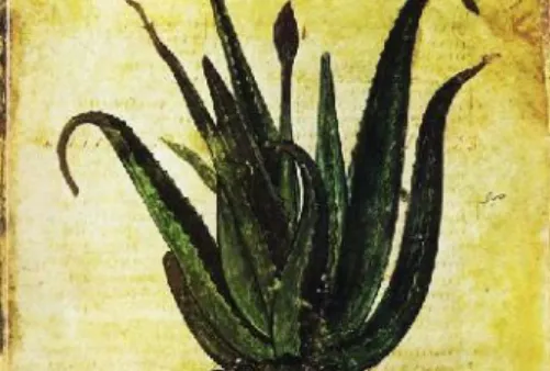
The genus Aloe has been one of the best known for medicinal purposes in several cultures for millennia and its uses are referred to as early as in Egyptian papyri or theMateria Medica of Dioscorides (Fig.1.1) and the Colloquies
of Garcia de Orta [1, 2]. Nonetheless, it was not until the last
decades that public interest in Aloe species grew tremendously, with such a vast array of proclaimed properties and applications, ranging from burns, skin damage, edema, pain and gastric ulcers, to cancer that it has been labeled a “cure-all miracle” plant. This boosted the investigator’s interest on this plant and consequently, a considerable amount of research about the various components of Aloe
began to arise, aiming to find out more about their properties and to pinpoint specific compounds which may be used/commercialized as pharmacological agents [3]. Indeed, Aloe extracts have been
reported to possess anti-inflammatory, anti-diabetes, cellular protection, restoration, and mucus-stimulating activities, among others, which might be related to its broad-range effects[4- 7].
Today, Aloe barbadensis (herein referred to as Aloe vera) is unquestionably the most popular species of Aloe used worldwide, often referred to as the one with the highest bioactivities in anecdotal reports. However, there are approximately 500 Aloe species documented, from which only a marginal fraction has recorded ethnomedicinal uses [8]. As a result of its huge medicinal demands and the
morphological similarities among several species, there is a high probability that certain species may often be erroneously identified for different medicinal uses. So in addition to the increasing interest in the phytochemical and pharmacological potential of Aloe, the regular evaluation and re-evaluation of existing bodies of knowledge are deemed necessary to provide an up-to-date information while identifying the current research gaps. In Portugal, access to different Aloe species is difficult, as they are mostly found in the African continent[8], but Instituto Superior de Agronomia (ISA) has the largest
collection of Aloe species in Europe, counting with more than 80 species, collecting species from Africa, identifying new taxa and establishing and maintaining cultures in the ISA´s botanical gardens.
1.1.1 Anatomy
The genus Aloe belongs to the Xanthorrhoeaceae family and subfamily Asphodeloideae, characterized by plants presenting a rosette of large and succulent leaves. The Aloe species are relatively similar regarding their anatomy. All plants have boat-shaped fleshy leaves with spiny margins, arranged in rosettes or spirals [9, 10]. These plants usually produce racemose inflorescences
with tubular flowers, with a yellow orange, pink or red color. They flower mainly during the winter season and produce large quantities of small, air-borne seeds, although there are many species that can also propagate asexually [9, 10]. Although Aloe plants are relatively similar in structure, they exhibit a
wide morphological variability and various growth forms [11], ranging in size from dwarf species, of
only a few cm high to giant, 20 m tree Aloes[9, 12].
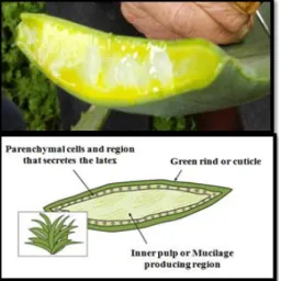
The leaf structure of the Aloe plant can be separate into three different components, from the exterior portion to the most interior: 1) a green rind or cuticle, with multiple layers interspersed with chloroplasts, 2) a region composed by vascular bundles with three tubular structures, which secretes a Figure 1.1 - Ancient Greek Dioscorides recorded his use of Aloe to treat war wounds as well as piles (Image: Vienna
~ 2 ~
latex or sap when cut, and 3) an inner pulp or gel [13]. The gel or pulp comprises the major part of the
leaf’s volume and is composed by large thin-walled parenchyma cells responsible for the clear mucilaginous aqueous extract obtained from this part of the plant [14, 15] (Fig.1.2). Carbohydrates
synthesized in excess are transported via phloem to these cells which also store water, minerals and malic acid. Thus, the parenchymal cells serve as a water and energy reservoir for the plant. The latex (yellow substance visible in Figure 1.2) is restricted to the margins of the leaves, suggesting that it may be a source of secondary metabolites [15].Although
there are several articles highlighting the pytochemical composition of Aloe vera, little is known about other species, such as A. arborescens and A. ferox, which have also been the target of several assays in the last years[16-19].
1.1.2 Reported groups of bioactive components and properties in the Aloe genus
According to Surjushe et al. (2008) [20] Aloe vera is by far the most thoroughly characterized
species within Aloe genus, contains many potentially active constituents: vitamins, enzymes, minerals, sugars, lignin, saponins, salicylic acids and amino acids [5, 22, 27]. Many of these compounds are
responsible for some pharmacological activities described, such as aloin, Aloesin, Aloenin, Aloeresin and Aloe-emodin, among others [8, 23]. It is known that these compounds differ greatly among species,
and some of them can be toxic and extremely allergenic[23, 24].
Below is a list of the main compounds present in A. vera:
1. Vitamins: vitamins A (β-carotene), C and E, which are antioxidants, are present in A. vera, which also contains vitamin B12, folic acid, and choline. Antioxidant compounds aid organisms coping with oxidative stress[7, 20, 21].
2. Enzymes: A. vera contains several enzymes such as alkaline phosphatase, amylase, bradykinase, carboxypeptidase, catalase, cellulase, lipase, and peroxidase. Bradykinase helps to reduce excessive inflammation when applied to the skin topically, while others help in the breakdown of sugars and fats[7, 20].
3. Minerals: A. vera is a good dietary source of calcium, chromium, copper, selenium, magnesium, manganese, potassium, sodium and zinc. These elements are essential for the proper functioning of various enzyme systems in different metabolic pathways and a few also participate in the organism’s antioxidants defenses[7, 20].
4. Sugars: A. vera provides essentially monosaccharides (glucose and fructose) and polysaccharides: (glucomannans/polymannose). These are derived from the mucilage layer of the plant and are known as mucopolysaccharides. The most prominent monosaccharide is mannose-6-phosphate, and the most common polysaccharides are glucomannans [β-(1,4)-acetylated mannan]. Acemannan, a prominent glucomannan has also been found. Recently, a glycoprotein with antiallergic properties, called alprogen and a novel anti-inflammatory compound, C-glucosyl chromone, have been isolated from Aloe vera gel[ 23, 25, 26, 29].
5. Anthraquinones: A. vera provides many anthraquinones, which are phenolic compounds traditionally known as laxatives [8, 20]. Aloe-emodin, Aloesinand barbaloin are associated to
Figure 1.2 - Components of the typical leaf structure of an Aloe plant. (Adapted from http://www.niehs.nih.gov/health/topics/agents/aloe/ index.cfm)
decelerate tumor growth and had been reported to have chemo-preventive effect in cancer cells
[21].
6. Fatty acids: A. vera provides 4 plant steroids, cholesterol, campesterol, β-sisosterol and lupeol. These exhibit anti-inflammatory action and lupeol also possesses antiseptic and analgesic properties[20].
7. Hormones: A. vera auxins and gibberellins help in wound healing and have anti-inflammatory action[7, 20].
8. Others: A. vera contains salicylic acid that possesses anti-inflammatory and antibacterial properties. Lignin, an apparently inert phenolic polymer, when included in topical preparations, enhances penetrative effect of the other ingredients into the skin. Saponins are the isoprenoid soapy substances which form about 3% of the gel and have cleansing and antiseptic properties[20].
1.2 Aloe as a pharmacological tool for cancer treatment
Albeit the widespread use of popular traditional medicine throughout the globe, Aloe species are used for several ailments, ranging from immunomodulatory, anti-inflammatory, antiulcer, antimicrobial and antifungal activity [7, 14, 18, 30]. One of its most popular attributes is proclaimed
anticancer activities, which have also been one of the most studied activities in this genus. Reported studies have shown the plant´s effectiveness towards various cancer types such as liver, colon, duodenal, skin, pancreatic, intestinal, lung and kidney, and these studies have been confirmed by several experimental in vitro and in vivo studies [31, 32]. Aloe vera extracted components have been
proven to decelerate tumor growth, especially the anthraquinones like Aloesin, Aloe-emodin and barbaloin [33]. These components have been reported to exert a chemo-preventive effect through the
regulation of several enzymes in the cell [33]. Aloe-emodin can induce cell apoptosis through S-phase
arrest in a dose- and time-dependent manner [33-36]. Acemannan extracted from A. vera and A.
arborescens parenchyma cells have an immunomodulatory activity and stimulate necrosis and
regression of tumors [25, 26, 37]. This polysaccharide also shows to have a significant effect on wound
healing, by accelerating the process and increasing epithelialization in burns[37].
1.3 Matrix metalloproteinases-2 and -9 as targets for cancer therapy
Matrix metalloproteinases (MMPs) are a multigene family of zinc-dependent extracellular matrix remodeling endopeptidases involved in several pathological processes like carcinogenesis, inflammation and auto-immune disorders. Their regulatory activity was found to be fundamental for tumor growth and for the multistep processes leading to invasion and metastasis[38, 39]. Several studies
demonstrated that a specific group of MMPs called gelatinases (MMP-2 and MMP-9) are highly related to cancer invasion both by degradation of the cellular matrix [40-43], and the subsequent release
of cancer cells through proteolysis [44]. These gelatinases contain a unique set of three repeats of
fibronectin that facilitate degradation of gelatinous substrates such as elastin, collagen type I and IV, gelatin and fibrinogen (Fig.1.3)[45-47].
~ 4 ~
Figure 1.3 - MMP-2 and MMP-9 structures (Adapted from 3http://arthritis-research.com/content/figures/ar2532-1-l.jpg). Regarding the wide variety of molecules that can be influenced by MMPs activity, their proteolytic function can result in both local and long-distance effects regarding the original site of product cleavage [48]. Also, considering the range of proteins influenced by MMPs, their pivotal role in a
cancer scenario can be easily comprehended.
1.3.1 MMP inhibition aimed at cancer prevention/treatment
Since death of cancer patients is usually caused by metastization rather than caused directly by the primary tumor itself[49, 50], and considering that cancer cell invasion is a key element in metastasis and
requires MMPs for focalized proteolysis [44, 51-53], in the last decades most anticancer drugs were
designed to target MMP activity as MMP inhibitors (MMPIs), which became an important branch of research in both academic and industrial settings [54, 55]. So far, numerous MMP inhibitors have been
tested in in vitro assays and in animal models, as well as in clinical trials [56-58]. However MMPIs with
high specificity and low side effects have been very hard to find, and most clinical trials yielded unsatisfactory results[50, 56, 57, 59]. A major problem associated to unspecific inhibition of MMP-9 is that
this enzyme is involved not only in various diseases but also beneficial in remodeling and scar tissue, so its complete inhibition causes several complications in the human body [60, 61]. A distinct and more
recent strategy in the search for novel MMP-9 inhibitors is to ‘look’ among the multitude of natural products that nature placed at our disposal. Under this context, there has been an intense search on various biological sources to develop novel anti-cancer drugs, particularly plant-food products or bioactive plant compounds[62, 63].
1.3.2 Can Aloe compounds target gelatinases MMP-9 and MMP-2?
Since MMP-9 and MMP-2 have been recently closely associated to inflammation, wound closure and also the formation of metastasis in several types of cancer [64-68], the wide range activities of Aloe
suggest its target might be a link between these two diseases, i.e. inflammation and cancer. But although it is known that MMPs play a critical role in both[69-71], to our knowledge there has been no
link established between MMP-9 inhibition induced by Aloe and cancer. So far, Aloe’s anticancer activity and antineoplastic properties is related to three main mechanisms: antiproliferation, immunostimulation and antioxidant effects. The antiproliferative action is known to be determined by anthracenic and antraquinonic molecules, while the immunostimulating activity is mainly due to acemannan[16].
Nonetheless, regarding inflammation, Aloe vera extracts are known to down regulate metalloproteinases expression, specifically MMP-9, which actively participates in the degradation of the extracellular matrix (ECM) when recruited by cytokines to inflammation sites [72]. So it seems
plausible that Aloe species might inhibit MMP-9 in cancer scenarios. This type of studies can be useful to ascertain the real effect of Aloe species on cancer and also lead to the identification of novel anticancer strategies which can be used in prevention, for assisting chemotherapy, or even to prevent re-incidence after surgery. Furthermore, the analysis of MMP inhibition may also be a novel perspective which can help to understand the mechanisms of action observed in A. vera.
1.4 The Aloe vera paradigm: friend or foe?
There is an intense debate whether the so called miracle plant exhibits all the proclaimed benefits. For example, whilst many papers described its anticancer properties [28, 31, 32, 73], other authors
suprisingly claim A. vera is in fact carcinogenic over a specific dose [23] or alert that some specific
compounds in Aloe may be severely toxic if not taken under caution [26]. Some countries such as the
USA have even forbidden the use of Aloe for consumption because of the lack of consensus towards this species’ activities (although it is still allowed as a topic agent). This dramatic imposition is highly disputed by communities worldwide, as the empirical experience and anecdotal results suggest that
Aloe has a high efficiency. This results in a generalized unsupervised use of Aloe, without a scientific
background. Furthermore, the phenotype similarities among species are extremely high, which induces people to use often the wrong species. Therefore, the current unsupervised use of A. vera can result in cases of allergies and toxic effects, and in scientific studies, can originate an erroneous evaluation of the real effective benefits of the species[71, 74, 75].
Many reasons can be advanced to explain these contradictory reports on A. vera: first and foremost, the overall misidentification of the Aloe genus’ species, not only because they can be erroneously mistaken with A. vera, but also because different species with potentially different and stronger bioactivities might be being neglected. In fact, ancient medical texts refer to A. perryi or A. ferox instead of A. vera [1, 64] for medicinal effects. Current research also supports other Aloe species do
contain more potent bioactivities than A.vera[8]. Nonethless these species continue to be neglected. A
search on PubMed (29.09.2016) gave a total of 2689 hits for Aloe (more than 96% involving A. vera) with only very few studies focusing on species other than A. vera (e.g. A. arborescens and A. ferox). So it seems plausible to infer that we have barely scratched the surface in exploring these other species as sources of pharmacological agents[8].
Secondly, in the last decades an effort has been made to isolate phytochemicals that can be used as a single chemical entity and as an alternative to synthetic drugs. However, in the last few years a different perspective has emerged suggesting that using crude and/or standardized extracts as opposed to single compounds might be an advantage since each component has its major effect when acting synergistically with other components in the plant [30, 75, 76]. In this context, it also important to refer
that there is an overall lack of consistency concerning the type of extractions to use. Some reports refer to aqueous extracts [77]whilst others use alcoholic or organic solvents such as methanol or acetone[78, 79]. Since the main phytochemical groups range from phenolic compounds to carbohydrates, proteins,
organic and inorganic compounds as well as vitamins [7, 21, 82-84], it becomes important to test different
types of extractions to identify which provide the highest amount of bioactive compounds and allows the better synergy among them.
1.5 Objectives
The genus Aloe has a high therapeutic potential and is often described as a cancer inhibitor. However, the mechanisms by which different species exert their effects it is not clear and there are many conflicting results. Also, less than 20 Aloe species have been evaluated under this perspective and their efficacy is highly debated throughout the scientific community. The fact that their effects are
~ 6 ~
associated with wound healing, anti-inflammatory and antitumor activities suggests that they may influence the activity of the gelatinases MMP-9 and MMP-2. However, there is a generalized lack of consensus towards the type of extraction one should use, the mechanism of action responsible for the proclaimed benefits of Aloe and furthermore, if A .vera is indeed the best species for cancer prevention / treatment. Hence, in this work, we aimed to evaluate and compare the antitumor activity of two well-known species of the genus Aloe: Aloe vera and Aloe arborescens and to ascertain if it is related to MMP inhibition. With that in mind, we set out to evaluate their content in several classes of potentially bioactive compounds, using different extraction procedures and to assess their impact on the growth and migration of colon adenocarcinoma cells, as well as their effect on 9 and MMP-2 activities.
The specific goals were:
1 – To compare the potentially bioactive compounds in both species, Aloe vera and Aloe arborescens; 2 – To determine which is the best extraction procedure to obtain higher amounts of the bioactive compounds;
2 – To test the effect of the same extracts in colon cancer cells, aiming to evaluate specifically cell proliferation, cell adhesion and cell invasion rates.;
4 – To test the potential of Aloe extracts as inhibitors of the gelatinases MMP-9 and MMP-2, using enzymatic and zymographic assays;
3 – To identify possible mechanisms of action in both Aloes, selecting the better extraction procedures and the best species to be used in prevention and therapy in colon cancer.
2 – Materials and Methods
2.1 Species selected and collection of plant samples
Leaves of A. vera, and A. arborescens were collected from the existing Collection of Aloe Plants of the Instituto Superior de Agronomia (Universidade de Lisboa), probably the biggest collection of this genus outside Africa, with over 80 species. All selected individuals had several years of existence and were not in the flowering season. Leaf samples were collected from at least three separate individuals.
2.2 Preparation of the leaf extracts
Fresh leaves were washed with running tap water and soap and chopped into small fragments of approximately 20 g each. Three different extraction methods were performed using different solvents: 1) 100% (v/v) methanol, 2) 50% (v/v) methanol and 3) 100 mM Tris-HCl buffer, pH 7. All extractions were performed grinding a fragment of 20 g with the respective solvent using an ULTRA-TURRAX T25 (IKA®Labortechnik) grinder, followed by agitation for 4h at a 4º C. Both extracts containing
methanol were evaporated in a bath at 60˚C (Kottermann) whereas the aqueous extracts were desalted through filtration using 3 kDa membrane centricons and centrifuged at 2.000 g. All extracts were then lyophilized (Edwards Modulyo EF4) for 24h and the obtained powder was weighted and stored at -20ºC.
2.3 Quantification of bioactive compounds
2.3.1 Proteins
Protein quantification was performed using the standard Bradford method as described by Bradford
et al. (1976) [85]. The samples were read in a spectrophotometer Syenery HT, Bio-TEK at 595 nm and
bovine serum albumin was used as standard. All samples used in protein quantification were treated with 1:4 (w/w) polyvinylpolypyrrolidone (PVPP) to eliminate the phenolic compounds that could interfere with the extraction of soluble proteins[86].
2.3.2 Phenolic compounds
The phenolic compounds were quantified using the Folin-Ciocalteau reagent using gallic acid as standard. The lyophilized powder (corresponding to 20g of fresh leaves) was treated with 10 µL of 70% (v/v) acetone, 10 µL of 0.5% (v/v) acetic acid and 80 µL of 7% (w/v) sodium carbonate. Subsequently, a volume of 100 µL of Folin-Ciocalteau was added to the solution and the mixture was vortexed. The solution of 200 µL was incubated for 8 min at room temperature and the absorbance was read in a Syenery HT Bio-TEK spectrophotometer at 765 nm[87].
2.3.2.1 Anthraquinone
To 300 µL of the previous extracts, 300 µL of pure benzene were added and the resulting solution incubated with agitation at 0˚C bath for 30 min. It was then centrifuged at 4.500 g for 20 min at 4˚C in a Beckman J2-21M/E centrifuge. To the recovered supernatant 500 µL of 10% (v/v) ammonia solution were added and the absorbance read at 515 nm using Aloe blue curacao aloin as a standard[88].
~ 8 ~
2.3.3 Total carbohydrates
Sugar quantification was performed utilizing the phenol/sulfuric acid method and mannose was used as the standard. To the previous extracts a 4% (v/v) phenol solution was added in a proportion of 1:5 and then incubated 5 min at room temperature. Afterwards, a 1:40 proportion of sulfuric acid was added and the absorbance read at 492 nm in the equipment mentioned above[89].
2.4 In vitro colon cancer cell assays
2.4.1 HT29 cell culture
The human colon adenocarcinoma cell line, HT29 (ECACC 85061109), obtained from a 44 year old Caucasian female, was used throughout this work. HT29 cell lines were maintained in RPMI medium supplemented with 10% (w/v) of heat-inactivated fetal bovine serum (FBS) and 200 mM glutamine, 2x10⁴UI/mL penicillin and 20 mg/mL streptomycin at 37 °C, in a humidified atmosphere
of 5% (v/v) CO2.
2.4.2 Cell proliferation assay
HT29 cultured cells were seeded on 96-well plates (2 x 10⁴ cells/well) and Aloe samples were
added to the growth media in different concentrations, and incubated for 24 h. After each treatment, the extracellular media was collected, and the wells were washed with PBS to remove unattached cells. Cell proliferation and viability was determined using the standard 3-(4,5-dimethylthiazol-2-yl)-2,5-diphenyltetrazolium bromide (MTT) assay as described by Carmichael et al. (1987)[90].
2.4.3 Minimal inhibitory concentrations (MICs)
Minimal inhibitory concentrations (MICs) were assessed in sterile 96-well plates (Greiner Bio-one, Germany), using the micro dilution method as described before [91]. Briefly, 50 μL of RPMI medium
was added to each well. Then, 50 μL of each Aloe sample (at a 100 μg/mL protein) was added to the first well and serially diluted 1:2 to each adjacent well, up to 10 dilutions. Subsequently, 50 μL of the HT-29 cell suspension with a concentration of 2x10⁵ cells/mL was added to the wells. Two controls
were performed: 1) 50 μL RPMI medium + 50 μL cell suspension, 2) 100 μL RPMI medium. Plates were incubated for 24 h, at 37 ºC, and cell growth was measured by the MTT assay[90].
2.4.4 Cell migration assay
For cell migration analysis, the wound healing assay was performed. HT29 cells (5x10⁵cells/well)
were seeded in 6-well plates and allowed to reach to 80% confluence. Wounds were performed by making a scratch across the cell monolayer to create an open gap, mimicking a wound. Cells were then washed twice with PBS to remove floating debris. Each well was subsequently filled with fresh media containing the samples under study, in a concentration of 100 μg/mL and allowed to grow for 48 h. The invaded area after 48 h was calculated in each treatment and compared to the initial area at 0 h, to determine the area covered by migrating cells into the denuded zone at the beginning of treatment. This comparison allowed us to assess the inhibitory effect (if any) exerted by each protein fraction on the HT29 cell migrating capacity.
2.5 MMP-9 and MMP-2 catalytic activities
2.5.1 Gelatinolytic activity
With commercial MMPs
The fluorogenic substrate dye-quenched (DQ)-gelatin was purchased from Invitrogen (Carlsbad, CA, USA) and dissolved in water at 1 mg/mL. All solutions and dilutions were prepared in assay-buffer (50 mM Tris-HCl assay-buffer, pH 7.6, containing 150 mM NaCl, 5 mM CaCl2and 0.01% v/v Tween
20). A 96-well micro-assay plate (chimney, 96-well, black) was used. Each well was loaded with 0.1 mM (for a final volume of 200 μL) MMP-9 (Sigma), to which 100 μg/mL protein of total Aloe extract (for a final volume of 200 μL) was added, and the plate was incubated for 1 h at 37 °C. Subsequently, DQ-gelatin (at a final concentration of 2.5 μg/mL) was added to each well and the plate was allowed to incubate again, for 1 h. Fluorescence levels were measured read in a spectrophotometer Syenery HT, Bio-TEK (ex. 485 nm/em. 530 nm). In each experiment, both positive (no protein fraction) and negative (no enzyme) controls were included for all samples, to correct for possible proteolytic activities present in the Aloe extracts. All data were corrected by subtraction of their corresponding negative controls.
With HT29 cell culture extracellular media
The same method described above was used, with some alterations. Roughly, each well was loaded with 100 μL of extracellular HT-29 media (containing MMP-9 and MMP-2) after exposure to the Aloe extracts. Subsequently, DQ-gelatin (at a final concentration of 2.5 μg/mL) was added to each well (for a final volume of 200 μL) and the plate was allowed to incubate again, for 1 h. Fluorescence levels were measured read in a spectrophotometer Syenery HT, Bio-TEK (ex. 485 nm/em. 530 nm).
2.5.2 Sodium dodecyl sulfate-polyacrylamide gel electrophoresis
Aloe samples were treated with 100 mM Tris-HCl buffer, pH 6.8, containing 100 mM
β-mercaptoethanol, 2% (w/v) SDS, 15% (v/v) glycerol and 0.006% (w/v) m-cresol purple, and heated at 100 °C for 5 min. One-dimension electrophoresis was carried out, following the method described by Laemmly et al. (1970) [92] in a 12,5% (w/v) acrylamide resolving gel and a 5% (w/v) acrylamide
stacking gel, and performed in a vertical electrophoresis unit at 100 V and 20 mA per gel. Gels were fixed for 20 min in 10% (w/v) TCA, and stained in 0.25% (w/v) Coomassie Brilliant Blue R-250, 25% (v/v) propanol and 10% (v/v) acetic acid. Destaining was carried in a solution of 25% (v/v) 2-propanol and 10% (v/v) acetic acid.
2.5.3 Reverse gelatin zymography
Reverse zymography, used to detect and quantify MMPI proteins in different samples was performed as described in Hawkes et al. (2001) [93], with some modifications. Protein samples were
treated with zymographic buffer (313 mM Tris-HCl buffer, pH 6.8, containing 10% (w/v) SDS, 50% (v/v) glycerol and 0.05% (w/v) bromophenol blue) and loaded in SDS-polyacrylamide (12.5% w/v acrylamide) gels copolymerized with gelatin (1% w/v) and 1 µmol/mL MMP-9. Electrophoresis was performed as described in 2.5.3 and the gels were washed three times in 2.5% v/v Triton X-100, for 60 min each, to remove SDS. Gels were then incubated overnight at 37 °C, with developing buffer (50 mM Tris-HCl buffer, pH 7.4, containing 5 mM CaCl2, 1 µM ZnCl2 and 0.01% w/v sodium azide),
~ 10 ~
stained with Coomassie Brilliant Blue G-250 0.5% (w/v) in 50% (v/v) methanol and 10% (v/v) acetic acid for 30 min, and destained with a solution of 50% (v/v) methanol, 10% (v/v) acetic acid in water. Dark bands visible against a white background marked the MMPI-mediated inhibition of gelatin degradation[93].
2.6 Statistical analysis
All experiments were performed in triplicate, in at least three independent times and the data are expressed as the mean ± standard deviation (SD). SigmaPlot software (version 12.5) was used for comparing different treatments, using one-way and two-way analysis of variance (ANOVA). Tukey’s test was used to compare differences between groups and the statistical differences with P value less
3 - Results and Discussion
3.1 Selected species: A. vera and A. arborescens
Evidence has shown that certain bioactive phytochemicals are often present in more than one Aloe species[82], an observation which inevitably supports the species-substitution hypothesis in traditional
medicine. Among the hundreds of identified Aloe species, there are two which stand out as the most common in European countries, specifically in Portugal. A. vera, because of its huge popularity, and
A. arborescens, because it is well adapted to the Portuguese climate and is widely spread across the
public gardens as an ornamental plant. Fig. 3.1 shows representative images of these two species, present in the herbarium collection at the Instituto Superior de Agronomia.
Figure 3.1 - Representative photos of the selected species A.arborescens and A. vera, from the herbarium collection at the Instituto Superior de Agronomia and during the flowering season.
Although there are many differences between both species, such as morphological features, flower color, sap color, smell and other histological features, both species are often mistaken, as many media and anecdotal reports show. Although there are several articles highlighting the pytochemical composition of Aloe vera, much less is known about other species, like A. arborescens. Nonetheless A.
arborescens has been the target of some studies in the last years, some of which related to its
anti-cancer properties [16-18]. For example, A. arborescens was already proven to be effective in aiding
chemotherapy when given orally at a dose of 10 mL thrice daily of a mixture consisting of 300 g of fresh leaves in 500 g of honey plus 40 mL of 40% (v/v) alcohol, every day without interruption, either during or after chemotherapy[16]. In this work we aimed to compare the potential anticancer activities
between these two well-known Aloe species, A. vera and A. arborescens. With that in mind, we set out to quantify the compounds with higher potential in cancer inhibition.
3.2 Bioactive compounds in A. vera and A. arborescens
The literature describes several compounds in A. vera[82]which can be responsible for the various
pharmacological activities previously described. The main phytochemical groups present in A. vera have been described as phenolic compounds (anthraquinones, anthrones), carbohydrates and proteins
[7, 21, 79, 82-84]. The main components related to bioactivity in cancer and are summarized in Table 3.1.
~ 12 ~
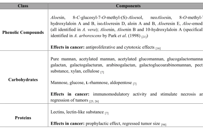
Table 3.1 – Summary of the potential bioactive compounds present in Aloe vera leaves (Adapted from Park et al. (1998) and Hamman et al. (2008)).
Class Components
Phenolic Compounds
Aloesin, 8-C-glucosyl-7-O-methyl-(S)-Aloesol, neoAloesin,
8-O-methyl-7-hydroxylaloin A and B, isoAloeresin D, aloin A and B, Aloeresin E, Aloe-emodin (all identified in A. vera); Aloenin, Aloenin B and 10-hydroxylaloin A (specifically identified in A. arborescens by Park et al. (1998)[21])
Effects in cancer: antiproliferative and cytotoxic effects[16]
Carbohydrates
Pure mannan, acetylated mannan, acetylated glucomannan, glucogalactomannan, galactan, galactogalacturan, arabinogalactan, galactoglucoarabinomannan, pectic substance, xylan, cellulose[7]
Mannose, glucose,L-rhamnose, aldopentose[7]
Effects in cancer: immunomodulatory activity and stimulate necrosis and
regression of tumors[25, 26]
Proteins Lectins, lectin-like substance[7]
Effects in cancer: prophylactic effect, regressed tumor size[94]
During evolution, these plants naturally evolved to produce important secondary metabolites in order to survive their environment and its predators[95]. These different metabolites or phytochemicals
may act individually, additively or in synergy to improve the plant’s health and its chances of survival in a given environment. Furthermore, it has been suggested that these combined actions of the phytochemicals usually tend to increase the bioactivity of the main medicinal constituents by influencing its assimilation in the body [77, 95]. Because of that, in this work, we not only aimed to
compare the amounts of the potentially bioactive compounds in the two selected species, but we also wanted to select the best extraction procedure, that would allow the highest yield in these compounds. Therefore, we set out to quantify the major classes of bioactive compounds: proteins, total phenolic compounds and specifically anthraquinones, and total polysaccharides.
3.2.1 The amount of bioactive compounds extracted from A. arborescens and A. vera is
influenced by the species and by the extraction procedure
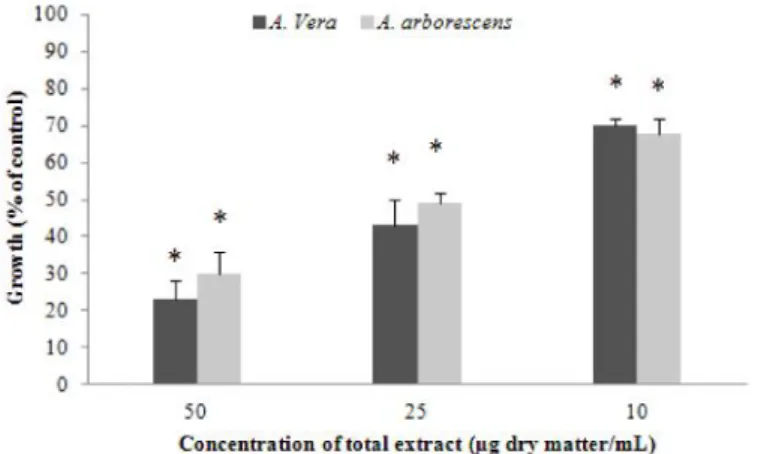
The amounts of the different classes of bioactive compounds in both Aloe species are present in Fig. 3.2. When comparing A. vera and A. arborescens we can easily identify differences between species regarding both the extraction methods for each component and the amount of each component per amount of fresh weight.
Proteins
Aloe plants contain around 95% (w/v) water but less than 0.1% (w/v) protein [94]. Although the
amount of total proteins present in Aloe is relatively small, its biological activities are meaningful as evidenced by their many applications, their major part related to wound healing and several skin diseases treatment[96-99]. Lectins are a class of proteins which have been recently identified in Aloe and
The different extractions yielded different types proteins, according to their solubility: water-soluble proteins, which are extracted in 100 mM Tris-HCl buffer, and the water inwater-soluble proteins, which encompass the membrane and wall-bound proteome and are extracted with methanol. It is important to note that in the protein quantifications, all samples were previously treated with PVPP to remove the majority of the phenolic components from the extract, which would otherwise interfere with protein quantifications. For the same reason, protein quantifications were also determined according to the method described by Bradford [85]because of the same reason. Results presented in
Fig. 3.1a corroborate the low amount of proteins present in both species, which were overall less than 0.1% (w/v) of fresh weight. Nonetheless, protein amounts and distribution varied significantly among both species.
Figure 3.2 – Quantitative characterization of A. vera and A. arborescens considering the main phytochemical groups defined by Hamman et al. 2008[7]. Each extraction was performed with 20g of fresh Aloe leaves for each of the three solvents: 100
mM Tris-HCl buffer, 50% (v/v) methanol and 100% (v/v) methanol. (a) Protein quantification using the Bradford method described by Bradford et al. (1976). All samples were treated with PVPP to eliminate the phenolic compounds that could interfere with the extraction of soluble proteins. (b) Phenolic compounds quantification with the Folin-Ciocalteau reagent and using gallic acid as standard. (c) Total carbohydrates quantification using phenol/sulfuric acid method and mannose as the standard. (d) Specific anthraquinone quantification (phenol component) using benzene, ammonia solution and Aloe blue
curacao aloin.* P<0.05; **P<0.001 when compared between extracts of the same species; # P<0.05; ## P<0.001 the same
extract when compared between species.
Whilst the amount of soluble protein was significantly higher (P<0.05) in A. arborescens (430 ng/g) than in A. vera (380 ng/g), A. vera presented the higher amounts of insoluble proteins than A.
arborescens (404.61 ng/g and 288.3 ng/g, respectively) (P<0.05). Phenolic compounds
Phenolic compounds were already described as the second major class of compounds found in A.
vera [100], second only to polysaccharides (Fig. 1.3.b). Phenolic compounds present in plants, result
from their secondary metabolism and are the main group of substances responsible for most of the plant medicinal properties, including Aloe species [27, 100, 101]. Regarding the results in Graphic 3.1.b,
corresponding to total phenols present per extraction, it is clear that the extraction with 50% (v/v) methanol yields higher amounts of total phenolic in A. vera extraction (26.19 µg/g), whereas for A.
a
c
b
d
The different extractions yielded different types proteins, according to their solubility: water-soluble proteins, which are extracted in 100 mM Tris-HCl buffer, and the water inwater-soluble proteins, which encompass the membrane and wall-bound proteome and are extracted with methanol. It is important to note that in the protein quantifications, all samples were previously treated with PVPP to remove the majority of the phenolic components from the extract, which would otherwise interfere with protein quantifications. For the same reason, protein quantifications were also determined according to the method described by Bradford [85] because of the same reason. Results presented in
Fig. 3.1a corroborate the low amount of proteins present in both species, which were overall less than 0.1% (w/v) of fresh weight. Nonetheless, protein amounts and distribution varied significantly among both species.
Figure 3.2 – Quantitative characterization of A. vera and A. arborescens considering the main phytochemical groups defined by Hamman et al. 2008[7]. Each extraction was performed with 20g of fresh Aloe leaves for each of the three solvents: 100
mM Tris-HCl buffer, 50% (v/v) methanol and 100% (v/v) methanol. (a) Protein quantification using the Bradford method described by Bradford et al. (1976). All samples were treated with PVPP to eliminate the phenolic compounds that could interfere with the extraction of soluble proteins. (b) Phenolic compounds quantification with the Folin-Ciocalteau reagent and using gallic acid as standard. (c) Total carbohydrates quantification using phenol/sulfuric acid method and mannose as the standard. (d) Specific anthraquinone quantification (phenol component) using benzene, ammonia solution and Aloe blue
curacao aloin.* P<0.05; **P<0.001 when compared between extracts of the same species; # P<0.05; ## P<0.001 the same
extract when compared between species.
Whilst the amount of soluble protein was significantly higher (P<0.05) in A. arborescens (430 ng/g) than in A. vera (380 ng/g), A. vera presented the higher amounts of insoluble proteins than A.
arborescens (404.61 ng/g and 288.3 ng/g, respectively) (P<0.05). Phenolic compounds
Phenolic compounds were already described as the second major class of compounds found in A.
vera [100], second only to polysaccharides (Fig. 1.3.b). Phenolic compounds present in plants, result
from their secondary metabolism and are the main group of substances responsible for most of the plant medicinal properties, including Aloe species[27, 100, 101]. Regarding the results in Graphic 3.1.b,
corresponding to total phenols present per extraction, it is clear that the extraction with 50% (v/v) methanol yields higher amounts of total phenolic in A. vera extraction (26.19 µg/g), whereas for A.
a
c
b
~ 14 ~
arborescens the extraction with 100% (v/v) methanol yielded the highest amounts (16.48 µg/g), in a
significant manner (P<0.05). Albeit in Fig. 1.3.b A. vera presented more phenolic compounds (P<0.05) when compared to A. arborescens, when evaluating the specific group of anthraquinones (Fig. 1.3.d) A. arborescens had a significant higher content in the methanolic extractions.
Total carbohydrates
It is well-known that polysaccharides are abundantly represented in A.vera, being the major compound class present in the mucilage of these plants [25, 26]. Partially acetylated mannan or
acemannan have been identified as the primary polysaccharides which are associated to the beneficial effects of A. vera as an anticarcinogenic agent. Acemannan for example was found responsible for the stimulation of the immune response in cancer scenarios, contributing to tumor weight reduction and the improvement of chemotherapy drugs[102, 103].
In the present work, regarding the composition in total polysaccharides between the two species in this work, the extraction with 50% (v/v) methanol presented a significantly higher yield than the other two extractions (P<0.001). For example, A.vera contains 7.72 ng/g against 0.25 ng/g for 100 mM Tris-HCl buffer and 0.14 ng/g for 100% (v/v) methanol. Similar results were found between both species for all tree extractions (P>0.05).
Overall, these results suggest that the composition in bioactive compounds is similar in both species, particularly in the phenolic compounds and total polysaccharides; there are significant differences that could be important for their bioactivities, namely the higher amount of anthraquinones and water-soluble proteins in A. arborescens when compared to A. vera extracts. A. vera nonetheless presented higher amounts of phenolics and wall and membrane-bound proteins.
3.2.2 The majority of the phytochemical components from A. vera are better extracted in
50% (v/v) methanol
When considering the whole leaf as an organ for phytochemical extraction, the possibility to combine several components has been suggested to potentiate its therapeutic activity [77, 95]. However,
the choice of the extraction method by which the compounds are obtained is essential. Results here presented show that the extraction method not only influences the amount of bioactive component extracted but it also differs among species. Hence, for A. vera, for all compounds classes except the proteins, the majority of the compounds were better extracted with 50% (v/v) methanol (phenolics and polysaccharides, with P<0.05) whereas for A. arborescens the highest yields were obtained in the 100% (v/v) methanol extraction. This suggests the presence of different compounds but more importantly, it arises the question of whether most literature using non-aqueous polar and non-polar solvents such as ethanol, acetone etc. are using the correct extractions to yield the better bioactivities with A. vera.
3.3 Effects of total Aloe extracts in colon cancer cells
An estimated 3.45 million new cases of cancer and 1.75 million deaths from cancer occurred in Europe in 2012 [104], with colorectal cancer (CRC) being the second most common cause of cancer
death in the European Union [104, 105]. CRC is often highly metastatic and resistant to anticancer
treatment strategies [106], and despite the intensive research made and the significant advances in
diagnosis, screening and treatment, the overall long-term outcome in patients has not significantly changed in the last decades; the five-year survival rate is approximately 60%[49 106]. MMP-9 inhibitors