Immunophenotypic Analysis of the TCR-V

Repertoire in 98 Persistent Expansions of CD3
⫹
/
TCR-
␣
⫹
Large Granular Lymphocytes
Utility in Assessing Clonality and Insights into the
Pathogenesis of the Disease
Margarida Lima,* Julia Almeida,
†Ana Helena Santos,* Maria dos Anjos Teixeira,*
Maria del Carmen Alguero,
†Maria Luı´s Queiro´s,*
Ana Balanzategui,
‡Benvindo Justic¸a,*
Marcos Gonzalez,
‡Jesu´s F. San Miguel,
‡and
Alberto Orfa˜o
†From the Servic¸o de Hematologia Clı´nica,* Unidade de Citometria, Hospital Geral de Santo Anto´nio, Porto, Portugal; and the Servicios de Citometrı´a†y Hematologı´a,‡Hospital
Universitario de Salamanca and Centro de Investigacio´n del Ca´ncer, Universidad de Salamanca, Salamanca, Spain.
At present , a major challenge in the initial diagnosis of leukemia of large granular lymphocytes (LGLs) is to establish the clonal nature of the expanded popula-tion. In the present study we have analyzed by flow cytometry immunophenotyping the TCR-V reper-toire of 98 consecutive cases of persistent expansions of CD4ⴙor CD8ⴙbrightCD3ⴙ/TCR-␣ⴙLGLs and com-pared the results with those obtained in molecular studies of TCR- gene rearrangements. Fifty-eight cases were considered to be monoclonal in molecular studies whereas in the remaining 40 cases there was no evidence for monoclonality (11 cases were consid-ered oligoclonal and 29 polyclonal). The TCR-V rep-ertoire was biased to the preferential use of one or more TCR-V families in 96% of cases, a total of 124 TCR-V expansions being diagnosed: one TCR-V ex-pansion in 71 cases and two or more TCR-V expan-sions in 23 cases. The highest TCR-V expansion ob-served in each case was higher among monoclonal (74 ⴞ 19%) as compared to nonmonoclonal cases (24ⴞ 14%) (P ⴝ 0.001), as did the fraction of LGLs that exhibited a TCR-V-restricted pattern (86 ⴞ 16% and 42ⴞ 23%, respectively; P ⴝ 0.0001); by contrast, the proportion of cases displaying more than one TCR-V expansion was higher in the latter group: 7%
versus 48% , respectively (P ⴝ 0.001). Results ob-tained in oligoclonal cases were intermediate be-tween those obtained in polyclonal and monoclonal
cases and similar results were observed for CD4ⴙas for CD8ⴙbright T-cell expansions. TCR-V families expressed in CD8ⴙbright T-cell-LGL proliferations showed a pattern of distribution that mimics the fre-quency at which the individual TCR-V families are represented in normal peripheral blood T cells. As-suming that a given proliferation of LGLs is monoclo-nal whenever there is an expansion of a given TCR-V family of at least 40% of the total CD4ⴙor CD8ⴙbright T-cell compartment , we were able to predict clonality with a sensitivity of 93% and a specificity of 80%. By increasing the cut-off value to 60% , sensitivity and specificity were of 81% and 100%. In summary , our results suggest that flow cytometry immunopheno-typic analysis of the TCR-V repertoire is a powerful screening tool for the assessment of T-cell clonality in persistent expansions of TCR-␣ⴙ LGLs. (Am J Pathol 2001, 159:1861–1868)
Leukemias of large granular lymphocytes (LGLs) repre-sent a well-recognized group of chronic T- and natural killer (NK)-cell neoplasias.1 Like their normal
counter-parts, leukemic LGLs are usually classified into two major groups based on the expression of both CD3 and the T-cell receptor (TCR) molecules: CD3⫹/TCR⫹ T- and CD3⫺/TCR⫺ NK-LGL leukemias.2 Among the former
group, monoclonal expansions of LGLs displaying the TCR-␣ represent the majority of cases, whereas
TCR-Supported in part by the Comissa˜o de Fomento da Investigac¸a˜o em Cuidados de Sau´de, Ministe´rio da Sau´de, Portugal (PI 51/99); Acc¸o˜es Integradas Luso-Espanholas do Conselho de Reitores das Universidades Portuguesas (E-31/99); and Accio´n Integrada Hispano-Portuguesa (HP 1998-0091), Direccio´n General de Ensen˜anza Superior e Investigacio´n Cientı´fica, Ministerio de Educacio´n y Cultura, Spain.
This work was done in the context of the BIOMED2 program (action BMH4-CT97-2611).
Accepted for publication August 3, 2001.
Address reprint requests to Margarida Lima, Clinical Hematology, Unit of Flow Cytometry, Hospital Geral de Santo Anto´nio, Rua D Manuel II, s/n, 4050 Porto, Portugal. E-mail: [email protected].
␥␦⫹T-LGL leukemias are relatively infrequent. From the
clinical point of view whereas most clonal T-LGL leuke-mias show a benign clinical outcome, in some cases they behave as an aggressive condition. Either transient or persistent expansions of polyclonal, oligoclonal, or even monoclonal LGLs are relatively common in various dis-ease conditions and such expansions of LGLs have also been reported in otherwise normal healthy individuals,3,4
although their neoplastic nature remains unclear.5,6 In
any case, at present, a major challenge in the initial diagnosis of LGL leukemias is to establish the clonal nature of the expanded population of LGLs, to distinguish between polyclonal, oligoclonal, and monoclonal LGL proliferations.
Molecular investigation into the existence of monoclo-nal rearrangements of the TCR- and TCR-␥ genes, us-ing Southern blot analysis is the preferred method for investigation of T-cell clonality.7Despite the high
reliabil-ity of the Southern blot, this method has some drawbacks that limit its routine use in diagnostic laboratories: it is a labor-intensive and time-consuming method and large quantities of high-quality DNA are needed to obtain reli-able results. In recent years alternative approaches have been developed for the assessment of T-cell clonality. Of them, the immunophenotypic analysis of the repertoire of the variable (V) regions of the TCR-␣, -, -␥, and -␦ chains represent one of the most attractive options. In humans, the V␣ and V gene segments are estimated to contain ⬃46 and 52 different functional members that, based on nucleotide homology, can be grouped into 32 and 25 different families, respectively.8,9Polymerase chain
reac-tion using TCR-V- and TCR-V␣-specific primers and, more recently, flow cytometry using TCR-V- or TCR-V␣-specific monoclonal antibodies (mAbs) can be used to investigate the TCR-V and TCR-V␣ gene usage. Flow cytometry is not only routinely available in many labora-tories but also offers several advantages: 1) it allows both a quantitative and qualitative characterization of the T-cell repertoire; 2) the T-T-cell repertoire can be specifically evaluated within the population of interest by combining TCR-V and TCR-V␣ specific with other mAbs; and 3) a large panel of mAbs against V-region determinants is now commercially available, making it possible to access a large fraction of the T-cell repertoire. Although the analysis of the TCR-V repertoire is now being used to indirectly assess T-cell clonality, the finding of an expan-sion of T cells restricted to a particular V-region family, does not necessarily mean the presence of underlying monoclonal TCR gene rearrangements. Thus, studies in which immunophenotyping is compared with molecular methods are necessary to establish the value of the TCR-V repertoire analysis in investigating T-cell clonality. Several studies have already been performed aimed at the characterization of both TCR-V and TCR-V␣ T-cell repertoires from normal healthy individuals10 –12and the
T-cell responses occurring in various pathological condi-tions, including autoimmune13–15and infectious
diseas-es,16tumors,17–19allogeneic transplants,20and other
dis-ease states.21However, only a few reports on the TCR-V
or TCR-V␣ repertoire in LGL leukemias are available and, furthermore, in the majority of these studies the numbers
of TCR-V families analyzed were limited.22–27Some of
these studies showed that several specific TCR-V and TCR-J genes are randomly used,23,24,27whereas others
suggested a preferential usage of a few TCR-V regions in LGL leukemias.22,24To the best of our knowledge, until
now no study has been reported in which the utility of using a relatively broad panel of anti-V mAbs for the assessment of T-cell clonality in a large series of consec-utive individuals showing a persistent expansion of pe-ripheral blood (PB) TCR-␣⫹LGLs has been evaluated in comparison to molecular techniques.
The aim of our study was to characterize the TCR-V repertoire from a group of 98 consecutive patients dis-playing a persistent expansion of either CD4⫹or CD8⫹ TCR-␣⫹T-LGLs in the PB and to establish its utility in the diagnosis of clonality. For that purpose, a large panel of 23 mAbs directed against 24 members of 19 different TCR-V families was used and these results were com-pared with those obtained with conventional molecular techniques.
Materials and Methods
Patients and Controls
Ninety-eight consecutive patients with persistent TCR-␣⫹T-cell proliferations of LGLs were studied (51 males
and 47 females; ages 12 to 90 years; median, 61 years). The diagnosis was established on the basis of a LGL morphology, together with a typical CD3⫹/TCR-␣⫹LGL phenotype, as defined by high FSC/SSC values, absence of expression of CD28, and reactivity for NK-associated antigens.2,3From the phenotypic point of view, LGL
pro-liferations were classified into two groups: TCR-␣⫹/ CD8⫹bright/CD4⫺ (n ⫽ 72 cases) and TCR-␣⫹/CD4⫹/ CD8⫺/⫹dim(n⫽ 26 cases). The mean absolute number of LGLs in the PB was 2856 ⫾ 3086 ⫻ 106/L (median,
1725⫻ 106/L) and the mean percentage of LGLs within
the CD4⫹ or CD8⫹bright T-cell population was of 78 ⫾ 19% (median, 83%). Increased numbers of LGLs were associated with absolute lymphocytosis (⬎3.5 ⫻ 109/L) in
60 cases (61%). Neutropenia (⬍1.5 ⫻ 109/L) was
ob-served in 34 cases (35%), anemia (Hb⬍ 10 g/dl) in 13 cases (13%), and thrombocytopenia (⬍100 ⫻ 109/L) in
11 cases (11%). Organomegalies were rarely detected (⬍10% of cases). Median follow-up for the patients ana-lyzed was of 29 months. During this period, 37 patients (38%) showed associated conditions, including autoim-mune disorders (14 cases), neoplastic diseases (15 cas-es), or other disease states (8 cases). Ten age- and sex-matched healthy individuals (six males and four fe-males; median age, 47 years) were used as controls.
Immunophenotypic Studies
Ethylenediaminetetraacetic acid-anti-coagulated PB samples were stained using a direct immunofluores-cence technique. Briefly, 100l of whole PB containing between 0.5 to 2.0⫻ 106nucleated cells were incubated
room temperature in the dark. Then 2 ml of fluorescence-activated cell sorting lysing solution (Becton Dickinson, San Jose, CA) were added to lyse nonnucleated red cells. After another 10 minutes incubation at room tem-perature in the dark, cells were washed once and resus-pended in 0.5 ml of phosphate-buffered saline (PBS).
The repertoire of the V chain of TCR-␣⫹/CD8⫹bright/ CD4⫺and TCR-␣⫹/CD4⫹/CD8⫺/⫹dimlymphocytes was analyzed by combining either anti-CD4 or anti-CD8 with the following panel of 23 mAbs specific against 24 mem-bers of a total of 19 families of variable regions of the TCR- chain: BV1S1 (V1.1), BV5S1 (V5.1), BV5S2 (V5.2), BV5S3 (V5.3), BV6S1 (V6.1), BV7S1 (V7.1), BV8S1⫹ BV8S2 (V8.1 ⫹ 8.2), BV9S1 (V9.1), BV11S1 (V11.1), BV12S2 (V12.2), BV13S1 (V13.1), BV13S6 (V13.6), BV14S1 (V14.1), BV16S1 (V16.1), BV17S1 (V17.1), BV18S1 (V18.1), BV20S1 (V20.1), BV21S3 (V21.3), BV22S1 (V22.1), BV23S1 (V23.1) (Beckman-Coulter Immunotech, Marseille, France), BV2S1 (V2.1) (Beckman-Coulter Immunotech or Biodesign Interna-tional, Kennebunk, ME), BV3S1 (V3.1) (Beckman-Coulter Immunotech or Endogen, Woburn, MA) and BV6S7 (V6.7) (Endogen). All anti-TCR-V reagents were tested in all samples except for the anti-V1.1, -V6.7, -V7.1, and -V9.1 that were tested in only 62%, 42%, 57% and 60% of the cases, respectively. In all cases, isotype-matched fluorochrome-conjugated nonspecific immunoglobulins were used as negative control.
Data acquisition was performed in two FACSCalibur flow cytometers (BD) using the Cell Quest software pro-gram (BD). Information on a minimum of 2⫻ 105events
was acquired for each reagent combination. Data analy-sis was performed using the Paint-a-Gate PRO software program (BD). For each TCR-V family, the proportion of positive cells within the CD8⫹brightor CD4⫹lymphocytes was calculated as the percentage of cells stained above the negative isotype control value.
For those TCR-V families that were assessed with the panel of mAbs used in this study (direct identification), we considered that there is a TCR-V expansion when-ever its representation exceeded by at least two standard deviations the mean value observed in CD4⫹or CD8⫹ circulating T cells in normal healthy individuals.16For the
remaining TCR-V families that were not explored with the panel of mAbs used in this study, criteria used to define a TCR-V expansion (indirect identification) was based on the observation of a relative decrease in the fraction of either CD4⫹or CD8⫹bright circulating T cells that were recognized with the panel of mAbs to values ⬍85% of those observed for the same T-cell subsets in PB samples from normal adult individuals. These criteria rely on previous studies demonstrating that a value of 15% should be considered a T-cell expansion.28,29
Molecular Biology Studies
Rearrangements of the TCR- chain genes were evalu-ated by conventional Southern blotting30 in all cases.
Briefly, mononuclear cells were obtained after fraction-ation on a Lymphoprep (Nycomed Pharma AS, Oslo,
Norway) density gradient, washed twice in PBS, and cryopreserved. DNA was extracted using the phenol/ chloroform method and digested with EcoRI and HindIII restriction enzymes. DNA fragments were separated by 0.8% agarose gel electrophoresis and transferred to ni-trocellulose membranes by vacuum blotting, UV fixed, and hybridized with 32P-labeled probes for the TCR-
gene region (C, TCRBC, and TCRBJ2; DAKO A/S, Glostrup, Denmark). In those cases (n⫽ 5) in which the CD4⫹or CD8⫹bright LGL population represented⬍10% of the total nucleated cells present in the sample and because all TCR-␣⫹ T-cell malignancies have rear-ranged TCR-␥ genes,31the clonality studies were
per-formed by polymerase chain reaction analysis of TCR-␥ gene rearrangements, using the strategies and primers previously described.32
Statistical Methods
For all variables under study, median, mean, SD, and range values were calculated. Comparison between groups was calculated using the Mann-Whitney U and chi-square tests for continuous and dichotomic variables, respectively (SPSS 9.0; SPSS, Chicago, IL). P values ⬍0.05 were considered to be associated with statistically significant differences.
Results
Immunophenotypic Analysis of the TCR-V

Repertoire of Both PB CD4
⫹and CD8
⫹brightT
Cells and Molecular Studies in Normal Healthy
Individuals
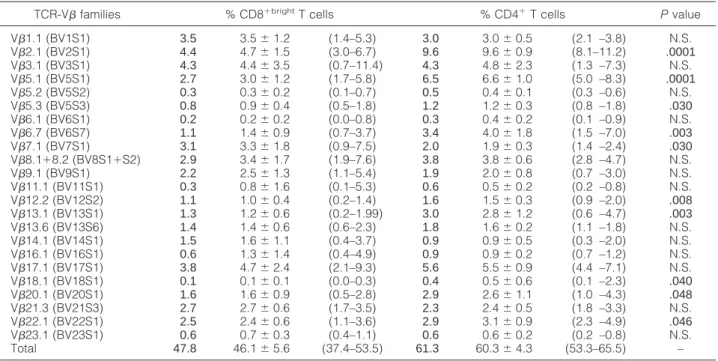
The panel of 23 mAbs used in the present work allowed us to identify 60.3⫾ 4.3% (range, 53.3 to 65.5%) and 46.1 ⫾ 5.6% (range, 37.4 to 53.5%) of all CD4⫹ and CD8⫹brightT cells in the PB of 10 normal healthy controls. The specific distribution of each of the different TCR-V families on both CD4⫹and CD8⫹brightT cells from normal individuals is displayed in Table 1. As shown in this table, certain TCR-V families are represented more than oth-ers, either within the CD4⫹ or the CD8⫹bright normal T lymphocytes. Overall, the relative distribution of the dif-ferent TCR-V families within the CD4⫹and CD8⫹ sub-sets was comparable; however, some TCR-V families were preferentially expressed either within the CD4⫹or the CD8⫹brightsubsets, with differences only statistically significant for a preferential expression of TCR-V2.1, -V5.1, -V5.3, -V6.7, -V12.2, -V13.1, -V18.1, -V20.1, and -V22.1 on CD4⫹T cells and TCR-V7.1 on CD8⫹brightT cells. Molecular studies showed a polyclonal pattern for the TCR- gene rearrangements in all control individuals.
Assessment of T-Cell Clonality by
Immunophenotypic Analysis of the TCR-V

Repertoire of PB CD4
⫹and CD8
⫹brightT Cells
and Molecular Studies in Patients Showing
Persistent Expansions of LGLs
From the molecular point of view, 58 of the 98 cases studied (59%) were found to be monoclonal by either Southern blot and/or polymerase chain reaction whereas in the remaining 40 cases there was no evidence of monoclonality. Among these latter 40 cases, 11 were classified as oligoclonal whereas 29 were classified poly-clonal.
The immunophenotypic analysis of the TCR-V repertoire of both CD4⫹and CD8⫹brightPB T cells from individuals
showing LGL expansions revealed the existence of an ex-pansion of at least one V family in all but four cases (n ⫽ 94; 96%), with a total of 124 expansions detected. Ninety-one of these TCR-V expansions (73%) were directly iden-tified with the panel of mAbs used whereas in the remaining 33 cases the identification was indirect. Seventy-one pa-tients (72%) showed an expansion of a single TCR-V fam-ily whereas 23 cases (23%) displayed expansions of two or more TCR-V families: two TCR-V families were simulta-neously expanded in 17 patients, three in 5 patients, and four in 1 case. The highest TCR-V family expansion found in each case was highly variable, ranging from 4 to 96% of the total PB CD4⫹and CD8⫹brightT cells (mean, 55⫾ 30%; median, 57%) (Figure 1). The other TCR-V expanded families represented 4 to 28% (mean, 9⫾ 5%) of the total PB CD4⫹and CD8⫹brightT cells. Interestingly, no
signif-Table 1. Usage of the TCR V- Families Tested in Peripheral Blood CD8⫹bright and CD4⫹ T Cells from Normal Healthy Adult Individuals (n ⫽ 10)
TCR-V families % CD8⫹brightT cells % CD4⫹T cells Pvalue V1.1 (BV1S1) 3.5 3.5⫾ 1.2 (1.4–5.3) 3.0 3.0⫾ 0.5 (2.1 –3.8) N.S. V2.1 (BV2S1) 4.4 4.7⫾ 1.5 (3.0–6.7) 9.6 9.6⫾ 0.9 (8.1–11.2) .0001 V3.1 (BV3S1) 4.3 4.4⫾ 3.5 (0.7–11.4) 4.3 4.8⫾ 2.3 (1.3 –7.3) N.S. V5.1 (BV5S1) 2.7 3.0⫾ 1.2 (1.7–5.8) 6.5 6.6⫾ 1.0 (5.0 –8.3) .0001 V5.2 (BV5S2) 0.3 0.3⫾ 0.2 (0.1–0.7) 0.5 0.4⫾ 0.1 (0.3 –0.6) N.S. V5.3 (BV5S3) 0.8 0.9⫾ 0.4 (0.5–1.8) 1.2 1.2⫾ 0.3 (0.8 –1.8) .030 V6.1 (BV6S1) 0.2 0.2⫾ 0.2 (0.0–0.8) 0.3 0.4⫾ 0.2 (0.1 –0.9) N.S. V6.7 (BV6S7) 1.1 1.4⫾ 0.9 (0.7–3.7) 3.4 4.0⫾ 1.8 (1.5 –7.0) .003 V7.1 (BV7S1) 3.1 3.3⫾ 1.8 (0.9–7.5) 2.0 1.9⫾ 0.3 (1.4 –2.4) .030 V8.1⫹8.2 (BV8S1⫹S2) 2.9 3.4⫾ 1.7 (1.9–7.6) 3.8 3.8⫾ 0.6 (2.8 –4.7) N.S. V9.1 (BV9S1) 2.2 2.5⫾ 1.3 (1.1–5.4) 1.9 2.0⫾ 0.8 (0.7 –3.0) N.S. V11.1 (BV11S1) 0.3 0.8⫾ 1.6 (0.1–5.3) 0.6 0.5⫾ 0.2 (0.2 –0.8) N.S. V12.2 (BV12S2) 1.1 1.0⫾ 0.4 (0.2–1.4) 1.6 1.5⫾ 0.3 (0.9 –2.0) .008 V13.1 (BV13S1) 1.3 1.2⫾ 0.6 (0.2–1.99) 3.0 2.8⫾ 1.2 (0.6 –4.7) .003 V13.6 (BV13S6) 1.4 1.4⫾ 0.6 (0.6–2.3) 1.8 1.6⫾ 0.2 (1.1 –1.8) N.S. V14.1 (BV14S1) 1.5 1.6⫾ 1.1 (0.4–3.7) 0.9 0.9⫾ 0.5 (0.3 –2.0) N.S. V16.1 (BV16S1) 0.6 1.3⫾ 1.4 (0.4–4.9) 0.9 0.9⫾ 0.2 (0.7 –1.2) N.S. V17.1 (BV17S1) 3.8 4.7⫾ 2.4 (2.1–9.3) 5.6 5.5⫾ 0.9 (4.4 –7.1) N.S. V18.1 (BV18S1) 0.1 0.1⫾ 0.1 (0.0–0.3) 0.4 0.5⫾ 0.6 (0.1 –2.3) .040 V20.1 (BV20S1) 1.6 1.6⫾ 0.9 (0.5–2.8) 2.9 2.6⫾ 1.1 (1.0 –4.3) .048 V21.3 (BV21S3) 2.7 2.7⫾ 0.6 (1.7–3.5) 2.3 2.4⫾ 0.5 (1.8 –3.3) N.S. V22.1 (BV22S1) 2.5 2.4⫾ 0.6 (1.1–3.6) 2.9 3.1⫾ 0.9 (2.3 –4.9) .046 V23.1 (BV23S1) 0.6 0.7⫾ 0.3 (0.4–1.1) 0.6 0.6⫾ 0.2 (0.2 –0.8) N.S. Total 47.8 46.1⫾ 5.6 (37.4–53.5) 61.3 60.3⫾ 4.3 (53.3–65.5) –
Results are presented as median values (bold), mean⫾ standard deviation, and range (minimum-maximum).
Figure 1. Percentage of TCR-␣⫹/CD4⫹or TCR-␣⫹/CD8⫹brightT cells from each individual patient displaying a single expanded TCR-V family, as represented by shaded bars superimposed by white bars in polyclonal, blue in oligoclonal, and red in monoclonal expansions of LGLs, as determined by molecular techniques.
icant expansions of TCR-␣⫹T cells not included in the suspected LGL population were detected in cases ana-lyzed here.
Figure 2 shows the relative incidence of expansions for each of the TCR-V families within the CD4⫹(Figure 2A) and the CD8⫹bright(Figure 2B) T cells. As may be seen, overall, the expanded TCR-V families within the CD8⫹bright LGLs showed a similar distribution to that observed in normal PB because the must frequently ex-panded TCR-V families were those usually highly
rep-resented in normal circulating CD8⫹brightT cells (Table 1 and Figure 2A). In contrast, among cases showing an expansion of CD4⫹ LGLs only a few TCR-V families were found to be expanded: TCR-V2.1, -V3.1, -V6.7, -V12.2, -V13.1, and -V17.1. When considering only monoclonal expansions of CD4⫹LGLs, two cases (22%) corresponded to TCR-V2.1, two cases (22%) to TCR-V3.1, and four cases (44%) to TCR-V13.1, a frequency that exceeds the frequency that was expected on the basis of the representation of each of these TCR-V
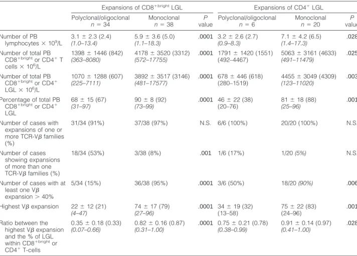
Table 2. Relative and Absolute Counts of PB Lymphocytes, LGL, and Expanded V-Families in Patients with Persistent Expansions of CD4⫹ and CD8⫹bright LGL According to the Presence or Not of a Monoclonal Expansion of PB T Cells by Molecular Techniques
Expansions of CD8⫹brightLGL Expansions of CD4⫹LGL Polyclonal/oligoclonal n⫽ 34 Monoclonal n⫽ 38 P value Polyclonal/oligoclonal n⫽ 6 Monoclonal n⫽ 20 P value Number of PB lymphocytes⫻ 109/L 3.1⫾ 2.3 (2.4) (1.0–13.4) 5.9⫾ 3.6 (5.0) (1.1–18.3) .0001 3.2⫾ 2.6 (2.7) (0.9–8.3) 7.1⫾ 4.2 (6.5) (1.4–17.3) .028 Number of total PB CD8⫹brightor CD4⫹T cells⫻ 106/L 1398⫾ 1446 (842) (363–8080) 4178⫾ 3520 (3312) (572–17755) .0001 1791⫾ 1420 (1551) (492–4467) 5063⫾ 3161 (4633) (491–11479) .025 Number of total PB CD8⫹brightor CD4⫹ LGL⫻ 106/L 1070⫾ 1288 (607) (225–7111) 3892⫾ 3517 (3146) (481–17577) .0001 678⫾ 446 (618) (280–1519) 4455⫾ 3049 (4309) (123–11020) .003 Percentage of total PB CD8⫹brightor CD4⫹ LGL 68⫾ 15 (67) (31–97) 90⫾ 8 (92) (73–99) .0001 46⫾ 22 (38) (20–76) 81⫾ 18 (88) (25–96) .001
Number of cases with expansions of one or more TCR-V families (%) 31/34 (91%) 37/38 (97%) N.S. 6/6 (100%) 20/20 (100%) N.S. Number of cases showing expansions of more than one TCR-V families (%)
18/34 (53%) 3/38 (8%) .001 1/6 (17%) 1/20 (5%) N.S.
Number of cases with at least one V expansion⬎ 40% 5/34 (15%) 36/38 (95%) .0001 3/6 (50%) 18/20 (90%) .006 Highest V expansion 22⫾ 12 (21) (4–47) 74⫾ 17 (79) (27–96) .0001 34⫾ 19 (32) (13–58) 75⫾ 22 (83) (24–96) .001 Ratio between the
highest V expansion and the % of LGL within CD8⫹brightor CD4⫹T-cells 0.35⫾ 0.18 (0.33) (0.07–0.66) 0.82⫾ 0.16 (0.87) (0.31–1.00) .0001 0.75⫾ 0.21 (0.78) (0.38–0.99) 0.91⫾ 0.14 (0.97) (0.41–1.00) .028
Results are presented as mean⫾ standard deviation, median (in brackets), and range (italic, in brackets). N.S.: no significant differences.
Figure 2. Incidence of expansions of each TCR-V family in proliferations of TCR-␣⫹/CD8⫹(A) and TCR-␣⫹/CD4⫹(B) LGLs in the whole series and in cases classified as monoclonal by molecular techniques (black bars).
families among normal PB CD4⫹ T cells (Table 1 and Figure 2B).
Correlation between Immunophenotypic and
Molecular Clonality Studies
Nonmonoclonal (polyclonal plus oligoclonal) and mono-clonal T-LGL expansions differed in a number of aspects including the number of expanded TCR-V families and the magnitude of the highest TCR-V expansion. Table 2 summarizes the immunophenotypic features of monoclo-nal versus nonmonoclomonoclo-nal expansions by considering separately the CD4⫹and CD8⫹brightLGL proliferations.
All but one monoclonal proliferation of LGLs showed expansion of at least one TCR-V family (98%). Further-more, in most of these cases expansion of a single TCR-V family was detected by immunophenotyping (53 of 57 cases; 93%), with other smaller V expansions being simultaneously detected in only four cases (7%). As a mean the highest TCR-V expansion accounted for 74 ⫾ 19% (range, 24 to 96%) of the expanded T-cell compartment: 74⫾ 17% of CD8⫹brightand 75⫾ 22% of CD4⫹T cells, for CD8⫹bright and CD4⫹LGLs, respec-tively, representing ⬎40% of the CD4⫹ or CD8⫹bright T-cell populations in all but four patients (93%).
Similarly, all but three LGL proliferations classified as not being monoclonal by molecular techniques displayed expansion of at least one TCR-V family (93%). However, the frequency of cases with expansion of more than one TCR-V family among nonmonoclonal proliferations of LGLs (19 of 40 patients; 48%)—18 of 34 patients (53%) and 1 of 6 patients (17%) for CD8⫹bright and CD4⫹ T-LGL, respectively—was much higher than that observed in monoclonal cases (P⫽ 0.001). In addition, the mag-nitude of the highest TCR-V expansion observed was also much lower (P ⫽ 0.001) among these cases as compared to the monoclonal cases, accounting for only 24⫾ 14% of the expanded T-cell population: 22 ⫾ 12% of CD8⫹brightand 34⫾ 19% of the CD4⫹T cells. Inter-estingly, the proportion of the TCR-V expanded T cells was higher among the oligoclonal as compared to the polyclonal cases (38⫾ 15% versus 18 ⫾ 9%). Also in contrast (P⫽ 0.0001) to what was observed in monoclo-nal proliferations, the percentage of CD4⫹or CD8⫹brightT cells expressing a single TCR-V family exceeded 40% of the total CD4⫹or CD8⫹brightT cells in only eight cases (20%), all of them being classified as oligoclonal by mo-lecular techniques. Interestingly, the TCR-V expansion did not exceed 60% in any of them. On comparing the proportion of T cells expressing the expanded TCR-V family from either the total CD4⫹or CD8⫹brightT cells that fulfilled the phenotypic criteria for LGLs in monoclonal and in oligoclonal/polyclonal T-LGL expansions (Figure 2), significantly higher numbers (P⫽ 0.0001) were ob-served in monoclonal cases— 86 ⫾ 16% versus 42 ⫾ 23%—the lowest values being observed for polyclonal T-LGL expansions (32⫾ 17%) whereas oligoclonal LGL expansions showed intermediate values (64⫾ 21%).
Taking 40% of the total CD4⫹or CD8⫹brightT cells as the cut-off value for diagnosis of a monoclonal T-cell
expansion, immunophenotyping showed a sensitivity of 93% and a negative predictive value of 89%, with a specificity and positive predictive value of 80% and 87%, respectively. By increasing the cut-off value to 60% both the specificity and positive predictive value increased to 100%, although a large proportion of cases displaying a monoclonal expansion of T-LGL proliferations were im-properly classified as nonmonoclonal by flow cytometry: sensitivity of 81% and negative predictive value of 78% (Table 3).
Discussion
The results presented here show that persistent expan-sions of TCR-␣⫹T-LGLs form a continuum spectrum of polyclonal, oligoclonal, and monoclonal proliferations of TCR-V-restricted T cells that differ both in type and number of expanded TCR-V families, as well as in the magnitude of the TCR-V expansion.
Overall, once cases displaying a monoclonal expan-sion of TCR-␣⫹T-LGLs based on molecular techniques were compared to the other patients, major differences in the immunophenotypic result were observed: 1) poly-clonal and oligopoly-clonal expansions of TCR-␣⫹LGLs fre-quently showed expansions of more than one TCR-V family; 2) the proportion of the most represented TCR-V family was usually much lower in these cases; and 3) even in the presence of a TCR-V dominance, nonmono-clonal LGL proliferations usually displayed a large frac-tion of residual LGLs with a highly diversified T-cell rep-ertoire.
Such observations would support the notion that monoclonal LGL proliferations may arise as a conse-quence of an antigen-driven immune response that would start as a polyclonal reactive T-cell response and could thereafter subsequently evolve into oligoclonal and monoclonal processes. This hypothesis would also be supported by the fact that TCR-V families expressed in CD8⫹brightLGL proliferations showed a pattern of distri-bution that mimics the frequency at which individual TCR-V families are represented in normal CD8⫹brightT cells. This would suggest that CD8⫹bright T-LGLs are clonally transformed in a random manner; the possibility of a bias to the preferential use of some TCR-V families in cases showing a monoclonal expansion of CD4⫹LGLs needs further evaluation using a larger number of cases. The notion that monoclonal expansions of T-LGLs may
Table 3. Sensitivity and Specificity of Immunophenotypic Analysis of the TCR-V Repertoire for the Diagnosis of (Mono)clonal Expansions of TCR␣⫹ LGL
% of the TCR-V expansion
⬎40% ⬎60%
Positive predictive value 87% 100% Negative predictive value 89% 78%
Sensitivity 93% 81%
represent an evolutionary end stage of an antigen-medi-ated proliferation of LGLs is not new.5,6In fact, there is a
great deal of evidence supporting this view: 1) clonal dominance with preservation of a polyclonal reservoir is a typical feature in the normal TCR-␣⫹T-cell repertoire;33
2) the immune TCR-V repertoire is dynamic and can be continuously modulated;343) in normal individuals,
TCR-V-restricted T-cell expansions accumulate with age;10 –12
4) TCR-V-restricted T-cell expansions are more frequently found in association with pathological conditions character-ized by chronic antigen stimulation;35,365) preliminary
re-ports suggest that a higher incidence of clonality seems to be a frequent finding once TCR-V-restricted T-cell reper-toires are observed;10,37,38this phenomenon seems to be
particularly frequent in the TCR-␣⫹/CD8⫹brightT-cell com-partment, although expansions of TCR-␣⫹/CD4⫹ cells, TCR-␣⫹/CD4⫺/CD8⫺39and TCR-␥␦⫹T cells40have also
been sporadically observed; 6) similarly to LGL-leukemia cells, polyclonal T-LGLs displaying a restricted usage of TCR-V families are usually CD28⫺,41express
NK-associ-ated antigens and NK receptors,42 suggesting that they
represent antigen-driven cytotoxic T cells; and 7) molecular studies provided evidence that TCR-V-restricted T-cell ex-pansions may depend on an antigen-mediated selection process.25,43,44
If this hypothesis is true, the higher the TCR-V expan-sion is, the greater the probability for a monoclonal T-cell proliferation, as found in the present study. In turn, this indicates that a large expansion of a TCR-V family would be highly predictive of clonal transformation of a chronic reactive process. Nevertheless, these phenomena do not always necessarily correlate. In fact,some cases display-ing LGL expansions in which a sdisplay-ingle TCR-V family account for more than a half of the CD4⫹or CD8⫹bright T-cell compartment proved to be oligoclonal by molecu-lar techniques. Moreover, we have found a case of mono-clonal T-cell proliferation in which a normal distribution of the different TCR-V families was observed in accor-dance to previous reports.45,46 It could be argued that
such discrepancies could be because of problems re-lated to technical uses such as the different sensitivity of the molecular and immunophenotypic techniques in cases in which minor populations of clonal LGLs are present.7 Alternatively, these observations could also
suggest that monoclonal T-cell rearrangements could theoretically occur at any of the stages of the process of continuous T-cell stimulation. Based on the results pre-sented here and in previous reports indicating that a TCR-V expansion representing ⬎40% of the overall population of CD4⫹and CD8⫹brightT cells is hardly ever found in normal individuals,10 –12 we propose that, for
routine purposes, expansions of a single TCR-V family representing ⬎60% of the overall population of CD4⫹ and CD8⫹bright T cells could be considered as highly suggestive of monoclonal whereas those of⬍40% would be nonmonoclonal, pending confirmation by molecular techniques. In those cases in which the TCR-V expan-sion represents between 40% and 60% of the total CD4⫹ and CD8⫹brightT cells, molecular studies are essential to establish clonality. Further studies are necessary to clar-ify the utility of the immunophenotypic assessment of
T-cell clonality based on the analysis of the TCR-V rep-ertoire together with a characterization of the phenotypic profile of the expanded TCR-V family aimed at the iden-tification of specific aberrant phenotypes, not only at the time of diagnosis, but also during the follow-up of pa-tients, in particular if therapy is required.
Acknowledgment
We thank Beckman/Coulter for providing part of the anti-TCR-V monoclonal antibodies used in this study.
References
1. Loughran TP: Clonal diseases of large granular lymphocytes. Blood 1993, 82:1–14
2. Scott CS, Richard SJ: Classification of large granular lymphocyte (LGL) and NK-associated (NKa) disorders. Blood Rev 1992, 6:220 – 233
3. Semenzato G, Pandolfi F, Chisesi T, De Rossi G, Pizzolo G, Zambello R, Trentim L, Agostini C, Dini E, Vespignani M, Cafaro A, Pasqualetti D, Giubellino MC, Migone N, Foa R: The lymphoproliferative disease of granular lymphocytes. A heterogeneous disease ranging from indolent to aggressive conditions. Cancer 1987, 60:2971–2978 4. Scott CS, Richards SJ, Sivakumaran M, Short M, Child JA´ , Hunt KM,
McEvoy M, Steed AJ, Balfour IC, Parapia LA: Transient and persistent expansions of large granular lymphocytes (LGL) and NK associated (NKs) cells: the Yorkshire Leukaemia Group Study. Br J Haematol 1993, 83:505–517
5. Semenzato G, Pizzoto G, Rannunci A, Agostini C, Chilosi M, Quinti I, De Sanctis G, Vercelli B, Pandolfi F: Abnormal expansion of poly-clonal large to small size granular lymphocytes: reactive or neoplastic process? Blood 1984, 63:1271–1277
6. Richards SJ, Short M, Scott CS: Clonal CD3⫹CD8⫹ large granular lymphocyte (LGL)/NK-associated (NKa) expansions: primary malig-nancies or secondary reactive phenomena? Leuk Lymphoma 1995, 17:303–311
7. Ryan DK, Alexander HD, Morris TC: Routine diagnosis of large gran-ular lymphocytic leukaemia by Southern blot and polymerase chain reaction analysis of clonal T cell receptor gene rearrangement. Mol Pathol 1997, 50:77– 81
8. Wei S, Charmley P, Robinson MA, Concannon P: The extent of the human germline T-cell receptor V beta gene segment repertoire. Immunogenetics 1994, 40:27–36
9. Wei S, Concannon P: Repertoire and organization of human T-cell receptor alpha region variable genes. Genomics 1996, 38:442– 445 10. Posnett D, Sinha R, Kabak S, Russo C: Clonal populations of T cells
in normal elderly humans: the T cell equivalent to “benign monoclonal gammapathy.” Exp Med 1994, 179:609 – 618
11. Wack A, Cossarizza A, Heltai S, Barbieri D, D’Addato S, Fransceschi C, Dellabona P, Casorati G: Age-related modifications of the human alphabeta T cell repertoire due to different clonal expansions in the CD4⫹ and CD8⫹ subsets. Int Immunol 1998, 10:1281–1288 12. van Den Beemd R, Boor PP, van Lochem EG, Hop WC, Langerak AW,
Wolvers-Tettero IL, Hooijkaas H, van Dongen JJ: Flow cytometric analysis of the vbeta repertoire in healthy controls. Cytometry 2000, 40:336 –345
13. Conrad B, Weldmann E, Trucco C, Rudert WA, Behboo R, Ricordi C, Rodriquez-Rilo H, Finegold D, Trucco M: Evidence for superantigen involvement in insulin-dependent diabetes mellitus aetiology. Nature 1994, 371:351–355
14. Holbrook MR, Tighe PJ, Powell RJ: Restrictions of T cell receptor beta chain repertoire in the peripheral blood of patients with systemic lupus erythematosus. Ann Rheum Dis 1996, 55:627– 631
15. Borgato L, Beri R, Biasi D, Testoni R, Cugola L, Ceru S, De Sandre G, Lunardi C: Analysis of the T cell receptor repertoire in rheumatoid arthritis. Clin Exp Rheumatol 1997, 15:475– 479
16. Roglic M, Macphee RD, Duncan SR, Sattler FR, Theofilopoulos AN: T cell receptor (TCR) BV gene repertoires and clonal expansions of
CD4 cells in patients with HIV infections. Clin Exp Immunol 1997, 107:21–30
17. Puisieux I, Even J, Pannetier C, Jotereau F, Favrot M, Kourilsky P: Oligoclonality of tumor-infiltrating lymphocytes from human melano-mas. J Immunol 1994, 153:2807–2818
18. Halapi E, Werner A, Wahlstrom J, Osterborg A, Jeddi-Tehrani M, Yi Q, Janson CH, Wigzell: T cell repertoire in patients with multiple my-eloma and monoclonal gammopathy of undetermined significance: clonal CD8⫹ T cell expansions are found preferentially in patients with a low tumor burden. Eur J Immunol 1997, 27:2245–2252 19. Goolsby CL, Kuchnio M, Finn WG, Peterson L: Expansions of clonal
and oligoclonal T cells in B-cell chronic lymphocytic leukemia are primarily restricted to the CD3(⫹)CD8(⫹) T-cell population. Cytom-etry 2000, 42:188 –195
20. Gorochov C, Debre´ P, Leblond V, Sadat-Sowti B, Sigaux F, Autran B: Oligoclonal expansion of CD8⫹CD57⫹ T cells with restricted T cell receptor chain variability after bone marrow transplantation. Blood 1994, 83:587–595
21. Probert CS, Chott A, Turner JR, Stevens AC, Bodinaku K, Elson CO, Balk SP, Blumberg RS: Persistent clonal expansions of peripheral blood CD4⫹ lymphocytes in chronic inflammatory bowel disease. J Immunol 1996, 157:3183–3191
22. de Totero D, Tazzari PL, DiSanto JP, di Celle PF, Raspadori D, Conte R, Gobbi M, Ferrara GB, Flomenberg N, Lauria F, Foa´ R: Heteroge-neous immunophenotype of large granular lymphocyte expansions: differential expression of the CD8␣ and CD8 chains. Blood 1992, 80:1765–1773
23. Kaneko T, Mizoguchi H, Oshimi K: Expression of T-cell receptor V regions in granular lymphocyte-proliferative disorders. Blood 1993, 81:3482–3483
24. Zambello R, Trentin L, Facco M, Cerutti A, Sancetta R, Milani A, Raimondi R, Tassinari C, Agostini C, Semenzato G: Analysis of the T cell receptor in the lymphoproliferative disease of granular lymphocytes: superantigen activation of clonal CD3⫹ granular lym-phocytes. Cancer Res 1995, 55:6140 – 6145
25. Kasten-Sportes C, Zahnoen S, Steis RG, Chan WCC, Winton EF, Waldmann TA: T-cell receptor gene rearrangement in T-cell large granular leukocyte leukemia: preferential V␣ but diverse J␣ usage in one of five patients. Blood 1994, 83:767–775
26. Bowman SJ, Bhavnani M, Geddes GC, Corrigall V, Boylston AW, Panayi GS, Lanchbury JS: Large granular lymphocyte expansions in patients with Felty’s syndrome: analysis using anti-T cell receptor V beta-specific monoclonal antibodies. Clin Exp Immunol 1995, 101: 18 –24
27. Davey MP, Starkebaum G, Loughran Jr TP: CD3⫹ leukemic granular lymphocytes utilize diverse T-cell receptor V beta genes. Blood 1995, 51:146 –150
28. Morley J, Batliwaua F, Hingorani R, Cregersen P: Oligoclonal CD8⫹ T cells are preferentially expanded in the CD57⫹ subset. J Immunol 1995, 154:6182– 6190
29. Halapi E, Werner A, Wahlstrom J, Osterborg A, Jeddi-Tehrani M, Yi Q, Janson CH, Wigzell H, Grunewald J, Mellstedt H: T cell repertoire in patients with multiple myeloma and monoclonal gammopathy of un-determined significance: clonal CD8⫹ T cell expansions are found preferentially in patients with a low tumor burden. Eur J Immunol 1997, 27:2245–2252
30. Garcia-Sanz R, Vargas Montero M, Gonzalez Diaz M, del Carmen M, Santos C, Balanzategui Echevarria A, Flores Corral T, Hernandez Martin JM, Caballero Barrigon MD, San Miguel JF: Action of single and associated lesions on the Bcl-l, Bcl-2, Bcl-6, c-Myc, p53, and p16 genes in B-cell non Hodgkin’s lymphomas. Value of molecular anal-ysis for a better assignment of the histologic subtype. Haematologica 1998, 83:209 –216 .
31. Van Dongen JJ, Wolvers-Tettero IL: Analysis of immunoglobulin and T cell receptor genes. Part II: possibilities and limitations in the diagnosis and management of lymphoproliferative diseases and re-lated disorders. Clin Chim Acta 1991, 198:93–174
32. Langerak AW, Szczepanski T, Van der Burg M, Wolvers-Tettero ILM, van Dongen JJM: Heteroduplex PCR analysis of rearranged T cell receptor genes for clonality assessment in suspect T cell prolifera-tions. Leukemia 1997, 1:2192–2199
33. Lantelme E, Granziero L, Angman L, Giachino C: Clonal predomi-nance, but preservation of a polyclonal reservoir, in the normal alpha beta T-cell repertoire. Hum Immunol 1997, 53:49 –56
34. Blish CA, Gallay BJ, Turk GL, Kline KM, Wheat W, Fink PJ: Chronic modulation of the TCR repertoire in the lymphoid periphery. J Immu-nol 1999, 162:3131–3140
35. Pannetier C, Even J, Kourilsky P: T-cell repertoire diversity and clonal expansions in normal and clinical samples. Immunol Today 1995, 16:176 –181
36. Grunewald J, Wigzell H: T cell receptors in health and disease. Introduction. Springer Semin Immunopathol 1999, 21:1– 4 37. Fitzgerald J, Ricalton N, Meyer A-C, West S, Kaplan H, Behrendt C,
Kotzin B: Analysis of clonal CD8⫹ T cell expansions in normal indi-viduals and patients with rheumatoid arthritis. J Immunol 1995, 154: 3538 –3547
38. Wang E, Moss P, Frodsham P, Lehner P, Bell J, Borvsiewicz L: CD8⫹highCD57⫹ T lymphocytes in normal, healthy individuals are oligoclonal and respond to human cytomegalovirus. J Immunol 1995, 155:5046 –5056
39. Niehues T, Gulwani-Akolkar B, Akolkar PN, Tax W, Silver J: Unique phenotype and distinct TCR V beta repertoire in human peripheral blood alpha beta TCR⫹, CD4⫺, and CD8⫺ double negative T cells. J Immunol 1994, 152:1072–1081
40. Giachino C, Granziero L, Modena V, Maiocco V, Lomater C, Fantini F, Lanzavecchia A, Migone N: Clonal expansions of V delta 1⫹ and V delta 2⫹ cells increase with age and limit the repertoire of human gamma delta T cells. Eur J Immunol 1994, 24:1914 –1918 41. Mugnaini EN, Egeland T, Spurkland A, Brinchmann JE: The T cell
receptor repertoire of CD8⫹CD28⫺ T lymphocytes is dominated by expanded clones that persist over time. Clin Exp Immunol 1999, 117:298 –303
42. Mingari MC, Schiavetti F, Ponte M, Vitale C, Maggi E, Romagnani S, Demarest J, Pantaleo G, Fauci AS, Moretta L: Human CD8⫹ T lym-phocyte subsets that express HLA class I-specific inhibitory recep-tors represent oligoclonally or monoclonally expanded cell popula-tions. Proc Natl Acad Sci USA 1996, 93:12433–12438
43. Sottini A, Bettinardi A, Quiros-Roldan E, Plebani A, Airo P, Primi D, Imberti L: Evidence for antigenic selection of large granular lympho-cytes in a patient with Wiskott-Aldrich syndrome. Blood 1995, 86: 2240 –2247
44. Quiros-Roldan E, Sottini A, Gulletta M, Stellini R, Puoti M, Primi D, Imberti L: The T-cell receptor repertoires expressed by CD4⫹ and CD4⫺ large granular lymphocytes derived from the same patients suggest the persistent action of an immune-mediated selection pro-cess. Blood 1996, 88:2133–2143
45. Hingorani R, Choi I-H, Akolkar P, Gulwani-Akolkar B, Pergolizzi R, Silver, Cregersen P: Clonal predominance of T cell receptors within the CD8⫹CD45R0⫹ subset in normal human subjects. J Immunol 1993, 151:5762–5769
46. Monteiro J, Flingorani R, Choi I-H, Silver J, Pergolizzi R, Cregersen P: Oligoclonality in the human CD8⫹ T cell repertoire in normal subjects and monozygotic twins: implications for studies of infectious and autoimmune diseases. Mol Med 1995, 1:614 – 624