HIV-specific T-cell responses, T-cell activation and
regulatory T-cells in AIDS pathogenesis: lessons
from the HIV-2 model.
“Respostas T específicas para o HIV, activação dos
linfócitos T, e células T reguladoras na patogénese
da SIDA: contributos do modelo HIV-2”
Russell Bourne Foxall
DOUTORAMENTO EM CIÊNCIAS BIOMÉDICAS
ESPECIALIDADE DE CIÊNCIAS BIOPATOLOÓGICASHIV-specific T-cell responses, T-cell activation and
regulatory T-cells in AIDS pathogenesis: lessons
from the HIV-2 model.
“Respostas T específicas para o HIV, activação dos
linfócitos T, e células T reguladoras na patogénese
da SIDA: contributos do modelo HIV-2”
Russell Bourne Foxall
Tese orientada por:
Prof. Doutora Ana Espada de Sousa
Prof. Doutor Rui MM Victorino
DOUTORAMENTO EM CIÊNCIAS BIOMÉDICAS
ESPECIALIDADE DE CIÊNCIAS BIOPATOLOÓGICASA impressão desta dissertação aprovada
pela
Comissão
Coordenadora
do
Conselho Científico da Faculdade de
Medicina de Lisboa em reunião de 22 de
Setembro de 2009.
As opiniões expressas nesta publicação
são da exclusiva responsabilidade do seu
autor.
Dissertação apresentada à Faculdade de
Medicina da Universidade de Lisboa,
para obtenção do grau de Doutor em
Ciências Biomédicas.
A presente dissertação foi realizada na Unidade de Imunologia Clínica do Instituto de Medicina Molecular, Faculdade de Medicina da Universidade de Lisboa.
O trabalho aqui apresentado foi co-financiado pelo POCI 2010 e o FSE.
Bolsa de Doutoramento da Fundação para a Ciência e a Tecnologia
To my parents And Anita
Things are as big as you make them- I can fill a whole body,
A whole day of life With worry
About a few words On one scrap of paper: Yet, the same evening, Looking up,
Can frame my fingers To fit the sky
In my cupped hands.
ACKNOWLEDGEMENTS... i ABBREVIATIONS...iii SUMÁRIO...ix SUMMARY...xv
CHAPTER 1:
INTRODUCTION... 11.THE HUMAN IMMUNODEFICIENCY VIRUS... 1
1.1 Emergence and origins ... 1
1.2 Structure of the HIV genome ... 2
1.3 Structure of the mature HIV virion ... 2
1.4 The HIV life cycle... 4
2.HIV INFECTION:EPIDEMIOLOGY AND NATURAL HISTORY... 7
2.1 Evolution of the pandemic ... 7
2.2 The natural history of HIV infection ... 8
2.3 HIV therapy...14
2.4 Viral Latency...15
2.5 Models of HIV infection ...16
3.HIVIMMUNOPATHOGENESIS...19
3.1 HIV-specific immunity ...19
3.2Immuno-homeostasis ...23
3.3 Immune Activation ...25
3.4 Regulatory CD4+ T-cells, a brief overview...27
3.5 Regulatory CD4+ T-cells in HIV immunopathogenesis...28
BIBLIOGRAPHY...31
CHAPTER 2:
AIM AND WORK PLAN...57GAG-SPECIFIC CD4 T-CELL FREQUENCY IS INVERSELY CORRELATED WITH PROVIRAL LOAD AND DIRECTLY CORRELATED WITH IMMUNE
ACTIVATION IN HIV-2, BUT NOT HIV-1 INFECTION...61
ABSTRACT...63
INTRODUCTION...64
METHODS...65
RESULTS AND DISCUSSION...67
REFERENCES...73
CHAPTER 4:
INCREASED FREQUENCY OF CD25DIMCD4+T-CELLS IN HIV-2 INFECTION, A NATURALLY OCCURRING ATTENUATED FORM OF HIV-1 ...77ABSTRACT...79
INTRODUCTION...80
MATERIALS AND METHODS...82
RESULTS...85
DISCUSSION...95
REFERENCES...99
CHAPTER 5:
COMPARATIVE ASSESSMENT OF REGULATORY CD4+T CELLS IN HIV-2 AND HIV-1 INFECTIONS, TWO ACQUIRED IMMUNE DEFICIENCIES WITH DIFFERING RATES OF DISEASE PROGRESSION...103ABSTRACT... 105
INTRODUCTION... 106
MATERIALS AND METHODS... 108
RESULTS... 112
DISCUSSION... 131
CHAPTER 1:
INTRODUCTION
FIGURE 1.GENOMIC STRUCTURE OF THE HIV VIRUSES... 3 FIGURE 2.THE MATURE HIVVIRION... 3 FIGURE 3.OVERVIEW OF THE NATURAL HISTORY OF HIV INFECTION...12
CHAPTER 3:
GAG-SPECIFIC CD4+T-CELL FREQUENCY IS INVERSELY CORRELATED WITH
PROVIRAL LOAD AND DIRECTLY CORRELATED WITH IMMUNE ACTIVATION IN
HIV-2, BUT NOT HIV-1 INFECTION
FIGURE 1.HOMOLOGOUS AND HETEROLOGOUS RESPONSES TO GAG PEPTIDES...68
CHAPTER 4:
INCREASED FREQUENCY OF CD25DIM
CD4+T-CELLS IN HIV-2 INFECTION,
A NATURALLY OCCURRING ATTENUATED FORM OF HIV-1
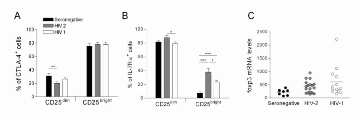
FIGURE 1. FREQUENCY OF CD25+ CELLS WITHIN THE CD4+T-CELL SUBSET...86 FIGURE 2. FREQUENCY OF CD25dim WITHIN CD4+T-CELL CELLS IN EARLY AND LATE STAGE
HIV INFECTION...88 FIGURE 3. EXPRESSION OF TREG MARKERS WITHIN CD25+ SUBPOPULATIONS...91 FIGURE 4.EXPRESSION OF ACTIVATION AND DIFFERENTIATION MARKERS...92 FIGURE 5. FLOW CYTOMETRIC ANALYSIS OF CYTOKINE PRODUCING CD4+T-CELLS ACCORDING TO CD25 EXPRESSION...94
CHAPTER 5:
COMPARATIVE ASSESSMENT OF REGULATORY CD4+T-CELLS IN HIV-2
AND HIV-1 INFECTIONS, TWO ACQUIRED IMMUNE DEFICIENCIES WITH DIFFERING RATES OF DISEASE PROGRESSION
FIGURE 1.FREQUENCY OF REGULATORY T-CELLS WITHIN THE CD4+T-CELL SUBSET... 114 FIGURE 2.IMPACT OF VIREMIA ON THE FREQUENCY OF REGULATORY T-CELLS... 116 FIGURE 3.NAÏVE-MEMORY CD4+T-CELL IMBALANCES AND
FIGURE 4.FREQUENCY OF CYCLING REGULATORY T-CELLS... 122 FIGURE 5.INCREASED FREQUENCY OF REGULATORY T-CELLS IN ART-DISCORDANTS... 126 FIGURE 6.LONGITUDINAL ASSESSMENT OF THE CD4+T-CELL SUBSETS IN
ART-DISCORDANT PATIENTS... 128 FIGURE 7.FREQUENCY OF CYCLING CELLS IN ART-DISCORDANTS... 129
CHAPTER 3:
GAG-SPECIFIC CD4+T-CELL FREQUENCY IS INVERSELY CORRELATED WITH
PROVIRAL LOAD AND DIRECTLY CORRELATED WITH IMMUNE ACTIVATION IN
HIV-2, BUT NOT HIV-1 INFECTION
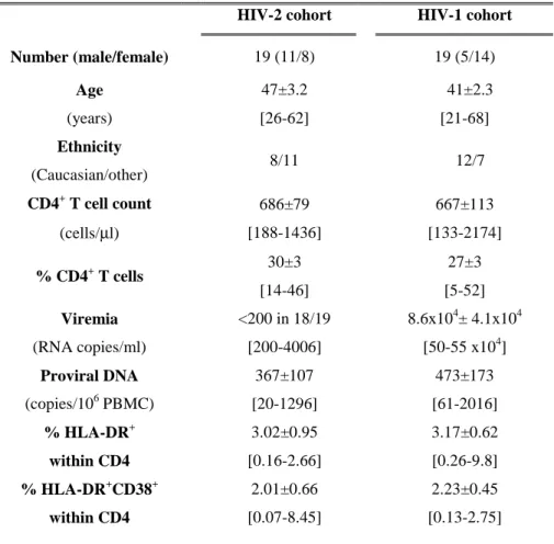
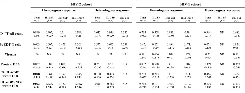
TABLE 1.HIV-2 and HIV-1 COHORT CHARACTERIZATION...65 TABLE 2:CORRELATION BETWEEN FREQUENCY OF GAG-SPECIFIC CD4 RESPONSES AND
POSSIBLE SURROGATE MARKERS OF HIV DISEASE PROGRESSION...71
CHAPTER 4:
INCREASED FREQUENCY OF CD25DIMCD4+T-CELLS IN HIV-2 INFECTION,
A NATURALLY OCCURRING ATTENUATED FORM OF HIV-1
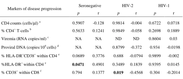
TABLE 1:CHARACTERISTICS OF THE STUDY COHORTS...83 TABLE 2.CORRELATION BETWEEN MARKERS OF DISEASE PROGRESSIONAND THE FREQUENCY OF CD25dim CELLS WITHIN THE CD4+T-CELL POPULATION...87
CHAPTER 5:
COMPARATIVE ASSESSMENT OF REGULATORY CD4+T-CELLS IN HIV-2
AND HIV-1 INFECTIONS, TWO ACQUIRED IMMUNE DEFICIENCIES WITH DIFFERING RATES OF DISEASE PROGRESSION
TABLE 1.CHARACTERISTICS OF THE STUDY COHORTS... 108 SUPPLEMENTAL TABLE 1:CHARACTERISTICS OF COHORTS ASSESSED
FOR FOXP3AND KI67 EXPRESSION CELLS... 109 TABLE 2.ASSOCIATION BETWEEN CD4+T-CELL AND Treg SUBSETS AND MARKERS OF
DISEASE PROGRESSION IN HIV INFECTED INDIVIDUALS AND SERONEGATIVE CONTROLS... 115 TABLE 3.ASSOCIATION BETWEEN THE PROPORTION OF CYCLING Treg, THE FREQUENCY OF Treg WITHIN CD4+T-CELLS AND MARKERS OF DISEASE PROGRESSION... 124
ACKNOWLEDGEMENTS
First, I want to thank my supervisors Prof. Doutora Ana Espada de Sousa (Unidade de
Imunologia Clínica) and Prof. Doutor Rui Victorino (Unidade de Imunologia Clínica & Clínica Universitária de Medicina 2, Hospital de Santa Maria) for their time and effort
invested in my project, for the ideas, creativity and scientific input and discussions. Also I want to acknowledge the Fundação para a Ciência e a Tecnologia for providing funding through the provision of my PhD scholarship.
I want to thank all my colleagues from the Unidade de Imunologia Clínica, all of whom over the years have made important contributions, both in terms of practical assistance, as well as theoretical input. Special mention must go to Adriana Albuquerque, Rita Cavaleiro and Catarina Cortesão who I have worked with since the start of my time in Portugal.
I would also like to thank Nuno Taveira (Faculdade de Farmácia) and everyone in his group that helped provide support for my work associated with primary HIV cultures, not least through the provision of P3 facilities; and Jaõ Gonçalves (Faculdade de Farmácia) for provision of cell lines.
I also want to thank Prof. Doutora Perpétua Gomes and Prof. Doutora M Helena Lourenço (Departamento de Microbiologia, Faculdade de Farmácia) for the quantifications of HIV-2 viremia, and all the doctors and nurses from the Departments of
Doenças Infecciosas and Medicina 2 from Hospital Santa Maria for their help in
I also want to offer special thanks to all the patients and healthy subjects recruited for my studies, because without their interest and commitment to science, none of this work would have been possible.
As relocating to a new laboratory, let alone a foreign country is not the easiest of processes, I want to thank all of the new friends that I have made, both within the unit and outside it, that have helped make the transition much easier than it could have been.
Within Unidade de Imunologia Clínica all my colleagues who thankfully became much more than co-workers, Adriana Albuquerque, Rita Cavaleiro, Rui Soares, Antonio Baptista, Rita Azevedo, Rita Tendeiro, Rita Barbosa, and Paula Matoso. Also not forgetting the new friends I made outside of work, that eased the acclimatization process, in particular Nikki, Belinda, Andy, Zoe, Rob, Mauritz, Rodrigo and Gonçalo.
I would like to give my heart-felt thanks to my Mother, whose understanding and support, even at such a distance, has been indispensible. Finally I must thank Anita Lopes, my long-suffering better-half, who has been a great source of support and strength over the years, and whose infinite patience and understanding when work has kept us apart more than is reasonable, is much appreciated.
ABBREVIATIONS
γc Common Cytokine Receptor Chain
7TM 7 Transmembrane
Ab(s) Antibody(ies)
Ag(s) Antigen(s)
AGM African Green Monkeys
AICD Activation Induced Cell Death
AIDS Acquired Immunodeficiency Syndrome
APC Allophycocyanin
APC-Cy7 Allophycocyanin-Cy7
APOBEC3G Apolipoprotein B mRNA-editing enzyme catalytic polypeptide-like 3G
ART Anti-retroviral Therapy
Bcl-2 B-Cell Lymphoma 2
BSA Bovine Serum Albumin
CA Capsid protein
CAF Cell Associated Factor
CCL Chemokine (C-C motif) Ligand
CCR Chemokine (C-C motif) Receptor
CD Cluster of Differentiation
CD4 Monomeric IgG superfamily protein expressed on CD4+ T cells
CD8 Homo or Heterodimeric protein co-receptor expressed on CD8+ T cells
CD25 The α-chain of the IL-2 receptor
CDC Centers For Disease Control and Prevention
CTLA-4 Cytotoxic T-lymphocyte Antigen 4
CXCR Chemokine (CXC motif) Receptor
DC(s) Dendritic cell(s)
DNA Deoxyribonucleic Acid
DP Double-Positive
EC Elite-controller
ELISA Enzyme-Linked Immunosorbent Assays
ELISPOT Enzyme-Linked Immunosorbent Spot Assay
Env Envelope
FCS Foetal Calf Serum
FITC Fluorescein
FIV Feline Iimmunodeficiency Virus
Foxp3 Forkhead-box p3
Gag Group-specific Antigen
GALT Gut-Associated Lymphoid Tissue
GAPDH Glyceraldehyde-3-Phosphate Dehydrogenase
GITR Glucocorticoid-induced Tumour Necrosis Factor Receptor
gp Glycoprotein
HAART Highly Active Anti-retroviral Therapy
HIV Human Immunodeficiency Virus
HLA Human Leukocyte Antigen
HTLV-III Human T-Lymphotropic Virus Type III
ICS Intracellular Cytokine Staining
ICAM-1 Intercellular Adhesion Molecule 1
IFN Interferon
IFN-α Interferon alpha
IL Interleukin
IL-2 Interleukin 2
IL-2R Interleukin 2 Receptor
IL-2Rα Interleukin 2 Receptor alpha-chain (CD25) IL-2Rβ Interleukin 2 Receptor beta-chain (CD122)
IL-7 Interleukin 7
IL-10 Interleukin 10
IL-15 Interleukin 15
IL-7Rα Interleukin 7 Receptor alpha chain (CD127)
IPEX Immune Dysregulation, Polyendocrinopathy, Enteropathy, X-linked syndrome
LAV Lymphadenopathy-Associated Virus
LPS Lipopolysaccharide
LTNP Long-Term Non-Progressors
LTR(s) Long Terminal Repeat Sequence(s)
MALT Mucosal Associated Lymphoid Tissue
MA Matrix protein
mDC(s) myeloid Dendritic cell
MFI Median Fluoresence Intensity
MHC Major Histocompatibility Complex
mRNA messenger RNA
MIP-1 α Macrophage Inflammatory Protein 1 alpha
MIP-1 β Macrophage Inflammatory Protein 1 beta
Nef Negative replication factor
NC Nucleocapsid protein
NFAT Nuclear Factor of Activated T Cells
NK Natural Killer
OI(s) Opportunistic infection(s)
pDC(s) Plasmacytoid Dendritic Cell(s)
PBMC Peripheral Blood Mononuclear Cells
PBS Phosphate Buffered Saline
PCR Polymerase Chain Reaction
PE Phycoerythrin
PE-Cy7 Phycoerythrin-Cy7
Pen/Strep Penicillin Streptomycin
PerCP Peridinin chlorophyll
PerCP Cy5.5 Peridinin chlorophyll-Cy5.5
PHI Primary HIV Infection
PIC Pre-integration Complex
PMA Phorbol 12-myristate 13-acetate
Pol Polymerase
PRR(s) Pattern-Recognition Receptor(s)
RAG Recombination activating gene
RANTES Regulated Upon Activation, Normal T Cell Expressed AndSecreted
Rev Regulator of Virion
RNA Ribonucleic Acid
RTC Reverse-transcription Complex
RT-PCR Reverse Transcription-Polymerase Chain Reaction
SDF-1α Stromal cell Derived Factor 1 alpha
SIDA Síndrome da Imunodeficiência Adquirida
SIV Simian Immunodeficiency Virus
SP-1 Specificity Protein 1
STI Structured Treatment Interruption
SU Surface unit
TCM Central Memory T cell
TEM Effector Memory T cell
Tat Transactivator
TAR Trans-activating Response Region
TCR T Cell Receptor
TGF-β Transforming Growth Factor Beta
Th T helper cell
Th1 T helper cell type 1: defined by their ability to produce Inteferon gamma
Th3 T helper cell type 3: defined by their ability to secrete TGF-β
Th17 T helper cell type 17: defined by their ability to produce IL-17 and IL21
TLR(s) Toll-like receptor(s)
TM Trans-membrane
TNF Tumour Necrosis Factor
Tr1 T regulatory cell type 1: defined by an ability to produce IL-10
Treg Regulatory CD4+ T cell: defined by expression of CD25 and/or Foxp3
TRIM5 Tripartite Motif Protein 5
Vif Virion infectivity protein
Vpr Viral-protein R
Vpu Viral-protein U
SUMÁRIO
A pandemia da infecção HIV/SIDA continua a representar um dos mais importantes
problemas mundiais de saúde. Desde os casos iniciais de SIDA descritos em 1981,
aproximadamente 70 milhões de novos casos de infecção pelo HIV foram diagnosticados
e mais de 25 milhões de mortes associadas à SIDA foram reportadas.
Os agentes causadores de SIDA são o HIV-1 e o HIV-2. A infecção pelo HIV-1 é
caracterizada por uma progressiva e, na ausência de tratamento, irreversível perda dos
linfócitos T CD4. Esta depleção não pode ser só atribuída a efeitos citopáticos directos do
vírus, sendo a hiper-activação crónica do sistema imunitário associada à infecção pelo
HIV um dos principais factores determinantes da progressão da doença. É por isso
fundamental investigar os mecanismos da imunidade celular que contribuem
potencialmente para o controlo da hiper-activação imunitária, tais como as células T
reguladoras (Treg), ou que favoreçam a manutenção das populações linfocitárias
relevantes, incluindo os linfócitos T específicos para o HIV. A infecção pelo HIV-2,
apesar da reduzida virémia, associa-se, tal como o HIV-1, a uma progressiva activação
imunitária ao longo do curso da doença. Contudo, o ritmo de aumento dos níveis de
hiper-activação imunitária e do consequente declínio dos linfócitos T CD4 é muito mais
lento na infecção pelo HIV-2 do que na pelo HIV-1. Assim, o HIV-2 é aqui explorado
como um “modelo” de doença HIV atenuada, com potencialidades únicas para analisar
aspectos da resposta vírus-hospedeiro que contribuam para controlar ou melhorar os
efeitos da hiper-activação crónica do sistema imunitário e desta forma modelar a
imunopatogénese da SIDA.
As respostas especificas dos linfócitos T CD4 foram aqui investigadas dados os poucos
estudadas em doentes infectados pelo HIV-1 e pelo HIV-2 com graus de depleção dos
linfócitos T CD4 comparáveis. A frequência e a magnitude das respostas foram
caracterizadas em termos das citocinas produzidas a nível celular individualizado.
As duas infecções apresentaram uma frequência semelhante de células T CD4+ capazes
de reconhecer péptidos homólogos do Gag. Contudo, nos doentes infectados pelo HIV-2
documentou-se uma melhor capacidade de responder a péptidos heterólogos. É
importante salientar que quer as respostas homólogas quer as heterólogas se
caracterizavam por uma maior produção de IL-2 na infecção pelo HIV-2. Além disso, as
respostas HIV-2 homólogas correlacionavam-se positivamente com os níveis de activação
dos linfócitos T CD4 e negativamente com a carga viral celular avaliada pelo DNA
proviral, sugerindo uma potencial relação entre as respostas CD4 especificas para o HIV,
a activação imunitária e o controlo da replicação viral que pode contribuir para o melhor
prognóstico associado à infecção pelo HIV-2.
De entre os múltiplos aspectos da desregulação do sistema imunitário associada à
infecção pelo HIV-1, a bem reconhecida disrupção das vias da IL-2 e do receptor da IL-2
tem um interesse particular uma vez que esta citocina tem um papel central quer na
homeostasia dos linfócitos T quer na função e sobrevivência das células T reguladoras. A
expressão diferencial da expressão da cadeia α do seu receptor, CD25, constitui um
importante mecanismo de modulação da função da IL-2. Os indivíduos infectados pelo
HIV-2 parecem ter a capacidade de contrariar a perda de CD4 associada à activação
imunitária persistente na ausência de terapêutica antiretroviral (ART). Assim, com o
objectivo de avaliar o potencial contributo da população CD4+CD25+ para a manutenção
dos linfócitos T CD4, esta população foi caracterizada em doentes infectados pelo HIV-1
Apesar do CD25 ter sido originalmente identificado como um marcador de activação T,
tal como previamente descrito, não foi encontrada qualquer diferença na proporção de
linfócitos T CD4 expressando CD25 em doentes infectados pelo HIV-1 e controlos
seronegativos. Em contraste, esta população estava significativamente aumentada em
indivíduos infectados pelo HIV-2. A divisão da população de células T CD4+CD25+ de
acordo com os níveis de expressão de CD25, revelou que este aumento era devido à
sub-população de células expressando baixos níveis de intensidade de CD25 (CD25dim). O aumento desta sub-população é aparentemente mantido ao longo de toda a história natural
da infecção pelo HIV-2 uma vez que não se observou qualquer relação com o grau de
depleção CD4. Importa realçar que estes níveis de expressão de CD25 não estavam
associados à expressão de outros marcadores de activação nos linfócitos T CD4 em
ambas as infecções. A caracterização fenotípica detalhada destas células permitiu ainda
demonstrar que esta sub-população CD25dim, ao contrário da população CD25bright, não está enriquecida em células reguladoras. Além disso, documentou-se uma elevada
frequência de células produtoras de IL-2, IL-4 e IFN-γ, confirmando que esta população CD25dim tem uma função predominantemente efectora e não supressora.
A proporção aumentada de células CD25dim nos linfócitos T CD4 em doentes infectados pelo HIV-2 era devido a células com um fenótipo de memória–efectoras
(CD45RA-CCR7-). Como a IL-15 é uma citocina crucial para a homeostasia das células de
memória-efectoras, os níveis séricos de IL-15 foram quantificados e documentou-se um
aumento significativo na infecção HIV-2 em comparação com a HIV-1. A IL-15, tal
como outras citocinas γc, induz a expressão de CD25 em células T CD4. Assim, a
proporção aumentada de CD25dim observada na infecção pelo HIV-2 pode estar relacionada com uma melhor capacidade de manutenção do compartimento de células T
determinante do ritmo de progressão da doença em modelos símios. Em conclusão,
identificámos uma expansão única da população CD25dim nos linfócitos T CD4 em doentes infectados pelo HIV-2, que poderá contribuir para a melhor manutenção das
células T CD4 de memória-efectoras e para a menor agressividade da imunopatogénese
da associada à infecção pelo HIV-2.
As células T CD4 reguladoras (Treg) têm um papel importante na homeostasia dos
linfócitos T e no controlo da imunopatologia associada às respostas imunitárias,
particularmente no contexto de infecções persistentes. A contribuição relativa desta
população na patogénese da infecção VIH/SIDA não está ainda clarificada. As Treg
foram avaliadas quer pela expressão de CD25bright quer do factor regulador da transcrição Foxp3, e o seu “turn-over” foi estimado utilizando o marcador de células em ciclo Ki67.
A selecção de “cohorts” de indivíduos infectados pelo HIV-2 e pelo HIV-1 sem
exposição prévia a ART, bem como doentes com infecção pelo HIV-1 tratados com ART
com diferentes graus de recuperação imunológica apesar da supressão da virémia
constitui uma estratégia inovadora para investigar as inter-relações entre Treg, activação
imunitária e carga viral/antigénica.
Na ausência de ART documentou-se em ambas as infecções uma aumento da proporção
de Treg nas populações de linfócitos T CD4 de memória e naïve com a progressão da
doença, que se associava a um aumento marcado da frequência de células Ki67+ na
população Treg de memória. Em doentes HIV-1 tratados com ART com fraca
recuperação imunológica, observou-se uma frequência reduzida de Ki67+ nas células
Treg. Apesar disso, o estudo longitudinal destes doentes documentou uma expansão
significativa mantida da população Treg, apontando para uma capacidade aumentada de
sobrevivência destas células neste contexto clínico em que foi demonstrado um
evidências suportando um contributo da diminuição da timopoiese na falência de
reconstituição imunológica nestes doentes.
Em conclusão, estes dados sugerem uma melhor preservação na infecção HIV/SIDA das
Treg circulantes naïve e de memória em relação a outras sub-populações T CD4, e
significativamente contribuem para a melhor compreensão dos processos envolvidos na
manutenção das células T reguladoras circulantes.
Palavras-chave: HIV/AIDS; HIV-2; Respostas T CD4 específicas para o HIV; CD25; Células T reguladoras.
SUMMARY
The AIDS pandemic continues to represent one of the most important health-care issues
world-wide. Since the initial case-reports describing AIDS in 1981, approximately 70
million cases of HIV infection were diagnosed and more than 25 million AIDS-related
deaths were reported.
The viral agents responsible for causing AIDS are HIV-1 and HIV-2. HIV-1 infection is
characterized by a progressive and, in untreated individuals, apparently irreversible loss
of CD4+ T-cells. This depletion cannot solely be attributed to direct virus-mediated cytopathicity. The progressive hyper-immune activation associated with HIV-infection is
thought to be a key factor in determining disease progression. It is, therefore, of relevance
to assess those aspects of cell-mediated immunity that possess the capability to regulate
hyper-immune activation, such as CD4+ regulatory T cells (Treg) or that contribute to increase the maintenance of relevant lymphocyte subsets including HIV-specific T-cells.
HIV-2 infection, like HIV-1, is characterized by similar levels of hyper
immune-activation throughout the course of the disease, despite reduced levels of viremia.
However, the rate of increase in the levels of immune activation and the consequent
progressive decline of the CD4+ T-cell population occur at a much slower rate than in HIV-1 disease. Thus, HIV-2 was utilized here as a model of “attenuated” HIV disease to
provide a system to analyse aspects of HIV-host responses that may control or ameliorate
the effects of hyper-immune activation currently thought to be a major factor associated
with HIV-immunopathogenesis.
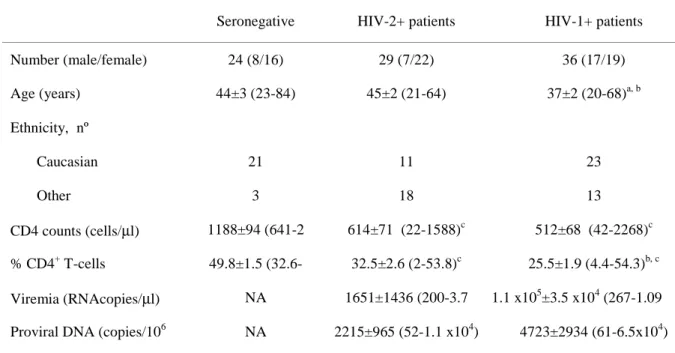
In view of the limited data available, HIV-2 specific CD4+ T-cell responses were assessed. Homologous and heterologous Gag-specific CD4+ T-cell responses were cross-sectionally assessed in cohorts of HIV-2 infected and HIV-1 infected patients, matched
for CD4 depletion. Frequency and magnitude of the responses were characterized in
terms of cytokine production at the single-cell level.
Similar frequencies of CD4+ T-cells able to recognise homologous gag peptides were observed in both cohorts. A clear difference in the frequency of CD4+ T-cells able to respond to heterologous peptides in HIV-2 infected compared to HIV-1 infected
individuals was also noted. Of note, HIV-2 specific CD4+ T-cell responses, whether homologous or heterologous, were characterized by a better maintained IL-2 response.
HIV-2 homologous responses alone correlated positively with CD4+ T-cell activation, and negatively with proviral load suggesting a potential link between HIV-2 specific CD4+ T-cell response, immune-activation and viral control, which may in turn relate to the better
prognosis associated with HIV-2 infection.
Amongst the many reported aspects of immune dysregulation associated with HIV-1
infection, the well-documented disruption of the IL-2/IL-2R network is of particular
interest given that this system is central to both immune regulation and T cell
homeostasis. The differential expression of CD25 molecule represents an important way
in which the cellular response to IL-2 can be modulated. Given that untreated HIV-2
infection features an apparent ability to counter-act CD4 loss in the face of persistent
immune activation, the CD25+ CD4+ T-cell population was characterized in cohorts of therapy naïve HIV-2 and HIV-1 infected individuals, to determine what if any potential
contribution this population may make to the maintenance of the CD4+ T-cell pool in HIV infection.
Although CD25 was originally identified as a marker of recent activation, we found, as
had others, no difference in the proportion of CD4+ T-cells expressing CD25 in HIV-1 infected individuals and seronegative controls. In contrast, the frequency of this
T-cells were subdivided according to degree of CD25 expression, HIV-2 infected patients
demonstrated an increase in a population of cells expressing low levels of CD25
(CD25dim). The increased frequency of this subset in HIV-2 infected patients appeared to be maintained throughout the natural history of the infection as it was shown to be
independent of the levels of CD4 depletion. It is also important to note that low-level
CD25 expression was dissociated from the expression of other activation markers within
CD4+ T-cells in both HIV-2 and HIV-1 infections. Further phenotypic characterization of the CD25dim CD4+ T-cell subset suggested that, unlike their CD25bright counterparts, they were not enriched in cells with a regulatory phenotype. Moreover, this population was
shown to be significantly enriched in IL-2, IL-4 and IFN-γ producing cells, particularly in the HIV-2 cohort, further supporting an enhanced effector rather than suppressor role.
The increased proportion of CD25dim within CD4+ T-cells in HIV-2 infected patients was apparently due to cells with an effector memory phenotype (CD45RA-CCR7-). As IL-15
is an important cytokine for effector-memory homeostasis, its levels were assessed and
found to be significantly increased in HIV-2 as compared to the HIV-1 cohort. This is of
particular interest as IL-15 along with other members of the γc using cytokine family is
able to induce CD25 expression on CD4+ T-cells. The increased proportion of CD25dim within the CD4+ T-cell subset observed in HIV-2+ subjects may, thus, be related to the better maintenance of the CD4+ effector-memory T-cell compartment as a whole in these individuals. An ability to preserve this T cell subset has been shown to determine the rate
of disease progression in SIV models. In conclusion, we identified a unique expansion of
CD25dim withinCD4+ T-cells in HIV-2 infected individuals, possibly contributing to the better maintenance of their CD4+ effector/memory pool and to the less aggressive HIV-2 immunopathogenesis.
Regulatory CD4+ T-cells (Treg) play an important role both in T-cell homeostasis and the control of immunopathology associated with immune responses, particularly in the
context of persistent infections. The relative contribution of this population to HIV/AIDS
pathogenesis has yet to be clarified. Treg subset imbalances were assessed both in terms
of CD25bright and of the fork-head box transcription factor Foxp3 expression. Their turn-over was estimated by the cell-cycle marker Ki67. The selection of cohorts of individuals
with untreated HIV-2 infection, untreated HIV-1 infection, and treated HIV-1 patients
that despite the suppression of viremia under antiretroviral drugs (ART) exhibited
variable degrees of CD4 recovery provided a novel strategy to assess the
inter-relationship of Treg, immune activation and viral/antigen load.
The proportion of Treg within the memory and naïve CD4+ T-cell populations increased with disease progression in both untreated HIV-1 and HIV2 infected individuals, and was
associated with a marked increase in cycling memory-Treg. In the context of poor
immunologic recovery under viral suppressive therapy for HIV-1 infection an increased
frequency of circulating Treg was observed in association with a reduced frequency of
Ki67+ Treg. Therefore the maintenance of this significantly expanded population suggests an increased ability of these cells to survive in this clinical context that has been reported
to be associated with impaired thymic function.
Overall, these data suggest a better preservation of circulating naïve and memory Treg as
compared to other CD4+ T-cell subsets in HIV/AIDS, and provide further insights into the processes involved in the maintenance of circulating human Treg.
Keywords: HIV/AIDS; HIV-2; HIV-specific CD4+ T-cell response; CD25; regulatory CD4+ T-cells.
CHAPTER 1:
INTRODUCTION
1. The Human Immunodeficiency Virus
1.1 Emergence and origins
In the early 1980’s the first reports of a newly emerging human disease began to be published. Patients typically presented with unusual infections, such as Pneumocystis
jiroveci (carinii) [1] and rare cancers such as Karposi’s Sarcoma [2; 3]. Two years after
the initial case reports, the causative agent, a human retrovirus belonging to the lentivirus genus was identified [4; 5; 6]. This new virus was eventually given the name Human Immunodeficiency virus (HIV) [7]. The discovery of a second, related virus in 1986 [8], resulted in the initially described HIV being renamed HIV-1.
As members of the lentivirus genus, both HIV-1 and HIV-2 belong to the retroviridae family of viruses all of which utilize single-stranded RNA as their genetic material. The other member of their group is Simian immunodeficiency virus (SIV), which infects various non-human primates, with the pathogenecity of the infection apparently dependent upon the host-strain relationship [9; 10]. Other groups of this family of retroviruses are associated with pathology in other species, e.g. Maedi-Visna virus in sheep and goats [11] and Feline Immunodeficiency Virus (FIV) in cats [12].
The origins of HIV-1 and HIV-2, revealed through comparative genomic analysis, lie in the zoonotic transmission of SIV to humans. This process has probably occurred several times already [13; 14], and may still be ongoing [15].
HIV-1 is believed to have arisen from cross-species transmission from the chimpanzee
Pan troglodytes. There are three groups of HIV-1: M, N and O [14; 16; 17], with a very
recent report identifying a fourth group, putatively named P, resulting from gorilla to human transmission [15].
The majority of HIV-1 cases result from infection by group M viruses. This group is divisible into nine subtypes A-D, F, H, J and K, with inter-subtype genetic differences ranging from 15-20% [18]. Of note, subtype B predominates in the Americas, Europe and
Australia, and subtype C in sub-Saharan Africa. Novel recombinant strains, such as A/G in West Africa, have also been reported [18].
HIV-2 has around 40-60% sequence homology at the nucleotide level with HIV-1 [19] and its genomic structure is very similar, as illustrated in Figure 1. HIV-2 is genetically closer to SIVSM, with over 75% nucleotide homology [20], a result of interspecies
transmissions from the natural host sooty mangabey (Cercocebus atys) [13; 21; 22; 23; 24]. HIV-2, though lacking subtypes, consists of five groups, of which A and B are the most clinically relevant [25; 26].
1.2 Structure of the HIV genome
The organization of the HIV-1 and HIV-2 genomes (Figure 1) follows a basic structure that is conserved throughout the Lentiviruses:
5’ and 3’ long terminal repeats (LTR) whose sequences are homologous and that serve both structural and regulatory functions, flanking the three main structural genes, group specific antigen (gag), polymerase (pol) and envelope (env). A varying number of genes encoding accessory proteins are also included in lenteviral genomes. In the case of HIV-1 and HIV-2 there are six: two encoding the regulatory proteins transactivator (Tat) and regulator of virion (rev); and four encoding the accessory proteins, negative replication factor (nef), virion infectivity factor (vif), viral protein R (vpr) in both viral genomes, and viral protein u (vpu) in HIV-1 or viral protein x (vpx) in HIV-2.
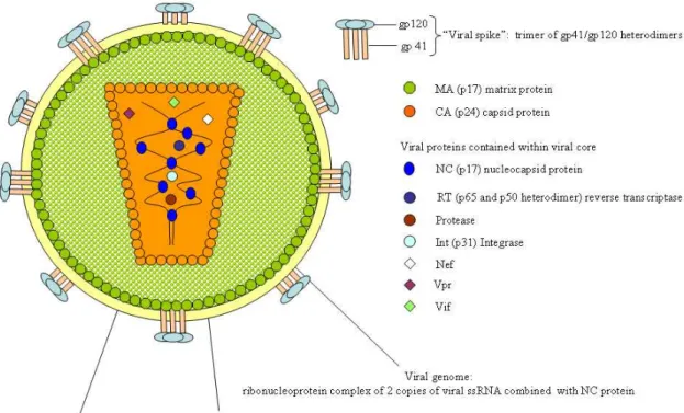
1.3 Structure of the mature HIV virion
The basic organization and composition of the HIV virion, as illustrated in Figure 2, is common to both HIV-1 and HIV-2, although some basic physical characteristics of viral proteins, such as molecular weight (e.g. gp140/160 rather than gp120 in HIV-2), may differ [23]. Thus, a basic overview of the HIV-1 virion structure will be provided.
Figure 1: genomic structure of the HIV viruses; modified from “Landmarks of the HIV-1, HIV2 and SIV genomes”, HIV molecular immunology 2008 Los Alamos database.
Figure 2: the mature HIV-1 virion
The mature HIV-1 virion is surrounded by a host-cell derived lipid bi-layer. This envelope is characteristically modified by the insertion of two viral glycoproteins: gp41
which spans the envelope, and gp120, which extends beyond the envelope surface. Heterodimers of the viral glycoproteins gp41 and gp120 associate to form trimeric complexes [27] often referred to as the viral spike [28], the complex structure of which has recently been reviewed [29]. It is important to note that the viral envelope can also contain several host-cell membrane-proteins such as Major Histocompatability Complex (MHC) class I and MHC class II molecules [30; 31], complement control proteins, such as CD55 [31], and ICAM-1 [32]. Lining the inner face of the viral membrane is a
multimeric protein shell constructed from the matrix protein (MA, p17). Located centrally within the virus particle is a conical capsid core constructed from the capsid protein (CA) p24. Contained within this viral core are two copies of the unspliced viral genome, which is stabilized as a ribonucleoprotein complex through interaction with the nucleocapsid protein (NC) p7. As well as the diploid viral genome the capsid contains six virally encoded proteins: the enzymes protease, reverse transcriptase and integrase; and the accessory proteins Nef, Vif, and Vpr. It is also important to note that several host-cell encoded proteins, such as Uracil DNA glycosylase [33] Staufen [34] and cyclophilin A [35], can be packaged into the mature virion, with only the latter being shown to be of importance for viral infectivity [36; 37]. Of note, the incorporation of apolipoprotein B mRNA-editing enzyme catalytic polypeptide-like 3G (APOBEC3G), a member of the cytidine deaminase family, into newly assembled virions [38] poses a sufficiently severe threat to HIV-1 [39], that the virus has evolved a specific countermeasure in the form of Vif, recently discussed in a review by Malim and Emerman [40].
1.4 The HIV life cycle
This is, in general terms, similar for both HIV viruses and can be broadly divided into two distinct stages, the early and late phase. The early phase encompasses the processes of virion binding and fusion and includes all the subsequent post-fusion events that eventually lead to the integration of viral genomic DNA into the chromosomes of the host cell. The late phase of the virus life-cycle begins with the initiation of regulated expression of the integrated provirus and ends with the budding of new virus particles and their subsequent maturation. As the general principles involved are similar for both viruses, an overview of the HIV-1 life-cycle is presented below.
The early phase of the HIV life cycle: virion binding and fusion
De novo infection of a susceptible target cell requires virion binding and membrane fusion, two processes that are intimately associated with the viral glycoproteins gp41 and gp120.
Viral entry is the initial step in the process of HIV-1 infection. Although receptor-mediated fusion is the primary method utilized by HIV-1 to gain entry into cells, other alternative pathways have been reported [41].
The HIV-1 Env protein mediates the process of viral entry into cells. Both post-translational glycosylation of Env and its cleavage, at the cell membrane, into its
functional moieties gp41 and gp120, are required for viral infectivity [42; 43]. The gp41 protein, through interaction of its C terminus with the viral matrix protein,
anchors the gp41/gp120 heterodimer to the viral envelope [44]. Whereas it is the the gp120 protein, which forms the surface unit (SU) of the gp41/gp120 heterodimer, that is important for binding specifically to the HIV-1 receptor, CD4, on the surface of target cells [45]. The structure of gp120 was solved several years ago [46], with the molecule shown to contain nine highly conserved interchain di-sulphide bonds and five hypervariable regions (V1-V5). The binding of trimeric gp120 on the virion surface to CD4 on the target cell induces a conformational change in the envelope proteins resulting in the exposure of the V3 loop [46]. This in turn leads to engagement of one of the two main HIV co-receptors, the chemokine receptors CCR5 [47] and CXCR4 [48]. Co-receptor engagement initiates the process of pore formation [49], through the induction of a conformational change in gp41 [50]. Assembled as a trimer on the virion membrane, the coiled-coil gp41 protein opens, projecting three peptide fusion domains that penetrate the lipid bilayer of the target cell. The fusion domains then form hairpin-like structures that draw the virion and cell membranes together to promote fusion, leading to the release of the viral core into the cell interior [50].
Of note, gp120 can also interact with the protein DC-SIGN which is expressed on the cell surface of dendritic cells [51]. Although this does not lead to productive infection of the DC-sign bearing cell, it may be important in dissemination of the virus and trans-infection of CD4+ lymphocytes [52].
These comprise of a series of sequential events, listed below, that eventually result in the integration of genomic viral DNA into the host cell genome. All of these events are dependent upon the interaction of virally encoded-proteins and a variety of host-cell factors, recently reviewed by Lehmann-Che [53].
Step 1 uncoating of the viral core
Step 2 reverse-transcription complex (RTC) formation Step 3 Reverse transcription
Step 4 pre-integration complex (PIC) formation Step 5 transport of the PIC into the host cell nucleus Step 6 Integration of genomic viral DNA.
The exact molecular processes underlying these events have yet to be definitively described, although some of the physical characteristics of specific complexes have been described [54; 55]. It is also clear that activities such as reverse transcription and integration are dependent upon specific viral enzymes, reverse transcriptase and integrase respectively, which has led to their selection as targets for therapeutic intervention.
The late phase of the HIV-1 life cycle: proviral transcription
The integrated form of HIV-1, called the provirus, is approximately 9.8 kilobases in length [56]. Its transcriptional activity is in part determined by the chromosomal environment [57] and regulated by cis-acting elements within the 5’LTR region. This region of the provirus is similar in structure to other eukaryotic transcriptional units and also contains transcriptional enhancers able to bind the transcription factors nuclear factor after cellular activation (NFAT) [58] and nuclear factor[kappa]B (NF-κB) [59]. The nuclear re-localisation of these factors is associated with T-cell activation, thus providing a link between viral transcription and the activation state of the cell. It is important to note that differences in the number and type of transcriptional enhancers in the 5’LTR of HIV-2 have been reported [60; 61].
Successful transcription is dependent upon the viral accessory protein Tat. This protein binds to the transactivation response element (TAR) on the nascent mRNA strand and
recruits several host-cell factors, leading to the successful elongation of the transcript [62].
Transcription of the viral genome produces a number of HIV-specific transcripts subject to varying degrees of splicing [63]. Cotranscriptional processing yields the multiply-spliced RNA’s that code for Nef, Tat and Rev. Their export to the cytoplasm is dependent on the cellular nuclear-export machinery. However, unspliced and singly-spliced viral mRNA’s, which codes for the structural proteins of the virus, are retained within the nucleus. The viral accessory protein Rev is able to facilitate their export into the cytoplasm through binding the Rev Response element contained within unspliced viral mRNA [64].
The late phase of the HIV-1 life cycle: viral assembly
Translation of singly-spliced mRNA yields both Gag and the Gag-pol polyprotein. Following their myristillation they localize to cholesterol- and glycolipid-rich membrane microdomains at the cell surface [65]. There they recruit both the viral and host-cell encoded constituents required to generate a new virus [66].
The late phase of the HIV-1 life cycle: budding and maturation
This is thought to involve one of two potential mechanisms, recently reviewed by Gomez and Hope [67]. One makes use of the multivascular body system, whilst the other involves direct budding from membrane. The particular system utilised may be cell type dependent with the latter being classically observed in infected CD4+ T-cells [68].
The maturation process occurs after viral budding and results in the structural rearrangement of the virion contents in such a way as to yield the mature virion particle [66].
2. HIV infection: Epidemiology and Natural history
2.1 Evolution of the pandemic
The initial reports of a novel syndrome involving a variety of rare infections and cancers [1; 2; 3] and associated with immunodeficiency [1; 69] gave no real suggestion of the
seriousness of what was to come. With reports of patients with a similar spectrum of infections in an ever-widening demographic [2; 70; 71; 72; 73; 74; 75; 76; 77; 78], it soon became obvious that this immunodeficiency syndrome, subsequently given the name AIDS, could potentially prove an important health-care issue.
Nearly thirty years after these events, and some 25 years following the identification of the eitiologic agents [4; 5; 6; 8] and its subsequent naming as HIV [7], the AIDS pandemic continues to represent one of the most important health-care issues world-wide. Between 1981 and 2007, approximately 65 million cases of HIV-1 infection were diagnosed and 25 million AIDS-related deaths reported. More importantly both the numbers of new infections (2.7 million) and AIDS deaths (2 million) in 2007 were very similar to those reported in 2001 [79], suggesting that although the pandemic may be relatively stable, it is still very far from eradication.
In terms of geographic spread HIV-1 is found all over the world, although the rates of prevelance vary from region to region, with sub-Saharan Africa forming the epicentre of the pandemic. As for HIV-2, there seems to be a much tighter geographic restriction, with the majority of infections being restricted to West Africa. Outside of this region HIV-2 tends to localize to those European countries with colonial links to this region of Africa, France [80] and in particular Portugal, which has the highest HIV-2 prevalance in Europe, with 3% of the notified cases of HIV infection (both asymptomatic and/or AIDS) between 1983 and 2005 attributable to HIV-2 [81]. It is also important to note that significant HIV-2 seroprevalence in several other ex-Portuguese colonies, has been reported [82; 83; 84].
2.2 The natural history of HIV infection
The main routes of HIV transmission were identified early in the epidemic, and are similar for both HIV-1 and HIV-2. Horizontal transmission can occur through exposure to contaminated body fluids, normally via sexual contact, and via contaminated blood and its products. Vertical, mother to child transmission defines a third route. This process can happen pre-partum, during delivery, or through the breast feeding of newborns. It is important to note that the route of transmission does not seem to impact on the course of the subsequent infection [85; 86].
Transmission risk seems to be determined by two factors; the infectiousness of the “index case” and the susceptibility of the host.
Although it may vary from body fluid to body fluid, the amount of circulating virus in an infected individual would appear to be the most important determinant of infectiousness, with respect to both horizontal [87] and vertical transmission [88]. The fact that HIV-2 infection, which is characterized by low to undetectable viremia [89; 90; 91], is also associated with lower efficacy of both horizontal [91; 92] and vertical transmission [93] directly supports this hypothesis. The reduced transmissibility of HIV-2 is also thought to underlie its previously mentioned geographic restriction.
An individual’s susceptibility to infection is determined by a number of variables. The presence of other infections [94; 95] has shown to have a direct impact, and, amongst the many host factors that modulate this process, genetic polymorphisms in the genes that encode the host proteins used by HIV as its co-receptors have been clearly demonstrated to have important effects.
Target cells, viral tropism and its relation to HIV susceptibility and rate of disease progression
As already stated in the previous section of this introduction, both HIV-1 and HIV-2 need to bind to, and fuse with, a target cell for productive infection to occur. This process is mediated by virally-encoded glycoproteins and defines the cell tropism of the virus, and, in turn, the immunopathology associated with HIV infection.
The primary receptor of both viruses is the CD4 molecule [23; 96]. As CD4+ T-cells and Macrophages express this molecule they represent potential targets for HIV infection [97; 98]. Following the initial interaction between virus-and host cell conformational changes in the viral glycoprotein occur exposing a second binding site that mediates the interaction between the virus and one of several co-receptors. This second interaction ultimately results in virus-cell fusion.
The two main co-receptors for HIV-1 were identified as CCR5 [47] and CXCR4 [48], both chemokine receptors belonging to the 7 transmembrane (7TM) spanning, G-protein coupled receptor family.
CCR5 is constitutively expressed on effector/memory CD4+ T-cells, where as CXCR4 expression is restricted to the naïve subset of CD4+ T-cells [99]. Various subsets of the monocyte/macrophage lineage can also express one or other or both of these chemokine receptors, although cell type and activation state determine the pattern of expression [100]. Though initially described as chemoattractants, chemokines have also been shown to have important immunoregulatory functions, reviewed recently by Wong and Fish [101]. CCR5 is degenerate in terms of chemokine binding and its ligands include CCL3 (Macrophage Inflammatory Protein 1 alpha; MIP-1α), CCL4 (Macrophage Inflammatory Protein 1 beta; MIP-1β) and CCL5 (Regulation upon Activation Normal T cell Expressed and Secreted; RANTES). However, only one ligand (stromal derived factor 1α; SDF-1α) for CXCR4 has so far been identified.
HIV tropism is currently defined according to which co-receptor a particular viral isolate utilizes in addition to CD4, although an alternative nomenclature, designed to distinguish co-receptor preference from target cell tropism, has been proposed [102]. Monotropic R5 and X4 viruses use either CCR5 or CXCR4 respectively, whereas R5X4 viruses can use both [103]. R5 viruses are generally associated with transmission and the early phase of infection [104] whereas a switch to X4 viruses is associated with, but not absolutely required for progression to AIDS during late stage disease in HIV-1 infected individuals [104; 105; 106; 107].
The situation is less clear cut for HIV-2. Not only have primary isolates of this virus been shown to utilize a broader range of chemokine receptors than HIV-1 [108] but some have also been shown to be able to infect target cells in a CD4-independent, co-receptor dependent manner [109; 110]. Despite these in vitro observations, data suggest that CCR5 and CXCR4 also serve as the main co-receptors for HIV-2 in vivo [111; 112; 113]. Of note, primary isolates of a particular co-receptor tropism do not appear to dominate, either during the asymptomatic [114; 115] or symptomatic phases of HIV-2 infection [109; 112; 114].
As previously mentioned, some of the host-genetic determinants of susceptibility to infection have been shown to be related to polymorphisms in chemokines and their receptors. The classic example is that of a 32 base-pair deletion (∆32) in the CCR5 locus that leads to a loss of function of the receptor, and almost total protection from infection in homozygous mutants [116; 117]. Although the rare individuals homozygous for the
∆32 mutation that become infected do not seem to progress differently to wild-type counterparts, there is some evidence that individuals heterozygous for this mutation may have a slower rate of disease progression [118]. In contrast, an increased density of CCR5 on CD4+ T-cells is associated with high viral loads and increased disease progression [119]. Other mutations in CCR5 have been reported, but are very rare in the general population [120; 121]. Data from HIV-2 infected individuals demonstrated that the levels of CCR5 on CD4+ T-cells were similar to that observed for HIV-1 infected individuals, matched for disease stage, suggesting that the slower rate of disease progression associated with HIV-2 infection is unrelated to coreceptor availability [113].
Of note, duplication of the CCL3L1 gene, a ligand of CCR5 was shown to be associated with protection from transmission [122], emphasizing the utility of targeting chemokine receptors as means of inhibiting HIV transmission.
Course of HIV infection
The course of HIV infection follows a similar pattern in most infected individuals. It can be roughly divided into three phases (discussed in more detail below): the acute phase, that lasts a matter of weeks, an asymptomatic phase that last several years and a final phase in which infected individuals succumb to infection by a variety of opportunistic infections (OI’s) and/or develop rare cancers, and as such meet the centre for disease control (CDC) criteria for AIDS (Figure 4).
Figure 3: Overview of the natural history of HIV infection: in terms of the commonly used prognostic markers: plasma viremia, CD4+ T-cell depletion and immune activation. The severity of an individual’s infection can generally be characterized in terms of the time to progression to AIDS. Highly variable disease progression rates between individuals infected with HIV-1 are well recognized and can be categorized as rapid (10%-15%, time to AIDS 2-4 years) and typical or intermediate progression (80%, time to AIDS 6-10 years), and late or long term non-progression (5%, AIDS free for more than ten years, and perhaps indefinitely) [123]. The situation for HIV-2 infected patients is similar, with rare, but documented cases of rapid progression [124; 125]. In the majority of HIV-2 infected individuals the asymptomatic phase is generally longer than for their HIV-1 infected counterparts [126].
The three distinct phases of HIV infection: acute/early infection
The acute phase of infection, corresponding to the first few weeks following successful transmission, is associated with rapid viral replication in actively infected cells resulting in widespread dissemination of the virus, and increasing viremia that can reach peak levels of several million viral copies, higher than any other stage of the disease [127]. A
concomitant depletion of both circulating and mucosal CD4+ T-cells and expansion of CD8+ T-cells occurs, leading to an inversion of the CD4+/CD8+ ratio. Of note, the recent discovery of an early and massive depletion of mucosal CD4+ T-cells in HIV-1 infected individuals [128], has had a major impact upon the understanding of the process of disease progression in HIV-1 infected individuals.
In HIV-1 infected individuals the acute phase of infection is generally accompanied by a series of symptoms, such as lymphadenopathy, mononucleosis-like syndrome, night sweats and generalized rash, often grouped together under the term “acute phase syndrome”. This syndrome is often not appropriately diagnosed [129], however, there is some evidence that the presence of seroconversion symptoms can relate to a more rapid rate of disease progression [130]. Of note there are no documented cases of this syndrome in HIV-2 infected individuals.
The period spanning the acute phase, and the months that follow is referred to the primary HIV infection (PHI), during which viremia levels drop and stabilize at a value referred to as the viral “set-point”. Concomitantly with this, CD4+ T-cell numbers rebound, but often to below preinfection levels. This value is referred to as the CD4+ T-cell set-point. The levels of immune activation at this stage of disease are also referred to as the immune activation “set-point”. Both CD4+ T-cell and, in particular, viral “set-points” have prognostic value in both HIV-1 [131; 132] and HIV-2 infections [133; 134; 135]. The immune activation “set point” has also been shown to have prognostic value in HIV-1 [136]. The reduction of viral load is closely related to the expansion of HIV-specific CD8+ T-cells, the breadth and magnitude of this response being directly correlated with the degree of viral control and inversely with the rapidity of disease progression [137; 138; 139]. This is not to say that other aspects of adaptive immunity such as HIV-specific CD4+ T-cell responses [140] do not have a role. Moreover, various aspects of innate immunity acting prior to the emergence of the HIV-specific adaptive immune response are also thought to have a role in the control of viral replication [141]. Of note seroconversion also occurs during this period, although the role of virus-specific antibodies in the establishment of the viral set-point is unclear [142].
The three distinct phases of HIV infection: asymptomatic infection
The asymptomatic phase is characterized by gradually increasing viral load and a decrease in the CD4+ T-cell count over time in both HIV-1 and HIV-2 infected individuals, although the rate of CD4+ T-cell loss is lower in HIV-2 infected individuals [143; 144], and the viremia may remain low to undetectable [90; 91; 143]. Mucosal CD4+ T-cells remain stable but still at very low frequencies in HIV-1 infected individuals [145]. Immune activation remains high during this phase, as do levels of circulating anti-HIV antibodies and HIV-specific CD8+ T-cells. However, despite an outward appearance of relative calm, this phase of disease is characterized by a wide-ranging immune dysregulation that will be described below.
The three distinct phases of HIV infection: late stage HIV infection and AIDS
The final stage of HIV infection is precipitated by the eventual collapse of the host immune system, and subsequently the rapid decline of CD4+ T-cells and concomittant increase of viremia. Exactly what causes this collapse is unclear and it likely may vary from one infected individual to another. As previously discussed a switch of viral co-receptor usage has been suggested as a precipitating event, although this phenomenon is not a prerequisite for progression. AIDS in HIV-1 and HIV-2 infected individuals can be defined clinically either by the appearance of OI’s such as CMV retinitis, Pneumonia, or in terms of peripheral CD4+ T-cell count, with less than 200 CD4+ T-cells/µl currently being a diagnostic criteria of the Centres for disease control (CDC).
Late stage HIV infection is also associated with increased viremia, but to levels on average two logs lower than those observed in HIV-1 infected individuals at a similar stage of disease [143].
2.3 HIV therapy
Antiretroviral therapy (ART) is often referred to as highly active antiretroviral therapy (HAART) in light of its ability to severely impact viral replication in appropriately treated fully compliant individuals. In first world countries it has had an important and obvious impact upon both morbidity and mortality of HIV-1 infected individuals [146; 147]. The situation in the third world is more complex, given the prohibitively high cost of the
standard multi-drug regimens. However, over the last few years steps have been taken towards providing ART in resource-limited settings [148].
Despite the success of ART it must be noted that it has several limitations, not least the selection for drug-resistant viral mutants. Long-term use of ART has been related to several side-effects of varying severity, recently reviewed by Hofman and Nelson [149]. Moreover its ability to correct some characteristic features of HIV-infection is questionable, with data to suggest it has limited ability to reverse the large-scale depletion of mucosal CD4+ T-cells [150].
Another problem for the current therapeutic regimes is there inability to target the pool of latently infected cells, referred to as the viral reservoir, within an infected individual. Their existence was first suggested when individuals undergoing structured treatment interruption (STI) of ART for 4 to 6 weeks, experienced rebound of plasma viremia, to levels equivalent to those measured prior to the initiation of therapy [151].
As latently infected cells remain transcriptionally silent they are resistant to therapies that target replicating virus. It is hoped that the development of therapies that prevent the establishment of this pool, such as the anti-integrase drug Raltegravir, currently in clinical trials [152], together with the use of immunotherapeutic treatment to purge the pool of latently infected cells [153] may help address this problem.
2.4 Viral Latency
Latent HIV infection results from the persistence of infected cells whose genome contains stably integrated, transcriptionally silent HIV provirus. This process has been shown to result in the creation of a pool of latently-infected cells, resulting in the generation of viral reservoirs. This pool mainly consists of rare, latently-infected resting CD4+ memory T-cells [154; 155; 156] as well as monocytes [157] and myeloid dendritic T-cells (mDCs) [158].
Differences in the ability of HIV-2 and HIV-1 to establish a pool of latently infected cells have not been established, although there is data suggesting that dendritic cells (DC’s) are a poor substrate for HIV-2 infection [159]. The recently described ability of HIV-2 vpx to
facilitate viral replication in both macrophages [160] and DC’s [161] may also reduce the contribution of these cell types to any pool of latently infected cells in HIV-2.
The persistence of a latent viral reservoir even following long-term anti-retroviral therapy has serious implications for the use and efficacy of current therapeutic regimens [154; 162; 163].
2.5 Models of HIV infection
A variety of model systems has been developed over the course of the pandemic and will be briefly summarised below
Animal models
The use of different animal models to study HIV infection and disease progression has yielded important insights into the course of this disease, as well as providing vital test beds for vaccine development.
The finding that Macaques, who are not natural hosts of SIV, would on infection develop a disease that followed a similar, if somewhat more rapid, disease course as HIV infected individuals [164] has provided the research community with a viable and important animal model for the study of HIV.
The subsequent discovery that some species of monkeys that are natural hosts of SIV, such as Sootey Mangabeys infected with SIVSM and African green monkeys infected with
SIVAGM failed to develop AIDS-like symptoms, despite sharing some characteristics of
HIV-infected humans has provided yet another system to study HIV immunopathogenesis [165; 166]. Of note, SIVAGM infection of several species of Asian macaques, such as the
rhesus macaque, causes immunodeficiency and an AIDS–like syndrome [167; 168] and
visa versa. Thus, the study of SIV infected macaques and comparative analyses of
pathogenic and non pathogenic SIV infections have provided important models for the study of HIV pathogenesis
The use of knock-out mice strains that allow efficient long-term engraftment of a human immune system, such as BALB/c-Rag2-/-γc-/-, may provide researchers with a humanized mouse model [169]. This has obvious benefits compared to the previously described
monkey models, not only in terms of cost, but also because several of the current monkey species utilized for HIV research are endangered.
So far it has been demonstrated that humanized mice can be infected with HIV and that the infected mice subsequently develop a disease with similar characteristics to HIV infection in humans [170]. This system has already been used to investigate certain aspects of HIV immunopathogenesis [171].
Finally, although a non-primate Lentivrus, the study of Feline immunodeficiency virus (FIV) infection in cats, has the potential to provide insights into those aspects of the immune response related to shared aspects of immunopathogenesis [172].
Human systems
With regard to human systems, these have mainly consisted of the study of rare individuals with a demonstrably different rate of disease progression following HIV-1 infection (the previously mentioned Rapid Progressors and Long term non-progressors (LTNP)) and those individuals who, despite repeated exposure to HIV, fail to become infected, the so-called “exposed but uninfected individuals”.
Rapid progressors represent a particular group of individuals who progress to AIDS in a
very short time. The underlying reasons can be both virological, as in the case of an individual infected with a particularly virulent drug-resistant virus [173], or relate in part to host genetics, exemplified by the increased rate of disease progression of HIV-1 infected individuals possessing HLA-B35. [174] and is likely a combination of the two. The corollary of the rapid progressors is the diverse group of individuals considered LTNP’s. They are generally defined as individuals with stable CD4+ counts, with low or undetectable viremia [175]. A subset of these individuals appear to be able to maintain both CD4+ T-cell frequency and control viremia in the absence of therapeutic intervention [176], and are termed “elite controllers”. As might be expected this subset of individuals has become the focus of a great deal of research, although this is hampered by their very low frequency within the general patient population. No single unifying factor has yet to be identified, although some observations, unique to these individuals have been reported [177].