PHYSIOLOGICAL, MOLECULAR AND PATHOGENIC DESCRIPTION Cecilia Armesto1, Fernanda Gonçalves Martins Maia2, Fernando Pereira Monteiro3,
Mário Sobral de Abreu4 (Recebido: 10 de novembro de 2015; aceito: 23 de maio de 2016)
ABSTRACT: Fungi of the genus Colletotrichum spp. are cosmopolitan and are responsible for disease in many plants of agronomic importance. In coffee crops, three species have been described: Colletotrichum acutatum, Colletotrichum kahawae and Colletotrichum gloeosporioides. In Brazil were only reported the species C. acutatum and C. gloeosporioides, which symptoms are attributed as antracnoses, dieback and blister spot. This study evaluates the population of Colletotrichum spp. through morpho-physiological, genetic and pathogenic characteristics, in several coffee plantations in the southern of Minas Gerais. All isolates showed similar morphological and physiological characteristics, being identified as C. gloeosporioides. The best medium for mycelial growth was agar-malt extract with pH 5.5 and for spore production oat-agar medium with pH 6.5. When inoculated on berries, all isolates were pathogenic with variable levels of aggressiveness, but only 57% of these were pathogenic for hypocotyls. Three isolates, I-9, I-24 and I-26, were identified featuring the highest rates of aggressiveness in berries and hypocotyls.
Index terms: Aggressiveness, anthracnose, blister spot.
COLLETOTRICHUM EM CAFEEIROS DO SUL DE MINAS GERAIS - BRASIL: DESCRIçãO MORFO-FISIOLóGICA, MOLECULAR E PATOGêNICA
RESUMO: Fungos do gênero Colletotrichum spp. são cosmopolitas, sendo responsáveis por doenças em diversas plantas de
importância agronômica. Na cultura do café três espécies foram descritas: Colletotrichum acutatum, Colletotrichum kahawae e Colletotrichum gloeosporioides. No Brasil ocorrem somente as espécies C. acutatum e C. gloeosporioides, as quais são atribuídos sintomas como: antracnoses, seca de ponteiro e mancha manteigosa O presente trabalho foi realizado com o objetivo de avaliar a população dos isolados de Colletotrichum spp. através de características morfo-fisiológicas, patogênicas e genéticas, em diversas lavouras cafeeiras do sul do estado de Minas Gerais. Todos os isolados apresentaram características morfo-fisiológicas semelhantes, sendo identificados como da espécie C. gloeosporioides. O melhor meio para crescimento micelial foi meio extrato de malte ágar com pH5,5. Já para produção de esporos destacou-se o meio aveia-ágar com pH 6,5. Os isolados quando inoculados em frutos apresentaram-se todos patogênicos, com níveis de agressividade variáveis, porem quando inoculados em hipocótilos verificou-se que apenas 57% destes foram patogênicos. Três isolados, I-9, I-24 e I-26, foram identificados apresentando os maiores índices de agressividade em frutos e hipocótilos.
Termos para indexação: Agressividade, antracnose, mancha manteigosa. 1 INTRODUCTION
Coffee is one of the most important activities in Brazil and have a large representation in the global economy. Brazil is the world’s largest coffee producer and second biggest consumer of the product and has an coffee crop estimated at 2.3 million hectares. There are around 287 thousands of producers, which are distributed in 15 states: Acre, Bahia, Ceará, Espírito Santo, Goiás, Federal District, Mato Grosso, Mato Grosso do Sul, Minas Gerais, Pará, Paraná, Pernambuco, Rio de Janeiro, Rondônia and São Paulo (BRASIL, 2016). The crop production and the benefited sacks of coffee in 2016 are estimated at 49,126.10 and 51,943.90, respectively.
1,3,4Universidade Federal de Lavras/UFLA - Departamento de Fitopatologia/DFP -Laboratório de Diagnose - 37.200-000 - Lavras/MG - [email protected], [email protected], [email protected]
2Universidade Federal de Uberlândia - Instituto de Ciências Agrarias -Uberlândia/MG - [email protected]
The development of the sustainable coffee production in Brazil depends of the profitability for producers, the minimum amount that will guarantee their permanence in the activity. The profit is dependent of the stable cultivation systems that provide longer life for the crops and frequent productions. Highly productive cultivars adapted to each edaphoclimatic conditions and cropping system, and resistant to pests and disease, are the main components of the coffee sustainability.
The incidence of microorganisms between pre and post harvest has emerged as a limiting factor to quality production, which filamentous fungi are the biggest factor responsible for damage to coffee culture. The most known fungal diseases that affect the production are rust (Hemileia vastatrix), Cercospora leaf spot
disinfected, for one minute, with alcohol 50% and 1% sodium hypochlorite and washed in distilled water. These sections were transferred to Petri dishes containing agar culture medium (AA) and incubated for seven days in a growth chamber at 25°C with 12 hour photoperiod. After the fungal growth, medium with the mycelial fragments were removed and carried under the microscope for the identification of the pathogen by the description of Sutton (1992). After identification, fragments of colonies were transferred to new plates to obtain purification and single spore cultures to preserved into microtubes.
Morpho-physiological characterization:
Mycelial growth was evaluated in: MEA (Malt Extract Agar); PDA (Potato Dextrose Agar); GYA (Glucose Yeast Agar); OA (Oat Agar). From the pure cultures of different isolates, mycelial discs of 6 mm diameter removed and deposited in the center of each Petri dish containing the different media. The incubation conditions were 25°C and photoperiod of 12 hours for seven days. The mycelial growth was evaluated by the average growth rate Index (AGRI), according to Souza, Souza and Mendes-Costa (2007), for 15 days.
Color of colonies was assessed by expression of the coloring of the aerial mycelium. For mycelial growth and formation of acervuli, colonies were observed for ten days, on the different culture media, incubated at 25°C and 12 hour photoperiod. Shape and size of conidia was obtained by measuring the length and width of 50 randomly selected conidia held on Zeiss microscope coupled to a camera, and measured by means of AxionVision 4.8 program. For determination of sporulation was prepared a spore suspension adjusted to 106 conidia.mL-1 in
Neubauer chamber, obtaining an average of five readings at the microscope for each replicates of treatments. The experimental design was completely randomized in a factorial design with three replicates (represented by a plate), in which the evaluated factors were isolated. The culture media used for growth and sporulation at different pH, were those that had the best AGRI and promoted production of cultures spores. After the culture media selection the pH levels were adjusted for 4.5, 5.5, 6.5 and 7.0. Into cultures were deposited mycelial disks, and they were incubated at 25°C with a photoperiod of 12 hours for ten days. The mycelial growth was evaluated by the average growth rate Index (AGRI) and sporulation. The experimental design was completely randomized in a factorial design. (Cercospora coffeicola ), Phoma leaf spot (Phoma
spp.), anthracnose and blister spot, the latter being attributed to Colletotrichum spp. complex.
Colletotrichum species occur in all
producing regions of the world, and are known three species related to coffee plants: C. kahawae - geographically restricted species to Africa,
C. gloeosporioides and C. acutatum. In Brazil
predominates species known as C. gloeosporioides, which is associated with a complex of diseases and symptoms throughout the plant: anthracnose with irregular and large patches, color brown to grayish, occurring commonly on edge leaves; die-back; brown blight and blister spot, which presents light green spots oily appearance on leaves and fruit (ABREU; FERREIRA; MARTINS, 2008).
The occurrence of Colletotrichum spp. is serious in coffee regions in Brazil and can cause significant losses in culture (ABREU; FERREIRA; MARTINS, 2008). Although, the importance of the C. gloeosporioides in coffee plantation in Brazil is still very questionable, due to the difficulty in inducing certain symptoms in controlled conditions, as the example of the blister spot (FERREIRA; ABREU; PEREIRA, 2009), and according Menezes (2006) for Colletotrichum spp. complex, it is desirable the integration of morphological and non–morphological methods to identify species and strains below the species level.
It is necessary to elucidate the fungus relationship with the coffee crop in Brazil, determining the species of Colletotrichum spp. usually associated with coffee. Therefore, the present work had as objective, the identification of Colletotrichum species isolated from coffee plants, through genetic characteristics and physiological studies which may be associated with pathogenicity.
2 MATERIALS AND METHODS
The experiments were carried out at the Plant Pathology Department of the Federal University of Lavras (UFLA) – Lavras, Minas Gerais, Brasil.
Collection and preservation of Colletotrichum spp. Leaves, branches and berries of coffee plants with symptoms of blister spot, anthracnose and dieback were collected, in different cities in the southern of Minas Gerais state (Table 1). The samples were placed in paper bags and then taken to the Diagnosis Lab-Department of Plant Pathology/UFLA. Sections of infected tissue were made on young lesions, which were superficially
Molecular analysis
DNA extraction was conducted from colonies at ten days of growth, according to the protocol provided by Promega® extraction kit - Wizard Genomic DNA Purification Kit (# A1120). DNA was quantified, and samples were diluted to the concentration of 10ng.μL-1.
For molecular amplification by PCR were used specific primers for the genus Colletotrichum spp., to confirm by sequencing that all isolates belonged to that genus. Then, specific oligonucleotides for C. gloeosporioides and C.
acutatum were used.
The identification of the genus was performed using pairs of oligonucleotide Cc1F1 (5’-ACCTAACTGTTGCTTCGGCG-3’) and Cc2R1 (5´-AAATTTGGGGGTTTTACGGC-3’) as Cullen et al. (2002). The reaction was performed according to protocol provided by Gotaq Green Master Mix kit - Promega® (# M7122). The program used by the thermocycler was a cycle of 1 minute at 94°C, 2 minutes at 65°C and 2 minutes at 72°C followed by a cycle of 1 minute at 94°C, 2 minutes at 63°C and 2 minutes at 72°C, finishing with 33 cycles of 1 minute at 94°C, 2 minutes at 61°C and 2 minutes at 72°C.
For the detection of C. gloeosporioides was used CgInt oligonucleotide (5’-GGCCTCCCGCCTCCGGGCGG-3’) described by Mills, Sreenivasaprasad and Brown (1993) and for C. acutatum, CaInt2 oligonucleotides (5’-GGGGAAGCCTCTCGCGG-3 ‘) both with ITS4 (5’-TCCTCCGCTTATTGATATGC-3’) described by Sreenivasaprasad et al. (1996). The reaction was performed according to protocol provided by Gotaq Green Master Mix kit - Promega® (# M7122). The program used was: one cycle of 30 seconds at 94°C, 45 seconds at 62°C and 90 seconds at 72°C followed by a cycle of 30 seconds at 94°C, 45 seconds at 60°C and 90
TABLE 1 - Level of symptom observed in seedlings.
Category Symptom
1 No visible reaction.
2 Superficial brown lesions.
3 Dark deep lesions.
4 Dark lesions with early bottlenecks.
5 Strong bottleneck.
6 Dead seedling.
Source: Varzea (1995).
seconds at 72°C, ending 33 cycles of 30 seconds at 94°C, 45 seconds at 58°C and 90 seconds at 72°C.
Each PCR (25 μl) contained 25 ng of DNA, 1 μL each primer, and 12.5 μl Gotaq Green Master Mix kit - Promega® (# M7122). Amplifications were performed in a thermal cycler (Hybaid, Ashford, UK) programmed for 1 cycle of 5 min at 95°C, 25 cycles of 30 s at 94°C, 30 s at 62°C, and 2 min at 72°C, ending with 1 cycle of 7 min at 72°C. After amplification, PCRs products were applied in agarose gel 1%. As a control for the reactions it used an isolated Cercospora spp. and sterile water.
Pathogenicity in berries and hypocotyls
Green berries of Red Catuaí cultivar were sterilized superficially, dried and placed in plastic boxes with sterile sand for support. In each box were conditioned 20 berries (Figure 2C), in which each fruit was considered a replicate. For inoculation, berries were drilled with sterile needle and inoculated with suspension of 106 conidia.mL-1, sterile
water was used as a control. The plates were kept in climatic chamber with temperature of 25°C and photoperiod of 12 hours. Berries were evaluated for 15 days. Berries were characterized according to the presence or absence of symptoms. Absence: berry without necrotic lesion at the inoculation point; Presence: berry with initial dry and necrotic lesions, with circular aspect and the depressed center.
For hypocotyls of Red Catuaí cultivar, inoculation was performed by spraying suspensions at a concentration of 106 conidia.mL-1. As a control
was used hypocotyls inoculated with sterile water. The hypocotyls were placed in trays with substrate in a growth chamber with 12 hours photoperiod at 25°C for 20 days. The symptoms were evaluated according to a scale described by Varzea (1995)
were mostly related to plants showing symptoms of blister spot.
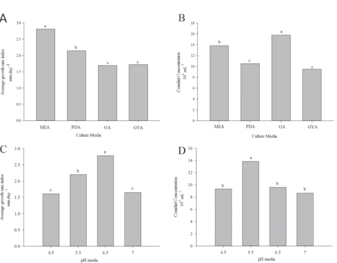
In relation to the speed of mycelial growth, was observed that the medium which had a higher carbon source to be metabolized by fungi, were the ones who promoted higher growth. The best performance observed was the MEA medium culture, with average growth of 2.85 µm, followed by PDA medium 2.15 µm (Figure 1). Conidia production was enhanced when the isolates were cultivated in OA, and each individual had different standard of sporulation (Figure 1).
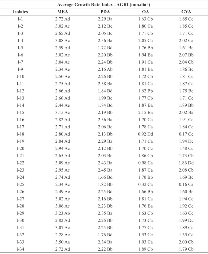
According to the variance analysis the interaction was significant for isolated x culture medium with a coefficient of variation of 7.7. In comparing mean values of the AGRI (Table 3) in accordance with Scott-Knott test (0.05), we can observe the formation of groups 4 to PDA and GYA means, and 5 groups for MEA and OA means. According to the composition of the medium, isolates have differentiated on the mycelial growth rate and sporulation, and the medium that favored the best mycelial growth rate for a particular isolate was not necessarily the best medium to produce conidia.
The culture media rich in carbon and nitrogen may provide a larger vegetative growth of fungi, while those deficient in nitrogen can increase conidiogenesis (LOUREIRO et al., 2011). According Menezes (2006) nutrition of C/N has an effect on the physiological processes of fungi, especially those related to growth, production of conidia, germination and dry weight, allowing also differentiate between isolates Colletotrichum spp. according to their ability to use particular source of carbon and nitrogen. In general, many fungi use glucose, but other sugars can serve as a carbon source with biosynthetic purposes, some culture media are more favorable for the sporulation of fungi than others, because they have complex carbohydrates which are less suitable for the production of vegetative hyphae, but more suitable to the production of spores (DIAS NETO et al., 2010).
In this work, all isolates of Colletotrichum spp. had better mycelial growth performance when cultured at pH 5.5, while for sporulation, the best result was observed at pH 6.5 (Figure 1). The pH is an important parameter in the development of phytopathogenic fungi, since this may affect physiological activities of microorganisms, such as enzyme production, that may determine the virulence or aggressiveness efficiency.
Deshmukh et al. (2012) noted that C. (Table 1), and the interpretation of results was
performed as McKinney (1923) index, adapted by Orozco Miranda et al. (2003), by calculating the intensity of the disease index (IDI). The experimental design was completely randomized in a factorial design with ten replicates, wherein each hypocotyl was considered a replicate.
Data analysis
Data were initially assessed by analysis of variance and F test, performed with the statistical program Sisvar (FERREIRA, 2000). The comparison between the averages when the F value was significant, was taken by Scott and Knott test (1974), 5%. Graphics were done through the demo version of the Sigma Plot 11.0 application (Systat Software Inc).
3 RESULTS AND DISCUSSION
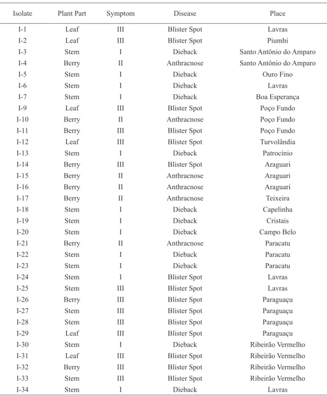
Thirty-three isolates were obtained from blister spot lesions, dieback and anthracnose in coffee from cities in southern Minas Gerais (Table 2). Three persistent symptoms related to isolates were identified: dry leaf and stems, depressive concentric necrotic lesions on fruit and yellow circular stain/spot with greenish halo in leaves and berries. These symptoms confer with those reported by Abreu, Ferreira and Martins (2008) for this pathosystem.
All isolates had homogeneous growth at 25°C, where the dominant mycelial color of the colony was white-gray cotton-like, with the exception of isolate I-14 which exhibited the salmon color when grown on MEA, PDA and GYA (Table 3). When grown in OA medium all isolates were superficial and have white growth. All isolates were able to produce acervuli during the twenty days of observation, with the exception of I-5 and I-25, distinguishing itself from other tested isolates.
The conidia isolates exhibited straight, cylindrical shape with rounded apexes and average length of 11.621 µm and width of 4.05 µm (Table 3). Among 33 isolates, 68% had the conidia with 12.0-17.0 µm in length and 3.5-6.0 µm in width. These values are proposed for C. gloeosporioides according to Sutton (1992). In this study, we observed the differentiation of two types of conidia, those in which there is a predominance of single conidia with length greater than 10 µm and those with the predominance of single conidia with less than 7 µm in length. This may be related to the existence of different strains into the species, since the isolates showed smaller sizes of conidia
TABLE 2 - Identification of Colletotrichum spp. in the south of Minas Gerais / Brazil.
Isolate Plant Part Symptom Disease Place
I-1 Leaf III Blister Spot Lavras
I-2 Leaf III Blister Spot Piumhi
I-3 Stem I Dieback Santo Antônio do Amparo
I-4 Berry II Anthracnose Santo Antônio do Amparo
I-5 Stem I Dieback Ouro Fino
I-6 Stem I Dieback Lavras
I-7 Stem I Dieback Boa Esperança
I-9 Leaf III Blister Spot Poço Fundo
I-10 Berry II Anthracnose Poço Fundo
I-11 Berry III Blister Spot Poço Fundo
I-12 Leaf III Blister Spot Turvolândia
I-13 Stem I Dieback Patrocínio
I-14 Berry III Blister Spot Araguari
I-15 Berry II Anthracnose Araguari
I-16 Berry II Anthracnose Araguari
I-17 Berry II Anthracnose Teixeira
I-18 Stem I Dieback Capelinha
I-19 Stem I Dieback Cristais
I-20 Stem I Dieback Campo Belo
I-21 Berry II Anthracnose Paracatu
I-22 Stem I Dieback Paracatu
I-23 Stem I Dieback Paracatu
I-24 Stem I Blister Spot Lavras
I-25 Stem III Blister Spot Lavras
I-26 Berry III Blister Spot Paraguaçu
I-27 Stem III Blister Spot Paraguaçu
I-28 Stem III Blister Spot Paraguaçu
I-29 Leaf III Blister Spot Paraguaçu
I-30 Stem I Dieback Ribeirão Vermelho
I-31 Leaf III Blister Spot Ribeirão Vermelho
I-32 Berry III Blister Spot Ribeirão Vermelho
I-33 Stem III Blister Spot Ribeirão Vermelho
I-34 Stem I Dieback Lavras
Symptoms: I - dry leaf and stems; II - concentric necrotic depressed lesions in berries; III - yellow circular spot with greenish halo on leaves and fruit.
TABLE 3 - Mycelial growth of Colletotrichum spp. isolates on different culture medium. Average Growth Rate Index - AGRI (mm.dia-1)
Isolates MEA PDA OA GYA
I-1 2.72 Ad 2.29 Ba 1.63 Cb 1.65 Cc I-2 3.02 Ac 2.12 Bc 1.80 Ca 1.85 Cc I-3 2.65 Ad 2.05 Bc 1.71 Cb 1.71 Cc I-4 3.08 Ac 2.36 Ba 2.05 Ca 2.02 Ca I-5 2.59 Ad 1.72 Bd 1.76 Bb 1.61 Bc I-6 3.02 Ac 2.20 Bb 1.94 Ba 2.07 Bb I-7 3.04 Ac 2.24 Bb 1.91 Ca 2.04 Cb I-9 2.34 Ae 2.16 Ab 1.81 Ba 1.86 Bc I-10 2.50 Ae 2.26 Bb 1.72 Cb 1.81 Cc I-11 2.75 Ad 2.38 Ba 1.81 Ca 1.87 Cc I-12 2.66 Ad 1.84 Bd 1.62 Bb 1.75 Bc I-13 2.66 Ad 1.99 Bc 1.77 Cb 1.71 Cc I-14 2.44 Ae 1.84 Bd 1.87 Ba 1.89 Bb I-15 3.15 Ac 2.19 Bb 2.15 Ba 2.02 Ba I-16 2.82 Ad 2.36 Ba 1.70 Ca 1.91 Cc I-17 2.71 Ad 2.06 Bc 1.78 Ca 1.84 Cc I-18 2.80 Ad 2.13 Bb 0.92 Dd 0.17 Ce I-19 2.84 Ad 2.29 Ba 1.71 Ca 1.94 Dc I-20 2.94 Ac 2.12 Bb 1.70 Cc 1.48 Cc I-21 2.65 Ad 2.03 Bc 1.86 Cb 1.73 Cb I-22 3.09 Ac 2.43 Ba 0.98 Ca 1.86 Dd I-23 2.95 Ac 2.45 Ba 1.87 Ca 2.08 Cb I-24 2.74 Ad 1.66 Bd 1.70 Bb 1.69 Bc I-25 2.34 Ac 1.82 Bb 0.32 Ca 0.16 Ca I-26 2.49 Ae 2.25 Bd 1.66 Bb 1.60 Bc I-27 3.02 Ac 2.16 Bb 1.81 Ca 1.94 Cc I-28 3.06 Ac 2.23 Bb 1.76 Ba 1.92 Cc I-29 3.23 Ab 2.35 Ba 1.63 Cb 1.63 Cc I-30 2.82 Ad 2.26 Bb 1.73 Ca 1.99 Dc I-31 3.07 Ac 2.25 Bb 1.77 Ca 1.89 Cc I-32 2.28 Ae 1.76 Bd 1.53 Cc 1.35 Cc I-33 3.50 Aa 2.34 Ba 1.93 Ca 2.00 Cb I-34 2.72 Ad 2.22 Bb 1.89 Cb 1.79 Cb
gloeosporioides sporulation occurred more
frequently when grown at pH 5.0, 5.5 and 6.0, and pH 5.5 to 6.0 for the best out of mycelial growth, also verified that the worst growth and sporulation rates were obtained when grown at pH 7.0 and 8.0. Kumara and Rawal (2010) in they studies with C. gloeosporioides isolates obtained from papaya fruits have also found that the favorable pH range for growth and sporulation was 5.0 and 6.0. Yakoby et al. (2000) found that expression of the pelB gene of C. gloeosporioides occurs at pH values above 5,1 and the secretion of enzymes transcribed by this occurs only above pH 5.8.
The confirmation of Colletotrichum spp. genus was performed by the pair of oligonucleotide CCF1 / Cc2R1 specific to the genus, amplifying a fragment of 447 bp for all the isolates (Figure 2). For oligonucleotides CanIt2/ITS4, which identifies
C. acutatum, no amplification was observed.
All isolates were positive for the primers CgInt/ ITS4, which amplify fragments of 450 bp, thus
FIGURE 1 - Mycelial growth and sporulation of Colletotrichum spp. (A-B) Mycelial growth and sporulation influenced by the culture media used for the fungi cultivation. (C-D) Mycelial growth and sporulation of the fungi influenced by the pH of the culture media.
the evaluated isolated belonged to the species C.
gloeosporioides. Freitas et al. (2011) also carried
out the molecular identification of Colletotrichum
gloeosporioides obtained from coffee, using the
same primers.
C. gloeosporioides isolates obtained from
coffee were capable of inducing symptoms in berries as well as in hypocotyls (Table 4). In both we observed variability in the induction of symptoms, because the material used (berry/ hypocotyl), as the isolates tested.
The first symptoms of necrosis in coffee green berries caused by isolates of C.
gloeosporioides were observed from the first
evaluation, five days after inoculation, causing depressed moist necrotic lesions that progressed over time. In some berries it was possible to observe a mass formation of conidia on the lesions. All isolates inoculated in berries differ statistically at a significance level of 5% from the control. Small brownish lesions on the stem of hypocotyls
aspects of Colletotrichum spp., as well as Coffea
arabica L., genotypes used, plant physiology
and their nutritional condition, influence the symptoms, such as: more vigorous trees are less susceptible to the pathogen, adult plants in the fruit maturation state tend to show higher incidence of the fungus (SERA et al., 2005). Rampazo, Marçal and Leite Júnior (2007) also demonstrated that the specialization of the pathogenic isolates of C.
gloeosporioides, since some isolates were not able
to induce lesions, depending of the susceptibility of coffee cultivars.
In this work was not observed the typical symptoms of blister spot (Figure 2) due to rapid development of necrosis in the inoculated materials presented. Blister spot symptoms are difficult to reproduce in controlled conditions, but manifest with great intensity on the field. The expression of these symptoms may not only restricted to the pathogen, but also conditioning by nutritional factors and plant age. Martins-Maia et al. (2012) working with Colletotrichum spp. inoculation in coffee seedlings obtained plants without symptoms of blister spot, also had no success in the reproduction of specific symptoms. There is a need for studies that report the association of some factor that predisposes to this pathogen, linked to growing conditions, resistance or susceptibility of these plants, since within a coffee plantation attacked plants are observed with generalized disease and others that do not have the disease incidence.
could be seen from the fifth day after inoculation, which evolved quickly throughout the stem, promoting the strangulation of seedling, with 12 days of evaluation. The isolates I-9, I-24 and I-26 were those with the highest disease intensity ratios when inoculated into seedlings and berries. None of the isolates showed index zero for berries, but in hypocotyls it was observed for various isolates. (Table 4). Isolates that showed the lowest disease indexes were I-28 and I-33 with 18 and 16% of IDI in berries and 0 in hypocotyls.
According to Ferreira et al. (2009), the mycelium of Colletotrichum spp. affects all the organs on coffee, systemically colonize the xylem tissue, the phloem, cortex and cells of endosperm thus causing death of branches, fruit and leaf drop mummification. The isolates in this work were more aggressive when inoculated into green berries, and all isolates showed symptoms of necrosis. In hypocotyls was observed that the number of isolates symptoms was reduced. Thus, berries can be considered more susceptible to C.
gloeosporiodes than seedlings. Such difference
may be due to composition of material. Fruits have high levels of starch (LAVIOLA et al., 2007) which may consists a nutritional source for pathogens.
The variability in the pathogenicity of C.
gloeosporioides isolates was observed by other
authors previously, showing that there is great diversity in the induction or incidence of symptoms in relation to the isolates tested (JULIATTI et al., 2006). This variability suggests that the genetic
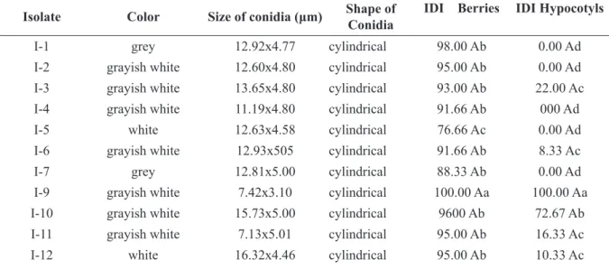
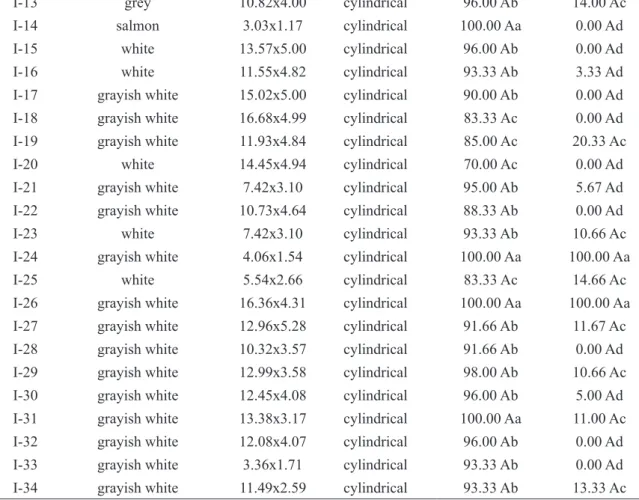
TABLE 4 - Morphological characteristics of Colletotrichum spp. and aggressiveness level in green berries and hypocotyls.
Isolate Color Size of conidia (μm) Shape of Conidia IDI Berries IDI Hypocotyls
I-1 grey 12.92x4.77 cylindrical 98.00 Ab 0.00 Ad
I-2 grayish white 12.60x4.80 cylindrical 95.00 Ab 0.00 Ad
I-3 grayish white 13.65x4.80 cylindrical 93.00 Ab 22.00 Ac
I-4 grayish white 11.19x4.80 cylindrical 91.66 Ab 000 Ad
I-5 white 12.63x4.58 cylindrical 76.66 Ac 0.00 Ad
I-6 grayish white 12.93x505 cylindrical 91.66 Ab 8.33 Ac
I-7 grey 12.81x5.00 cylindrical 88.33 Ab 0.00 Ad
I-9 grayish white 7.42x3.10 cylindrical 100.00 Aa 100.00 Aa
I-10 grayish white 15.73x5.00 cylindrical 9600 Ab 72.67 Ab
I-11 grayish white 7.13x5.01 cylindrical 95.00 Ab 16.33 Ac
I-13 grey 10.82x4.00 cylindrical 96.00 Ab 14.00 Ac
I-14 salmon 3.03x1.17 cylindrical 100.00 Aa 0.00 Ad
I-15 white 13.57x5.00 cylindrical 96.00 Ab 0.00 Ad
I-16 white 11.55x4.82 cylindrical 93.33 Ab 3.33 Ad
I-17 grayish white 15.02x5.00 cylindrical 90.00 Ab 0.00 Ad
I-18 grayish white 16.68x4.99 cylindrical 83.33 Ac 0.00 Ad
I-19 grayish white 11.93x4.84 cylindrical 85.00 Ac 20.33 Ac
I-20 white 14.45x4.94 cylindrical 70.00 Ac 0.00 Ad
I-21 grayish white 7.42x3.10 cylindrical 95.00 Ab 5.67 Ad
I-22 grayish white 10.73x4.64 cylindrical 88.33 Ab 0.00 Ad
I-23 white 7.42x3.10 cylindrical 93.33 Ab 10.66 Ac
I-24 grayish white 4.06x1.54 cylindrical 100.00 Aa 100.00 Aa
I-25 white 5.54x2.66 cylindrical 83.33 Ac 14.66 Ac
I-26 grayish white 16.36x4.31 cylindrical 100.00 Aa 100.00 Aa
I-27 grayish white 12.96x5.28 cylindrical 91.66 Ab 11.67 Ac
I-28 grayish white 10.32x3.57 cylindrical 91.66 Ab 0.00 Ad
I-29 grayish white 12.99x3.58 cylindrical 98.00 Ab 10.66 Ac
I-30 grayish white 12.45x4.08 cylindrical 96.00 Ab 5.00 Ad
I-31 grayish white 13.38x3.17 cylindrical 100.00 Aa 11.00 Ac
I-32 grayish white 12.08x4.07 cylindrical 96.00 Ab 0.00 Ad
I-33 grayish white 3.36x1.71 cylindrical 93.33 Ab 0.00 Ad
I-34 grayish white 11.49x2.59 cylindrical 93.33 Ab 13.33 Ac
Lowercase letters compare isolated within each mode of material evaluated and uppercase letters compare pathogenicity within each individual, at 5% probability by Scott-Knott test.
FIGURE 2 - Symptoms of diseases caused by Colletotrichum spp. (A) Blister spot symptoms on coffee leaves. (B) Symptoms in hypocotyls caused by C. gloeosporioides . (C) Symptoms in green berries caused by the isolate I-24.
4 CONCLUSIONS
In this study were obtained 33 isolates of
Colletotrichum spp., which were identified as
belonging to the species C. gloeosporioides. Malt extract agar (MEA) and oat agar (OA) medium culture are the most appropriate for mycelial growth and sporulation, respectively, and develops efficiently in the pH range between 5.5 and 6.5. Through the pathogenicity test was able to detect different levels of aggressiveness in berries as in hypocotyls, but the morphological and cultural characteristics are not sufficient to identify such variations among the isolates of C. gloeosporiodes related to coffee plants.
5 ACKNOWLEDGEMENTS
The State Funding Agency of Minas Gerais (FAPEMIG) and National Counsel of Technological and Scientific Development (CNPq) for project funding.
6 REFERENCES
ABREU, M. S.; FERREIRA, J. B.; MARTINS, F. G. Mancha manteigosa no contexto do complexo Colletotrichum em cafeeiros. In: SIMPóSIO DE MANEJO DE PLANTAS: MANEJO FITOSSANITáRIO DO CAFEERIO, 2008, Lavras. Anais... Lavras: UFLA, 2008. p. 105-126.
BRASIL. Ministério da Agricultura. Vegetal: culturas - café. Brasília, 2012. Disponível em: <http://www. agricultura.gov.br/>. Acesso em: 3 maio 2016.
COMPANHIA NACIONAL DE ABASTECIMENTO. Indicadores da agropecuária. Disponível em: <http:// www.conab.gov.br>. Acesso em: 3 maio 2016.
CULLEN, D. W. et al. Detection of Colletotrichum coccodes from soil and potato tubers by conventional PCR and real-time quantitative PCR. Plant Pathology, London, v. 51, p. 281-92, 2002.
DESHMUKH, A. J. et al. In vitro effect of various nitrogen, carbon sources and pH regimes onthe growth and Sporulation of Colletotrichum gloeosporioides Penz. and Sacc causing anthracnose of Indian bean. Journal of Biopesticides, Palayamkottai, v. 5, n. 46, p. 46-49, 2012.
DIAS NETO, J. J. et al. Influência do meio de cultura na esporulação de magnaporthe grisea e da concentração de conídios na severidade da brusone do arroz. Bioscience Journal, Uberlândia, v. 26, n. 2, p. 173-179, 2010.
FERREIRA, D. F. Manual do sistema SISVAR para análises estatísticas. Lavras: UFLA, 2000. 66 p. FERREIRA, J. B.; ABREU, M. S.; PEREIRA, I. S. Análise da dinâmica, estrutura de focos e arranjo espacial da Mancha Manteigosa em campo. Ciência e Agrotecnologia, Lavras, v. 33, p. 24-30, 2009.
FERREIRA, J. B. et al. Aspectos morfológicos da colonização de Colletotrichum gloeosporioides em órgãos de plantas de cafeeiro e com sintomas de mancha manteigosa. Ciência e Agrotecnologia, Lavras, v. 33, n. 4, p. 956-964, jul./ago. 2009.
FREITAS, R. L. et al. Colletotrichum gloeosporioides e C. boninense associados à antracnose do café no Brasil. In: SIMPóSIO DE PESQUISA DOS CAFÉS DO BRASIL, 7., 2011, Araxá. Anais... Araxá, 2011. 1 CD-ROM.
JULIATTI, F. C. et al. Agressividade e divergência genética por RAPD de isolados de Colletotrichum gloeosporioides coletados em lavouras cafeeiras de Minas gerais. Bioscience Journal, Uberlândia, v. 22, p. 159-169, 2006.
KUMARA, K.; RAWAL, R. Influence of carbon, nitrogen, temperature and PH on the growth and sporulation of some Indian Isolates of Colletotrichum gloeosporioides causing Anthracnose disease of Papaya (Carrica papaya L). Tropical Agricultural Research and Extension, Sri Lanka, v. 11, p. 7-12, Apr. 2010. LAVIOLA, B. G. et al. Alocação de fotoassimilados em folhas e frutos de cafeeiro cultivado em duas altitudes. Pesquisa Agropecuária Brasileira, Brasília, v. 42, n. 11, p. 1521-1530, nov. 2007.
LOUREIRO, A. et al. Isoenzymatic characterization of Colletotrichum kahawae isolates with different levels of aggressiveness. Tropical Plant Pathology, Brasília, v. 36, n. 5, p. 287-293, 2011.
MARQUES, V. V. Patogenicidade e variabilidade genética de Colletotrichum spp. em cafeeiro (Coffea arabica L.). 2008. 108 p. Tese (Doutorado em Agronomia) - Universidade Estadual de Londrina, Londrina, 2008.
MARTINS-MAIA, F. G. et al. Pigments, total soluble phenols and lignin levels of coffeeSeedlings inoculated with Colletotrichum gloeosporioides. Coffee Science, Lavras, v. 7, n. 2, p. 152-159, maio/ago. 2012.
MCKINNEY, H. H. Influence of soil temperature and moisture on infection of wheat seedlings by Helminthosporium sativum. Journal Agricultural Research, Washington, v. 26, p. 195-217, 1923.
MENEZES, M. Aspectos biológicos e taxonômicos de espécies do gênero Colletotrichum. Anais da Academia Pernambucana de Ciência Agronômica, Recife, v. 3, p. 170-179, 2006.
MILLS, P. R.; SREENIVASAPRASAD, S.; BROWN, A. E. Detection and differentiation of Colletotrichum gloeosporioides isolates using PCR. FEMS Microbiology Letters, Malden, v. 98, p. 137-143, 1992.
OROZCO MIRANDA, E. F. Caracterização morfológica, molecular, bioquímica e patogênica de isolados de Colletotrichum spp. associados ao cafeeiro em Minas Gerais e comparação com
Colletotrichum kahawae. 2003. 147 p. Tese (Doutorado
em Fitopatologia) - Universidade Federal de Lavras, Lavras, 2003.
RAMPAZO, L. G. L.; MARÇAL, V. V. M.; LEITE JÚNIOR, R. P. Caracterização patogênica de isolados de Colletotrichum spp. obtidos de cafeeiro e outras culturas no Estado do Paraná. In: SIMPóSIO DE PESQUISA DOS CAFÉS DO BRASIL, 2007, Brasília. Anais... Brasília: EMBRAPA Café, 2007. p. 5.
SERA, G. H. et al. Correlação entre a ocorrência de Colletotrichum spp. e outras características agronômicas em cafeeiros. Bragantia, Campinas, v. 64, n. 3, p. 435-440, 2005.
SOUZA, B. O.; SOUZA, E. A.; MENDES-COSTA, M. C. Determinação da variabilidade em isolados de Colletotrichum lindemuthianum por meio de marcadores morfológicos e culturais. Ciência e Agrotecnologia, Lavras, v. 31, n. 4, p. 1000-1006, jul./ ago. 2007.
SREENIVASAPRASAD, S. et al. PCR-based detection of Colletotrichum acutatum on strawberry. Plant Pathology, Saint Paul, v. 45, p. 650-655, 1996.
SUTTON, B. C. The genus Glomerella and its anamorph. In: BAILEY, J. A.; JEGUER, M. J. (Ed.).
Colletotrichum: biology, pathology and control.
London: CBA, 1992. p. 1-26.
VARZEA, V. M. P. Variabilidade em Colletotrichum spp. de cafeeiro: pesquisa de fontes de resistência ao C. Kahawae. 1995. 128 p. Dissertação (Mestrado em Investigador Auxiliar) - Instituto de Investigação Cientifica Tropical, Lisboa, 1995.
YAKOBY, N. et al. pH regulation of pectate lyase secretion modulates the attack of Colletotrichum gloeosporioides on avocado fruits. Applied and Environmental Microbiology, Washington, v. 6, p. 1026-1030, 2000.