S H O R T C O M M U N I C A T I O N
The recombinant protein rSP03B is a valid antigen
for screening dog exposure to Phlebotomus perniciosus
across foci of canine leishmaniasis
T. K O S T A L O V A
1, T. L E S T I N O V A
1, C. M A I A
2,3, P. S U M O V A
1,
M. V L K O V A
1, L. W I L L E N
1, N. P O L A N S K A
1, E. F I O R E N T I N O
4,
A. S C A L O N E
4, G. O L I V A
5, F. V E R O N E S I
6, J. M. C R I S T Ó V Ã O
2,
O. C O U R T E N A Y
7, L. C A M P I N O
2,8, L. G R A D O N I
4, M. G R A M I C C I A
4and P. V O L F
11Department of Parasitology, Faculty of Science, Charles University in Prague, Prague, Czech Republic,2Global Health and Tropical
Medicine, Medical Parasitology Unit, Instituto de Higiene e Medicina Tropical, New University of Lisbon, Lisbon, Portugal,3Faculty
of Veterinary Medicine, Lusophone University of Humanities and Technologies, Lisbon, Portugal,4Unit of Vector-Borne Diseases
and International Health, Istituto Superiore di Sanità, Rome, Italy,5Department of Veterinary Medicine and Animal Production,
University Federico II, Naples, Italy,6Department of Veterinary Medicine, University of Perugia, Perugia, Italy,7Warwick Infectious
Disease Epidemiology Research (WIDER), School of Life Sciences, University of Warwick, Coventry, U.K. and8Department of
Biomedical and Medical Sciences, University of Algarve, Faro, Portugal
Abstract. The frequency of sandfly–host contacts can be measured by host antibody
levels against sandfly salivary proteins. Recombinant salivary proteins are suggested to represent a valid replacement for salivary gland homogenate (SGH); however, it is necessary to prove that such antigens are recognized by antibodies against various populations of the same species. Phlebotomus perniciosus (Diptera: Psychodidae) is the main vector of Leishmania infantum (Trypanosomatida: Trypanosomatidae) in southwest Europe and is widespread from Portugal to Italy. In this study, sera were sampled from naturally exposed dogs from distant regions, including Campania (southern Italy), Umbria (central Italy) and the metropolitan Lisbon region (Portugal), where P. perniciosus is the unique or principal vector species. Sera were screened for anti-P. perniciosus antibodies using SGH and 43-kDa yellow-related recombinant protein (rSP03B). A robust correlation between antibodies recognizing SGH and rSP03B was detected in all regions, suggesting substantial antigenic cross-reactivity among different P. perniciosus populations. No significant differences in this relationship were detected between regions. Moreover, rSP03B and the native yellow-related protein were shown to share similar antigenic epitopes, as canine immunoglobulin G (IgG) binding to the native protein was inhibited by pre-incubation with the recombinant form. These findings suggest that rSP03B should be regarded as a universal marker of sandfly exposure throughout the geographical distribution of P. perniciosus.
Key words. Leishmania infantum, Phlebotomus spp., antibody response, dog, markers of exposure, Mediterranean region, salivary proteins, sandflies.
Correspondence: Tatiana Kostalova, Department of Parasitology, Faculty of Science, Charles University, Vinicna 7, 128 44 Prague 2, Czech Republic. Tel.: + 420 2 2195 1826; E-mail: [email protected]
Leishmaniasis is a widely distributed disease caused by Leish-mania protozoans and transmitted by phlebotomine sandfly vectors. During blood feeding, sandflies inoculate saliva into the host. Bitten hosts then develop a species-specific antibody response against salivary antigens that reflects the intensity of sandfly exposure and thus provides a useful marker of expo-sure to generate epidemiological data (Vlkova et al., 2011; Martín-Martín et al., 2014; Kostalova et al., 2015).
Large-scale serological studies using total sandfly salivary gland homogenate (SGH) are currently impractical because it is difficult to dissect the high numbers of sandflies necessary to obtain sufficient amounts of SGH. Another potential complica-tion refers to variability in the protein composicomplica-tion of sandfly saliva, which has been found to fluctuate depending on physi-ological factors such as sandfly age and diet (Volf et al., 2000; Prates et al., 2008). Studies in Old World sandfly species also revealed a certain degree of intra- and inter-population vari-ability in protein and mRNA levels (Rohousova et al., 2012; Ramalho-Ortigão et al., 2015). Therefore, salivary recombi-nant proteins have been suggested to represent valid replace-ments for the whole salivary gland protein cocktail, and some have already been validated in the field (Drahota et al., 2014; Martín-Martín et al., 2014; Kostalova et al., 2015). The use of specific recombinant salivary antigen circumvents the neces-sity for the laborious maintenance of sandfly colonies, and potentially provides a more refined way to minimize antigenic cross-reactivity with taxonomically close sandfly relatives. A useful recombinant salivary protein would demonstrate anti-genicity comparable with that of SGH, share similar antigenic epitopes with the native proteins, and demonstrate similar anti-genic patterns throughout the geographical distribution of a par-ticular sandfly vector.
This study follows the canine longitudinal study conducted in southern Italy by Kostalova et al. (2015), which described the dynamics and diagnostic potential of antibodies recogniz-ing Phlebotomus perniciosus (Larroussius subgenus) salivary recombinant proteins in dogs following natural exposure to sandflies over 2 years. Factors such as salivary antigens, age and expected sandfly dynamics were considered as variables and were therefore carefully evaluated. The most reactive and reproducible antigen was found to be the 43-kDa yellow-related recombinant protein (rSP03B) from P. perniciosus saliva. In view of these promising results, the rSP03B antigen was tested in canine sera samples collected cross-sectionally in canine leish-maniasis (CanL) endemic settings in Italy and Portugal. The study evaluated levels of individual canine antigenic responses to P. perniciosus rSP03B compared with P. perniciosus SGH, and the degree of similarity in these antigenic associations, across endemic canine populations in Portuguese and Italian foci in order to assess the universal use of rSP03B as a marker of natural sandfly exposure. Previous research had confirmed the presence of two native yellow-related proteins in P. perniciosus salivary gland transcriptome and proteome (Anderson et al., 2006). Therefore, the antigenic similarity of rSP03B to its native form was studied and the specificity of the anti-rSP03B immunoglobulin G (IgG) antibody response was confirmed by the inclusion of 42-kDa yellow-related recombinant protein (rSP03).
Canine sera originated from three regions: (a) Campania (n = 118), a traditional high-risk area for CanL in southern con-tinental Italy (Oliva et al., 2006); (b) Umbria (n = 96), an inland area of central Italy recently recorded as a medium- to high-risk area for CanL (Di Muccio et al., 2012), and (c) the metropoli-tan Lisbon region (n = 341), which is well known as a CanL endemic locality on the west coast of Portugal (Cortes et al., 2012). In all three areas, P. perniciosus is the only or princi-pal vector of CanL (Bongiorno et al., 2003; Rossi et al., 2007; Alten et al., 2016). Phlebotomus perfiliewi (Larroussius sub-genus), another vector of Leishmania infantum, was found to be abundant in some areas in Umbria (Maresca et al., 2009). However, P. perfiliewi is found in association with large ani-mals (cattle and equine species) in rural habitats (Bongiorno et al., 2003). Dogs examined in Umbria included urban pets and animals hosted in kennels, but all lived in populated areas including residential zones surrounding urban centres, which represent typical habitats for P. perniciosus (Maroli et al., 1994). Additionally, sampled dogs may have been exposed to sand-flies from other subgenera occurring in study localities (Cortes et al., 2007; Rossi et al., 2007; Maresca et al., 2009). A previ-ous study by Volf & Rohprevi-ousova (2001) suggested there was no cross-reaction of Larroussius species with other sandflies present in these study regions, namely Phlebotomus papatasi (Phlebotomus subgenus), Phlebotomus sergenti (Paraphleboto-mus subgenus) and members of the genus Sergentomyia.
Single sera samples from Campania and Umbria were pur-posely selected from archived samples collected in 2007–2013 to represent the period from July (i.e. at least 2 months after the beginning of the sandfly season) to October (i.e. the end of the sandfly season). The selected sera were collected from dogs ranging in age from 1.5 to 13 years. The dogs from both Italian regions represented a mixture of hunting breeds and mongrels. Single sera samples from the metropolitan Lisbon region were randomly collected from kennelled dogs (mostly mongrel) at the beginning of the sandfly season in May 2012. These dogs ranged from young (6–12 months) to more senior (> 7 years) dogs.
Samples from Campania consisted of stored sera sent by veterinary clinics to the Istituto Superiore di Sanità for routine serological diagnosis of suspected CanL in owned dogs. Sera from Umbria were collected from healthy dogs that were enrolled on a voluntary basis in the Perugia University CanL surveillance programme. Blood sampling was performed in accordance with the Italian guidelines for animal welfare, following owners’ consent, and did not include additional or unnecessary invasive procedures. The collection of sera in the metropolitan Lisbon region was ethically approved by the board of the Institute of Hygiene and Tropical Medicine, New University of Lisbon (IHMTUNL) (authorization no. 8 2011-PI) in compliance with Portuguese legislation for the protection of animals (Law 113/2013).
Anti-Leishmania IgG in canine sera from Campania and Umbria was detected with an in-house indirect fluorescent anti-body test (IFAT) using L. infantum promastigotes as antigen, as described in Gradoni & Gramiccia (2008). Samples show-ing an IFAT titre of 1 : 40 or greater were considered to indicate exposure to Leishmania. Immunoglobulin G antibodies against Leishmania in canine sera from the metropolitan Lisbon region were detected using an enzyme-linked immunosorbent assay
(ELISA) kit (Bordier Affinity Products SA, Crissier, Switzer-land) according to the manufacturer’s guidelines (Maia et al., 2010). The result was considered positive when the absorbance of the analysed sample was higher than the absorbance of the weak positive control serum provided with the kit.
A longterm established laboratory colony of P. perniciosus originating from Spain (Murcia) was reared under standard conditions as described in Volf & Volfova (2011). Salivary glands, rSP03B (GenBank accession no. DQ 150622) and rSP03 (GenBank accession no. DQ 150621) from P. perniciosus were obtained for this study as previously described (Kostalova et al., 2015) and used as antigens for testing the canine sera.
Antibodies against P. perniciosus SGH and rSP03B protein were measured by ELISA as described by Kostalova et al. (2015). Each serum was tested in duplicate. Test absorbance values were reported as optical densities (ODs) with subtracted blanks (the ELISA plate background mean absorbance value measured in control wells).
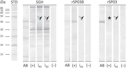
Western blot analysis was used to confirm the similarity of antigenic epitopes between the native yellow-related protein found in P. perniciosus SGH and the corresponding recombi-nant protein rSP03B. Sodium dodecyl sulphate polyacrylamide gel electrophoresis (SDS-PAGE) of SGH (equivalent to 4 μg total salivary proteins per lane) and rSP03B (2 μg per lane) was run on a 12% gel and blotted onto the nitrocellulose mem-brane using the iBLOT instrument (Invitrogen Corp., Carlsbad, CA, U.S.A.). Membrane with separated proteins was cut into strips and blocked in 5% milk diluted in Tris-buffered saline with 0.05% Tween 20 (Tris-Tw) overnight at 4 ∘C. For the inhi-bition test, three Italian canine sera possessing high levels of anti-P. perniciosus IgG against SGH and rSP03B were pooled. The positive serum pool was diluted 1 : 50 in Tris-Tw and split into halves. The first half was incubated for 2 h on a shaker with rSP03B (20 μg/mL) and the second half was incubated without rSP03B. Negative control sera (canine sera from a non-endemic locality) were diluted 1 : 50 in Tris-Tw and incubated without rSP03B on a shaker for 2 h. In the next step, part of the positive sera pool, incubated either with or without rSP03B protein, and part of the negative control sera was incubated with strips of sep-arated P. perniciosus SGH. The same procedure was repeated for strips containing rSP03B, except that sera were diluted 1 : 100 in Tris-Tw. After 1 h, all strips were rinsed in Tris-Tw and subse-quently incubated for 1 h with peroxidase-conjugated anti-dog IgG (1 : 3000) (Bethyl Laboratories, Inc., Montgomery, TX, U.S.A.). The colour reaction was developed by substrate solu-tion containing 3,3′-diaminobenzidine (Sigma-Aldrich Corp.,
St Louis, MO, U.S.A.). Furthermore, in order to confirm the specificity of the western blot analysis, the same procedure was repeated for rSP03 protein.
Statistical analyses were carried out using R software (http:// cran.r-project.org/) and stata Version 13.1 (Stata Corp., Col-lege Station, TX, U.S.A.). Correlations were analysed using the Spearman rank correlation test and medians were compared between groups using a Wilcoxon rank sum test. Optical density values were logarithmized (natural logarithm) for better read-ability. Statistical analyses of the relationships between SGH and rSP03B OD values among the canine populations were statistically tested by fitting Poisson general linearized models (GLMs) with an ln link function as the right-skewed frequency
distributions were found not to follow a negative binomial dis-tribution (deviance goodness-of-fit𝜒2> 56.2; P = 1, d.f. = 549,
for each antibody). The full Poisson GLMs included interaction terms to test differences between the regions, both in terms of baseline anti-rSP03B value (intercept where anti-SGH equals 0) and the relationship between antibodies against SGH and rSP03B (slopes). A P-value of< 0.05 was considered to indicate statistical significance.
The use of P. perniciosus rSP03B as an epidemiological tool was tested for investigations of canine exposure to sandfly bites in geographically distinct localities in which P. perniciosus is the prevalent phlebotomine vector. The recombinant protein rSP03B used in this study was obtained from the salivary glands of P. perniciosus in a laboratory-reared colony originating from Murcia in Spain, and was used as an antigen in the serology of dogs living in the Campania and Umbria regions of Italy and in the metropolitan Lisbon region in Portugal.
Levels of canine IgG antibodies reacting with SGH and rSP03B were measured by ELISA. Positive but variable corre-lations between antibody responses to SGH and rSP03B anti-gens were observed in sera from all three localities [Campania: r = 0.73, 95% confidence interval (CI) 0.62–0.82 (P< 0.001); Umbria: r = 0.56, 95% CI 0.38–0.71 (P< 0.001); metropoli-tan Lisbon: r = 0.81, 95% CI 0.76–0.84 (P< 0.001)] (Fig. 1). Table 1 summarizes the OD values for each region and indi-cates that OD frequency distributions were over-dispersed. To query possible differences in the relationships between SGH and rSP03B antibody responses between geographical regions, the equality of the population-specific regression slopes was tested by fitting a Poisson model. No significant differences were detected (population × antigen interaction terms: Z> −0.85, P> 0.365). Relative to the metropolitan Lisbon region, both the Campania and Umbria populations tended to produce higher baseline antibody responses against rSP03B, although these dif-ferences failed to reach significance at the 5% level (Campania: Z = 1.66, P = 0.097; Umbria: Z = 1.95, P = 0.051). One plausi-ble explanation for the putative differences in baseline rSP03B antibody levels among populations is that the populations dif-fer in their condition or past history of infections and that these differences affect general immunological responses to certain antigens, and/or that sandfly biting pressure differs across these populations. The seasonal exposure of dogs to sandflies has been found to lead to antibody response fluctuations related to the period of activity and abundance of vectors (Vlkova et al., 2011; Kostalova et al., 2015). Secondly, as age is a frequent covariate of cumulative exposure used to model cross-sectional age-related prevalence data of Leishmania infection (Courtenay et al., 1994), the average older dog is expected to have experi-enced more sandfly seasons (Kostalova et al., 2015). Dogs from Campania and Umbria were sampled from July (i.e. during the period of highest sandfly abundance in Italy). All of the animals tested from these two regions had experienced at least two con-secutive transmission seasons. Sera from dogs in the metropoli-tan Lisbon region were sampled in May, which is the beginning of the sandfly season, and were sourced mainly from dogs aged > 1 year. Thus these dogs had experienced at least one transmis-sion season. According to reactivity data shown by Kostalova et al. (2015), dogs will be reactive to saliva at the beginning of the transmission season if they have already been ‘primed’ in the
r = 0.73 (95% CI 0.62–0.82); P < 0.001 1.0 (A) (B) (C) 1.0 0.8 0.8 0.6 0.6 0.4 0.4 0.2 In anti-rSP03B antibodies
In anti-SGH antibodies In anti-SGH antibodies In anti-SGH antibodies
In anti-rSP03B antibodies In anti-rSP03B antibodies
0.2 0.0 0.0 0.0 0.2 0.4 0.6 0.8 1.0 0.0 0.2 0.4 0.6 0.8 1.0 1.0 0.8 0.6 0.4 0.2 0.0 1.0 0.8 0.6 0.4 0.2 0.0 1.0 0.8 0.6 0.0 r = 0.56 (95% CI 0.38–0.71); P < 0.001 r = 0.81 (95% CI 0.76–0.84); P < 0.001
Fig. 1. Correlations between antibodies recognizing salivary gland homogenate (SGH) and rSP03B in dogs naturally bitten by Phlebotomus
perniciosus in (A) Campania, (B) Umbria and (C) the metropolitan Lisbon region. Correlations were ascertained using Spearman rank correlation. r, correlation index; 95% CI, 95% confidence interval.
Table 1. Summary of optical density (OD) values recorded by
enzyme-linked immunosorbent assay (ELISA) using Phlebotomus
per-niciosus salivary antigens.
OD values
Antigen Region Dogs, n Median (IQR) Min–max SGH Campania∗ 118 0.131 (0.073–0.241) 0.011–1.899 Umbria† 96 0.218 (0.133–0.409) 0.005–1.652 Lisbon‡ 341 0.221 (0.165–0.311) 0.081–1.390 rSP03B Campania∗ 118 0.407 (0.311–0.516) 0.091–1.761 Umbria† 96 0.495 (0.386–0.649) 0.026–1.925 Lisbon‡ 341 0.323 (0.234–0.436) 0.092–1.766 ∗Southern Italy. †Central Italy.
‡Metropolitan Lisbon region (Portugal).
IQR, interquartile range; SGH, salivary gland homogenate.
previous season. These results indicate substantial salivary anti-gen cross-reactivity amongst P. perniciosus populations from Campania, Umbria and the metropolitan Lisbon region. Strong antigenic cross-reactivity between populations of the same sand-fly species was similarly observed between two geographically distant colonies of Phlebotomus orientalis (Larroussius sub-genus) in Ethiopia (Vlkova et al., 2014), and among colonies of P. sergenti originating from Israel and Turkey (Rohousova et al., 2012).
The similarity of antigenic epitopes between native yellow-related proteins in Spanish P. perniciosus SGH and rSP03B was demonstrated by an inhibition test (Fig. 2). For this analysis, sera of dogs from Campania and Umbria with high levels of specific antibodies were selected and pooled. The inhibition test showed that all IgG antibodies specific for the native yellow-related protein bind to the recombinant form dur-ing pre-incubation of the sera, which resulted in the complete disappearance of the corresponding band on western blotting
(Fig. 2). This demonstrated that rSP03B shares antigenic epi-topes with the native yellow-related protein contained within P. perniciosus saliva and presumably identifies the proportion of bitten dogs in a manner similar to the use of SGH. By contrast, when the inhibition test was performed with rSP03 protein, intended to confirm the specificity of the western blot analysis, no band appeared and no inhibition was observed (Fig. 2). Therefore, rSP03 is considered to be a non-immunogenic antigen. These results show that the band observed in western blotting with SGH as antigen corresponds to the native 43-kDa yellow-related protein and that the anti-SP03B IgG antibodies are highly specific for the tested rSP03B protein.
Italy and Portugal are generally assumed to show endemic CanL transmission (Oliva et al., 2006; Cortes et al., 2012; Di Muccio et al., 2012). In this study, CanL seropositivity ranged from 5% to 30%, with the lowest prevalence in Umbria and the highest in Campania (Table 2). The use of antibodies against sandfly salivary proteins as risk markers of L. infantum infection has been tested earlier for SGH (Vlkova et al., 2011), as well as for salivary recombinant proteins, among which rSP03B proved to be a powerful marker of host exposure to sandflies (Kostalova et al., 2015). Therefore, the present study analysed the rela-tionship between anti-P. perniciosus antibodies and Leishmania serological status. When using rSP03B antigen, significantly higher levels of specific IgG in Leishmania-seropositive dogs [median = 0.346, interquartile range (IQR) 0.257–0.536] than in Leishmania-seronegative dogs (median = 0.320, IQR 0.229–0.422) were found only in the metropolitan Lisbon region (Wilcoxon rank sum test, W = 5391.5, P = 0.025). In Campania, the differences in antibodies against rSP03B between Leish-mania-seropositive (median = 0.457, IQR 0.357–0.550) and Leishmania-seronegative (median = 0.379, IQR 0.303–0.499) dogs were marginally significant (Wilcoxon rank sum test, W = 1123.5, P = 0.053). Previous studies on the relationship between anti-P. perniciosus antibodies and seropositivity to L. infantum show variable correlations. In Kostalova et al.
Fig. 2. Western blot analysis of salivary gland homogenate (SGH), rSP03B and rSP03 and inhibition test. A mixture of canine sera positive to
Phlebotomus perniciosus SGH was pre-incubated with rSP03B or rSP03 and then tested in western blotting against SGH. Arrows indicate the points
at which inhibition should take place. The star indicates the position of rSP03. STD, standard; AB, strip stained by Amido black; (+), positive control strip; i43, inhibition strip for rSP03B; i42, inhibition strip for rSP03; (−), negative control strip.
Table 2. Frequencies of Leishmania seropositivity and seronegativity in dogs from different regions.
Anti-L. infantum IgG positive/total animals sampled, n (%) Diagnostic method Cut-off Serological status∗ Campania Umbria Lisbon
IFAT 1 : 40 Positive 35/118 (30%) 5/96 (5%) —
Negative 83/118 (70%) 91/96 (95%) —
ELISA 0.26 Positive — — 46/341 (13%)
Negative — — 295/341 (87%)
∗As determined by the IFAT titre or ELISA cut-off.
IgG, immunoglobulin G; IFAT, indirect fluorescent antibody test; ELISA, enzyme-linked immunosorbent assay.
(2015), a positive association was observed between levels of canine IgG antibodies against sandfly saliva and active CanL infection in dogs sampled longitudinally over 2 years. By con-trast, the study by Vlkova et al. (2011) described a negative correlation between levels of specific IgG2 and risk for Leish-mania infection. Comparisons between studies are difficult following observations that anti-saliva antibodies wax and wane with sandfly exposure and seasonality (Kostalova et al., 2015). In actively infected dogs, anti-Leishmania antibodies tend to persist after an initial increase, whereas in exposed resistant animals they tend to fluctuate or convert to negative (Oliva et al., 2006). As studies tend to be cross-sectional and use different approaches to determine Leishmania infection status, cross-study comparisons are difficult. Although longitudinal studies have already demonstrated the potential usefulness of the sandfly saliva antigenic response in dogs as a marker for Leishmania infection (Kostalova et al., 2015; R. J. Qinnell, personal communication, 2016), the possibility of using sandfly salivary recombinant proteins in a similar way in cross-sectional surveys still needs to be validated.
In conclusion, this study showed that P. perniciosus rSP03B, the 43-kDa yellow-related recombinant protein, possesses the same antigenic epitopes as its native form in salivary glands, and binds similarly in canine sera from foci in Italy and Portugal. Therefore, it could serve as a universal marker of
sandfly exposure across the entire geographical distribution of P. perniciosus, even in dogs of various breeds and ages.
Acknowledgements
This study was partially funded by Charles University (GAUK 1642314/2014) and by a European Union (EU) grant (FP7-261504 EDENext). The paper is catalogued by the EDENext Steering Committee as EDENext450 (http:// www.edenext.eu). This project received funding from the EU’s Horizon 2020 research and innovation programme under the Marie Sklodowska-Curie grant agreement (N∘ 642609). The Portuguese authors thank the animal shelters that con-tributed to the collection of samples. CM holds a fellowship (SFRH/BPD/44082/2008) from Fundação para a Ciência e a Tecnologia, Ministério da Educação e Ciência (Foundation for Science and Technology, Ministry of Education and Science), Portugal.
References
Alten, B., Maia, C., Afonso, M.O. et al. (2016) Seasonal dynamics of phlebotomine sand fly species proven vectors of Mediterranean leish-maniasis caused by Leishmania infantum. PLoS Neglected Tropical
Anderson, J.M., Oliveira, F., Kamhawi, S. et al. (2006) Comparative salivary gland transcriptomics of sandfly vectors of visceral leishma-niasis. BMC Genomics, 7, 52.
Bongiorno, G., Habluetzel, A., Khoury, C. & Maroli, M. (2003) Host preferences of phlebotomine sand flies at a hypoendemic focus of canine leishmaniasis in central Italy. Acta Tropica, 88, 109–116.
Cortes, S., Afonso, M.O., Alves-Pires, C. & Campino, L. (2007) Stray dogs and leishmaniasis in urban areas, Portugal. Emerging Infectious
Diseases, 13, 1431–1432.
Cortes, S., Vaz, Y., Neves, R., Maia, C., Cardoso, L. & Campino, L. (2012) Risk factors for canine leishmaniasis in an endemic Mediterranean region. Veterinary Parasitology, 189, 189–196. Courtenay, O., Macdonald, D.W., Lainson, R., Shaw, J.J. & Dye,
C. (1994) Epidemiology of canine leishmaniasis: a comparative serological study of dogs and foxes in Amazon Brazil. Parasitology,
109, 273–279.
Di Muccio, T., Veronesi, F., Antognoni, M.T., Onofri, A., Piergili Fioretti, D. & Gramiccia, M. (2012) Diagnostic value of conjunctival swab sampling associated with nested PCR for different categories of dogs naturally exposed to
Leishma-nia infantum infection. Journal of Clinical Microbiology, 50,
2651–2659.
Drahota, J., Martín-Martín, I., Sumova, P. et al. (2014) Recombinant antigens from Phlebotomus perniciosus saliva as markers of canine exposure to visceral leishmaniases vector. PLoS Neglected Tropical
Diseases, 8, 2597.
Gradoni, L. & Gramiccia, M. (2008) Leishmaniosis. OIE Manual of
Diagnostic Tests and Vaccines for Terrestrial Animals (Mammals, Birds and Bees), 6th edn, pp. 240–250. Office International des
Epizooties, Paris.
Kostalova, T., Lestinova, T., Sumova, P. et al. (2015) Canine antibodies against salivary recombinant proteins of Phlebotomus perniciosus: a longitudinal study in an endemic focus of canine leishmaniasis. PLoS
Neglected Tropical Diseases, 9, e0003855.
Maia, C., Nunes, M., Cristóvão, J. & Campino, L. (2010) Experi-mental canine leishmaniasis: clinical, parasitological and serological follow-up. Acta Tropica, 116, 193–199.
Maresca, C., Scoccia, E., Barizzone, F. et al. (2009) A survey on canine leishmaniasis and phlebotomine sand flies in central Italy. Research
in Veterinary Science, 87, 36–38.
Maroli, M., Bigliocchi, F. & Khoury, C. (1994) Sandflies in Italy: obser-vations on their distribution and methods for control. Parassitologia,
36, 251–264.
Martín-Martín, I., Molina, R., Rohoušová, I., Drahota, J., Volf, P. & Jiménez, M. (2014) High levels of anti-Phlebotomus perniciosus saliva antibodies in different vertebrate hosts from the re-emerging leishmaniasis focus in Madrid, Spain. Veterinary Parasitology, 202, 207–216.
Oliva, G., Scalone, A., Foglia Manzillo, V. et al. (2006) Incidence and time course of Leishmania infantum infections examined by parasitological, serologic, and nested-PCR techniques in a cohort of naive dogs exposed to three consecutive transmission seasons.
Journal of Clinical Microbiology, 44, 1318–1322.
Prates, D.B., Santos, L.D., Miranda, J.C. et al. (2008) Changes in amounts of total salivary gland proteins of Luzomyia longipalpis (Diptera: Psychodidae) according to age and diet. Journal of Medical
Entomology, 45, 409–413.
Ramalho-Ortigão, M., Coutinho-Abreu, I.V., Balbino, V.Q. et al. (2015)
Phlebotomus papatasi SP15: mRNA expression variability and amino
acid sequence polymorphisms of field populations. Parasites and
Vectors, 8, 298.
Rohousova, I., Volfova, V., Nova, S. & Volf, P. (2012) Individual variability of salivary gland proteins in three Phlebotomus species.
Acta Tropica, 122, 80–86.
Rossi, E., Rinaldi, L., Musella, V. & Maroli, M. (2007) Mapping the main Leishmania phlebotomine vector in the endemic focus of the Mt. Vesuvius in southern Italy. Geospatial Health, 1, 191–198.
Vlkova, M., Rohousova, I., Drahota, J. et al. (2011) Canine antibody response to Phlebotomus perniciosus bites negatively correlates with the risk of Leishmania infantum transmission. PLoS Neglected
Tropical Diseases, 5, e1344.
Vlkova, M., Sima, M., Rohousova, I. et al. (2014) Comparative analysis of salivary gland transcriptomes of Phlebotomus orientalis sand flies from endemic and non-endemic foci of visceral leishmaniasis. PLoS
Neglected Tropical Diseases, 8, e2709.
Volf, P. & Rohousova, I. (2001) Species-specific antigens in salivary glands of phlebotomine sandflies. Parasitology, 122, 37–41.
Volf, P. & Volfova, V. (2011) Establishment and maintenance of sand fly colonies. Journal of Vector Ecology, 36, S1–S9.
Volf, P., Tesarová, P. & Noh´ynková, E.N. (2000) Salivary proteins and glycoproteins in phlebotomine sandflies of various species, sex and age. Medical and Veterinary Entomology, 14, 251–256.
Accepted 5 June 2016