Mechanical flexural
ex vivo
study of osteotomized swine femurs stabilized
with two types of polyamide 12 rods
Estudo mecânico de flexão, ex-vivo, de fêmures suínos osteotomizados, estabilizados com dois tipos de haste de poliamida 12
Juliana Scarpa da Silveira Almeida1 Débora de Oliveira Garcia1 Renato Camargo Bortholin2 Carlos Amaral Razzino3 Cristiane dos Santos Honsho1 Fernanda Gosuen Gonçalves Dias1 Ewaldo de Mattos-Junior1 Luis Gustavo Gosuen Gonçalves Dias4
ISSNe 1678-4596
INTRODUCTION
Long bone fracture is one of the main orthopaedic conditions in Veterinary Medicine, with femur fractures representing 20 to 25% of all
long bone fractures in cats and dogs. Unfortunately, there is currently no stabilization method that can be applied to all types of long bone fractures but every procedure has its pros and cons (DIAS, 2006). However, the Interlocking Nail technique described
1Universidade de Franca (UNIFRAN), Franca, SP, Brasil.
2Instituto Federal de Educação, Ciência e Tecnologia de São Paulo (IFSP), Araraquara, SP, Brasil.
3Faculdade de Engenharia, Universidade Estadual Paulista (UNESP), Bauru, SP, Brasil.
4Faculdade de Ciências Agrárias e Veterinárias (FCAV), Universidade Estadual Paulista (UNESP), Rod. Paulo Donato Castellane, s/n,
14884-900, Jaboticabal, SP, Brasil. E-mail: [email protected]. Corresponding author.
ABSTRACT: Long bone fractures are commonly in surgery routine and several bone imobilization techniques are currently available. Technological progress has enabled to use low cost materials in surgical procedures. Thus, the aim of this study was to evaluate the applicability
of polyamide 12 rods, solid and hollow in swine femurs, comparing them through flexion strength. This study had as second aim to fix the locking errors, commom place in interlocking nails, once polyamide 12 allows perforation in any direction by orthopaedic screw. Six groups
were used: G1 - eight whole swine femurs; G2 - eight whole swine femurs with drilled medullary canal; G3 - two solid polyamide 12 rods; G4 - two hollow polyamide 12 rods; G5 - eight osteotomized drilled swine femurs with a solid polyamide 12 rod implanted in the medullary canal
and locked by four 316L stainless steel screws; and G6 - similar to G5 but using hollow rods instead of solid ones. No significant differences
were observed for the modulus of rupture between solid and hollow rods, demonstrating that both rods had similar performances. These results led to the speculation that the addition of other polymers to the hollow rods could increase their strength and thus the bone-implant system.
Furthermore, the comparison between G1, G5 and G6 could be analyzed using the finite element method in future. New polymeric materials
may be developed based on the data from this study, strengthening the bone-implant system and making possible screws to be placed in any direction, nullifying the detrimental forces on the fracture site.
Key words: biomechanical test, implants, interlocking nail, prototyping, laser sintering.
RESUMO: Fraturas em ossos longos são comumente encontradas na rotina cirúrgica e várias técnicas de imobilização óssea estão
disponíveis. Com o avanço tecnológico, tornou-se viável utilizar materiais de baixo custo nos procedimentos, portanto esse estudo objetivou
avaliar a aplicabilidade de hastes de poliamida 12, sólidas e vazadas, implantadas em fêmures suínos, comparando-as segundo as forças de
flexão e aos erros de bloqueio, corriqueiros nesse implante, uma vez que a poliamida 12 permite sua perfuração em qualquer direção por meio
de parafusos ortopédicos. Seis grupos foram usados: G1 - oito fêmures suínos íntegros; G2 - oito fêmures suínos, fresados intramedularmente; G3 - duas hastes maciças de poliamida 12; G4 - duas hastes vazadas de poliamida 12; G5 - oito fêmures suínos osteotomizados e fresados,
com haste de poliamida 12 maciça implantada no canal medular e bloqueada com quatro parafusos de aço inoxidável 316L e G6 - diferente de G5 apenas por utilizar hastes vazadas. Não foram observadas diferenças significativas no módulo de ruptura entre hastes sólidas e vazadas,
demonstrando que ambas apresentaram o mesmo desempenho. Estes resultados levaram à especulação de que adicionar outros polímeros às hastes vazadas aumentaria sua força e, portanto, do sistema osso-implante. Além disso, a comparação entre G1, G5 e G6 poderia no futuro
ser analisada utilizando o método dos elementos finitos. Novos polímeros podem ser desenvolvidos baseando-se nos dados deste estudo,
reforçando o sistema osso-implante e também possibilitando o uso de perfurações para o bloqueio no transoperatório em qualquer direção, anulando as forças deletérias atuantes no sítio de fratura.
Palavras-chave: ensaio biomecânico, implantes, haste bloqueada, prototipagem, sinterização a laser.
block the forces on the fracture site (GUPTA, 2001). Forces acting on the fracture site are minimized by
fixing the locked rod to the bones with screws. Thus,
interlocked rods are considered to be biomechanically superior to intramedullary rods, plates or external
fixators (HORSTMAN et al., 2004).
There is a call for rods made of light resistant low cost materials that are biocompatible with the bone tissue (VAN Der ELST et al., 1999). In this context, polymeric materials have been continuously discovered, tested and incorporated into surgical procedures, both in human and animal medicine
(SPADETO JUNIOR et al., 2011). According to
WIEBECK & HARADA (2005), polymeric materials can be manufactured by Selective Laser Sintering (SLS). Furthermore, polyamides are of low cost, easy acquisition, versatile and can be moulded into various shapes and sizes. The polyamide 12 stands out in the manufacturing of surgical materials due to its low absorption of water, thus not requiring drying prior to
processing; low absorption of body fluids; therefore,
minimizing the chances of contamination and for its easiness of perforation during the surgical procedure. The aim of this study was to analyse the applicability of solid and hollow polyamide 12 locked rods in osteotomized swine femurs, as well as
compare the differences in the bending force (flexion)
of both rods through mechanical tests.
MATERIALS AND METHODS
Thirty-two swine femurs obtained at a commercial abattoir were radiographed and selected for their structural integrity. Femurs of homogeneous weight, size and shape were selected. The bones were dissected and cryopreserved in a conventional freezer at -20 to -25ºC until needed, as recommended
by GALUPPO et al. (2002) and DALLABRIDA
et al. (2005). The polyamide 12 rods, with 15mm diameter and 150mm in length, were manufactured by Selective Laser Sintering (prototyping) at the Centro de Tecnologia da Informação Renato Archer, Divisão de Tecnologia Tridimensional, Campinas – SP, Brazil. They were produced in solid and hollow forms. The hollow rods were longitudinally perforated in the centre and had an internal diameter of 1/3 of the external one. For the mechanical tests, the bodies of proof (femurs and rods) were distributed into six experimental groups: G1 - eight whole femurs, not osteotomized or drilled; G2 - eight whole femurs,
1C); G5 - eight osteotomized drilled femurs, with a solid polyamide 12 rod implanted in the medullary canal and interlocked by four 316L stainless steel orthopaedic screws; and G6 - different from G5 due to the use of hollow rods instead of solid ones.
The femurs were removed from the freezer
and submerged in filtered water, at room temperature,
for 24 hours. The body of proof of all groups, after their respective preparations, were submerged in a similar manner for 24 hours to ensure that the materials were saturated due to their hygroscopic
properties. Once the preparation was over, the flexion
tests were applied according to the ASTM D790-00 guidelines – “Standard Test Methods for Flexural Properties of Unreinforced and Reinforced Plastics and Electrical Insulating Materials”. An Emic®
DL10000 universal test machine, with 100kN load cell and speed of 2mm min-1 was used for the
three-point flexion tests, with the third loading pin being
placed equidistant to the two supporting pins (span). The span was chosen to prevent the body of proof from sliding and so that it’s central region was free for test. A distance of 100mm was adopted for all groups. Tests were undertaken at the Laboratório de Engenharia Mecânica da Universidade de Franca (UNIFRAN), Franca-SP, Brazil.
Osteotomy was performed in groups
G5 and G6 using an orthopaedic saw and creating a transversal fracture in the middle of the femoral diaphysis. The medullar cavity was drilled continuously and progressively with 5 to 15nm manual drills, on both osteotomized bone fragments. Subsequently, the solid (G5) and hollow (G6) rods were manually inserted into the drilled cavity so as to ensure an anatomical reduction of the fracture. As they were of the same diameter as the drilled hole,
the rods fitted tightly, with no room for movement.
Four points (two in each bone fragment), equidistant from each other and from the osteotomy line, were selected for insertion of the screws. A pneumatic orthopaedic drill containing a 2.5mm diameter orthopaedic drill piece was used to make the screw
holes, which went through the first cortical bone, the
rod and the opposite cortical bone. The bone-implant system was locked by tightening the 3.5mm diameter 316L stainless steel orthopaedic screws of adequate length. Drilling and positioning of screws followed the lateromedial disposition, parallel to the osteotomy line as described by DUELAND et al. (1996) and
As the drilling of the femoral intramedullary canal was necessary for the insertion of the rods in groups G5 and G6, it was proposed that a test should be performed to verify the fragility
of the bone following the surgical procedure. Thus, non-osteotomized swine femurs with (G2, Figure 1E) and without drilling (G1, Figure 1D) were used to test whether there was a weakening of the bone after 1
2 3 4
1 2 3 4
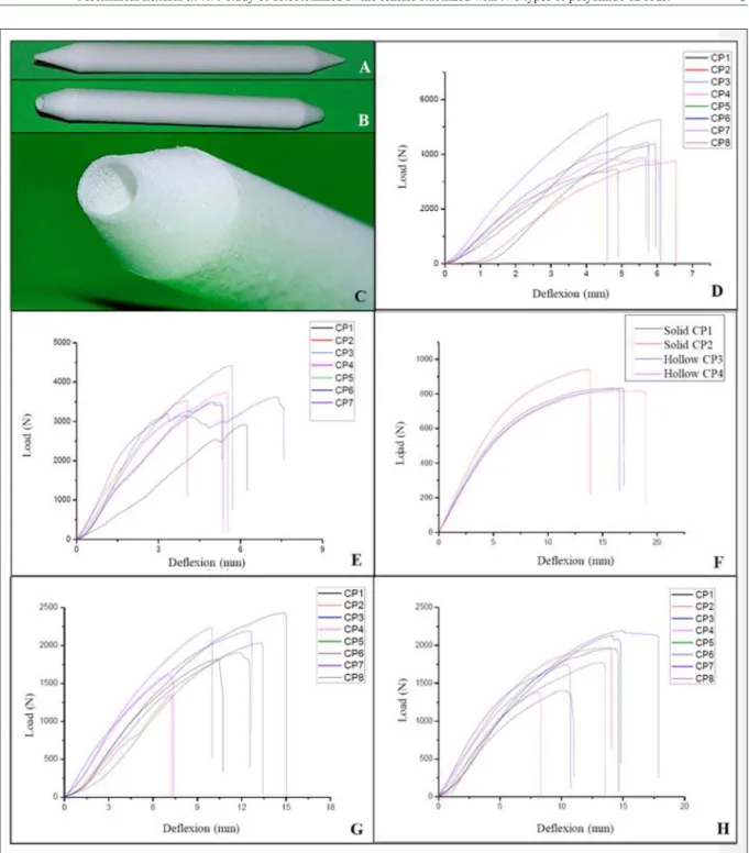
Figure 1 - Photographic images of polyamide 12 rods and the load deflection curves of the experimental groups. A) Solid polyamide 12 rod
from group G3; B and C) Hollow polyamide 12 rod from group G4; D) Load deflexion curves of intact femurs not subjected to drilling or osteotomy, group G1; E) Load deflexion curves of intact femurs subjected to drilling but not osteotomy, group G2; F) Load deflexion curves of the solid and hollow rods, G3 (n=2) and G4 (n=2), respectively; G) Load deflexion curves of group
G5, bone-implant system with solid polyamide 12 intramedullary rod interlocked with 2316L stainless steel orthopaedic screws
in each osteotomized bone fragment; H) Load deflexion curves of group G6, bone-implant system with hollow polyamide 12
on solid (G3) and hollow (G4) polyamide 12 rods (Figure 1F) in order to verify if alterations in
their geometry led to significant changes in their
mechanical properties, modulus of elasticity and
modulus of rupture during flexion. Lastly, tests were
performed on the bone-implant system containing solid (G5) and hollow (G6) rods (Figure 1G and 1H) to compare their mechanical properties with those of whole bones. Flexural modulus for the solid rods (G3) was determined by equation 1: EM = (4 x L3 x
m)/(3 x π x D4). For G4 (hollow rods) equation 2 was
used: EV = (4 x L3 x m) / [3 x π x (D4 - d4)], where “L”
corresponds to the distance between supports (span); “m” the gradient of the initial straight line portion of
the load deflexion curve; “d” the internal diameter of
the body of proof; and “D” the diameter of the body of proof (GARCIA et al., 2012).
Tension at rupture during flexion was calculated using equation 3: Ϭmax = Mf Max/Wf, where
MfMax corresponds to the maximum bending moment and Wf to the modulus of resilience to flexion. Maximum bending moment occurred half way
between the fixed support points of the span machine and; therefore, equation 4 was used: MfMax = (F x L)/4, where “F” corresponds to the load applied in the test and “L” the distance between the support points. Equation 5 was used to determine the modulus of
resilience to flexion for solid circular section with D diameter: Wf = π x (D3/32). For circular hollow
section, with external diameter “D” and internal
diameter “d”, equation 6 was used: Wf = π x (D4 -
d4)/32 x D (GARCIA et al., 2012).
After the tests, the internal and external diameters of the ruptured transversal section from bones and rods were measured in order to calculate the geometric parameters of each sample so that the mechanical properties could be determined. The Student t-test was used to compare the means from the modulus of elasticity and tension at rupture
between G1 x G2, G3 x G4 and G5 x G6. Significance
was considered at 5% (P<0.05).
RESULTS AND DISCUSSION
Swine femurs were chosen for this study for the easiness and speed to which they could be obtained, especially as standardized sizes had to be used. Furthermore, obtaining the same material from dog could have proven to be complicated and comparison with the literature would have been
way (G5 and G6), which is the most commonly used procedure for canine femur fractures when conventional rods are used, as recommended by
SPADETO JUNIOR et al. (2011).
Contrary to SPADETO JUNIOR et al.
(2011), who used electric drills and drill bits, the bones in this study were drilled manually. Nevertheless, the present study and those mentioned above are in agreement with FAIRBANK et al. (1995), who recommend the removal of all soft tissue to maximize the contact surface between the cortical bone and the implant, thus strengthening the bodies of proof.
Flexion tests for rigid and semi-rigid materials often determine the modulus of elasticity
and resilience to flexion; the former being used as the
most important criteria when evaluating the stiffness of polymeric materials and the later being equal to the
maximum tension in the external fibres of the body of
proof of a polymer at the time of rupture (GARCIA
et al., 2012). In the present study, the flexion test was applied to all study groups. Similarly to SPADETO JUNIOR et al. (2011), whole non-drilled bones were
used as controls (G1); however, this study also used as controls whole bones that had been drilled but not osteostomized (G2) in order to analyse a potential weakening of the body of proof due to drilling.
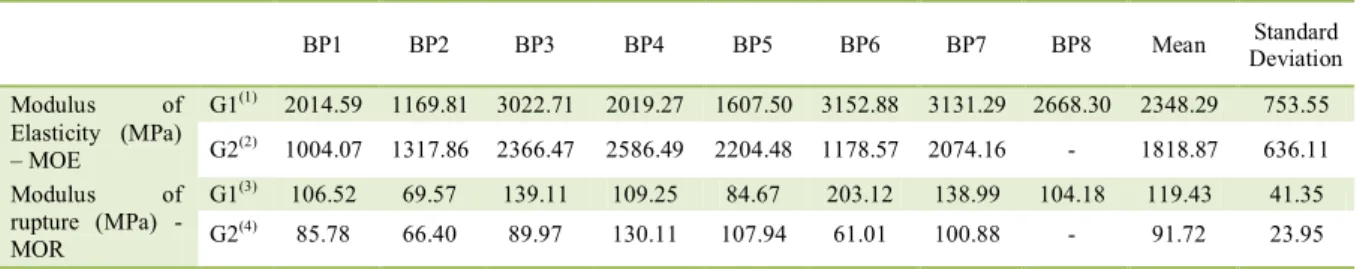
No significant difference (P=0.30) was observed in the flexion tests between G1 and G2
(Figure 1D and 1E, Table 1), demonstrating that the drilling did not affect the resilience of the body of proof. Thus, G1 (intact bone) was used as the control for the comparison analysis between the groups (G3 to G6). Results from the body of proof 8 (BP8) from G2 were disregarded as its values differed extremely from the rest. Results for the modulus of elasticity
under flexion of the polyamide 12 rods manufactured by SLS (G3 and G4) can be reported on figure 1F and
table 2. The data obtained by ASTM D790 was the same as that provided by the distributer (3D Systems).
Sintered polyamide 12 was chosen for this study based on the reports from several other studies that demonstrated its superiority to other materials, such as polyacetal and polypropylene. Furthermore, when compared to previous studies, the manufacturing method used in this experimental
model is still unprecedented (SPADETO JUNIOR et
al., 2010; SPADETO JUNIOR et al., 2011).
No significant difference (P=0.046) was
due to the lack of significant differences in their
geometrical dimensions in spite of the difference in their transversal sectioning. Interestingly, groups G3 and G4 demonstrated similar results for resilience to
flexion. This could be considered an advantage when
using hollow rods, as the same resistance of a solid rod can be obtained with less material. Moreover,
other materials could be used to fill the hollow space
of these rods and potentially increase their resilience while maintaining the ability of being perforated for the insertion of the locking screws.
A comparison between the mechanical
properties of flexion tests from groups of femurs
without osteotomy (G1 and G2) and those with rods (G3 and G4) demonstrated that the main mechanical property to be improved was the modulus of elasticity
of rods. This property could be significantly improved
by using reinforcing materials such as glass and
carbon fibres, manufactured in a similar way or by
other polyamides synthesized by other processes,
such as injection moulding. SPADETO JUNIOR et
al. (2011) reported faults on the rods at the site of
the screw closest to the fracture line and fissures in
the bone. In the present study; conversely, the tests from G5 and G6 have shown that faults on the rods
occurred close to the osteotomy site and not where the screws were inserted.
During a flexion test the bone is
simultaneously compressed on one side and tractioned on the other. Due to its anisotropy, when
flexed the fault, it will always be present on the side subjected to the traction (SPADETO JUNIOR et al.,
2010). In this study, crack nucleation (bone-rod) was observed on the area tractioned, as the bone is less resistant to traction than compression. Similarly,
SPADETO JUNIOR et al. (2010), in a study (in vivo) that used polyamide for the stabilization of induced fractures in the femurs of calves, have reported that half of the inserted rods had a fracture on the interface containing the screws close to the osteotomy line. In the present study; however, the rupture was observed in the region of the osteotomy
and not by the screws. These findings are thought
to be due to the good drilling technique applied, the tight insertion of the rods and the coaptation of the fracture to the type of polyamide and its manufacturing; which minimized the fatigue on the screws and evenly distributed the stress throughout the system, resulting on the fracture of the rod at its weakest point: the osteotomy site.
Table 1- Flexion test results (Modulus of elasticity and rupture) of G1 (eight whole swine femurs, not osteotomized or drilled) and G2 (seven whole swine femurs, not osteotomized but with a 15mm diameter drilled medullary canal).
BP1 BP2 BP3 BP4 BP5 BP6 BP7 BP8 Mean Standard
Deviation
Modulus of Elasticity (MPa) – MOE
G1(1) 2014.59 1169.81 3022.71 2019.27 1607.50 3152.88 3131.29 2668.30 2348.29 753.55
G2(2) 1004.07 1317.86 2366.47 2586.49 2204.48 1178.57 2074.16 - 1818.87 636.11
Modulus of rupture (MPa) - MOR
G1(3) 106.52 69.57 139.11 109.25 84.67 203.12 138.99 104.18 119.43 41.35
G2(4) 85.78 66.40 89.97 130.11 107.94 61.01 100.88 - 91.72 23.95
(1)Obtained using Equation 1; (2)Obtained using Equation 2; (3)
and (2) obtained using Equation 3; BP: body of proof.
Table 2 - Flexion test results (Modulus of elasticity and rupture) of G3 (two solid polyamide 12 rods) and G4 (two hollow polyamide 12 rods).
---Modulus of elasticity (MPa) – MOE---
BP1 BP2 Mean Standard deviation
G3 (1) 1260.78 1501.84 1381.31 170.45
G4 (2) 1349.45 1267.32 1308.39 58.07
G3 (3) 77.20 87.36 82.28 7.18
G4 (4) 79.36 77.91 78.64 1.02
(1)Obtained using Equation 1; (2)Obtained using Equation 2; (3)
rods (Figure 1G and 1H). This could be due to the fact that there were no significant differences in their geometrical dimensions in spite of the difference in their transversal sectioning. Groups G5 and G6 (bone-implant) did not reach the mechanical resistance levels of G1 (control). However, there was a synergism between the use of polyamide 12 rods and the interlocking system with screws, as the bone-implant system resisted load levels greater than those by rods alone (G3
and G4). These observations, in conjunction with
the possibilities of reinforcing polyamide 12, provide great scope for future research, which could analyse the use of reinforced polyamide 12 rods manufactured and implanted in a similar manner to the ones described in this study.
The modulus of elasticity and the modulus
of rupture during flexion could not be calculated for
G5 and G6 as the intricate geometry of the structure did not allow the determination or application of a mathematical model that could accurately represent the physical state of the loads, geometry, boundary conditions and behaviour of the materials. Thus, approximate models, in which the principles of the elasticity theory could be applied in an accessible and precise manner, should be used. According to LEE (2015), the most commonly used method for this is the Finite Element Method (FEM), which subdivides the domain into simpler parts to solve the problem. Thus, herein a suggestion for a future
study with the use of specific software, such as the
ANSYS mentioned by LEE (2015), to correctly determine the values of the mechanical properties of the elements of the bone-implant system and also of the stress throughout the system.
CONCLUSION
The polyamide 12 rods proved to be adequate for implantation following bone drilling, as well as for interlocking with screws, thus making its use in live animals promising. The resilience of hollow rods did not differ from that of solid ones; therefore, future studies should look into incorporating other materials to give even greater resilience to the bone-implant system. The bone-implant system withstood load levels greater than the rods alone and
these findings, in conjunction with the possibility of
reinforcing the polyamide with other materials, could generate interesting studies.
The study was approved and conducted under the guidance of the Ethics Committee in the Use of Animals of the Universidade de Franca (UNIFRAN), protocol number 026/12.
REFERENCES
DALLABRIDA, A.L. et al. Biomechanical analisis ex vivo of two osteosynthesis methods for transversal diaphiseal fracture in canine femur. Ciência Rural, v.35, n.1, p.116-120, 2005. Available
from: <https://www.scielo.br/scielo.php?script=sci_arttext&pid =S0103-84782005000100018>. Accessed: Aug. 10, 2016. doi: 10.1590/S0103-84782005000100018.
DIAS, L.G.G.G. Osteossíntese de tíbia com uso de fixador
esquelético externo conectado ao pino intramedular “Tie-in” em cães. 2006. 79f. Dissertação (Mestrado em Cirurgia Veterinária) - Programa de Pós-graduação em Cirurgia Veterinária, Universidade Estadual Paulista, SP.
DUELAND, R.T. et al. Structural properties of interlocking nails, canine femoral, and femur-interlocking nail constructs. Veterinary
Surgery,v.25, n.5, p.386-396, 1996. Available from: <https://
www.ncbi.nlm.nih.gov/pubmed/8879110>. Accessed: May 07, 2017. doi: 10.1111/j.1532-950X.1996.tb01432.x.
DUHAUTOIS, B. Use of veterinary interlocking nails for diaphyseal fractures in dogs and cats: 121 cases. Veterinary
Surgery, v.20, n.1, p.8-20, 2003. Available from: <https://www.
ncbi.nlm.nih.gov/pubmed/12520485>. Accessed: May 07, 2017.
doi: 10.1053/jvet.2003.50008.
FAIRBANK, A.C. et al. Long-term results of core decompression for ischaemic necrosis of the femoral head. Journal of Bone and
Joint Surgery, v.77B, n.1, p.42-49, 1995. Available from: <http://
www.bjj.boneandjoint.org.uk/content/jbjsbr/77-B/1/42.full.pdf>.
Accessed: Aug. 10, 2016.
GALUPPO, L.D. et al. An in vitro biomechanical investigation of an MP35N intramedullary interlocking nail system for repair of third metacarpal fractures in adults horses. Veterinary Surgery, v.31, n.3, p.211-225, 2002. Available from: <https://www.ncbi.
nlm.nih.gov/labs/articles/11994848/>. Accessed: May 07, 2017.
doi: 10.1053/jvet.2002.32399.
GARCIA A. et al. Ensaio dos materiais. 2.ed. Rio de Janeiro:
Livros Técnicos e Científicos, 2012. 247p.
GUPTA, A. Dinamic compression nail: a preliminary report. BMC
Musculoskeletal Disorders, v.2, p.6, 2001. Available from: <http://
www.ncbi.nlm.nih.gov/pmc/articles/PMC59672/pdf/1471-2474-2-6.pdf>. Accessed: May 07, 2017. doi: 10.1186/1471-2474-2-6.
HORSTMAN, C.L. et al. Biological osteosynthesis versus traditional anatomic reconstruction of 20 long-bone fractures using an interlocking nail: 1994-2001. Veterinary Surgery, v.33, n.3, p.232-237, 2004. Available from: <https://www.ncbi.nlm.nih.gov/
pubmed/15104629>. Accessed: May 07, 2017. doi: 10.1111/j.1532-950X.2004.04034.x.
LEE, H.H. Finite element simulations with ANSYS Workbench
SPADETO JUNIOR, O. et al. Failures in the use of polyacetal
and polyamide in the form of intramedullary interlocking nail for immobilization of induced femoral fracture in young cattle. Ciência Rural, v.40, p.907-912, 2010. Available from:
<http://www.scielo.br/scielo.php?script=sci_arttext&pid =S0103-84782010000400025>. Accessed: Aug. 10, 2016. doi:
10.1590/S0103-84782010005000038.
SPADETO JUNIOR, O. et al. Ex vivo bone-implant systems using
polymeric intramedullary nails for fixation of femoral fractures in
young calves. Ciência Rural,v.41, n.2, p.301-306, 2011. Available
from: <http://www.scielo.br/scielo.php?script=sci_arttext&pid
=S0103-84782011000200020>. Accessed: Aug. 10, 2016. doi:
0.1590/S0103-84782011000200020.
VAN DER ELST, M. et al. Bone tissue response to biodegradable
polymers used for intra medullary fracture fixation: a long-term
in vivo study in sheep femoral. Biomaterials, v.20, n.2, p.121-128, 1999. Available from: <https://www.ncbi.nlm.nih.gov/
pubmed/10022781>. Accessed: May 07, 2017. doi: 10.1016/ S0142-9612(98)00117-3.