Mestre em Engenharia Química e Bioquímica
Functional monolithic platforms for antibody purification
Dissertação para obtenção do Grau de Doutor em
Química Sustentável
Orientadores: Prof. Doutora Ana Aguiar-Ricardo
Prof. Doutora Ana Cecília Roque
“Copyright”
Telma Godinho Barroso
Faculdade de Ciências e Tecnologia
Universidade Nova de Lisboa
The PhD was more than four years of scientific research combined with my intellectual skills. In fact, it was a test to my personal resistance (Márcio Temtem you were completely right! Thanks very much for your advices, and for all friendship and teachings! You were a great professor). Fortunately, I believe that I passed it with a feeling of “job done”. However, PhD was not possible without the support and reinforcement of several people that I would like to acknowledge.
First of all, I would like to acknowledge my supervisors Prof. Ana Aguiar-Ricardo and Prof. Cecilia Roque that gave me the opportunity to work on this project. I am very grateful for all their effort and dedication to me, and to the work that together we were able to develop. Prof. Ana Aguiar-Ricardo many thanks to be an exigent and outstanding supervisor, and an especial friend when I needed. I will always remember our brainstorming, the phone calls after hours, and the happy and frustrating moments together, but the true is: I got here because you never gave up of me. Prof. Cecilia Roque, I am very thankful to your patience with me as well as to your good vibes that you always transmitted to me (yûûûû ooouuu). Without you, the biochemical world would be utopic for me! If I have biochemistry in my education, I owe it to you.
I also would like to acknowledge to Polymer Synthesis and Processing Group and to Biomolecular Engineering Group. I could find numerous words to say to each one however, it will become another thesis. Thus, I reserved some special words for each one of you. Starting with Polymer Synthesis and Processing Group: Raquel Viveiros (Yesterday, today and
tomorrow…you will be forever in my heart, thanks for everything), Vanessa Correia (I will miss you, and I will keep great memories of us including brainstorming ), Rita Restani (“Gazeada Girl”: sometimes the life is hard, but at same time is amazing…please hold it), Patrícia Morgado (Thanks for your sympathy and friendship), Ana Silva (For you, the sky is the limit…close your eyes, close your mouth and forget the others, you are “the important” because
you have everything to be it), Anita Lourenço (Thank you for your participation in this chapter of my life), Márcia Tavares (“Hard Rock Girl”; It was a pleasure to work with you), Renato Cabral (“Great Engineer”: it was a pleasure to meet you), Vasco Bonifácio (“The Chemist Man”: thanks
for all your patience in try to explain me organic chemistry, it was not easy, but it was possible ) and Teresa Casimiro (“Great scientist and friend”: Thanks to clean my tears and support my
euphoric moments. You had a fundamental role on this chapter of my life). Now the Biomolecular Engineering Group: Íris Batalha (“Keep Calm Girl”: thank you very much for all
funny and zen moments that only you could give me), Margarida Dias (“Special friend and great professional”: I will miss you a lot), Susana Palma (It was a pleasure to meet you), Henrique
Carvalho (“Alternative Guy”: I always remember your good mode), Vijaykumar Dhadge
(“Patents Man”: Great Doctor! One day we will be rich!!!), Abid Hussain (“Great colleague, researcher and English professor”: Thanks for everything that you taught me), Ricardo Branco
all words are not enough. During the last four years you were one of the main pieces of this game and when the motivation disappeared, you were the reason that made me to continue. I
will remember all nights that we spent in the lab, all the “balcony moments” where we laugh and
where we cried, all conferences, all days, everything…You are simply the best.
Sincerely, thank you all for everything. This thesis has pieces of you thus, it is also yours.
To Professor Manuel Nunes da Ponte I would like to express my sincere acknowledgments for all teachings in green chemistry which formatted me to be a sustainable engineer. A special thanks also to professor Ana Maria Rego for all XPS discussions and friendship.
A special thanks to Maria José and Isabel Rodrigues for their assistance in bureaucratic work, and to Maria de Palma, Idalina and Conceição for providing me clean laboratory material to develop me research. For all, a distinctive thank also for your love and kindness. I want also to acknowledge the Analytical Services Laboratory of REQUIMTE for the characterization of materials, and to the Animal Cell Technology Unit of ITQB-UNL/IBET (Dr. Paula M Alves and Dr. Ana Teixeira) for providing the cell culture bulks of antibodies.
To all my family, I am very grateful for all your support and patience. Due to all of you (father, sister, nephew (Ti), cousins, uncles, aunts, brothers-in-law, Manuela e Armando) I am the person that was able to get here. I am sorry for the moments that I missed with you (especially with my nephew, Tiago) but this work was hard. However, believe in me, you were always in my heart. Specially, I would like to acknowledge two persons: Sónia Barroso that more than a sister, she was and is the mother that I never had. I love you! The second person is my husband Nuno Fernandes that was the light that appeared in my life, and make me believe that I was able to do everything that I wanted. Thank you for your love, friendship, patience and encouraging words. Without you my life will be uncoloured. Love you! To all my friends, that are a lot, but you know who I am referring, many thanks for always being there for me! Beetocada
(all members and staff) thanks very much for all sportive and relax weekends and moments.
“Nephews” (Joana, Porco, Li, Central, Pêlo, Caixinha, and all others) for you a special thanks for all happy moments that you gave me, and that I will always keep in my heart. I love you all in the same way. For the ones that life turned difficult their presence (Bru, Inês, Meguy and Tropa), a special kiss. You were also my driving force to continue. You are great friends.
This work aimed at the development of monolithic chromatographic platforms for antibody purification. A sustainable strategy, comprising the use of natural polymers such as chitosan, agarose and dextran, was employed to create 3D porous structures. In order to improve the mechanical properties and biodegradability of monoliths, natural polymers were physically blended with synthetic ones. All supports were, in a first stage, produced by freeze-drying methods while in a second attempt were prepared by an integrated approach involving gelation process, water-acetone substitution and scCO2 drying. A further optimization for opening the porous network was evaluated involving swelling and freeze-drying procedures. To optimize the efficacy of monoliths, magnetic nanoparticles were embedded in monoliths structure to confer them a magnetic responsive behaviour. This additional feature improved antibody recovery when performing a magnetically-assisted elution (93% recovery of bound IgG) complemented to less time processing. The selectivity of monoliths for antibody, IgG, was guaranteed by the immobilization of ligand 22/8 (artificial Protein A) and a new triazine-based ligand (TPN-BM) onto their surfaces. The functionalization strategy of TPN-BM, which synthesis followed the principles of green chemistry, was induced by plasma technology. This alternative strategy allowed the reduction of time and solvents consumption while maximizing the functionalization yield of supports (2-fold, comparing to the traditional procedures). Moreover, the binding/elution mechanism between TPN-BM and IgG at a molecular level was validated through molecular docking studies and dynamic simulations.
Overall, TPN-BM functionalized natural-based monoliths revealed values of pore size diameter, porosity, and flux between 1-96 µm, 28-88 % and 3-220 (L m-2 h-1). Chitosan/poly(vinyl alcohol)-based monoliths revealed the best binding and elution capacities, 160 mg IgG g-1 support and 97%, respectively, at least over four consecutive cycles. Moreover, tested with crude samples, supports exhibited a good specificity for mAbs, recovering them with 96-98% of purity.
KEYWORDS: Biopolymers, monoliths, affinity ligand, plasma technology, supercritical carbon
Este trabalho teve como objectivo o desenvolvimento de suportes monolíticos para a purificação de anticorpos por cromatografia de afinidade. Para tal, utilizou-se uma estratégia sustentável para produzir essas estruturas porosas 3D (monólitos), envolvendo polímeros naturais tais como quitosano, dextrano e agarose. Para melhorar as propriedades mecânicas e biodegradáveis dos monólitos, os polímeros naturais foram misturados fisicamente com polímeros sintéticos. Primeiramente, todos os suportes foram produzidos por liofilização, e numa segunda fase por processos de gelificação, substituição de água por acetona e secagem por scCO2. A optimização da abertura da rede porosa foi efectuada recorrendo à capacidade de inchamento dos suportes e subsequente liofilização. Para melhorar a performance dos monólitos, incorporaram-se nanopartículas magnéticas nas redes monolíticas por forma a conferir-lhes a capacidade de resposta magnética e consequente deformação quando sob acção de um campo magnético. Realizaram-se assim eluições assistidas por campo magnétco o que permitiu o aumento do rendimento de recuperação de anticorpo (IgG) ligado (93%) e diminuir o tempo do passo de eluição. A selectividade dos monólitos para o anticorpo foi garantida através da imobilização de dois ligandos sintéticos mimetizando a Proteína A (ligando 22/8 e o novo ligando TPN-BM) na superfície dos suportes. A estratégia de funcionalização do TPN-BM, cuja síntese seguiu os princípios da química verde, foi feita utilizando a tecnologia de plasma. Esta estratégia permitiu reduzir tempo e uso de solventes bem como maximizar todo o processo (em 2 vezes) comparativamente aos procedimentos tradicionais. O mecanismo de ligação/eluição do TPN-BM e IgG foi validado através de estudos de acoplamento molecular e simulações dinâmicas.
Em geral, todos os monólitos TPN-BM-funcionalizados exibiram valores de diâmetro de poro, porosidade e fluxo entre 1-96 µm, 28-88 % e 3-220 (L m-2h-1), respectivamente. Os monólitos de quitosano/poli(vinil álcool) revelaram as melhores capacidades de ligação e de eluição, 160 mg IgG g-1 suporte e 97%, respectivamente, pelo menos durante quatro ciclos consecutivos. Adicionalmente, quando testados com extractos brutos, exibiram uma boa especificidade para mAbs, recuperando-os com 96-98% de pureza.
.
PALAVRAS-CHAVE - Biopolímeros, monólitos, ligandos de afinidade, tecnologia de plasma,
ACKNOWLEDGMENTS ... III
ABSTRACT ... V
RESUMO ... VII
TABLE OF CONTENTS ... IX
INDEX OF FIGURES ... XIII
INDEX OF TABLES ... XXI
ABBREVIATIONS ... XXIII
BACKGROUND ... XXVII
CHAPTER 1: FUNCTIONAL MONOLITHIC PLATFORMS: CHROMATOGRAPHIC TOOLS
FOR ANTIBODY PURIFICATION ... 1
1.1 INTRODUCTION ... 2
1.2 MONOLITHIC PLATFORMS ... 3
1.2.1. SYNTHETIC POLYMER MONOLITHS ... 4
1.2.1.1. Hydrogels and cryogels ... 7
1.2.2. MONOLITHS BASED ON NATURALLY OCCURRING POLYMERS ... 8
1.3 STRUCTURAL CHARACTERIZATION OF MONOLITHS ... 11
1.4 PERFORMANCE EVALUATION OF MONOLITHIC PLATFORMS ... 11
1.5 SUMMARY, CONCLUDING REMARKS AND FUTURE TRENDS ... 13
CHAPTER 2: BIOINSPIRED AND SUSTAINABLE CHITOSAN-BASED MONOLITHS FOR ANTIBODY PURIFICATION ... 15
2.1. INTRODUCTION ... 16
2.2. EXPERIMENTAL AND METHODS ... 17
2.2.1. MATERIALS ... 17
2.2.2. MONOLITHS PREPARATION ... 17
2.2.3. MONOLITHS CHARACTERIZATION ... 18
2.2.4. MONOLITHS FUNCTIONALIZATION ... 19
2.2.5. STATIC PARTITION EQUILIBRIUM EXPERIMENTS ... 20
2.2.6. FRONTAL ANALYSIS-BREAKTHROUGH CURVES AND BINDING CAPACITY ... 21
2.2.7. CHROMATOGRAPHIC EXPERIMENTS ... 21
MONOLITHS ... 22
2.3.2 PREPARATION AND CHARACTERIZATION OF AFFINITY CHITOSAN-BASED MONOLITHS ... 25
2.3.3. EVALUATION OF AFFINITY MONOLITHS FOR ANTIBODY PURIFICATION ... 29
2.3.4. OPTIMIZATION OF AN AFFINITY MONOLITH FOR ANTIBODY RECOVERY ... 31
2.4. CONCLUDING REMARKS ... 34
CHAPTER 3: A SUSTAINABLE BIOMIMETIC LIGAND FOR DIRECT IMMOBILIZATION ON (BIO)POLYMERIC SUPPORTS ... 35
3.1. INTRODUCTION ... 36
3.2. EXPERIMENTAL AND METHODS ... 37
3.2.1. MATERIALS ... 37
3.2.2. LIGAND SYNTHESIS AND CHARACTERIZATION ... 38
3.2.3. MONOLITHS PREPARATION AND FUNCTIONALIZATION WITH TPN-BM ... 39
3.2.4. BIOMIMETIC MONOLITHS CHARACTERIZATION ... 40
3.2.5. STATIC PARTITION EQUILIBRIUM STUDIES ... 40
3.2.6. FRONTAL ANALYSIS – BREAKTHROUGH CURVES AND BINDING CAPACITY .... 41
3.2.7. CHROMATOGRAPHIC EXPERIMENTS WITH PURIFIED PROTEIN SOLUTIONS ... 41
3.2.8. PURIFICATION OF MONOCLONAL ANTIBODIES FROM MAMMALIAN CRUDE EXTRACTS ... 41
3.3. RESULTS AND DISCUSSION ... 42
3.3.1. TPN-BM SYNTHESIS ... 42
3.3.2. IMMOBILIZATION OF LIGAND TPN-BM ONTO NATIVE CHITOSAN-BASED MONOLITHS ... 45
3.3.3. EVALUATION OF TPN-BM MONOLITHS AS AFFINITY DEVICES FOR hIgG PURIFICATION ... 50
3.3.4. OPTIMIZATION OF TPN-BM AFFINITY MONOLITH FOR ANTIBODY PURIFICATION ... 52
3.4. CONCLUDING REMARKS ... 55
CHAPTER 4: STRUCTURAL EVALUATION OF AN ALTERNATIVE PROTEIN A BIOMIMETIC LIGAND TOWARDS ANTIBODY PURIFICATION ... 57
4.1. INTRODUCTION ... 58
4.2 METHODS ... 59
4.2.1. MOLECULAR MODELLING ... 59
4.2.2. MOLECULAR DOCKING ... 59
4.2.3. MD SIMULATIONS ... 60
4.3. RESULTS AND DISCUSSION ... 61
4.3.1. INTERACTIONS OF LIGAND TPN-BM WITH IgG FRAGMENTS ... 62
4.3.2. pH DEPENDENCE ON THE AFFINITY BETWEEN TPN-BM AND IgG ... 66
5.1. INTRODUCTION ... 72
5.2. EXPERIMETAL AND METHODS ... 73
5.2.1. MATERIALS ... 73
5.2.2. PREPARATION OF MAGNETIC NANOPARTICLES ... 73
5.2.3. EVALUATION OF POLYMERS ADSORPTION ON MNPs ... 74
5.2.4. PREPARATION OF NATIVE AND MAGNETIC MONOLITHS ... 74
5.2.5. PREPARATION OF HYBRID MONOLITHS ... 76
5.2.6. CHARACTERIZATION OF NATIVE, MAGNETIC AND HYBRID MONOLITHS ... 77
5.2.7. DESIGN OF A PERMANENT MAGNET ... 78
5.2.8. DETERMINATION OF STATIC AND DYNAMIC BINDING CAPACITIES ... 78
5.2.9. CAPTURE AND RELEASE OF IgG FROM PURE SOLUTIONS ... 79
5.2.10. PURIFICATION OF MONOCLONAL ANTIBODIES, mAbs, DIRECTLY FROM CRUDE SAMPLES ... 79
5.3. RESULTS AND DISCUSSION ... 80
5.3.1. CHARACTERIZATION OF NATIVE AND MAGNETIC MONOLITHS ... 80
5.3.2. PREPARATION AND CHARACTERIZATION OF HYBRID MONOLITHS ... 86
5.3.3. HYBRID MONOLITHS IN Ab PURIFICATION ... 92
5.4. CONCLUDING REMARKS ... 100
CHAPTER 6: POROUS CHITOSAN-BASED MONOLITHS PREPARED FROM THE BEST COMBINATION OF SUSTAINABLE MATERIALS AND TECHNIQUES ... 101
6.1. INTRODUCTION ... 102
6.2. EXPERIMENTAL AND METHODS ... 103
6.2.1. MATERIALS ... 103
6.2.2. MONOLITHS PREPARATION ... 103
6.2.3. PREPARATION OF AFFINITY MONOLITHS ... 105
6.2.4. CHARACTERIZATION OF NATIVE AND FUNCTIONALIZED MONOLITHS ... 106
6.2.5. DETERMINATION OF STATIC BINDING CAPACITIES ... 106
6.2.6. DETERMINATION OF DYNAMIC BINDING CAPACITIES ... 107
6.2.7 MONOLITHS PERFORMANCE OVER CYCLES OF PROTEIN CAPTURE AND RELEASE ... 107
6.2.8. PURIFICATION OF mAbs DIRECTLY FROM CRUDE SAMPLES ... 108
6.3 RESULTS AND DISCUSSION ... 108
6.3.1. MONOLITHS PREPARATION AND FUNCTIONALIZATION ... 108
6.3.2. CHARACTERIZATION OF CP MONOLITHS BEFORE AND AFTER TPN-BM COUPLING ... 110
6.3.3. EVALUATION OF TPN-BM FUNCTIONALIZED MONOLITHS FOR ANTIBODY PURIFICATION ... 118
6.3.4. REPRODUCIBILITY AND OPTIMIZATION OF TPN-BM FUNCTIONALIZED CP MONOLITHS FOR ANTIBODY PURIFICATION ... 121
6.4. CONCLUDING REMARKS ... 127
7.1.1.1. GREEN METRICS ... 132
7.1.2. LIFE CYCLE ASSESSMENT (LCA) ... 134
7.2. CASE STUDY 1: EVALUTION OF TPN-BM LIGAND SYNTHESIS ... 139
7.3 CASE STUDY 2: EVALUATION OF PLASMA TREATMENT AS A METHOD FOR SURFACES MODIFICATION ... 141
7.4 CONCLUDING REMARKS ... 144
CHAPTER 8: CONCLUDING REMARKS AND FUTURE PERSPECTIVES ... 145
8.1 CONCLUDING REMARKS ... 145
8.2. FUTURE PERSPECTIVES ... 147
PAGE
Figure 1.1 – Schematic representation of opportunity window for polymer monolith
incorporation into chromatographic bioseparation processing technology on the basis of target molecule size. 2 Figure 1.2 – Preparation of GMA-EDMA monoliths (A) and of AA-AGE cryogels (B) and their
chemical structures. 5 Figure 1.3 - (A) Schematic preparation of chitosan-based monoliths. (B1, B2) SEM micrographs
of chitosan monoliths and chitosan cryopolymerized with glycidyl methacrylate (GMA) respectively,82 adapted with the permission of The Royal Society of Chemistry.
9 Figure 2.1 – Schematic representation showing the functionalization strategy of chitosan-based monoliths with ligand 22/8 using non-thermal plasma surface activation. 20 Figure 2.2 – SEM images of chitosan (CHT) based-monoliths before and after functionalization
with the ligand 22/8: (A) CP_N, native monolith prepared from chitosan and polyvinyl alcohol (CP); (B) CG_N, native monolith prepared from chitosan and glycidyl methacrylate (CG); (C) CP_22/8, CP monolith functionalized with ligand 22/8 and (D) CG_22/8, CG monolith functionalized with ligand 22/8. All the images have a magnification of 300 and the scale bar in white indicates 10 µm. 23 Figure 2.3 – Stability evaluation of CP_22/8 (A and B) and CG_22/8 (C and D) monoliths
immersed over 12 h in solutions typically used during cleaning-in-place (CIP) procedures, including solutions with pH values between 1 and 12. All data was obtained from duplicated measurements with errors of ±5. 28 Figure 2.4 – Langmuir–Freundlich adsorption isotherms for (A) CP and (B) CG monoliths: (♦)
native and (▲) functionalized. (C) Summarizes the estimated parameters of the Langmuir–
Freundlich isotherms and standard errors for CP and CG monoliths before and after functionalization with ligand 22/8. 29 Figure 2.5 – Breakthrough profiles for human IgG upon (A) CP_22/8 and (B) CG_22/8 monoliths at different flow rates: (♦) 1 mL min-1
and (▲) 2 mL min-1. All data was obtained from duplicated measurements with errors of ±0.05. 30 Figure 2.6 – Evaluation of chromatographic performance for (A, B) native and (C, D)
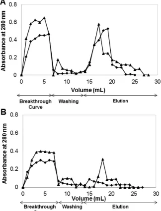
functionalized CP monoliths using pure IgG and BSA solutions, respectively. The chromatographic procedures (bind, elution and regeneration steps) were performed consecutively along four cycles at a flow rate of 2 mL min-1. The last cycle was performed after autoclaving (After_AC). 31 Figure 2.7 - Evaluation of chromatographic performance for (A, B) native and (C, D)
native and (▲) functionalized, at a flow rate of 1 mL min
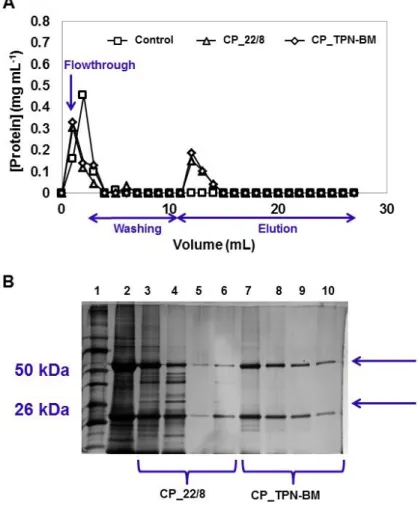
flowthrough (F.T.) followed by the washing and elution steps. The acrylamide gel from SDS-PAGE performed with the fractions collected during the mAbs purification (B): lane 1 corresponds to the molecular weight marker, lane 2 represents the loading, lane 3 is the flowthrough, lane 4 corresponds to the first wash (phosphate buffer (50 mM, pH 8.0)), and lane 5 and lane 6 are the first and second elution fractions (sodium citrate buffer (50 mM, pH 3.0)).33 Figure 3.1 - Chemical structures of ligand 22/8 and TPN-BM. 37 Figure 3.2 - (A) 1H-NMR spectrum of TP-BM in CDCl3 and (B) FT-IR spectrum of TP-BM. 43 Figure 3.3.- (A) 1H-NMR spectrum of TPN-BM in CDCl3 and (B) FT-IR spectrum of TPN-BM. 44 Figure 3.4 - SEM images of chitosan based-monoliths before and after functionalization of ligand TPN-BM: (A) CP_N, native monolith prepared from chitosan and polyvinyl alcohol; (B) CG_N, native monolith prepared from chitosan and glycidyl methacrylate; (C) CP_TPN-BM, CP monolith functionalized with TPN-BM and (D) CG_TPN-BM, CG monolith functionalized with TPN-BM. All the images have a magnification of 300 and the scale bar in white corresponds to 50 µm. 47 Figure 3.5 - Stability evaluation of CP_TPN-BM (A and B) and CG_TPN-BM (C and D)
monoliths immersed, over 12 hours, in solutions typically used during cleaning-in-place (CIP) procedures, including solutions with pH values between 1 and 12. All data was obtained from duplicated measurements with errors of ±6. 49 Figure 3.6 - Langmuir-Freundlich adsorption isotherms for (A) CP and (B) CG monoliths: (♦) native and (●) functionalized with TPN-BM. In (C) are summarized the estimated parameters of Langmuir-Freundlich isotherms and standard errors, for CP and CG monoliths before and after functionalization with ligand TPN-BM and ligand 22/8 (a).82 51 Figure 3.7 - Breakthrough profiles for human IgG upon CP_TPN-BM (♦) and CG_TPN-BM (▲)
monoliths at a flow rate of 1 mL min-1. All data was obtained from duplicated measurements with errors of ±0.05. 52 Figure 3.8 - Evaluation of chromatographic performance for CP_TPN-BM monoliths using pure
human IgG (A) and BSA (B) solutions, respectively. The chromatographic procedures (binding, elution and regeneration steps) were performed consecutively along four cycles at a flow rate of 1 mL min-1. The last cycle was performed after autoclaving (After_AC).
53
(Software used: ChemBioDraw Ultra 13.0). 59 Figure 4.2 - Image showing the preferential binding site of ligand TPN-BM in the Fab fragment
of IgG (PDB code 1HZH). Highlighted regions in the Fab represent residues that are within 5Å from the ligand, colored by hydrophobicity. (Software used: Pymol 1.3. and VMD 1.9.1). 64 Figure 4.3 - Image showing alternative binding sites of ligand TPN-BM in the Fc fragment of
IgG (PDB code 1HZH). Highlighted region in the Fc represents the residues that are within 5Å from TPN-BM, colored by hydrophobicity. (Software used: Pymol 1.3. and VMD 1.9.1). 65 Figure 4.4– pH dependence of ligand binding to the Fc fragment of IgG (PDB code 1HZH).
Protonation state of the protein residues adjusted to pH 7 (A) where the naphtol ring of the ligand is anchored within 5 Å to the polar and hydrogen bonding interaction with the Fc domain; and pH 3 (B), where main hydrogen bond interactions were disrupted forcing the ligand to drift away from the receptor (distances above 8 Å). Both regions of interactions are colored by hydrophobicity of the correspondent residues. (Software used: Pymol 1.3 and VMD 1.9.1). 68 Figure 4.5 - Graphical representation of the distance between the His 466 (A) and His 302 (B)
with the ligand atom type OAV and OHAZ, respectively at both pH (pH 7 line colored in black and pH 3 line colored in gray), monitored along the 10 ns of simulation time. 69 Figure 5.1 - Schematic representation of the natural-based monoliths design and composition. CHT indicates chitosan monolith, CP means chitosan/poly(vinyl alcohol) monolith, AA represents agarose/acrylamide monolith and DXT designates dextran-based monolith. 75 Figure 5.2 - Illustration of the materials produced in this work (A) and schematic representation
of the procedures applied for the production and functionalization of the magnetic monoliths (B).
76
Figure 5.3 - SEM images of natural-based monoliths before (native, N) and after MNP’s
incorporation (magnetic, M) and hybrid monoliths (magnetic with ligand TPN-BM coupled, M_TPN-BM): (A) native chitosan monolith (CHT_N), (B) magnetic chitosan monolith (CHT_M), (C) hybrid chitosan monolith (CHT_M_TPB-BM), (D) native chitosan blended with poly(vinyl alcohol) monolith (CP_N), (E) magnetic chitosan blended with poly(vinyl alcohol) monolith (CP_M), (F) hybrid chitosan blended with poly(vinyl alcohol) monolith (CP_M_TPN-BM), (G) native agarose-based monolith (AG_N), (H) magnetic agarose-based monolith (AG_M), (I) hybrid agarose-based monolith (AG_M_TPN-BM), (J) native dextran-based monolith (DXT_N), (K) magnetic dextran-based monolith (DXT_M) and (L) hybrid dextran-based monolith (DXT_M_TPN-BM). All the micrographs have a magnification of 300 and the scale bar in white indicates 10 µm. 81 Figure 5.4 - Magnetic deformations of different magnetic natural-based monoliths: chitosan,
CHT_M (A), chitosan- poly(vinyl alcohol), CP_M (B), agarose, AG_M (C) and dextran, DXT_M (D). All data was obtained from triplicated measurements with errors of ±5. 84 Figure 5.5 - Evaluation of magnetic nanoparticles (MNPs) leaching from chitosan, CHT_M (A,
from duplicated measurements with errors of ±8. 85 Figure 5.6 - Schematic representation of: (A) the amination procedure, assisted by plasma
technology, of magnetic natural-based monoliths. (B) Graphical representation of the amination and TPN-BM immobilization yields obtained for all magnetic monoliths. 87 Figure 5.7 - XPS regions C 1s, N 1s, and Fe 2p of native monoliths (black), magnetic and
aminated (green) and hybrid monoliths (blue). 89 Figure 5.8 – Evaluation of TPN-BM leaching from hybrid chitosan, CHT_M_TPN-BM (A, B),
chitosan- poly(vinyl alcohol), CP_M_TPN-BM (C, D), agarose, AG_M_TPN-BM (E, F) and dextran, DXT_M_TPN-BM (G, H) monoliths immersed over 12 h in solutions with pH values between 1 and 12 and typically used during cleaning-in-place (CIP) procedures, respectively. All data was obtained from duplicated measurements with errors of ±11. 91 Figure 5.9 - Graphical representation of experimental adsorption isotherms (Exp) fitted by
Langmuir-Freundlich (LF) model for magnetic (M) and hybrid (M-TPN-BM) monoliths: (A) CHT_M and CHT_M_TPB-BM (B) CP_M and CP_M_TPN-BM (C) AG_M and AG_M_TPN-BM (D) DXT_M and DXT_M_TPN-BM. 93 Figure 5.10 - Breakthrough profiles for human IgG upon CHT_M_TPB-BM (▲), CP_M_TPN -BM (♦), AG_M_TPN-BM (●) and DXT_M_TPN-BM (■) monoliths, performing the elution at pH 3 (A) and pH 11 (B). (C) Summarizes the binding and elution capacities estimated through breakthrough curves at different elution conditions. 94 Figure 5.11 Map of the magnetic flux density strength in the z-direction, highlighting the key
components of the setup. Magnet dimensions: internal radius r_int = 5.5 mm, external radius r_ext = 20 mm, height h = 15 mm. 95 Figure 5.12 - (A) Schematic representation of the porous network availability of hybrid
monoliths during typical and magnetically-assisted elution of chromatographic experiments. (B) Graphical representation of binding (black), normal elution (grey) and magnetically-assisted elution (white) of hybrid natural-based monoliths. 96 Figure 5.13 – Evaluation of chromatographic performance of (A) CHT_M_TPN-BM, (B)
CP_M_TPN-BM and (C) AG_M_TPN-BM monoliths using pure IgG solutions. The chromatographic procedures, bind (black), elution (grey) and regeneration (white) steps, were performed consecutively along four cycles at a flow rate of 1 mL min-1. The last cycle was performed after autoclaving (After_AC). The elution was assisted by the permanent magnet (0.5 T). 97 Figure 5.14– Chromatographic performance of CP_M_TPN-BM and AG_M_TPN-BM in mAbs
Figure 6.1 – Schematic representation of the chitosan-poly(vinyl alcohol) (CP) hydrogels drying under supercritical carbon dioxide (scCO2 drying). 104 Figure 6.2 – Schematic representation of the additional procedure for the opening of porous
network of chitosan-poly(vinyl alcohol) (CP) monolith: after obtaining CP monoliths by scCO2 drying, monoliths swelled 2 hours in water and then were frozen at -20 ºC and lyophilized. 105 Figure 6.3 – SEM images of bare and magnetic chitosan-poly(vinyl alcohol) (CP) monoliths
before and after functionalization with TPN-BM. Particularly, (A) bare CP monolith, CP 25:75, (B) magnetic CP monolith CP 25:75_M and (C) magnetic CP monolith submitted to an additional treatment for opening porous network involving swelling and freeze-drying procedures, CP 25:75_MFD. The corresponding SEM images obtained after functionalization with TPN-BM are shown in D, E and F respectively: (D) CP 25:75_TPN-BM, (E) CP 25:75_M_TPN-BM and (F) CP 25:75_M_TPN-BMFD. All the images have a magnification of 500 and the scale bar in white indicates 10 µm. 110 Figure 6.4 - Distributions of pore size diameter of all chitosan-poly(vinyl alcohol) (CP)
monoliths. Particularly, (A) represents bare CP monoliths: (●) CP 25:75, (■) CP 14:86 and, (▲) CP 50:50; (B) represents magnetic CP monoliths: (Δ) CP 25:75_M and (▲) CP 25:75_MFD
and (C) represents CP monoliths functionalized with TPN-BM: (○) CP 25:75_TPN-BM and (●) CP
25:75_M_TPN-BMFD. FD means that monoliths were submitted to an additional treatment for opening porous network involving swelling and freeze-drying procedures. 111 Figure 6.5 – Water fluxes of bare and magnetic chitosan-poly(vinyl alcohol) (CP) monoliths
before and after functionalization with ligand TPN-BM at the absence and presence (represented by bars with strikes) of a permanent magnet of 0.5 T. FD means that monoliths were submitted to an additional treatment for opening porous network involving swelling and freeze-drying procedures. 113 Figure 6.6 – Magnetic deformation of magnetic chitosan-poly(vinyl alcohol) (CP) monolith, CP
25:75_M, magnetic CP monolith submitted to an additional treatment for opening porous network involving swelling and freeze-drying procedures, CP 25:75_MFD, and CP 25:75 and CP 25:75_MFD monoliths after functionalization with TPN-BM, CP 25:75_M_TPN-BM and CP 25:75_M_TPN-BMFD, respectively, in dry and wet states, at the presence of a permanent magnet of 0.5 T. 115 Figure 6.7 - Stability evaluation of magnetic chitosan-poly(vinyl alcohol) (CP) monolith
functionalization strategy based on plasma technology. refers to monoliths that have undergone further swelling and freeze-drying procedures for additional opening of porous network. 117 Figure 6.9 - (A) Langmuir–Freundlich adsorption isotherms for bare and magnetic
chitosan-poly(vinyl alcohol) (CP) monoliths before, CP 25:75 and CP 25_75_MFD, and after TPN-BM immobilization (CP 25:75_TPN-BM and CP 25_75_M_TPNBMFD and (B) summary of the estimated affinity parameters of Langmuir–Freundlich isotherms for bare and magnetic chitosan-poly(vinyl alcohol) (CP) monoliths before and after TPN-BM immobilization. FD refers to monoliths that have undergone further swelling and freeze-drying procedures for additional opening of porous network. 119 Figure 6.10 - Breakthrough profiles for human IgG upon chitosan-poly(vinyl alcohol) (CP) 25:75
monoliths: (A) bare and (B) magnetic, before and after TPN-BM coupling. Bare CP monoliths before and after TPN-BM coupling, CP 25:75 and CP 25:75_TPN-BM respectively, were tested in a column with 1.5 cm of diameter and thus, in the absence of a permanent magnet (A) while magnetic CP monoliths before and after TPN-BM coupling, CP 25:75_MFD and CP 25:75_M_TPN-BMFD respectively, were tested in a column with 1 cm of diameter and under magnetic elution conditions of 0.5 T (B). FD refers to monoliths that have undergone further swelling and freeze-drying procedures for additional opening of porous network. 120 Figure 6.11 - Evaluation of chromatographic performance of TPN-BM functionalized
chitosan-poly(vinyl alcohol) (CP) monoliths: (A) CP 25:75_TPN-BM and (B) CP 25:75_M_TPN-BMFD. FD refers to monoliths that have undergone further swelling and freeze-drying procedures for additional opening of porous network. The chromatographic procedures (bind, elution and regeneration steps) were performed consecutively along three cycles at a gravitational flow rate. 122 Figure 6.12 - Images of gravitational chromatographic apparatus employed for bare and
magnetic chitosan-poly(vinyl alcohol) (CP) monoliths before, CP 25:75 (A), and after TPN-BM coupling, CP 25:75_TPN-BM (B) and CP 25:75_M_TPN-BMFD (C). A and B are performed in the absence of a permanent magnet and C in the presence of a permanent magnet of 0.5 T. FD refers to monoliths that have undergone further swelling and freeze-drying procedures for additional opening of porous network. 123 Figure 6.13 - The acrylamide gels from SDS-PAGE performed with the fractions collected
opening porous network involving swelling and freeze-drying procedures, CP 25:75_M_TPN-BMFD, using two different crude extracts: one contain only the single chain fractions (scFv) and another one containing monoclonal antibodies (mAbs). 125 Figure 7.1 - Schematic representation of the meaning of IMPROVEMENT and
PRODUCTIVELY from the green chemistry point of view.242 132 Figure 7.2 - Schematic representation of a typical diagram of LCA.244 135 Figure 7.3 - Schematic representation of the two main processing stages of pharmaceutical
processes: primary processing (A) and second processing (B).5 136 Figure 7.4 - Schematic representation of chemical synthetic route followed for the preparation of (A) ligand 22/8: (i) 3-aminophenol, NaHCO3, acetone, water, 0 ºC, 2h; (ii) 4-amino-1-naphthol hydrochloride, NaHCO3, acetone, water, 45 ºC, 5h, and (B) ligand TPN-BM: (i) resorcinol, DIPEA, dry THF, 0 ºC, 2h; (ii) 1,4-dihydroxynaphthalen, DIPEA, dry THF, 0 ºC, 2h. DIPEA= diisopropylethylamine. 139 Figure 7.5 - Schematic representation of two methods of materials functionalization: (1)
PAGE
Table 1.1 - Overview of commercially available monoliths for applications in bioseparation.29 4 Table 1.2 - Morphological features of different types of porous structure.33 4 Table 1.3 - Monolithic materials for antibody purification. 10 Table 1.4 - Summary of monolithic platforms in antibody separation: pros and cons. 13 Table 2.1 - Morphological and mechanical characterization of chitosan-based monoliths before
and after functionalization with ligand 22/8. All data was obtained from triplicated measurements. 24 Table 2.2. Functionalization of monoliths with amine groups using either the traditional or the
plasma activation routes, and surface density of affinity ligand achieved by non-thermal plasma activation of the supports. 25 Table 3.1 - Comparison between synthetic routes of ligand 22/8 and TPN-BM from a green
chemistry point of view. 45 Table 3.2 - Amination and ligand densities of chitosan-based monoliths. 45 Table 3.3 - Morphological and mechanical characterization of chitosan-based monoliths before
and after functionalization of ligand TPN-BM. 47 Table 4.1 - Properties of molecular system used on MD simulation. 61 Table 4.2 - Experimental and theoretical values of affinity constants for Immunoglobulin G and Protein A, or ligand 22/8 or ligand TPN-BM. 63 Table 4.3 - Resume of the main type of interactions and their contributions over simulation time
for TPN-BM_IgG complexes. 66 Table 5.1 - Morphological and mechanical characterization of natural-based monoliths before
(native (N)) and after magnetic nanoparticles embedding (magnetic (M)). All data was obtained from duplicated and triplicated measurements. 83 Table 5.2 - Morphological and mechanical characterization of hybrid monoliths. All data was
obtained from duplicated and triplicated measurements. a Determined for dried monoliths by mercury porosimetry analysis. 83 Table 5.3 –Binding Energies (eV) ± standard deviations and quantitative results obtained for
magnetic (M) chitosan/ polyvinyl alcohol (CP) and agarose based-monolith (AG) in native (N), aminated and TPN-BM functionalized conditions. 90 Table 5.4 - Summary of the estimated parameters of the Langmuir–Freundlich isotherms for all
magnetic and hybrid monoliths. 92 Table 6.1 - Morphological and mechanical characterization of bare and magnetic
chitosan-poly(vinyl alcohol) (CP) monoliths before and after functionalization with ligand TPN-BM. All data was obtained from triplicated and triplicated measurements. FD means that monoliths were submitted to an additional treatment for opening porous network involving swelling and freeze-drying procedures. 112 Table 6.2 - Binding and elution dynamic capacities of chitosan-poly(vinyl alcohol) monoliths,
human IgG. 121 Table 7.1 - 12 Principles of Green Chemistry.235 131 Table 7.2 - LCA tool to perform sustainability evaluations of pharmaceutical processes based
on indicators.5 137
Table 7.3 - Mass indicators for ligands 22/8 and TPN-BM. 139 Table 7.4 - Comparison of ligand 22/8 and ligand TPN-BM synthesis.161 140 Table 7.5 - Qualitative evaluation of ligand 22/8 and ligand TPN-BM synthesis. The colours
represent different scenarios: green denotes alternatives with significant advantages, red means alternatives with significant disadvantages and yellow suggests alternatives that do not exhibit significant advantages or disadvantages. 141 Table 7.6 - Mass indicators for both strategies of monoliths functionalization: traditional and
induced by plasma treatment. 142 Table 7.7 - Qualitative evaluation of both strategies of monoliths functionalization: traditional
and induced by plasma treatment. The colours represent different scenarios: green denotes alternatives with significant advantages, red means alternatives with significant disadvantages and yellow suggests alternatives that do not exhibit significant advantages or disadvantages.
142
AAm - Acrylamide Ab - Antibody
AC - Affinity chromatography AE - Atom economy
AG - Agarose
AG_M - Magnetic agarose-based monolith
AG_M_TPN-BM - Magnetic agarose-based monolith functionalized with ligand TPN-BM AG_M_Amine - Magnetic and aminated agarose-based monolith
AGE - Allyl glycidyl ether Ala - Alanine
AN - Adsorption nitrogen
API - Active pharmaceutical ingredients APS - Ammonium peroxodisulphate Ar - Argon
Asn - Aspargine BE - Binding energy BCA - Bicinchoninic acid BSA - Bovine serum albumin CBS - Consensus binding site CE - Carbon efficiency CIP – Cleaning-in-place
CIM - Convective interaction media CG - Chitosan-glycidyl methacrylate
CG_N - Native chitosan-glycidyl methacrylate monolith
CG_22/8 - Chitosan-glycidyl methacrylate monolith functionalized with ligand 22/8
CG_TPN-BM - Chitosan-glycidyl methacrylate monolith functionalized with ligand TPN-BM CHT - Chitosan
CHT_M_TPN-BM - Magnetic chitosan monolith functionalized with ligand TPN-BM CHT_M - Magnetic chitosan monolith
CHT_N - Native chitosan monolith
C-NMR - Carbon nuclear magnetic resonance CO2 - Carbon dioxide
CP - Chitosan-poly(vinyl alcohol) monolith
CP_M - Chitosan-poly(vinyl alcohol) magnetic monolith
CP_M_BM - Magnetic chitosan-poly(vinyl alcohol) monolith functionalized with ligand TPN-BM
CP_M_Amine - Magnetic and aminated chitosan-poly(vinyl alcohol) monolith CP_N - Native chitosan-poly(vinyl alcohol) monolith
replacement and scCO2 drying
CP 25:75_TPN-BM - Chitosan-poly(vinyl alcohol) monolith prepared by gelation, water-acetone replacement and scCO2 drying, and functionalized with ligand TPN-BM
CP 25:75_M - Magnetic chitosan-poly(vinyl alcohol) monolith prepared by gelation, water-acetone replacement and scCO2 drying
CP 25:75_MFD - Magnetic chitosan-poly(vinyl alcohol) monolith prepared by gelation, water-acetone replacement and scCO2 drying, and submitted to an additional treatment for opening porous network involving swelling and freeze-drying procedures
CP 25:75_M_TPN-BMFD - Magnetic chitosan-poly(vinyl alcohol) monolith prepared by gelation, water-acetone replacement and scCO2 drying, and submitted to an additional treatment for opening porous network involving swelling and freeze-drying procedures, and functionalized with ligand TPN-BM
DMAEMA - N’,N’-dimethylaminoethyl methacrylate
DMF - N,N-dimethylformamide
DMSO - Dimethylsulfoxide DN – Desorption of nitrogen DXT - Dextran
DXT_M - Dextran-based monolith
DXT_M_TPN-BM - Magnetic dextran-based monolith functionalized with ligand TPN-BM EDMA - Ethylene glycol dimethacrylate
FAT - Fixed analyzer transmission
FT-IR - Fourier transform infrared spectroscopy Gln - Glutamine
Glu - Glutamic acid
GMA - Glycidyl methacrylate HCl - Hydrochloric acid
HEMA - Hydroxyethyl methacrylate
HIC - Hydrophobic interaction chromatography hIgG - Human Immunoglobulin G
His - Histidine 1
H-NMR – Proton nuclear magnetic resonance IgG - Immunoglobulin G
IgM - Immunoglobulin M IDA - Iminodiacetate
IEC - Ion exchange chromatography
ISEC - Inverse size inclusion chromatography
Ka - Affinity constant
mAbs - Monoclonal antibodies
MAH - N-methacryloyl-(L)-histidine methyl ester MBA -N’,N’-methylenebisacrylamide
MD - Molecular dynamics simulations MI - Mass intensity
MIP - Mercury intrusion porosimetry MNPs - Magnetic nanoparticles MP - Mass productivity
NaOH - Sodium hydroxide
n - Langmuir–Freundlich coefficient PDB - Protein data bank
PGPGE - Poly(glycerol polyglycidyl ether) PVA - Poly(vinyl alcohol)
QA - Quaternary amine
Qmax - Theoretical maximum capacity
REACH - Registration evaluation and authorization of chemicals RME - Reaction mass efficiency
SCF - Supercritical fluids
scCO2 - Supercritical carbon dioxide
SDS-PAGE - Sodium dodecyl sulfate-polyacrylamide gel electrophoresis SEM - Scanning transmission electron microscopy
SGE - Sun grid engine SIP - Sterilization in place
SpA - Staphylococcus aureus Protein A TEMED - N,N,N’,N’-tetramethylene diamine TEM - Transmission electron microscopy THF - Tetrahydrofuran
TP-BM - 3-((4,6-dichloro-1,3,5-triazin-2-yl)oxy)phenol
TPN-BM - (4-((4-chloro-6-(3-hydroxyphenoxy)-1,3,5-triazin-2-yl)oxy)naphthalen-1-ol) Tyr - Tyrsine
Trp - Tryptophan
The European market for therapeutic monoclonal antibodies (mAbs) is one of the fastest growing in the pharmaceutical sector.1,2 Currently, about 25% of commercial pharmaceuticals are biopharmaceuticals and about half of the worldwide sales are referred to mAbs, representing the majority. Therefore, these values translate greatly the importance of these proteins.2
Nowadays, the high value of mAbs as therapeutic drugs is possible due to established synergy between the development of hybridoma technology and subsequent advancements in molecular biology and genetic engineering.3 Presently, mAbs are employed on the treatment of several diseases being cancer and autoimmune disorders the most common targets. Thus, mAbs with high purity level are required and thus, considerable efforts have been made to restructure the purification process in terms of specificity, selectivity, reproducibility, economy, product recovery, storage and maintenance.3,4,5 Although different downstream processes have been established for mAbs, affinity chromatography is still the most widely used technique.4,6,7 This process relies on a chromatographic matrix which has covalently immobilized an affinity ligand able to establish highly specific and selective interactions to the target.
Up to now, different chromatographic supports and affinity ligands have been explored in order to improve antibody purification processes.8 The most common affinity ligand used is the biological ligand Protein A. Although the biological ligands present highly biding and selectivity, their use presents several drawbacks such as: high associated costs, low stability and re-usability.9 In order to overcome this, biomimetic ligands have emerged based on triazine and boronic molecules, and Ugi reactions based products.9,10,11,12 An artificial protein A has been developed based on the triazine scaffold which showed great affinity and stability towards mAbs.10 On the other side, also chromatographic matrices have been target of exhaustive optimization studies in order to reduce or eliminate mass diffusion and pressure drop problems associated to traditional chromatographic fillings (agarose beads or other polymeric particles).13,14 Thus, membranes and more recently monoliths have been developed as the most attractive generation of chromatographic platforms able to overcome chromatography drawbacks.15 Monoliths are 3D porous structures known by presenting an outstanding porous networks with well-defined and interconnected pores that enable to process faster, by convection transport, different viscous fluids, than typical chromatographic platforms that operate mainly by diffusion.16
use of supercritical fluids (SCF) in the preparation of 3D porous platforms. SCF-based processes offer significant environmental benefits, the drying steps are energy intensive and pores structure collapse is avoided.22
Once established, is essential that the support should be uniform, macroporous, hydrophilic, chemically and mechanically stable, selective and insoluble in the solvent used in purification.14 Additionally, it must exhibit minimum non-specific absorption and ideal flow characteristics, and provide a large surface area for ligand attachment. Different methods for ligand coupling have been frequently used based on chemical approaches (e.g. epoxyactivation followed by amination procedures or aldehyde functionalization processes), to facilitate further ligand attachment.23 Since these sequential methods also involve large time and solvent consumptions, it is required novel, sustainable and robust methods for ligand coupling, able to avoid ligand leaching, and reduce costs.
To date, there is no purification alternative that combines a sustainable approach with the most attractive features of a chromatographic support and affinity ligand. More important than creating something totally new, it is the redesign of processes already established using natural resources and minimizing the waste, the energy and solvents feedings. The regulatory laws call for a business strategy regarding environment, health, safety and social issues. This has been working as the most important driving force to change mentality in academy and industry to reinvent greener and sustainable processes and products. Based on this, herein it is intended to prepare, through greener methods, monoliths based on natural polymers, with well-defined pores architecture, tuneable mechanical properties and able to operate over a wide range of chemical conditions (e.g. pH and solution compositions) for antibody purification. Also the redesign, following green metrics, of a biomimetic synthetic ligand is envisioned as well as its study at atom level to predict the promising binding sites with the antibody. Moreover, the strategy of ligand attachment to the chromatographic matrices pretends to be optimized regarding time saving and the reduction of organic species involved. Thus, this greener and integrated strategy aims to offer a sustainable solution for antibody purification
processes, that can also be extended to other applications such as drug or cells delivery
CHAPTER 1
FUNCTIONAL MONOLITHIC PLATFORMS: CHROMATOGRAPHIC TOOLS FOR
ANTIBODY PURIFICATION
SUMMARY
Polymer monoliths are an efficient platform for antibody purification. The use of monoclonal antibodies (mAbs) and engineered antibody structures as therapeutics has increased exponentially over the past few decades. Several approaches use polymer monoliths to purify large quantities of antibody with defined clinical and performance requirements. Functional monolithic supports have attracted a great deal of attention as they offer practical advantages for antibody purification, such as faster analysis, smaller sample volume requirements and the opportunity for a greater target molecule enrichment. This chapter focuses on the development of synthetic and natural polymer-based monoliths for antibody purification. The materials and methods employed in monolith production are discussed, highlighting the properties of each system. It is also presented the structural characterization techniques available using monolithic systems and their performance under different chromatographic approaches to antibody capture and release. Finally, a summary of monolithic platforms developed for antibody separation is offered, as well as expected trends in research to solve current and future challenges in this field.
1.1 INTRODUCTION
In recent decades, the use of antibodies, monoclonal antibodies (mAbs) and engineered antibody structures for cancer, autoimmune, inflammation and infectious disease therapy has increased exponentially, with an overall annual market worth tens of billions of US dollars.7,23,24 Therefore, innovative platforms for large scale antibody production and purification are required.25 Current research is aimed at developing more selective isolation methods for antibody purification, rather than relying on traditional chromatographic techniques.7,23,24 A chromatographic process can be defined as a separation process which allows the isolation of a target molecule from a complex mixture. This is enabled through the different chemical interactions between a specific ligand immobilized onto a chromatographic support and the target molecule. Presently, chromatographic methods such as hydrophobic interaction chromatography (HIC), ion exchange chromatography (IEC) and affinity chromatography (AC) dominate the manufacturing of biopharmaceuticals.16,26 Numerous biological (antibodies, peptides, proteins, lectins) and non-biological (synthetic dyes, ion exchangers, metal chelates) ligands, materials and geometries (agarose beads, polymeric membranes, monoliths) may be incorporated into chromatographic separation matrices. The plethora of options available make chromatography the most commonly used technique for antibody purification.9,26
Figure 1.1 - Schematic representation of opportunity window for polymer monolith incorporation into chromatographic bioseparation processing technology on the basis of target molecule size.
the efficiency of the established processes while improving their associated limitations. One of these new generation of alternatives are monolithic supports, herein referred to as monoliths,27
which were introduced in the early 1990’s.26
1.2 MONOLITHIC PLATFORMS
We define monolith here as a porous, single-unit material introduced into a chromatographic device.28 Individual monoliths are characterized by a network of large interconnected pores (or channels) which allow high operational fluxes and consequently lead to rapid processing times.7 Due to their excellent morphological and mechanical properties, monoliths have attracted attention for use in antibody purification14 (Fig. 1.1) both at research and industrial scale (table 1). Incorporation of monoliths into chromatography stationary phase also avoids a high-shear fractionation atmosphere, which is crucial for optimal recovery of shear-sensitive molecules such as viruses, sensitive proteins, DNA and cells.27 A vital requirement for implementation within the pharmaceutical industry is translation to large-scale operation. High-throughput processing must be enabled at moderate pressure drops without sacrificing the product purity. In this respect, the main advantages associated with monoliths (convention dominated mass transport, high porosity, low cost preparation and simple column filling) has encouraged several manufacturers to examine monoliths as potential supports.29 Nowadays there are several commercially available polymeric monolith based supports, for both small scale and analytical purposes, offering a wide range of pore diameters. These allow the purification of a large number of biomolecules ranging in size and features in a simple and effective way (Fig. 1.1 and Table 1.1).
Monoliths employed in antibody purification have been prepared using inorganic materials as well as natural and synthetic polymers. Recently, Arrua et al.30 reviewed current developments
and future possibilities for polymeric monolithic structures. Depending on the material, different manufacturing routes can be followed, including polymerization initiated by different stimuli,31 sol-gel32 and cryogelation,19 creating porous networks with distinguishing structural properties. Since these polymers and materials adopt the format of the mould used, monolithic materials can be prepared in different formats, such as large rod polymers (used in standard HPLC/capillary columns), monolithic disks, cylinders and flat sheet polymers.30 A classification according to the morphological features of different monolithic supports is indicated in Table 1.2 agreeing to the commonly defined literature criteria.33
Since the optimal performance of monoliths depends on the balance between morphological, mechanical and physicochemical properties, it is difficult to single out any specific parameter
range to be set as the “gold standard”. It is crucial to establish first whether the monolith will be
for analytical or large scale applications. For analytical purposes, pore size diameter can be designed according to the target antibody. In contrast, at large scale pore size must be considered in light of the contaminants which also residing in the load solution, so that all components are able to permeate freely through the support. Hence, in general, a monolith for
of around 60±10%, a surface area within 10–400 m2 cm-3 with a permeability and a binding capacity of up to 100 L m-2h-1atm-1 and 50 mg mL-1, respectively.34,35 This range of values can be tuned according to the components of the load solution by the methods selected to prepare the monoliths. Thus, different types of monoliths can be generated and customized to ensure maximum efficiency in the capture of the target antibody.
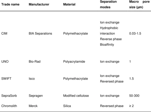
Table 1.1 - Overview of commercially available monoliths for applications in bioseparation.29
Trade name Manufacturer Material Separation modes
Macro pore size (µm)
CIM BIA Separations Polymethacrylate
Ion exchange Hydrophobic interaction Reverse phase Bioaffinity
0.03-1.5
UNO Bio-Rad Polyacrylamide Ion exchange 1
SWIFT Isco Polymethacrylate Ion exchange
Reversed phase 1.5
SepraSorb Sepragen Modified cellulose Ion exchange 50-300
Chromolith Merck Silica Reversed phase ≥ 2
Table 1.2 - Morphological features of different types of porous structure.33
Micropore Mesopore Macropore
Pore Diameter (nm) < 2 2-50 > 50
Porosity (%) ≤ 25 25-65 ≥ 65
Surface Area (m2cm-3) ≥ 1000 1000-350 ≤ 350
1.2.1. SYNTHETIC POLYMER MONOLITHS
Polymer monoliths produced by organic synthesis were first used in chromatography columns in
monovinyl monomer in the presence of a crosslinker, radical initiator and porogen (responsible for pore formation) (Fig. 1.2 A). Inspired by this straightforward strategy of monolith production, different monomers such as acrylamide (AAm), methacrylate and styrene were then employed to create rigid monoliths with desired morphological properties and dimensions.14,39,40,41,42,43,44
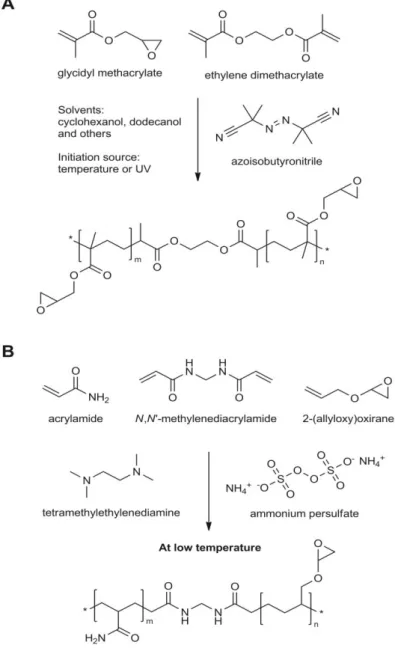
Figure 1.2 - Preparation of GMA-EDMA monoliths (A) and of AA-AGE cryogels (B) and their chemical structures.
In particular, glycidyl methacrylate (GMA) and ethylene glycol dimethacrylate (EDMA) have become the most commonly employed monomers for the preparation of synthesized monoliths.31,45 The great advantages of using these monomers is that GMA, which carries the very reactive epoxy group, facilitates further functionalization for target molecule capture; while EDMA, as an excellent crosslinker, confers mechanical stability to the final monolith. As an example, Hahn et al.46 developed an affinity poly(GMA-co-EDMA) monolith using a simple
reaction with the epoxy groups on the GMA chains incorporated into the matrix of monolith. Spacer arms can be introduced between the ligand and the support to promote the accessibility of the ligand functional groups to interact with the target biomolecule. As an example, reactive macroporous monoliths of poly(GMA-co-EDMA) were prepared by in situ copolymerization of
GMA and EDMA in the presence of porogenic agents, followed by Protein A and L-histidine linkage to the monoliths either directly or through the use of a spacer arm. The IgG adsorption capacity of the monolith functionalized with Protein A was greatly increased with the introduction of the spacer.47 Poly(GMA-co-EDMA) monoliths have also been functionalized with Protein L and Protein G with promising results,48,49,50 sufficient to justify their commercialization.51,52
To evaluate the application of synthetic monoliths for antibody purification, several studies have been performed. Lokman et al.53 developed a novel porous monolithic system for effective IgG
purification from human plasma based on the preparation of porous monoliths through the bulk polymerization of (hydroxyethyl) methacrylate HEMA and Nmethacryloyl(L)histidine -methylester (MAH). An upper adsorption value (>96.5 mg g–1) was achieved from human plasma with an associated purity value of 95.3%. Moreover, the authors verified that IgG could be reversibly adsorbed using poly(HEMA-MAH) monolith. Another strategy from the same group54 involved the preparation of imprinted poly(hydroxyethyl methacrylate-N-methacryloyl-l-tyrosine methyl ester) particles using hepatitis B antibody as the surface template. These particles demonstrated spectacular binding specificity, adsorbing an amount of hepatitis B antibody 18.3 times greater than anti-hepatitis A antibody, and 2-fold greater than immunoglobulin E. The self-polymerization of poly(glycerol poly(glycidyl ether) (PGPGE) using methyl tert-butyl ether as a porogenic agent resulted in the formation of a particularly rigid monolith where the epoxy groups of the poly(glycerol polyglycidyl ether) served a dual purpose: firstly, to provide functional groups for the polymerization reaction, and secondly to allow direct binding of Protein A to the monolith surface. Capillary columns loaded with this monolith allowed
the isolation of IgG (5.3±0.9 μg) and presented a capacity of 0.44±0.08 mg mL-1 within a
capillary volume of 12 μL.50
abundance plasma proteins.59 Other approaches regarding IgM isolation have also been developed31,60,61 using synthetic polymer monoliths.
1.2.1.1. Hydrogels and cryogels
Cryogels and hydrogels are synthetic polymer monoliths which can be defined as supermacroporous gels.19 In cryogels, networks are formed by the cryogelation of monomers (e.g. GMA, allyl glycidyl ether (AGE)) at sub-zero temperatures using ammonium persulfate (APS) as an initiator and N,N,N’,N’-tetramethylene diamine (TEMED) as the catalyst (Fig. 1.2 B). Hydrogels are formed by the polymerization of acrylamide (AAm), N’,N’ -methylenebisacrylamide (MBA) and AGE in an aqueous buffer which works as a porogen, just as in the formation of acrylamide gels for gel electrophoresis assays.26 The use of GMA and AGE allows a direct introduction of epoxy groups enabling further functionalization with ligands or other synthetic and natural species.
The macroporous network of hydrogels and cryogels makes them very attractive for cell and antibody separation,61 due to their higher porosity (up to 90%) and larger pore size (0.1
–200
μm).26,62,63,64 Unlike methacrylate or silica monoliths, cryogels and hydrogels have poor mechanical behaviour. Low material rigidity can be minimized through crosslinking procedures, physical blends or the addition of stiff polymers to the initial casting solution.30
Over the past decade, different polymeric cryogel systems such as AAm and MBA grafted with N,N-dimethylaminoethyl methacrylate (DMAEMA) and poly(methacrylic acid (MAA)-co-polyethylene glycoldiacrylate) embedded with polystyrene or poly(EDMA) nanoparticles have been prepared at sub-zero temperatures. Due to the large porous network, efficient separation of highly purification antibody from fermentation broth was achieved using affinity supermacroporous monolithic cryogels functionalized with Protein A.64,65,66
1.2.2. MONOLITHS BASED ON NATURALLY OCCURRING POLYMERS
Societal, environmental, and regulatory drivers are pressing industry to design engineered
products from “cradle to grave”.71 This has been a driving force for the use of natural and biodegradable polymers at an industrial level. The most widespread natural polymers are polysaccharides, such as cellulose, chitosan and agarose.71 The popularity of agarose beads as first-choice supports for traditional affinity chromatography stems from bead hydrophilicity and good chemical stability, even under extremes of pH.9 Thus, it is not surprising that agarose has been used for monolith preparation.72 Unfortunately, agarose based monolith supports exhibit poor mechanical properties, and at the time of writing they are only known as porous particles confined in a mold or as a macroporous gel.72,73
Chitosan (CHT) is also a natural polymer obtained by deacetylation of chitin originated from the xoskeleteon of crustaceans.74 Chitosan has been extensively investigated in diverse fields of work73,75 due to its nontoxic, antimicrobial, biocompatible, and biodegradable properties and sensitivity towards changes in pH.76 Due to its high molecular weight, chitosan yields viscous solutions can be utilized to produce porous gels and structures through methodologies such as freeze drying and supercritical fluid technology.20,75,77,78,79,80 Sun et al.81 prepared
chitosan-agarose cryogels in situ through cryopolymerization and linked 2-mercaptopyridine onto
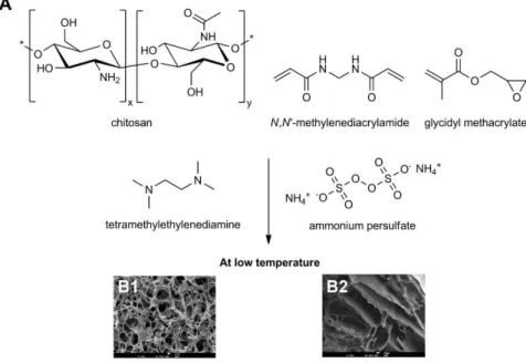
divinylsulfone-activated matrix, producing cryogels used to purify IgG. Cryogels presented interconnected pores of 10-100 μm size, a specific surface area of 350 m2 g-1 and a high adsorption and elution capacity for IgG of 71.4 mg g-1 and 90%, respectively. These supports proved to be stable and reusable for more 10 cycles without substantial loss in their performance. More recently, Barroso et al.82 prepared chitosan-based monoliths for IgG
purification by combining freezing and lyophilization methods. The authors were able to improve the mechanical properties of chitosan through blending with poly(vinyl alcohol )(PVA) and by cryopolymerizing with GMA at sub-zero temperatures (Fig. 1.3). The supports were functionalized with a Protein A biomimetic ligand, through plasma technology, a free solvent technique. This sustainable and faster approach allowed high binding capacities (150±10 mg IgG g-1 support), and 90±5% recovery of the bound protein with 98% purity directly from cell-culture extracts.
Cellulose has also been employed in chromatographic procedures using cellulose derivatives in the form of discs/membranes retaining the possibility for further functionalization with different type of molecules for protein separation, and evaluation of affinity interactions.83,84,85,86,87 Recently, Barroso et al.88 prepared cellulose membranes/discs using an alternative approach to
Figure 1.3 - (A) Schematic preparation of chitosan-based monoliths. (B1, B2) SEM micrographs of chitosan monoliths and chitosan cryopolymerized with glycidyl methacrylate (GMA) respectively,82 adapted with the permission of The Royal Society of Chemistry.
Table 1.3 - Monolithic materials for antibody purification.
Material Mode Ligand Target Surface area
(m2 g-1)
Flow rate
(mL min-1) Capacity
Recovery
(%) Purity Ref’s
GMA-EDMA
Affinity
Protein A IgG, IgM, IgA 89.1 0.05; 1.0 20 (IgG) mg g-1 99 High [47] [49]
Protein L IgG n.a. 1×10⁻⁵ n.a. n.a. High [45]
Protein G IgG n.a. 0.05; 2.5 20 mg g-1 n.a. High [49] [50]
L-histidine IgG 89.1 1.0 22.0 mg g-1 n.a. n.a. [47]
Anion Exchange
DEAE
MAbs n.a. 1.0 n.a. 95.0 High [51]
EDA n.a. 1.0 n.a. 91.4 High [51]
Ion Exchange MAA IgG 57.1 1.0-2.0 n.a. 98.8 Good [48]
PG-PGE Affinity Protein A IgG from rabbit serum n.a. n.a. 0.44 mg mL-1 n.a. n.a. [49]
HEMA-MAH Pseudo-affinity (MAH) IgG 145.8 1.0 96.5 mg g-1 n.a. 95.3% [53]
CIM-IDA IMAC Cu2+/Ni2+/Zn2+/Co2+ IgG/ Mab’s n.a. 2.0 n.a. 63/41/85/40 n.a. [55] IMAC Cu2+/Ni2+/Zn2 IgG and Mab’s n.a. 3.0 0.5 mg mL-1 82.4 n.a. [89]
CIM
Pseudo-affinity Peptide IgM/IgG/ Mab’s n.a. 1.0-10 n.a. 83/67/95 n.a. [56]
Ion Exchange QA/DEAE/EDA IgM/IgG n.a. 1.0-2.0 ≈ 20 mg g-1 n.a. n.a. [90]
IgM n.a. n.a. 16-36 mg g-1 n.a. n.a. [57]
DMAA-AGE cryogel
IMAC IDA-Cu2+ Fv antibody fragments from
E. Coli cell culture n.a. n.a. n.a. 84-96 High
[91]
Affinity Protein A Cells bearing IgG antibodies 20.2 0.5 1.6×10adsorbent 6 cells mL-1 60-70 High [68]
AAm-AGE cryogel Affinity Conc A IgG n.a. 1.0 25.6 mg g
-1 94 85% [67]
Afinity Protein A IgG labeled inclusion bodies n.a. 0.5 n.a n.a. n.a. [92]
HEMA-cryogel
Affinity Protein A
IgM 20.2 0.5 42.7 mg g
-1 ≥ 90 - [68]
IMAC PGMA-IDA-Cu2+ n.a. 0.5-2.0 257 mg g-1 89.4 - [93]
Affinity PGMA-Cibracron Blue F3GA
IgG n.a. 0.5-2.0 342 mg g
-1 93.6 - [93]
Affinity Protein A n.a. 0.5-3.0 83.2 mg g-1 85 85% [69]
HEMA-MAH
cryogel Pseudo- affinity (MAH) IgG n.a. 0.5-3.0 97.3 mg g-1 80.7 94,6% [93]
Chitosan-agarose Affinity 2-mercaptopyridine IgG 350 1.0 71.4 mg g-1 90 High [81]