ABSTRACT
Apoptosis is an essential physiological process of elimination of destined cells during the development and differentiation or after damage from external stresses such as ionizing radiation or chemotherapeutic agents. Disruption of apoptosis is proved to cause various diseases including cancer. Among numer-ous molecules involved in diverse anti- or pro-apoptotic signaling pathways, NF-κB is one of the key factors controlling apoptotic responses. Its anti-apoptotic effect is thought to be mediated through not only transcriptional acti-vation of dependent genes but also by crosstalking with the JNK pathway. Oncogenic proteins such as Ret/PTC, Ras and BRAF can induce NF-κB activa-tion making it an important change in thyroid cancer. A number of specific or non-specific NF-κB inhibitors have been tried to take over the cascade in in vitro and in vivo experiments. These agents can induce massive apoptosis especially in combination with radio- or chemotherapy. Current results suggest that the inhibition of the NF-κB may be a promising strategy for advanced thy-roid cancer treatment but further investigations are warranted to develop spe-cific and clinically effective NF-κB inhibitors in future. (Arq Bras Endocrinol Metab 2007;51/5:843-851)
Keywords:Thyroid cancer; Apoptosis; NF-κB; Molecular target therapy
RESUMO
Fator Nuclear-κB na Carcinogênese de Tireóide e sua Progressão: um Novo Alvo Terapêutico para Câncer da Tireóide Avançado.
A apoptose é um processo fisiológico essencial destinado a eliminar células durante o desenvolvimento e diferenciação ou após danos decorrentes de estresses externos com a radiação ionizante ou agentes quimioterápicos. Distúrbios na apoptose têm sido demonstrados como causadores de várias doenças, incluindo câncer. Entre as inúmeras moléculas envolvidas nas várias vias de sinalização anti- ou pró-apoptoticas, NF-κB é um dos fatores-chave que controlam as respostas apoptóticas. Acredita-se que seu efeito anti-apoptótico seja mediado não apenas pela ativação transcricional de genes dependentes mas também por crosstalking com a via JNK. Proteínas oncogênicas como Ret/PTC, Ras e BRAF podem induzir ativação de NF-κB promovendo importante transformação no câncer da tireóide. Uma série de inibidores específicos e não-específicos do NF-κB tem sido usada em experimentos in vitro e in vivo procurando inibir a cascata. Esses agentes podem induzir apoptose maciça especialmente em combinação com radio ou quimioterapia. Resultados atuais sugerem que a inibição de NF-κB pode ser uma estratégia promissora no tratamento do câncer da tireóide avançado, mas novas investigações são necessárias para desenvolver inibidores específicos e clinicamente efetivos do NF-κB. (Arq Bras Endocrinol Metab 2007; 51/5843-851)
Descritores:Câncer da tireóide; Apoptose; NF-κB; Terapia de alvo molecular
perspectiva
HIROYUKINAMBA VLADIMIRSAENKO SHUNICHIYAMASHITA
Department of Molecular Medicine (HM & SY), and Department of International Health and Radiation Research (VS & SY), Nagasaki University Graduate School of Biomedical Sciences 1-12-4, Sakamoto, Nagasaki 852-8523, Japan.
A
POPTOSIS, OR PROGRAMMED CELL DEATH, is an active process of cell destruction which requires the activation of genetic and signaling suicidal pro-grams manifesting in membrane blebbing, DNA frag-mentation, shrinking and condensation of the cell and its organelles (1,2). This process is obligatory in vari-ous physiological and pathological conditions in mul-ticellular organisms. An appropriate balance between apoptosis and cell proliferation is essential in the devel-opment and differentiation in the body. It also con-tributes to normal tissue functioning by the elimina-tion of “worn” cells or those damaged by various external stresses such as ionizing radiation, UV, chem-ical substances and some cytokines. Considering an indispensable importance of the process in the body, one can easily imagine that the dysfunction or defect of the apoptotic machinery may result in disease.This review describes how deregulation of apoptosis is involved in thyroid carcinogenesis. Partic-ular attention is given to the nuclear factor kappa B (NF-κB), as a key regulatory molecule in anti-apop-totic pathways and a new possible therapeutic molecu-lar target for advanced thyroid cancer.
APOPTOSIS IN THYROID CANCER CELLS
Proteins encoded by oncogenes or tumor suppressor genes are an integral part of carcinogenesis and tumor progression. Among them, many molecules, e.g. Bcl-2 and p53, play an important role in the regulation of apoptosis. It has long been speculated that carcino-genesis starts when inadequate apoptosis occurs due to dysregulation of pro-apoptotic or anti-apoptotic intra-cellular signaling (3). The acquired resistance to an insult in cancer cells would contribute to escaping from apoptotic process, which could be switched on by cytokines released by immunocompetent cells and malnutrient or anoxic condition arising during the tumor growth. Furthermore, in the cancer treatment setting, the aberrant apoptotic signaling might result in the resistance to chemo- or radiation therapy. This knowledge naturally brought up the idea that the restoration of functional apoptosis in cancer cells could be a useful therapeutic means, which can enhance the efficacy of treatment.
Advanced thyroid cancers, such as undifferenti-ated and anaplastic thyroid carcinomas, are clinically characterized by the rapid growth, concomitant inflammation and resistance to conventional therapy. Consistent with these clinical observations, anaplastic thyroid cancer cells have been found to be extremely
resistant to apoptosis after exposure to ionizing radia-tion in vitrocompared with normal thyroid cells (4). Furthermore, thyroid cancer cells also show unusual responses to TNF family proteins TNF-α, FasL and TRAIL, which were originally identified as proteins that kill tumor cells. Of them, only TRAIL can induce cell death whereas TNF-αand FasL fail to do so. Since these cytokines are usually produced by macrophages or lymphocytes, the phenomenon suggested that thy-roid cancer cells might not be efficiently eliminated through anti-tumor immune response (5-8).
NF-κB AS A KEY-REGULATORY MOLECULE OF ANTI-APOPTOTIC SIGNALING
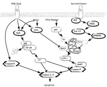
Cell fate is determined by numerous molecules in diverse intracellular pro-apoptotic and anti-apoptotic pathways (figure 1). Among those molecules, the tran-scription factor NF-κB is well recognized as a central activator of the anti-apoptotic cascades in response to external stimuli or intrinsic immune reactions (9).
There are five known members of the NF-κB family: NF-κB1 (p50/p105), NF-κB2 (p52/p100), c-Rel, RelB, and RelA (p65), each distinguished by its Rel homology domain, which mediates DNA binding and dimerization. Usually ubiquitous NF-κB dimers interact with inhibitory factors known as the IκB pro-teins (IκBα, IκBβand IκBε), binding to which retains the complex in the cytoplasm. Phosphorylation of IκB proteins by IκB kinases (IKKαand IKKβand the asso-ciated modulatory protein IKKγor NEMO) results in
their rapid ubiquitination and proteolysis by the 26S proteasome (10-12). The degradation of IκB proteins liberates the NF-κB complex, which is subsequently translocated from the cytoplasm to the nucleus. Then, NF-κB activates the expression of a wide spectrum of genes involved in inflammation, immune response, cell proliferation and resistance to apoptosis (13). The anti-apoptotic genes transcriptionally activated by NF-κB include the cellular inhibitors of apoptosis (c-IAP1, c-IAP2 and XIAP), TNF receptor-associated factors (TRAF1and TRAF2), Gadd45β, the Bcl-2homologue A1/Bfl-1, IEX-IL and Bcl-xL (14,15). There are two types of NF-κB activation pathways. The classical path-way, which is mainly mediated by the p50/p65 and p52/c-Rel dimers, is responsible for inhibition of apop-tosis under most conditions including ionizing radia-tion, UV, cytotoxic agents and cytokines (16). Another is the alternative pathway, which depends on the selec-tive activation of the p52/RelB dimer after processing of the NF-κB2/p100 precursor protein triggered by certain members of the TNF cytokine family (17).
CROSSTALK BETWEEN THE NF-κB AND JNK PATHWAYS
JNK, a member of the MAP kinase superfamily (18), is involved in stress-induced apoptosis via the mito-chondrial pathway (19). In contrast, some studies have suggested that activation of JNK contributes to cell survival (18). A number of studies helped to dissect this problem by showing that the duration of JNK activation affects its role in apoptosis. It was found that prolonged, but not transient, JNK activation promotes cell death induced by external stimuli such as TNF-α (20,21), although sustained JNK activation alone is insufficient to induce cell killing (21). These data sug-gested that JNK may cooperate with other pro- or anti-apoptotic signals to evoke apoptosis. Several groups have demonstrated a crosstalk between the NF-κB and JNK pathways seen as an inhibitory effect of NF-κB on TNF-α-induced apoptosis due to the suppression of JNK activity (20-22). The findings implied that the balance between JNK and NF-κB activities is crucial to determine the cell fate, i.e. sur-vival or death, in response to external stimuli (23,24). Indeed, the prolonged JNK activation occurs in the cells that cannot activate NF-κB by external stimuli. Suppression of NF-κB by p65 ablation, forced expres-sion of IKKβor super-repressor mutant form of IκBα,
mut-IκBα, leads to persistent JNK signaling following TNF-αtreatment.
Two NF-κB-dependent factors that mediate JNK activity have been identified so far (20,21). One is GADD45β, which activates a selective positive JNK reg-ulator MKK7/JNKK2. Suppression of the latter in fibroblasts abolishes JNK induction by TNF-α (18,19, 24) and blocking of MKK7 alone seems to be sufficient to account for the specific and near-complete inhibition of the JNK cascade. Another molecule, XIAP, which par-ticipates in the negative modulation of JNK activity, has been identified in the study of murine embryonic fibrob-lasts deficient in either IKKβor p65 (25).
Overall, data indicate that the mechanism by which NF-κB affects cell survival and death is influ-enced by the NF-κB-dependent factors’ ability to con-trol JNK activation. Particularly, the pro-survival effect of NF-κB depends on the prevention of prolonged JNK activation (26,27).
JNK ACTIVITY IN THYROID CELLS
JNK activity was examined in thyroid cells after various external stimuli including ionizing radiation, UV, ceramide, and growth factors (28-30). The result showed that each of the stimuli led to different dura-tion of JNK activadura-tion. We have demonstrated that sustained JNK activation after exposure to UV or ceramide correlated with cell death, whereas its tran-sient activation after either X-ray irradiation or treat-ment by growth factors did not (29,30). Moreover, transient activation of JNK in thyroid cells may be mediated by the activation of protein kinase C (PKC)-MKK7-JNK cascade after radiation exposure (31). Despite a high basal level ofGADD45mRNA in thy-roid cancer cells harboring TP53 gene mutation, its level is not augmented following irradiation (4). Thus, it is possible that the aberrant GADD45 response might result only in the transient JNK activation con-tributing to cell survival. Since ionizing radiation is the only known etiological factor of papillary thyroid can-cer, it could be speculated that the resultant transient JNK activation after radiation exposure results in an incomplete elimination of damaged cells that might eventually manifest in thyroid carcinogenesis.
NF-κB AND CANCER
aberrant regulation of NF-κB signaling in a variety of human cancers including leukemia, lymphoma, head and neck squamous carcinoma, and ovarian, prostate, colon and breast cancers (33-35). The genes encoding NF-κB family members p52/p100, c-Rel, p65 and IκB-like pro-tein Bcl-3 are frequently rearranged or amplified in human lymphoma and leukemia, and inactivating muta-tions of IκBα gene can cause Hodgkin’s lymphoma (16,36,37). Moreover, most oncogene products, includ-ing the Tax protein of human T-cell leukemia virus type 1 (HTLV-1), Bcr-Abl, Her-2/Neu and oncogenic vari-ants of Ras, can induce overexpression of p65 in cancer cells (38) and NF-κB activation (36,37,39,40). The acti-vation of NF-κB can contribute to the oncogenesis in several ways: by driving cell proliferation (41) perhaps through increased transcription of cyclin D1, which mediates G1/S progression of the cell cycle (13), by enhancing cell survival (42), and by promoting angio-genesis and metastasis (43). The in vivostudies discov-ered a link of NF-κB activation with inflammation-asso-ciated tumor promotion, progression and metastasis in mouse models (44,45). For example, the role of the IKKβ-dependent NF-κB activation in cancer initiation was demonstrated in an experiment in which inactivation of IKKβin enterocytes resulted in a dramatic decrease in the number of tumor foci due to increased apoptosis (45). In another study, the inflammatory process trig-gered chronic activation of NF-κB in hepatocytes and resulted in cholestatic hepatitis followed by the develop-ment of hepatocellular carcinoma in the Mdr2-deficient mice. Suppression of the chronic NF-κB activation at later stage of carcinogenesis resulted in the apoptosis of inflamed hepatocytes and a failure to progress to malig-nancy (46). The critical role of NF-κB activation in the inflammation-driven tumor progression was also shown in a colon and mammary cancer xenograft model (44). Cancer cells were inoculated into syngeneic immuno-competent mice to form metastases in the lungs. Once metastases were established, mice were given a sublethal dose of LPS to elicit systemic inflammation that pro-moted tumor growth. Inhibition of NF-κB in cancer cells converted LPS-induced tumor growth to tumor regression without any effect on the metastatic ability to the lung.
NF-κB AND MAPK PATHWAY IN PAPILLARY THYROID CANCER
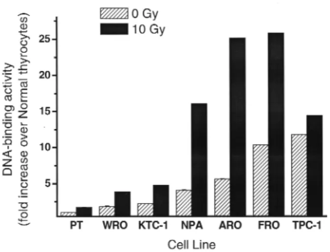
Several studies have suggested that the increased NF-κB activity is associated with thyroid carcinogenesis and tumor progression. A significant increase of p65 mRNA
and protein expression, compared to normal thyroid cells, was found in thyroid cancer cell lines (47). Consis-tent with the elevated expression level, NF-κB DNA binding and reporter assays showed the increased tran-scriptional activities in the cultures of thyroid cancer cells (48,49) (figure 2). The activation of NF-κB was also observed in the papillary, follicular, and anaplastic cancer tissue specimens by immunohistochemical staining using an anti-p65 antibody (49,50).
The products of RET/PTCs, activated mutant RASand BRAFV600Egenes involved in pathogenesis of
papillary thyroid carcinoma can potently activate the MAPK pathway (51-53). In its turn, activated MAPK has been shown to induce activation of NF-κB signaling and associated NF-κB-mediated transcriptional activity (54,55). In addition, we have observed that degradation of IκBαtakes place shortly after ectopic accumulation of the BRAFV600E protein, resulting in the activation of NF-κB signaling via MEK-ERK independent pathway (56). In line with these findings, previous study demon-strated that oncogenic Ret-induced NF-κB activity depends on IKK-mediated IκBα degradation and requires functional Ras, Raf and MEKK1 in a medullary thyroid cancer cell line TT, and that NF-κB activation is not accomplished by MEK/ERK activation (57).
NF-κB AND PI3K PATHWAYS IN THYROID CANCER
PI3-kinase pathway is an important signaling cascade that regulates cell survival and apoptosis. Cell stimula-tion with various growth factors or cytokines recruits a
lipid second messenger phosphatidylinositol 3,4,5-triphosphate [PI(3,4,5)P3] from membrane by the action of PI3-kinase, which then activates a serine/threonine kinase Akt/PBK. Akt/PBK relays signal to a number of molecules involved in cell prolif-eration, resistance to apoptosis and angiogenesis. The sequential activation of PI3K-Akt/PBK is negatively regulated by the PTEN phophatase.
Akt/PBK and PTEN have been implicated in fol-licular thyroid cell carcinogenesis. Germline mutations in the PTENgene have been associated with Cowden’s drome, an autosomal dominant multiple hamartoma syn-drome in which more than 50% of patients develop fol-licular thyroid cancer (58). The abnormal activation of Akt/PBK is often found in thyroid cancer harboring RAS mutation or RET/PTC rearrangements (59-61). The pro-survival ability of Akt/PBK is likely to be medi-ated through increased NF-κB activity (62). It has been shown that the inhibition of endogenous Akt by overex-pression of PTEN resulted in the decrease of NF-κB tran-scriptional activity and sensitization of cells to TNF-induced apoptosis (63). Interestingly, PTEN itself is downregulated by p65 but not p50 subunit, forming a negative regulatory loop between PTEN and NF-κB. Therefore, the findings suggest that depressed PTEN or activated Akt/PBK in cancer cells might contribute to carcinogenesis by activation of the NF-κB pathway.
NF-κB ACTIVATION BY PAX8-PPARγAND CYTOKINES IN THYROID CANCER
PAX8-PPARγ fusion gene was initially identified in follicular thyroid tumors (64). When fused to PAX8, PPARγ not only loses the ability to stimulate thiazo-lidinedione-induced transcription, but also inhibits intrinsic PPARγ transcriptional activity. In an in vivo model study, in the progeny from the cross of PPARγ+/-and TRβPV/+mice, the reduced PPARγ pro-tein led to the activation of NF-κB, resulting in the ele-vation of cyclin D1 level and decreased apoptosis (65). Similar to oncogenes, some cytokines have been associated with thyroid carcinogenesis through NF-κB activation. For example, overexpression of TGF-β is often observed in thyroid cancer cells. The study sug-gested that TGF-βinduces apoptosis in normal thyroid cells via p27 reduction, but NF-κB activation in neo-plastic thyroid cells blocks the apoptotic effect (66).
In case of TNF-α, which cannot induce cell death in thyroid cancer cells as mentioned above, resis-tance to apoptosis is thought to be due in part to NF-κB activation (67).
NF-κB AND THYROID CANCER PROGRESSION
The close interrelation between oncogenes and NF-κB activity raises the possibility that NF-κB activity not only promotes cell survival in early stage of thyroid carcinogenesis, but also may facilitate tumor progres-sion and metastasis. Supportive to the idea, NF-κB inhibition by overexpression of IκBαin a lung cancer cell line was found to decrease the frequency of metas-tases in model experiments (68).
The activated NF-κB transcriptionally controls Cox-2, ICAM-1 and matrix metalloproteinase 9 (MMP-9) genes, whose products are known to corre-late with metastasis, poor prognosis and reduced dis-ease-free interval (69). Accordingly, we recently found that forced expression of BRAFV600Eby an adenovirus
vector in wild-type BRAF thyroid cancer cells could elevate MMP-1 and MMP-9 levels in parallel with NF-κB activation, resulting in the increased cancer cells migration through extracellular matrix that could be reverted by NF-κB inhibition (56).
NF-κB AS A THERAPEUTIC TARGET IN CANCER
A number of studies have been conducted to examine whether suppression of NF-κB activity promotes apop-tosis in cancer cells. A super-suppressor mutant form of IκBαor a dominant-negative form of IKK can block the NF-κB pathway and enhance cell killing (55,70). The specific antisense oligonucleotides against NF-κB p65 gene greatly reduced the ability of two undifferentiated thyroid cancer cell lines to form colonies in soft agar and reduced their growth rate (47). RNA interference (siRNA) specific to p65 with or without concomitant administration of a chemotherapeutic agent, irinotecan, significantly impaired drug-induced NF-κB activation, enhanced apoptosis, and reduced colony formation in soft agar in HCT116 colon cancer cell line (71).
regard to ATO, which is clinically applied for treatment of promyelocytic leukemia and multiple myeloma (77-79), it has been shown that the anticancer effect is achieved not only because of NF-κB suppression but also through upregulation of JNK activity.
Further studies, however, have found that clin-ical use of NF-κB inhibitors in cancer therapy may not be as effective as expected. For example, it was shown that the inhibition of NF-κB alone can retard cancer cell growth, but not necessarily leads to massive cell death in most solid cancers because of the coexistence of other, sometimes aberrant, NF-κB-independent anti- or pro-apoptotic pathways (40,80).
Yet, even though an NF-κB inhibitor per se may have a moderate potential to induce apoptosis, it might play an apoptosis-permissive role in the cells treated with cytokines, chemotherapeutic drugs or by ionizing radia-tion, which induce NF-κB activation increasing the resis-tance to an insult (40). Indeed, inhibition of NF-κB by expression of the mutant IκBαor other NF-κB inhibitors has resulted in model experiments in a dramatic improve-ment of the apoptotic response to ionizing radiation or to drugs as compared with the control cells (14).
NF-κB CASCADE INHIBITION AS A NEW THERAPEUTIC APPROACH TO ADVANCED
THYROID CANCER
Advanced thyroid cancers are well known to be refracto-ry to conventional chemo- or radiation therapy and usu-ally result in a fulminant fatal outcome. Therefore, effec-tive therapeutic modalities are being continuously sought for to achieve better treatment results. Since the increased NF-κB activity at least partly reinforces the intrinsic radio- or chemo-resistance of thyroid cancer cells, its inhibition could be a potential novel therapeutic approach. Effects of inhibition of the NF-κB pathway were experimentally examined in one study in an anaplas-tic thyroid cancer cell line stably expressing mutant IκBα. The mutant IκBα could enhance sensitivity to drug-induced apoptosis, led to the loss of cells’ ability to form colonies in soft agar and to give rise tumors in nude mice (49). Of note, whereas parental anaplastic thyroid cancer cell line displayed a very low JNK expression, inhibition of NF-κB by the mutant IκBαrestored its levels. In line with these results, the beneficial effects of NF-κB inhibi-tion by mutant IκBαwere found in the in vivomodels of anaplastic thyroid carcinoma. The most pronounced inhibitory effect on the tumor growth after radiation therapy was observed in the animals with mutant IκBα -harboring tumors (figure 3).
Several NF-κB inhibitory reagents have been test-ed for the effectiveness in anaplasitc thyroid cancer cells. The cell-permeable peptide SN50, which acts as an inhibitor of NF-κB nuclear translocation, markedly pro-moted cell death in vitroin combination with radiation exposure (48). Bortezomib, a proteasome inhibitor, also induced apoptosis in anaplastic thyroid cancer cell lines accompanied by the decrease in IκB degradation and increase of p53 expression and JNK activity (50).
Recently, a number of low molecular weight NF-κB inhibitors have been designed as anticancer, immuno-suppressant and anti-inflammatory agents. Most of them target factors upstream IκB (81). One such compound, dehydroxymethylepoxy quinomicin (DHMEQ), a deriv-ative of the antibiotic epoxyquinomicin C (82), has been tested in anaplastic thyroid cancer cells (83). After DHMEQ treatment, we observed the activation of cas-pases followed by massive apoptosis of thyroid cancer cells both in vitroand in vivo (figure 4). The mechanism of induction of apoptosis by DHMEQ was proved to be the inhibition of NF-κB translocation to the nucleus, the decrease of expression level of several downstream anti-apoptotic proteins and the prolonged JNK activation.
CONCLUSION
Accumulated somatic mutations in oncogenes or tumor-suppressor genes in thyroid cancer cells cause resistance to apoptosis by unbalancing anti- and pro-apoptotic pathways. NF-κB is a key molecule to induce the dominance of the former thus contributing to tumor growth and progression. The sustained ele-vation of NF-κB activity is often found in thyroid
cer and it can be further augmented by chemo- or radi-ation-therapy, perhaps accruing the therapy-resistant phenotype of advanced cancers. Preliminary experimen-tal studies show that the inhibition of NF-κB or inter-ference with the pathway might be a promising treat-ment strategy for thyroid cancer, especially when com-bined with conventional chemo- and radiation therapy. Further extended large-scale experiments to discover specific NF-κB inhibitors and clinical studies to confirm their safety and effectiveness in the patients with advanced thyroid cancer are urgently needed.
ACKNOWLEDGEMENTS
Our studies were supported in part by Grants-in-Aid for Scientific Research (A) 15256004 and (B) 15390295 from the Ministry of Education, Culture, Sports, Science and Technology of Japan. We also would like to thank Professor Kazuo Umezawa (Keio University, Yokohama) for collaboration in the study of an NF-κB inhibitor, DHMEQ, effects on thyroid cancer cells.
REFERENCES
1. Kerr JF, Wyllie AH, Currie AR. Apoptosis: a basic biological phenomenon with wide-ranging implications in tissue kinet-ics. Br J Cancer 1972;26:239-57.
2. Wyllie AH, Morris RG, Smith AL, Dunlop D. Chromatin cleav-age in apoptosis: association with condensed chromatin mor-phology and dependence on macromolecular synthesis. J Pathol 1984;142:67-77.
3. Zhivotovsky B. More than one road to kill tumor cells — why are they not always successful? Cell Cycle 2003;2(1):31-3. 4. Namba H, Hara T, Tukazaki T, Migita K, Ishikawa N, Ito K, et
al. Radiation-induced G1 arrest is selectively mediated by the p53-WAF1/Cip1 pathway in human thyroid cells. Cancer Res 1995;55:2075-80.
5. Mitsiades CS, Poulaki V, Mitsiades N. The role of apoptosis-inducing receptors of the tumor necrosis factor family in thy-roid cancer. J Endocrinol 2003;178:205-16.
6. Basolo F, Pollina L, Fontanini G, Fiore L, Pacini F, Baldanzi A. Apoptosis and proliferation in thyroid carcinoma: correlation with bcl-2 and p53 protein expression. Br J Cancer 1997;75:537-41.
7. Mitsiades N, Poulaki V, Tseleni-Balafouta S, Chrousos GP, Koutras DA. Fas ligand expression in thyroid follicular cells from patients with thionamide-treated Graves’ disease. Thy-roid 2000;10:527-32.
8. Arscott PL, Stokes T, Myc A, Giordano TJ, Thompson NW, Baker JR, Jr. Fas (CD95) expression is up-regulated on papil-lary thyroid carcinoma. J Clin Endocrinol Metab 1999;84:4246-52.
9. Barkett M, Gilmore TD. Control of apoptosis by Rel/NF-κB transcription factors. Oncogene 1999;18:6910-24.
10. Ghosh S, May MJ, Kopp EB. NF-κB and Rel proteins: evolu-tionarily conserved mediators of immune responses. Annu Rev Immunol 1998;16:225-60.
11. Zandi E, Karin M. Bridging the gap: composition, regulation, and physiological function of the IκB kinase complex. Mol Cell Biol 1999;19:4547-51.
12. Karin M, Delhase M. The IκB kinase (IKK) and NF-κB: key ele-ments of proinflammatory signalling. Semin Immunol 2000;12:85-98.
13. Baldwin AS, Jr. Series introduction: the transcription factor NF-κB and human disease. J Clin invest 2001;107:3-6. 14. Wang CY, Mayo MW, Korneluk RG, Goeddel DV, Baldwin AS,
Jr. NF-κB antiapoptosis: induction of TRAF1 and TRAF2 and c-IAP1 and c-IAP2 to suppress caspase-8 activation. Science 1998;281:1680-3.
15. Wu MX, Ao Z, Prasad KV, Wu R, Schlossman SF. IEX-1L, an apoptosis inhibitor involved in NF-κB-mediated cell survival.
Science 1998;281:998-1001.
16. Karin M, Lin A. NF-κB at the crossroads of life and death. Nat Immunol 2002;3:221-7.
17. Senftleben U, Cao Y, Xiao G, Greten FR, Krahn G, Bonizzi G, et al. Activation by IKKαof a second, evolutionary conserved, NF-κB signaling pathway. Science 2001;293:1495-9. 18. Davis RJ. Signal transduction by the JNK group of MAP
kinases. Cell 2000;103:239-52.
19. Tournier C, Hess P, Yang DD, Xu J, Turner TK, Nimnual A, et al. Requirement of JNK for stress-induced activation of the cytochrome c-mediated death pathway. Science 2000;288:870-4.
20. De Smaele E, Zazzeroni F, Papa S, Nguyen DU, Jin R, Jones J, et al. Induction of gadd45βby NF-κB downregulates pro-apoptotic JNK signalling. Nature 2001;414:308-13. 21. Tang G, Minemoto Y, Dibling B, Purcell NH, Li Z, Karin M, et
al. Inhibition of JNK activation through NF-κB target genes.
Nature 2001;414:313-7.
22. Javelaud D, Besancon F. NF-κB activation results in rapid inactivation of JNK in TNFα-treated Ewing sarcoma cells: a mechanism for the anti-apoptotic effect of NF-κB. Oncogene 2001;20:4365-72.
23. Zhang Y, Chen F. Reactive oxygen species (ROS), trouble-makers between nuclear factor-κB (NF-κB) and c-Jun NH(2)-terminal kinase (JNK). Cancer Res 2004;64:1902-5. 24. Papa S, Zazzeroni F, Pham CG, Bubici C, Franzoso G. Linking
JNK signaling to NF-κB: a key to survival. J Cell Sci 2004;117:5197-208.
25. Kaur S, Wang F, Venkatraman M, Arsura M. X-linked inhibitor of apoptosis (XIAP) inhibits c-Jun N-terminal kinase 1 (JNK1) acti-vation by transforming growth factor beta 1 (TGF-β1) through ubiquitin-mediated proteosomal degradation of the TGF-beta1-activated kinase 1 (TAK1). J Biol Chem 2005;280:38599-608.
26. Sakon S, Xue X, Takekawa M, Sasazuki T, Okazaki T, Kojima Y, et al. NF-κB inhibits TNF-induced accumulation of ROS that mediate prolonged MAPK activation and necrotic cell death.
EMBO J 2003;22:3898-909.
27. Luo JL, Kamata H, Karin M. IKK/NF-κB signaling: balancing life and death — a new approach to cancer therapy. J Clin Invest 2005;115:2625-32.
28. Shklyaev SS, Namba H, Sautin Y, Mitsutake N, Nagayama Y, Ishikawa N, et al. Involvement of wild-type p53 in radiation-induced c-Jun N-terminal kinase activation in human thyroid cells. Anticancer Res 2001;21:2569-75.
29. Shklyaev SS, Namba H, Mitsutake N, Alipov G, Nagayama Y, Maeda S, et al. Transient activation of c-Jun NH2-terminal kinase by growth factors influences survival but not apopto-sis of human thyrocytes. Thyroid 2001;11:629-36.
30. Sautin Y, Takamura N, Shklyaev S, Nagayama Y, Ohtsuru A, Namba H, et al. Ceramide-induced apoptosis of human thy-roid cancer cells resistant to apoptosis by irradiation. Thy-roid 2000;10:733-40.
31. Mitsutake N, Namba H, Shklyaev SS, Tsukazaki T, Ohtsuru A, Ohba M, et al. PKC delta mediates ionizing radiation-induced activation of c-Jun NH(2)-terminal kinase through MKK7 in human thyroid cells. Oncogene 2001;20:989-96.
32. Gilmore TD. Multiple mutations contribute to the oncogenicity of the retroviral oncoprotein v-Rel. Oncogene 1999;18:6925-37. 33. Rayet B, Gelinas C. Aberrant rel/nfkb genes and activity in
human cancer. Oncogene 1999;18:6938-47.
34. Morin CI, Huot J. Recent advances in stress signaling in can-cer. Cancer Res 2004;64:1893-8.
35. Courtois G, Gilmore TD. Mutations in the NF-κB signaling pathway: implications for human disease. Oncogene 2006;25:6831-43.
36. Kucharczak J, Simmons MJ, Fan Y, Gelinas C. To be, or not to be: NF-κB is the answer — role of Rel/NF-κB in the regula-tion of apoptosis. Oncogene 2003;22:8961-82.
37. Orlowski RZ, Baldwin AS, Jr. NF-κB as a therapeutic target in cancer. Trends Mol Med 2002;8:385-9.
38. Romieu-Mourez R, Kim DW, Shin SM, Demicco EG, Landes-man-Bollag E, Seldin DC, et al. Mouse mammary tumor virus c-rel transgenic mice develop mammary tumors. Mol Cell Biol 2003;23:5738-54.
39. Podolsky DK. Inflammatory bowel disease (1). N Engl J Med 1991;325:928-37.
40. Wang CY, Cusack JC, Jr., Liu R, Baldwin AS, Jr. Control of inducible chemoresistance: enhanced anti-tumor therapy through increased apoptosis by inhibition of NF-κB. Nature Med 1999;5:412-7.
41. Bargou RC, Emmerich F, Krappmann D, Bommert K, Mapara MY, Arnold W, et al. Constitutive nuclear factor-κB-RelA acti-vation is required for proliferation and survival of Hodgkin’s disease tumor cells. J Clin Invest 1997;100:2961-9. 42. Duffey DC, Chen Z, Dong G, Ondrey FG, Wolf JS, Brown K, et
al. Expression of a dominant-negative mutant inhibitor-κBαof nuclear factor-κB in human head and neck squamous cell car-cinoma inhibits survival, proinflammatory cytokine expres-sion, and tumor growth in vivo. Cancer Res 1999;59:3468-74. 43. Huang S, Pettaway CA, Uehara H, Bucana CD, Fidler IJ. Block-ade of NF-κB activity in human prostate cancer cells is asso-ciated with suppression of angiogenesis, invasion, and metastasis. Oncogene 2001;20:4188-97.
44. Luo JL, Maeda S, Hsu LC, Yagita H, Karin M. Inhibition of
NF-κB in cancer cells converts inflammation-induced tumor growth mediated by TNFαto TRAIL-mediated tumor regres-sion. Cancer Cell 2004;6:297-305.
45. Greten FR, Eckmann L, Greten TF, Park JM, Li ZW, Egan LJ, et al. IKKβlinks inflammation and tumorigenesis in a mouse model of colitis-associated cancer. Cell 2004;118:285-96. 46. Pikarsky E, Porat RM, Stein I, Abramovitch R, Amit S, Kasem
S, et al. NF-κB functions as a tumour promoter in inflamma-tion-associated cancer. Nature 2004;431:461-6.
47. Visconti R, Cerutti J, Battista S, Fedele M, Trapasso F, Zeki K, et al. Expression of the neoplastic phenotype by human thy-roid carcinoma cell lines requires NFκB p65 protein expres-sion. Oncogene 1997;15:1987-94.
48. Starenki D, Namba H, Saenko V, Ohtsuru A, Yamashita S Inhi-bition of nuclear factor-κB cascade potentiates the effect of a combination treatment of anaplastic thyroid cancer cells. J Clin Endocrinol Metab 2004;89:410-8.
49. Pacifico F, Mauro C, Barone C, Crescenzi E, Mellone S, Monaco M, et al. Oncogenic and anti-apoptotic activity of NF-κB in human thyroid carcinomas. J Biol Chem 2004;279:54610-9.
50. Mitsiades CS, Kotoula V, Poulaki V, Sozopoulos E, Negri J, Charalambous E, et al. Epidermal growth factor receptor as a therapeutic target in human thyroid carcinoma: mutational and functional analysis. J Clin Endocrinol Metab 2006;91:3662-6.
51. Fusco A, Grieco M, Santoro M, Berlingieri MT, Pilotti S, Pierotti MA, et al. A new oncogene in human thyroid papillary carcinomas and their lymph-nodal metastases. Nature 1987;328:170-2.
52. Kimura ET, Nikiforova MN, Zhu Z, Knauf JA, Nikiforov YE, Fagin JA. High prevalence of BRAF mutations in thyroid can-cer: genetic evidence for constitutive activation of the RET/PTC-RAS-BRAF signaling pathway in papillary thyroid carcinoma. Cancer Res 2003;63:1454-7.
53. Namba H, Gutman RA, Matsuo K, Alvarez A, Fagin JA. H-ras protooncogene mutations in human thyroid neoplasms. J Clin Endocrinol Metab 1990;71:223-9.
54. Tuyt LM, Dokter WH, Birkenkamp K, Koopmans SB, Lummen C, Kruijer W, et al. Extracellular-regulated kinase 1/2, Jun N-terminal kinase, and c-Jun are involved in NF-κB-dependent IL-6 expression in human monocytes. J Immunol 1999;162:4893-902.
55. Bochner BS, Undem BJ, Lichtenstein LM. Immunological aspects of allergic asthma. Annu Rev Immunol 1994;12:295-335.
56. Palona I, Namba H, Mitsutake N, Starenki D, Podtcheko A, Sedliarou I, et al. BRAFV600Epromotes invasiveness of thyroid cancer cells through nuclear factor κB activation.
Endocrinology 2006;147:5699-707.
57. Ludwig L, Kessler H, Wagner M, Hoang-Vu C, Dralle H, Adler G, et al. Nuclear factor-κB is constitutively active in C-cell car-cinoma and required for RET-induced transformation. Can-cer Res 2001;61:4526-35.
58. Liaw D, Marsh DJ, Li J, Dahia PL, Wang SI, Zheng Z, et al. Germline mutations of the PTEN gene in Cowden disease, an inherited breast and thyroid cancer syndrome. Nat Genet 1997;16:64-7.
59. Vasko V, Saji M, Hardy E, Kruhlak M, Larin A, Savchenko V, et al. Akt activation and localisation correlate with tumour inva-sion and oncogene expresinva-sion in thyroid cancer. J Med Genet 2004;41:161-70.
60. Kim D, Chung J. Akt: versatile mediator of cell survival and beyond. J Biochem Mol Biol 2002;35:106-15.
61. Jung HS, Kim DW, Jo YS, Chung HK, Song JH, Park JS, et al. Regulation of protein kinase B tyrosine phosphorylation by thyroid-specific oncogenic RET/PTC kinases. Mol Endocrinol 2005;19:2748-59.
62. Van Antwerp DJ, Martin SJ, Kafri T, Green DR, Verma IM. Suppression of TNF-α-induced apoptosis by NF-κB. Science 1996;274:787-9.
63. Vasudevan S, Garneau N, Tu Khounh D, Peltz SW. p38 mito-gen-activated protein kinase/Hog1p regulates translation of the AU-rich-element-bearing MFA2 transcript. Mol Cell Biol 2005;25:9753-63.
64. Kroll TG, Sarraf P, Pecciarini L, Chen CJ, Mueller E, Spiegel-man BM, et al. PAX8-PPARγ1 fusion oncogene in human thy-roid carcinoma [corrected]. Science 2000;289:1357-60. 65. Kato Y, Ying H, Zhao L, Furuya F, Araki O, Willingham MC, et
al. PPARγinsufficiency promotes follicular thyroid carcino-genesis via activation of the nuclear factor-κB signaling path-way. Oncogene 2006;25:2736-47.
66. Bravo SB, Pampin S, Cameselle-Teijeiro J, Carneiro C, Dominguez F, Barreiro F, et al. TGF-β-induced apoptosis in human thyrocytes is mediated by p27kip1 reduction and is overridden in neoplastic thyrocytes by NF-κB activation.
67. Pang XP, Ross NS, Park M, Juillard GJ, Stanley TM, Hersh-man JM. Tumor necrosis factor-αactivates nuclear factor κB and induces manganous superoxide dismutase and phos-phodiesterase mRNA in human papillary thyroid carcinoma cells. J Biol Chem 1992;267:12826-30.
68. Jiang Y, Cui L, Yie TA, Rom WN, Cheng H, Tchou-Wong KM. Inhibition of anchorage-independent growth and lung metas-tasis of A549 lung carcinoma cells by IκBβ. Oncogene 2001;20:2254-63.
69. Baldwin AS. Control of oncogenesis and cancer therapy resis-tance by the transcription factor NF-κB. J Clin Invest 2001;107:241-6.
70. Feldmann M, Brennan FM, Maini RN. Role of cytokines in rheumatoid arthritis. Annu Rev Immunol 1996;14:397-440. 71. Guo J, Verma UN, Gaynor RB, Frenkel EP, Becerra CR. Enhanced chemosensitivity to irinotecan by RNA interfer-ence-mediated down-regulation of the nuclear factor-κB p65 subunit. Clin Cancer Res 2004;10:3333-41.
72. Yamamoto Y, Gaynor RB. Therapeutic potential of inhibition of the NF-κB pathway in the treatment of inflammation and cancer. J Clin Invest 2001;107:135-42.
73. Yin MJ, Yamamoto Y, Gaynor RB. The anti-inflammatory agents aspirin and salicylate inhibit the activity of IκB
kinase-β. Nature 1998;396:77-80.
74. Adams J, Palombella VJ, Sausville EA, Johnson J, Destree A, Lazarus DD, et al. Proteasome inhibitors: a novel class of potent and effective antitumor agents. Cancer Res 1999;59:2615-22.
75. Holmes-McNary M, Baldwin AS, Jr. Chemopreventive prop-erties of trans-resveratrol are associated with inhibition of activation of the IκB kinase. Cancer Res 2000;60:3477-83. 76. Monks NR, Biswas DK, Pardee AB. Blocking anti-apoptosis as
a strategy for cancer chemotherapy: NF-κB as a target. J Cell Biochem 2004;92:646-50.
77. Niu C, Yan H, Yu T, Sun HP, Liu JX, Li XS, et al. Studies on treatment of acute promyelocytic leukemia with arsenic tri-oxide: remission induction, follow-up, and molecular moni-toring in 11 newly diagnosed and 47 relapsed acute promye-locytic leukemia patients. Blood 1999;94:3315-24.
78. Rousselot P, Larghero J, Arnulf B, Poupon J, Royer B, Tibi A, et al. A clinical and pharmacological study of arsenic trioxide in advanced multiple myeloma patients. Leukemia 2004;18:1518-21.
79. Cavigelli M, Li WW, Lin A, Su B, Yoshioka K, Karin M. The tumor promoter arsenite stimulates AP-1 activity by inhibit-ing a JNK phosphatase. EMBO J 1996;15:6269-79. 80. Chen S, Fribley A, Wang CY. Potentiation of tumor necrosis
factor-mediated apoptosis of oral squamous cell carcinoma cells by adenovirus-mediated gene transfer of NF-κB inhibitor. J Dent Res 2002;81:98-102.
81. Erkel G, Anke T, Sterner O. Inhibition of NF-κB activation by panepoxydone. Biochem Biophys Res Commun 1996;226:214-21.
82. Matsumoto N, Ariga A, To-e S, Nakamura H, Agata N, Hirano S, et al. Synthesis of NF-κB activation inhibitors derived from epoxyquinomicin C. Bioorg Med Chem Lett 2000;10:865-9. 83. Starenki DV, Namba H, Saenko VA, Ohtsuru A, Maeda S, Umezawa K, et al. Induction of thyroid cancer cell apoptosis by a novel nuclear factor κB inhibitor, dehydroxymethyle-poxyquinomicin. Clin Cancer Res 2004;10:6821-9.
Address for correspondence:
Hiroyuki Namba