Egg envelopes and cuticle renewal in
Porcellio
embryos
and marsupial mancas
Polona Mrak1, Nada Žnidaršič1, Magda Tušek-Žnidarič1, Waltraud Klepal2, Daniela Gruber2, Jasna Štrus1
1 Department of Biology, Biotehnical faculty, University of Ljubljana, Večna pot 111, SI-1000 Ljubljana, Slovenia 2 Department of Ultrastructural Research, University of Vienna, Althanstrasse 14, A-1090 Vienna, Austria
Corresponding author:Polona Mrak (polona.mrak@bf.uni-lj.si)
Academic editor:S. Sfenthouraki | Received 22 November 2011 | Accepted 26 January 2012 | Published 20 March 2012
Citation: Mrak P, Žnidaršič N, Tušek-Žnidarič M, Klepal W, Gruber D, Štrus J (2012) Egg envelopes and cuticle renewal in Porcellio embryos and marsupial mancas. In: Štrus J, Taiti S, Sfenthourakis S (Eds) Advances in Terrestrial Isopod Biology. ZooKeys 176: 55–72. doi: 10.3897/zookeys.176.2418
Abstract
An important adaptation to land habitats in terrestrial isopod crustaceans is development of embryos in a luid-illed female brood pouch, marsupium. he study brings insight into the structure and protec-tive role of egg envelopes and cuticle renewal during ontogenetic development of Porcellio embryos and marsupial mancas. Egg envelopes cover embryos, the outer chorion until late-stage embryo and the in-ner vitelline membrane throughout the whole embryonic development. Egg envelopes of Porcellio have relatively simple ultrastuctural architecture compared to Drosophila egg envelopes. Exoskeletal cuticle is produced in late embryonic development by hypodermal cells of the embryo and is renewed in further development in relation to growth of developing embryos and mancas. Cuticle structure and renewal in prehatching late-stage embryos and marsupial mancas exhibit main features of cuticle in adults. Epicuticle is thin and homogenous. he characteristic arrangement of chitin-protein ibers and the dense distal layer in exocuticle are hardly discernible in prehatching embryo and distinct in marsupial mancas. Endocuticle consists of alternating electron dense and electron lucent sublayers and is perforated by pore canals in both stages. Diferences from adult cuticle are evident in cuticle thickness, ultrastructure and mineralization. Signs of cuticle renewal in prehatching embryo and marsupial mancas such as detachment of cuticle from hypodermis, partial disintegration of endocuticle and assembly of new cuticle are described.
Keywords
Chorion, vitelline membrane, cuticle, molting, ontogenetic development, terrestrial isopods www.zookeys.org
Copyright Polona Mrak et al. This is an open access article distributed under the terms of the Creative Commons Attribution License 3.0 (CC-BY), which permits unrestricted use, distribution, and reproduction in any medium, provided the original author and source are credited.
introduction
he unique feature of embryonic development in isopod and amphipod crustaceans (Peracarida) is its location in the brood pouch on the ventral side of female body (mar-supium). he marsupium has likely been of a great adaptive signiicance in the colo-nization of land by crustacean species as it allows embryonic development to occur in aqueous environment within the protected chamber (Hornung 2011, Warburg 2011). Two main types of marsupium are distinguished in Oniscidea, the amphibian or open type and the terrestrial or closed type marsupium (Hoese and Janssen 1989). In the for-mer, marsupium is partly open and water from the external environment can pass into the marsupium. In the closed type, marsupium is a watertight structure, provisioned only with luid from the mother. Marsupial luid is persistently osmotic and ionic regu-lated, probably via transporting epithelia of segmental cotyledons, which hang down from the overlying sternites (Surbida and Wright 2001). Depending on the level of maternal control of the marsupial environment, osmotic tolerance of the embryos and marsupial mancas is of adaptive importance. Protective envelopes between embryo/ manca and marsupial luid function against potential physiological stresses, including osmotic and ionic variation and desiccation in marsupial environment (Charmantier and Charmantier-Daures 2001, Surbida and Wright 2001). Ontogenetic development of terrestrial isopod crustacean Porcellio scaber,from released fertilized eggs to embryos and marsupial mancas, occurs in the terrestrial type marsupium. Intramarsupial de-velopment of P. scaber lasts approximately 35 days (Milatovič et al. 2010), and during development individuals in diferent stages are coated by diferent protective envelopes - egg envelopes (chorion and vitelline membrane) and cuticle. During growth of em-bryos and mancas egg envelopes are shed and cuticle is renewed.
Two egg envelopes, the outer chorion and the inner vitelline membrane, are pro-duced during oogenesis by the somatic follicle cells of the female reproductive system and cover the embryo until the transition to the late-stage embryo and hatching, re-spectively. Arthropods evolved egg envelopes of diferent morphologies and complexi-ties as a consequence of embryonic development in diferent environments. he data on egg envelopes (chorion and vitelline membrane) structure derive mainly from the studies on the fruit ly Drosophila melanogaster, as this model species ofers wide op-portunities for genetic studies (Margaritis et al. 1980, Jagadeeshan and Singh 2007), while there are no data on isopod chorion and vitelline membrane ultrastructure. In contrast to Porcellio scaber embryonic development in the female protected environ-ment, Drosophila embryonic development takes place independently of the female. he egg envelopes of Drosophila, especially the chorion, are structurally complex and their proteins show signs of evolving under selection by ecological factors. At the mor-phological level, diferences in egg envelopes surfaces between specialists and general-ists of Drosophila species were found, particularly in the surface ridges and surface porosity (Jagadeeshan and Singh 2007).
cuticle is a complex hierarchically structured extracellular matrix, consisting of chitin, proteins and lipids, hardened mostly by mineralization in crustaceans in contrast to insect cuticle which is only sclerotized. It comprises the distal epicuticle, the exocuticle in the middle and the proximal endocuticle. Several reports were published on cuticle structure in adult isopods, mostly in Porcellio scaber (Price and Holdich 1980, Štrus and Compere 1996, Ziegler 1997, Glötzner and Ziegler 2000, Štrus and Blejec 2001, Hild et al. 2008, Hild et al. 2009). he thin and non-calciied epicuticle is composed mainly of lipoproteins and consists of thinner 5-layered outer epicuticle and thicker inner epicuticle. he exocuticle, comprising sublayers of chitin–protein ibers arranged in characteristic pattern and the endocuticle, consisting of lamellar chitin–protein sub-layers, are calciied. In addition, the thin non-calciied membranous layer lies between the endocuticle and the epithelial cells. he ultrastructure and composition of the cuticle in isopod embryos and marsupial mancas have not been studied in detail, while cuticle structure is well described in Drosophila melanogaster embryos (Locke 2001, Payre 2004, Moussian et al. 2006, Moussian 2010). Fully formed cuticle in Drosophila
embryo is organized in distinct horizontal layers. he distal lipoprotein epicuticle is subdivided in the outer thin epicuticle (cuticulin layer) and the inner thick epicuticle and the proximal chitin-protein procuticle consists of several lamellae.
Cuticle renewal is related to growth in arthropods. In crustaceans molting frequent-ly recurs during adult life. Isopod crustaceans molt in two phases, separatefrequent-ly molting posterior and anterior parts of the body. Molt cycle begins with premolt stage, when remarkable morphological changes of the integument occur. he old cuticle separates from the underlying epithelium (apolysis). Epithelial cells secrete a new cuticle, starting with the epicuticle and followed by pre-ecdysal exocuticle. he old and the new cuticles are separated by an extracellular compartment, the ecdysal space, containing diferent material involved in cuticle renewal. At molting the old cuticle is shed and the new cu-ticle is further produced, forming post-ecdysal endocucu-ticle. Postmolt stage is marked by soft body surface with progressive hardening of the exoskeleton until the intermolt stage.
In this study we present new data on the ultrastructural architecture of egg en-velopes, including chorion and vitelline membrane, and on the ultrastructural char-acteristics of cuticle renewal in embryos and marsupial mancas of isopod crustaceans
Porcellio scaber and Porcellio dilatatus. Comparison of envelopes structure in terrestrial crustaceans and insects will bring new insights into the protective role of egg envelopes and cuticle renewal in developing embryos of these two terrestrial arthropod groups with diferent developmental strategies.
Methods
he embryos and marsupial mancas of diferent developmental stages are shown in Figures 1A, 2A, 3A, 4A and 5A-C in the Results. he term early-stage embryo is used for embryos with large amount of yolk mass in the central part and no visible limb buds. A mid-stage embryo has visible developing limb buds and two midgut glands primordia, which enclose yolk. After bending ventrally and shedding of cho-rion, embryos are termed late-stage embryos. Prior to hatching swelled embryo inside the vitelline membrane is described as a prehatching late-stage embryo. When late embryos hatch from the vitelline membrane they become marsupial mancas. he pro-gress of development of P. scaber marsupial mancas is characterized by the following morphologically discernible modiications: reduction of the midgut glands size due to yolk consumption, increase in exoskeleton pigmentation, enlargement of body size and pronounced locomotion (Wolf 2009, Milatovič et al. 2010). In previous studies stages of marsupial mancas were not precisely determined. For this reason and accord-ing to the morphological characteristics listed above, we determined three sequential developmental stages of marsupial mancas, early-stage, mid-stage and late-stage mar-supial mancas. he term early-stage manca is used for 1.5 – 1.6 mm long mancas with no or very little locomotion inside the marsupium, with scarce chromatophores on the body surface and with the midgut yolk extending into the pleon. Mid-stage mancas are 1.7 - 1.8 mm long, with darker pigmentation on the head region and tergites and with the midgut yolk only partly extending into the pleon. Late-stage mancas are 1.9 - 2.0 mm long, with pronounced locomotion of the whole body and pereopods. he yolk in the midgut extends only to the end of the pereon.
Embryos and mancas at diferent stages of development were isolated from the mar-supium and ixed in 2.5% glutaraldehyde in 0.1 M cacodylate bufer (pH 7.2). Prior to ixation, the egg envelopes of embryos were either carefully perforated with a thin needle or removed. After washing in cacodylate bufer, the samples were postixed in 1% os-mium tetroxide for 2 hours, washed again and dehydrated in a graded series of ethanol. Embryos and mancas of P. scaber for light microscopy (LM) and transmission
electron microscopy (TEM) were embedded in Agar 100 resin. Prior to embedding, mancas were perforated with a thin needle for better iniltration of resin. Semithin sections were made with a glass knife, stained with Azure II - Methylene Blue and im-aged by Zeiss AxioImager Z.1 light microscope, equipped with a HRC Axiocam cam-era. Ultrathin sections were made with a Reichert Ultracut S ultramicrotome (Leica), contrasted with 4% uranyl acetate for 10 minutes and 10% lead citrate for 5 minutes and inspected with a Philips CM100 transmission electron microscope, equipped by BioScan 792 camera (Gatan).
After dehydration in a graded series of ethanol and in acetone, P. dilatatus speci-mens for scanning electron microscopy (SEM) were transferred into hexamethyldisila-zane (HMDS) to perform chemical drying. Mounted specimens were coated with gold and observed with scanning electron microscopes (Philips XL20 and Philips XL30).
medium (Jung). Transversal sections (10 µm) were cut with a Leica CM1850 cryostat at –18°C and stained with Alizarin red S solution in 0.2 M Trihydroxylmethyl ami-nomethane (Tris) - HCl bufer (pH 9). Cuticle ofadult P. scaber was used as a positive
control. Sections were imaged by Zeiss AxioImager Z.1 light microscope, equipped with a HRC Axiocam camera.
Results
Ontogenetic development of Porcellio scaber in the marsupium, from released ferti-lized eggs to marsupial mancas, was recently described morphologically (Wolf 2009, Milatovič et al. 2010), but the issue of protective envelopes was not addressed speciical-ly. We present here the ultrastructural architecture of chorion, vitelline membrane and cuticle in embryos and marsupial mancas of isopod crustaceans P. scaber and P. dilatatus.
Ultrastructure of chorion and vitelline membrane
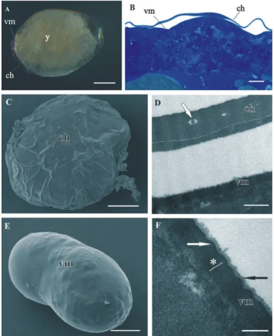
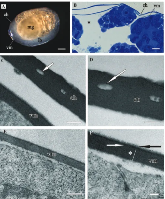
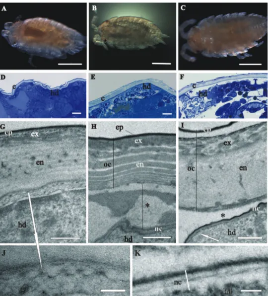
Both egg envelopes, distal chorion and proximal vitelline membrane, surround early-stage (Figs 1A, B, C, E) and mid-early-stage embryos (Figs 2A, B). Chorion has a similar ap-pearance in both stages. It is a one-layered envelope, separated from the embryo surface and is approximately 500 nm thick. Ultrastructurally chorion consists of an electron dense matrix with sparse electron lucent “lacunae” (Figs 1D, 2C, 2D).
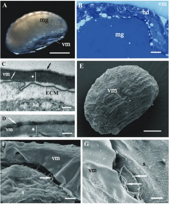
he vitelline membrane surrounds embryos throughout the whole developmental period (Figs 1A, 1 B, 1E, 2A, 2B, 3A, 3B, 3E, 4A). It maintains the same thickness of approximately 200 nm from early-stage till late-stage embryo. It is closely apposed to the embryo surface in early-stage embryos (Figs 1B, D, F), while it is slightly detached from the embryo surfaces of mid-stage embryos (Figs 2B, E, F) and late-stage embryos (Figs 3B, C, F, G). Above the embryo limb buds a wider space appears between embryo surface and vitelline membrane due to intense cell rearrangement during limb buds formation (Fig. 2B). he vitelline membrane consists of a thick proximal homogenous electron dense matrix superposed by a thin middle electron dense layer and a super-icial corrugated lucent layer (Figs 1F, 2E, 2F, 3C). In non-osmicated specimens of late-stage embryo, the main thick layer is evidently lighter and supericial layer is not discerned (Fig. 3D). Between the outer embryo surface and the vitelline membrane of late-stage embryo a network of ibers was observed by SEM, presumably functioning as connective elements (Figs 3F, G).
Structure and renewal of cuticle
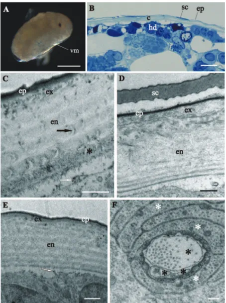
stage, in which the overall ultrastructural architecture of the cuticle is similar to the adult crustacean cuticle and irst morphological evidence of cuticle renewal is evident. Cuticle is from 2 to 3 µm thick, which is signiicantly thinner than in adults. It is com-posed of three layers, the outermost thin electron dense epicuticle, the middle exocuti-cle and the innermost endocutiexocuti-cle (Figs 4C, D, E). Cuticular scales are fully elaborated (Fig. 4B, D). Several transverse sections of completely structured sensilla are observed in the hypodermis (Fig. 4F). Dendritic outer segments and enveloping cells are clearly diferentiated. he characteristic pattern of chitin-protein ibers arrangement in the exocuticle is resolved in some regions and hardly discernible in other regions of the same specimen. he endocuticle is subdivided in several electron dense sublayers alter-nating with electron lucent sublayers (Figs 4C, D, E). In some regions pore canals are visible running through the endocuticle, consisting of electron lucent central part and electron dense margins (Fig. 4C). Several morphological features in prehatching late-stage embryo show the exoskeletal cuticle renewal already in this late-stage of development: partly disintegrated proximal portion of endocuticle in some regions (Fig. 4C); cuticle detachment from the hypodermis (Figs 4B, C, D, E); rough apical plasma membranes of hypodermal cells and irregularly arranged electron dense particles on their outer surface (Figs 4C, E).
synthesis are clearly visible underneath the assembling cuticle in all stages of marsupial mancas (Figs 5G, I, J).
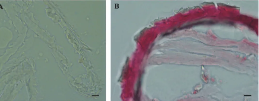
he results of histochemical reaction with Alizarin red S for calciied tissues lo-calization indicate that the cuticle of marsupial mancas is not strongly mineralized (Fig. 6A) in comparison to adult cuticle (Fig. 6B). he ventral calcium deposits, char-acteristic for adult premolt animals, were not observed in molting marsupial mancas.
Figure 6. Histochemical reaction for calcium – Alizarin red S. A No reaction in P. scaber late marsupial manca B Positive control (red-pink) in adult P. scaber cuticle. Bars: 10 µm.
Discussion
he structure of egg envelopes and the ultrastructural architecture of the exoskeletal cuticle in Porcellio embryos and marsupial mancas share some common features with those described in other arthropods, but there are also some signiicant diferences.
In comparison to egg envelopes of Drosophila eggs from ovaries, described by Margaritis et al. (1980), the egg envelopes of Porcellio embryos are thinner and have
considerably less complex ultrastructure. Chorion of Porcellio is approximately two times thinner than chorion of Drosophila. he chorion of Drosophila eggs is dife-rentiated into several layers, a thin innermost chorionic layer, a complex fenestrated endochorion and a ibrous exochorion,while Porcellio chorion in embryos consists of a single homogenous layer. Electron lucent “lacunae” observed in the Porcellio
chori-on resemble the cavities in the inner part of the Drosophila chorion. hese complex cavities are thought to be air-illed in laid eggs and involved in respiration (Margaritis et al. 1980). Study of embryo tolerance to physiological stresses in terrestrial isopod
membrane in Drosophila egg (Margaritis et al. 1980). he authors presume that this layer in Drosophila consists of wax, functioning to reduce water loss. his is not expec-ted for Porcellio embryos since they are less vulnerable to desiccation due to their
deve-lopment in aqueous environment. he comparison of egg envelopes ultrastructure in
Porcellio embryos and in Drosophila eggs reveals several diferences. We consider these dissimilarities the consequence of diferent environments of embryonic development in Porcellio embryos which develop in a protected luid-illed maternal brood pouch and in Drosophila embryos which are directly exposed to external environment during
development. Here we also report on a network of ibers between embryo surface and vitelline membrane in late-stage embryo. We presume that these ibers function as con-nective elements between embryo and envelope, but no comparative data on vitelline membrane attachment were found in the literature.
We show here that the late embryo is already covered with a homogenous extra-cellular matrix, possibly irst cuticle. In the prehatching late-stage embryo of P. scaber
the exoskeletal cuticle has already main features of the adult crustacean cuticle, but minor diferences are evident. It is composed of three principal layers - epicuticle, exo-cuticle and endoexo-cuticle. Endoexo-cuticle shows the typical arrangement of chitin lamellae in sublayers which are not so distinct as in adult cuticle. he characteristic pattern of chitin-protein ibers arrangement in the exocuticle of adults is not discernible in exo-cuticle of the prehatching late-stage embryo. A distal layer, similar to the dense distal exocuticular layer, described in adult isopods (Hild et al. 2009, Huber and Ziegler 2011, Seidl and Ziegler 2011), is not observed in the cuticle of prehatching embryo. Sensilla in hypodermis are already very elaborated and have similar ultrastructure as tricorn sensilla described in adult P. scaber (Ziegler and Altner1995). Previous studies of cuticle diferentiation during embryonic development of Drosophila melanogaster
Parhyale hawaiensis embryo, but it is not known which larval stage was observed. Our research, using histochemical approach to localize calciied tissue, indicates that the exoskeletal cuticle of P. scaber marsupial mancas is not strongly, if at all mineralized.It
could be possible that amorphous calcium carbonate was dissolved during preparation, since it has relatively high solubility.
Next, the issue of cuticle renewal during intramarsupial development was ad-dressed in this study. Partly disintegrated proximal endocuticle and cuticle detach-ment from the underlying hypodermis indicate that cuticle renewal takes place already in the prehatching embryonic stage. Prehatching embryo is thus the earliest stage of P. scaber development, where renewal of exoskeleton, i.e. initiation of molting, was ob-served so far. hese results are in agreement with the previous observation of apolysis on appendage tips in late-stage embryo of P. scaber (Milatovič et al. 2010). Apolysis and cuticle disintegration were not observed in the studies of cuticle structure during embryonic development in insect Drosophila melanogaster and amphipod crustacean
Parhyale hawaiensis (Locke 2001, Moussian et al. 2006, Havemann et al. 2008, Mous-sian 2010). We observed that the old cuticle is detached from the hypodermis and the new cuticle is produced in marsupial mancas of diferent stages, which is a sign of premolt. Several similarities and diferences with respect to molting process in adult isopods are described. Similarities in apical protrusions of hypodermal cells during cu-ticle synthesis, and appearance of ecdysal space are very explicit. Regarding synthesis of newly assembling cuticle prior to ecdysis we show that preecdysal cuticle in mancas has mostly homogenous procuticle, with no distinct chitin-protein arrangement, al-though in certain regions a helicoidal arrangement of chitin-protein ibers is evident. In adult P. scaber it is reported that several exocuticular lamellae are deposited in premolt stage (Ziegler 1997). Advanced stages of new cuticle formation in marsupial mancas need to be investigated in further research. Molting in two phases is typical in adult terrestrial isopods, while this is still not conirmed for developing marsupial mancas. In adult isopods some other morphological changes accompany molting pro-cess, particularly concerning calcium dynamics. In adult premolt stage ecdysal space in-between the old and new cuticle contains calcium storage granules (Ziegler 1994, Štrus and Blejec 2001). In mancas no similar granules were observed in the ecdysal space. In adults appearance of calcium deposits on the irst four anterior sternites is a clear indication of the premolt stage (Steel 1982, Zidar et al. 1998). In our study cal-cium deposits in molting marsupial stages were not observed, indicating diferences in calcium dynamics compared to adults. here are no data on calcium dynamics during marsupial development in terrestrial isopods.
Conclusions
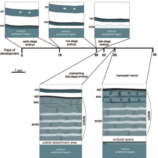
During marsupial development of Porcellio scaber embryos and marsupial mancas in
diferent stages are coated by diferent protective envelopes (Fig. 7).
Egg envelopes of isopod crustacean Porcellio scaber embryos are thinner and struc-turally less complex in comparison to egg envelopes of insect Drosophila melanogaster.
hese are expected diferences due to diferent embryonic developmental strategies of these arthropods. Similarities in egg envelopes of these two species appear particularly in their inner egg envelope, the vitelline membrane.
Exoskeletal cuticles of Porcellio scaber prehatching late-stage embryo and marsupial mancas have already some features of the adult crustacean cuticle, but are signiicantly thinner. hree principal layers are distinguished, the outermost epicuticle, the mid-dle exocuticle and the innermost endocuticle. Characteristic chitin-protein patterns of adult cuticle, particularly regarding the exocuticle, are not very distinct in prehatching late-stage embryo and become progressively more explicit in marsupial mancas. Cuti-cular scales and sensilla are fully elaborated already in prehatching late-stage embryo. In the distal portion of the exocuticle a dense layer is observed in marsupial mancas, which could correspond to the dense distal exocuticular layer of adult cuticle. Marsu-pial manca cuticle is not strongly calciied.
Cuticle renewal takes place already in prehatching late-stage embryo, where deta-chment of cuticle from hypodermis and partial disintegration of proximal endocuticle occur. Old cuticle detachment and new cuticle assembly appear in marsupial mancas of several stages. Morphological changes, related to calcium storage during molt cycle in adult isopods, were not observed in premolt marsupial stages.
Acknowledgements
We would like to thank Jožica Murko Bulić, Miloš Vittori and dr. Rok Kostanjšek for their laboratory assistance and all help and suggestions. his work was inanced by the Slovenian Research Agency (ARRS), grand No. P1-0184 and 1000-11-310087.
References
Charmantier G, Charmantier-Daures M (2001) Ontogeny of osmoregulation in crustaceans: he embryonic phase. American zoologist 41: 1078–1089. doi: 10.1093/icb/41.5.1078 Glötzner J, Ziegler A (2000) Morphometric analysis of the calcium-transporting sternal
epithe-lial cells of the terrestrial isopods Ligia oceanica, Ligidium hypnorum, and Porcellio scaber during molt. Arthropod Structure & Development 29 (3): 241–257. doi: 10.1016/S1467-8039(00)00030-X
Havemann J, Müller U, Berger J, Schwarz H, Gerberding M, Moussian B (2008) Cuticle dif-ferentiation in the embryo of the amphipod crustacean Parhyale hawaiensis. Cell Tissue Research 332 (2): 359–70. doi: 10.1007/s00441-007-0571-7
Hild S, Marti O, Ziegler A (2008) Spatial distribution of calcite and amorphous calcium car-bonate in the cuticle of the terrestrial crustaceans Porcellio scaber and Armadillidium vul-gare. Journal of Structural Biology 163 (1): 100–108. doi: 10.1016/j.jsb.2008.04.010 Hild S, Neues F, Žnidaršič N, Štrus J, Epple M, Marti O, ZieglerA (2009) Ultrastructure
Hoese B, Janssen HH (1989) Morphological and physiological studies on the marsupium in terrestrial isopods. Monograia Monitore zoologico Italiano 4: 153–173. http://cat.inist.fr /?aModele=aicheN&cpsidt=6769791
Hornung E (2011) Evolutionary adaptation of oniscidean isopods to terrestrial life: Struc-ture, physiology and behavior. Terrestrial Arthropod Reviews 4 (2): 95–130. doi: 10.1163/187498311X576262
Huber J, Ziegler A (2011) Structure and element distribution within the pars incisiva of the mandibles of Porcellio scaber Latr., 1804. In: Zidar P, Štrus J (Eds) Proceedings of the ISTIB 2011. 8th International Symposium of Terrestrial Isopod Biology, Bled (Slovenia), June 2011. University of Ljubljana, Biotechnical faculty, Department of Biology, Ljubljana, 57–58. http://www.istib-2011.si/uploads/media/ISTIB_2011.pdf
Jagadeeshan S, Singh RS (2007) Rapid evolution of outer egg membrane proteins in the
Dros-ophila melanogaster subgroup: A case of ecologically driven evolution of female reproductive
traits. Molecular biology and evolution 24 (4): 929–938. doi: 10.1093/molbev/msm009 Locke M (2001) he Wigglesworth Lecture: Insects for studying fundamental problems in
biology. Journal of Insect Physiology 47: 495-507. doi: 10.1016/S0022-1910(00)00123-2 Margaritis LH, Kafatos FC, Petri WH (1980) he eggshell of Drosophila melanogaster. I. Fine
structure of the layers and regions of the wild-type eggshell. Journal of Cell Science 43: 1–35. http://jcs.biologists.org/content/43/1/1.full.pdf+html
Milatovič M, Kostanjšek R, Štrus J (2010) Ontogenetic development of Porcellio scaber: Stag-ing based on microscopic anatomy.Journal of Crustacean Biology 30 (2): 225–235. doi: 10.1651/09-3189.1
Moussian B, Seifarth C, Müller U, Berger J, Schwarz H (2006) Cuticle diferentiation
dur-ing Drosophila embryogenesis. Arthropod Structure & Development 35: 137–152. doi:
10.1016/j.asd.2006.05.003
Moussian B (2010) Recent advances in understanding mechanisms of insect cuticle difer-entiation. Insect Biochemistry and Molecular Biology 40: 363–375. doi: 10.1016/j. ibmb.2010.03.003
Ouyang D, Wright J (2005) Calcium accumulation in eggs and mancas of Armadillidium vul-gare (Isopoda: Oniscidea). Journal of Crustacean Biology 25 (3): 420–426. http://www. jstor.org/stable/4540248
Payre F (2004) Genetic control of epidermis diferentiation in Drosophila. International Journal of Developmental Biology 48: 207–215. http://www.ncbi.nlm.nih.gov/pubmed/15272387 Price JB, Holdich DM (1980) An ultrastructural study of the integument during the moult
cycle of the woodlouse, Oniscus asellus (Crustacea, Isopoda). Zoomorphology 95 (3): 250– 263. doi: 10.1007/BF00998125
Steel CGH (1982) Stages of the intermoult cycle in the terrestrial isopod Oniscus asellus and their relation to biphasic cuticle secretion. Canadian Journal of Zoology 60 (3): 429–437. doi: 10.1139/z82-058
Surbida KL, Wright JC (2001) Embryo tolerance and maternal control of the marsupial en-vironment in Armadillidium vulgare Brandt (Isopoda: Oniscidea). Physiological and Bio-chemical Zoology 74: 894–906. doi: 10.1086/324474
Štrus J, Compere P (1996) Ultrastructural analysis of the integument during the moult cycle
in Ligia italica (Crustacea, Isopoda). Plügers Archiv - European Journal of Physiology 431
(6): 251–252. doi: 10.1007/BF02346363
Štrus J, Blejec A (2001) Microscopic anatomy of the integument and digestive system during the molt cycle in Ligia italica (Oniscidea). In: Kensley B, Brusca RC (Eds) Isopod system-atics and evolution.Crustacean Issues 13: 343–352. http://www.vliz.be/imis/imis.php?mo dule=ref&reid=11315&request=11315
Warburg MR (2011) he oniscid isopod female reproductive system and gestation, with a partial review. Invertebrate, Reproduction and Development: 1–24. doi: 10.1080/07924259.2011.573812
Wolf C (2009) he embryonic development of the malacostracan crustacean Porcellio scaber (Isopoda, Oniscidea). Development Genes and Evolution 219: 545–564. doi: 10.1007/ s00427-010-0316-6
Zidar P, Drobne D, Štrus J (1998) Determination of moult stages of Porcellio scaber (Isopoda) for routine use. Crustaceana 71(6): 646–654. doi:10.1163/156854098X00644
Ziegler A (1994) Ultrastructure and electron spectroscopic difraction analysis of the sternal calcium deposits of Porcellio scaber Latr. (Isopoda, Crustacea). Journal of Structural Biology 112 (2): 110–116. doi: 10.1006/jsbi.1994.1012
Ziegler A, Altner H (1995) Are the most numerous sensilla of terrestrial isopods hygrorecep-tors? Ultrastructure of the dorsal tricorn sensilla of Porcellio scaber. Cell and Tissue Rese-arch 282 (1): 135–145. doi: 10.1007/BF00319140