An Overview: Treatment of Lung Cancer
on Researcher Point of View
Javeria Amin
Department of Computer Science, COMSATS University, Wah Campus- Pakistan Email: javeriacomsat@gmail.com
---ABSTRACT ---Cancers is defined as the uncontrolled cell divisions. Cell does not grow maturely and destined to uncontrolled cell growth. When these cells of lungs grow uncontrolled it is called lung cancer. Nowadays mortality rate due to lung cancer is increasing day by day. Many treatment and diagnoses are now a day’s available to deal with lung cancer. Here we disused different method for diagnosis the common types of lung cancer Non-Small Cell Lung Cancer, Small Cell Lung Cancer, Small Cell Lung Cancer Limited Stage, Small Cell Lung Cancer - Extensive Stage, Lung Adenocarcinoma, Squamous Cell Carcinoma,Bronchioloalveolar carcinoma (BAC), Metastatic lung cancer.
Keywords -EGFR gene, LCINS,NSCLC,Quantitative positron imaging technique, SCLC
---Date of Submission: August 31, 2014 Date of Acceptance: January 03, 2015
---1. INTRODUCTION
L
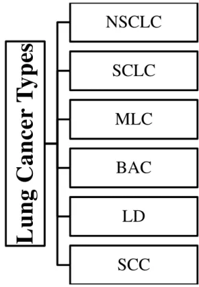
ung cancer leads to death including both man & woman .In diagnostic view PET is a quantitative positron imaging technique which is helpful in early diagnosis of lung cancer in patient. The risk of most redundant outcome of this disease (death) can be minimized by the early detection in proper diagnosis. The main developers of lung cancer are K-RAS and EGFR which are present in NSCLC patients 10% to 15%.5% of NSCLC patients represent PI3KCA, ERBB2, and B-RAF which is identify by efforts on DNA sequencing [1-2]Lung cancer is developed by the abnormal behavior of cells which causes tumor. [3] In blood, these cancer cells can be removed from lungs. Lymph flow through the vessels which are called Lymphatic vessels [4] .A computer with magnet gives the detailed pictures inside the body, in clinical practice Diffusion-weighted MRI (DW-MRI) is useful to detect tumor and its related therapy [5-9]. Due to cancer, Lung cancer moves to the center of chest and their diagnosis have been discussed here. In figure different type of lung cancer are shown.There are some types of lung cancer,non-small cell lung cancer (NSCLC) and small cell lung cancer (SCLC) etc.2. Literature Review
Lung cancer is the main cause of death, as more people are dying due to the lung cancer ascompared to other cancers. Initial treatment of lung cancer can easily recover the infected, tumor patient. That’s why there is a huge demand for the latest technologies andmethodologies to analyze
the lung correctly in its initial stage. In this paper we have described the diagnosis of different types of lung cancer. Figure 1.Different types of Lung Cancer
L
u
n
g
C
a
n
ce
r
T
y
p
es
NSCLC
SCLC
MLC
BAC
LD
3.Non-Small Cell Lung Cancer
It is a type of cancer which leads to death causing both man & woman. The subgroup of its patients has mutations in the gene of EGFR. This correlates with the responsiveness of clinical to the inhibitor of gefitinib this Non–small-cell lung cancer is the leading cause of death from cancer among both man and woman.A subgroup of patients with non–small-cell lung cancer has specific mutations in the EGFR gene,
Correlate the inhibitor of gefitinib this inhibitor for signaling to increase grows. The patients who are affected by gefitinib are exposed to screening for identification of lung cancer [10]. Production of lung cancer for a patient is better against prognostic factors and NSCL stages across the year. These probability of detection for lung cancer from ACOSOG Z0030 and CALGB 9761 is 72% and 79%.A subgroup of patient with disease at 1A Stage were at high risk and these may be treated by chemotherapy[11]The patient who were better than positron Emission Tomography for wide range of cancers PET will play valuable role for RT planning. In RT planning the physician should be aware when request is for PET scan [12] Non- small cell lungcancer
theFDG-PET the patients must safely decrease the radiotherapy volume experimentally the radiation loss within the tumor. The role of FDG-PET is emerging in some disease [13]the survival rate for patient of stage 1 disease is 64.6% and 41.2%. The aim here is to find mechanism of the function related to cancer. 2D-DIGE analysis was performed on tumor from the patients having NSCLC and HEK293 cells andencoded SEC62proteomiceffects ofsiRNA were analyzed with depletion of SEC62interactome and protein SEC63[14].The most common cause of lung cancer is the tobacco smoking, but just abut 10–25% of patients with this disease are lifelong never smoker. With lung cancer, mutations involvingTP53andKRASgenes are more common in tobacco a smoker which is well explained
from many studies, while LCINS is described by EGFR TK mutations, ALK.RET and ROS fusions. The genome of LCINS is considerably different from the tumor genome of a tobacco smoke with lung cancer. For lung cancer the LCINS genome may offer us with a relatively enhanced and easily identifiable set of oncogenic drivers. Against LCINS the relatively small number of genomic alterations also provides some better opportunities for the growth of targeted therapies.
Figure 2.Tumour genome from a never smoker with lung cancer and a tobacco smoke with lung cancer [15]
4.Small Cell Lung Cancer
This is aggressive type of lung cancer. This causes the
brain metastasis early. The interaction between SCLC and brain metastasis affected patients is poor. Therefore the mechanism of metastasis is required to be improved the present therapies and treatment by new modalities .elevated level PLGF are identified which are associated with SCLC brain metastasis and are inversely related with SCLC outcome of the clinical results [20] an effective treatment for SCLC the response rate of 50% and 10.3 month in patient for refractory disease isimportant ,the activity of additional studies in patients with SCLC, especially for patients who have done therapy as a single agent or combination is important for target base agent [21].
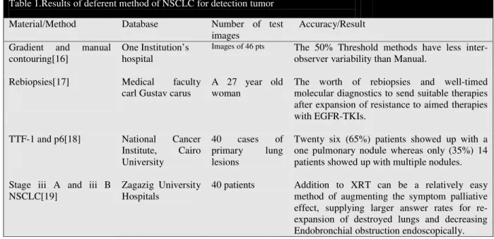
Table 1.Results of deferent method of NSCLC for detection tumor
Material/Method Database Number of test
images
Accuracy/Result Gradient and manual
contouring[16]
One Institution’s hospital
Images of 46 pts The 50% Threshold methods have less inter-observer variability than Manual.
Rebiopsies[17] Medical faculty carl Gustav carus
A 27 year old woman
The worth of rebiopsies and well-timed molecular diagnostics to send suitable therapies after expansion of resistance to aimed therapies with EGFR-TKIs.
TTF-1 and p6[18] National Cancer Institute, Cairo University
40 cases of primary lung lesions
Twenty six (65%) patients showed up with a one pulmonary nodule whereas only (35%) 14 patients showed up with multiple nodules. Stage iii A and iii B
NSCLC[19]
Zagazig University Hospitals
4.1 Small Cell Lung Cancer - Limited Stage
Limited stage of small cell lung cancer patients with modern cohort, brain imaging MRI prior to PCI the survival advantage of thoracic chemoradiation is conferred by PCI alone. Chemotherapy with response of small cell lung cancer is offered with slandered of care [22].This study presents the attempt to correlate between FDG avidity with the outcome of LS-SCLC in both pre and post CRT settings. Meaningful prognostic type information is unlikely to provide in PET and patients who are treated by CRT [23].
4.2Small Cell Lung Cancer - Extensive Stage
Extensive stage of lung cancer (SCLC), the technique of efficacy and AZD-0530 is more helpful which four cycles of platinum-based. Saracatinib at a dose of 175 mg/day by mouth is well tolerated the PFS rate pragmatic at the pre-planned interim analysis did not meet the criteria for additional enrollment. [24] In small cell lung cancer tumor occupied area is highly vascularized. Vascular agent of tumor is ASA404 with carboplatin and ASA404 with PSF was not prolonged [25].
5. Metastatic Lung Cancer
Metastatic lung cancer is the type of lung cancer in which cancer cells from any part or any other organ of the body spreads through towards the lungs. Organ which firstly cancer began is called primary cancer. General symptom of metastatic cancers are (1)Fatigue(2)weakness(3)weight loss(4)Metastatic cancer to the lungs is the spread of cancer from another region of Loss of appetite etc. A burden of substantial symptoms occurs in non- small lung cancer and goes through the end of life. In early disease well care leads to improvement in quality and quantity of life as compared with patients’ receiving less aggressive care at the end of life have longer survival [26].An examination of mice having lung cancer tumors. When treated with inhalational formulation of DIM5 and C-DIM8. These treatments results in the less protein expression of mediators of tumors initiation, metastasis and other forms that procedures cancer. A tumor marker (CD31, VEGF) shows the suppression of angiogenesis and metastasis. So C-DIM5 and C-DIM8 are antitumor’s [27].A metastatic adenocarcinoma the lungs multiple lesions have cystic appearance are found [28].
6. Bronchioloalveolar carcinoma (BAC)
Antifolate drugs is an active agent in lung cancers specially adenocarcinoma Pemetrexed (antifolate drug) is active in patients of Bronchioloalveolar carcinoma (BAC) to underline mechanism of action as an antifolate drug [29] Tumor marker detect LIPH expression in Bronchioloalveolar carcinoma (BAC). LIPH protein early and late phase lung cancer patients by high serum level LIPH protein have better survival chances after surgery. So LIPH is tumor markers of lung cancer that is for adenocarcinoma and Bronchioloalveolar carcinoma (BAC) [30].
7. Squamous Cell Carcinoma
In lung cancer Squamous cell carcinoma (SCC) is the most common type of lung cancer. NCI-H69 cells express choline transporter which in turn gives a force in relation with NHE1. This system of choline is used for synthesis of Ach apoptosis (cell death).This choline transport system is used as chemotherapy [31].Profiling of radiation survival should facilitative discovery of protective measures [32]. 8. Lung Adenocarcinoma
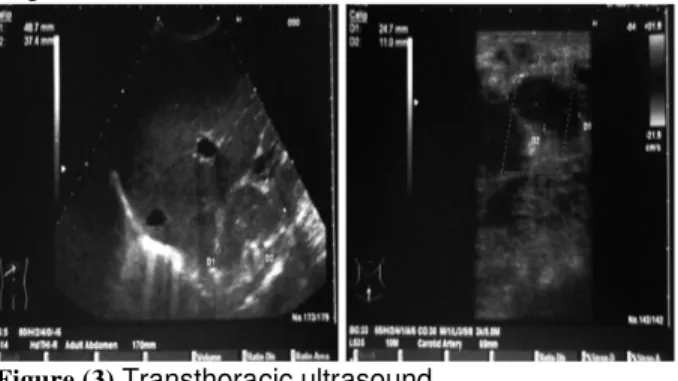
In lung adenocarcinoma platinum based chemotherapy is the most common treatment. In stage three and is non prognostic mutation treatment decision are based on patients sub stages or relevant subgroups of disease [33] .There is no association found by clinicians to exclude malignancy in patients with sarcoidosis [34].Transthoracic ultrasound can be used in different stages of lung cancer and protect chest physicians in defining the modality of diagnosis in every single patient dependent on his/her stage [35].
Figure (3).Transthoracic ultrasound
DW-MR imaging offers expensive material not attained by conventional MR and can be helpful for variations of central lung carcinoma from atelectasis [36-37].
Figure (4). MR and PET/CT images of a 39-year-old woman with lung adenocarcinoma.
9.Conclusion
Table2. Different Method base Comparison for Treatment of LungCancer
Application Advantege Limitations Result
Chain reaction of digital polymers [38]
It enables the quantification of DNA from peripheral blood.
- It identified PIK3CA
circulating in patients of breast cancer and blood cancer.
PI3K/Akt/mTOR[39] The inhibitor of PI3K emerged to the problem of EGFR TKI resistance.
Resistance to EGFR TKIs is related with extensive, heterogeneity and complexity.
The inhibitors have paired with other agents if they are effective. Application metastatic
adenocarcinoma of the lung is associated with NCS[40]
In the absence of mandibular or base of skull metastatic lesion, NCS can be present.
- NCS is the sign of
metastatic cancer.
In metastatic
adenocarcinoma Docetaxel induced hypersensitivity pneumonitis mimicking lymphangitic carcinoma [41].
Docetaxel is one of the anticancer drugs.
The patients presents with cough, fever, dyspnea.
Docetaxel also effect in response to steroids.
Lung adenocarcinoma classification[42]
Survival chances. - Disease prognosis and
mediator gene. Lepidic component [43] Patients with less papillary
structure have shorter disease chances than those of structure less.
- Low papillary structure
is associated with high cancer behavior lung adenocarcinoma.
ESD[44] ESD with the flex Knife
make it easier to control the knife during ESD and any difficulties such as severe bleeding and perforation.
Endoscopic mucosal resection (EMR) is also a less-invasive treatment
By EMR and additional therapies it is complex able to get en bloc resection for larger lesions.
LSD1[45] As compared to ESCC
patients with lower expression of LSD1 were considerably inferior to overall survival.
With any of the following clinic pathologic characteristics sex, age, infiltration and differentiation ,LSD1 expression was not connected
In the cases with LNM than those without LNM (p < 0.05) the expression level of LSD1 was higher
[46]In the human lung adenocarcinoma cells (A549) are showing to Pem, MX, and/or cisplatin (CP) (24 or 48)
At the G1/S border the irradiation protocol was considered to get advantage from the radio sensitivity of cells.
The cell cycle Supply of A549 cells and no clear radio sensitization MX (3 or 6 mM) alone had no outcome.
Before RT could further extend the reply line of the drugs allow the cycling cells to build up at the G1/S border.
SIB integrated with WBRT [47]
The patients with many Brain metastases have particular option of WBRT.
Further studies with more patients are necessary by the limitation of the small sample size.
It is efficient in volume reduction and displayed excellent intracranial control.
FDG-PET and CIMs[48] In the detection of primary tumor FDG-PET and CIMs were equally effective
- When analyzed
alongside with CIM FDG-PET scanning depicts important additional in sequence and has a relevant impact on therapy planning.
FDG-PET evaluation for grading, staging ,post therapeutic evaluation and response assessment in children affected by Wilms tumor (WT).[49]
FDG-PET is found to be superior with recurrent WI for 1/3 cases.
Additional information is not provided by FDG-PET to the traditional work of imaging for WT patients. Clinical outcomes and pre-operative response
References
[1]. Hoda A. Abu-Youssef ,Khaled M. Kamel ,Samah Selim,Sally M. Gamal El-Deen ,Study of the added value of transthoracic ultrasound in staging of lung cancer,Egyptian Journal of Chest Diseases and Tuberculosis, 2014,1-10.
[2]. Greenman, C., Stephens, P., Smith, R., Dalgliesh, G.L., Hunter, C, Bignell, G., Davies, H., Teague, J., Butler, A., Stevens, C., et al, Patterns of somatic mutation in human cancer genomes, Nature 446, 2007,153–158. [3]. Mokhled S. AL-TARAWNEH, Lung Cancer Detection
Using Image Processing Techniques, Leonardo Electronic Journal of Practices and Technologies,2012 , 147-158
[4]. Kumar, Vineet, Nucleus Extraction from Sputum Images for Early Lung Cancer Detection,CT International Journal of Information & Communication Technology 1( 1), 2013.
[5]. Masood, Saleha, Muhammad Sharif, MussatYasmin, MudassarRaza, and SajjadMohsin, Brain Image Compression: A Brief Survey, ResearchJournal of Applied Sciences, (5), 2013, 49-59.
[6]. Yasmin, Mussarat, SajjadMohsin, Muhammad Sharif, MudassarRaza, and SalehaMasood, Brain Image Analysis: A Survey, World Applied Sciences Journal, 19(10),2012, 1484-1494.
[7]. Yasmin, Mussarat, Muhammad Sharif, SalehaMasood, MudassarRaza, and SajjadMohsin, Brain image enhancement: A survey,World Applied Sciences Journal, 17 (9),2012,1192-1204.
[8]. Yasmin, Mussarat, Muhammad Sharif, SalehaMasood, MudassarRaza, and SajjadMohsin, Brain image reconstruction: A short survey, World Applied Sciences Journal, 19(1), 2012, 52-62.
[9]. Raza, Mudassar, Muhammad Sharif, MussaratYasmin, SalehaMasood, and SajjadMohsin, Brain Image Representation and Rendering: A Survey, Research Journal of Applied Sciences, (4), 2012.
[10]. Lynch, Thomas J., Daphne W. Bell, RaffaellaSordella, SaradaGurubhagavatula, Ross A. Okimoto, Brian W. Brannigan, Patricia L. Harris et al. ,Activating mutations in the epidermal growth factor receptor underlying responsiveness of non–small-cell lung cancer to gefitinib,New England Journal of Medicine 350( 21) ,2004, 2129-2139
[11]. Potti, Anil, Sayan Mukherjee, Rebecca Petersen, Holly K. Dressman, Andrea Bild, Jason Koontz, Robert Kratzke et al,A genomic strategy to refine prognosis in early-stage non–small-cell lung cancer,New England Journal of Medicine 355(6), 2006,570-580.
[12]. MacManus, Michael, Ursula Nestle, Kenneth E. Rosenzweig, IgnasiCarrio, Cristina Messa, OtakarBelohlavek, Massimo Danna et al,Use of PET and PET/CT for radiation therapy planning: IAEA expert report 2006–2007, Radiotherapy and oncology 91( 1) ,2009, 85-94.
[13]. De Ruysscher, Dirk, and Carl-Martin Kirsch.,PET scans in radiotherapy planning of lung cancer,Radiotherapy and Oncology 96(3),2010, 335-338.
[14]. Johannes Linxweiler, LaxmikanthKollipara, René P. Zahedi, PavelLampel, Richard Zimmermann,Markus Greiner ,Proteomic insights into non-small cell lung cancer: New ideas for cancer diagnosis and therapy from a functional viewpoint,EuPA Open Proteomics 4,2014, 25– 39
[15]. Janakiraman Subramanian, RamaswamyGovindan, Molecular profile of lung cancer in never smokers,European Journal of Cancer Supplements, 11 (
2), 2013, 248–253
[16]. DanijelaJelovac,Julia A.
Beaver, SasidharanBalukrishna, Hong Yuen Wong, Patricia Valda Toro, Ashley Cimino-Mathews, PedramArgani, Vered Stearns, Lisa Jacobs, Dustin VanDenBerg, Jill Kessler, Stacie Jeter, Ben H. Park, Antonio C. Wolff, A PIK3CA mutation detected in plasma from a patient with synchronous primary breast and lung cancers, Human Pathology45( 4),2014, 880–883. in patients having Wilms
tumors [50]
with Wilms tumors. However the small pulmonary may be visualize easily.
for the lungs with 10mm or smaller
helpful in assessing the response for treatment.
PET/CT optimized with dual contrast[51]
It can be optimized for molecular imaging to be used in humans.
- Dual contrast PET/CT
having early post contrast with 3h delay. It provides the better way to detect early tumor lesions.
Positron emission tomography for F-18-fluoro-2-deoxy-d-glucose [52]
In chemotherapy it was better to evaluate the TBR for SUVmax.
Distinguishing b//w good and poor response was difficult by SUV2/SUV1
The promising tool of FDG-PET is easily to access the response of chemotherapy for noninvasively.
IFRT[53] Prescribed doses of radiation and lymph failure nodes were mainly developed by field of radiation.
The meditational lymph node displacements, It might not be appropriate to use an isotropic margin in applying IFRT to NSCLC.
The nodes with Incidental radiation are delivered by using the node region of IRFT. PCI [54] The disease free survival and
overall survival of patients after complete response to chemo-radiation therapy.
- PCI is beneficial over
the earlier
[17]. Shirish M. Gadgeel1, 2, Antoinette Wozniak1, Preclinical Rationale for PI3K/Akt/mTOR Pathway Inhibitors as Therapy for Epidermal Growth Factor Receptor Inhibitor-Resistant Non–Small-Cell Lung Cancer, Clinical Lung Cancer14( 4),2013, 322–332. [18]. M. Werner-Wasik1, P. Kang2, W. Choi2, N. Ohri3, P.
Faulhaber4, D. Nelson5, A. Nelson5, J. Piper5, Z.Shen5, S. Pirozzi5 ,Comparison of PET Contouring Methods in Patients With Early-Stage Resected Non-Small Cell Lung Cancer (NSCLC): A Pathologic–Imaging Correlation, Proc in American Society for Radiation Oncology 55th Annual Meeting ASTRO's 55th Annual Metting, International Journal of Radiation Oncology Biology Physics 87( 2),2013,S540-S540
[19]. Jan Stoehlmacher-Williamsa, Gerhard Ehningera, Dieter R. Zimmermannb, Sabine Merkelbach-Brusec, Hans-Ulrich Schildhausc, ReinhardBuettnerc, Targeting TKI-resistance in NSCLC: Importance of rebiopsy and molecular diagnostics A case study, Cancer Treatment Communications1(1),2013, 1–5
[20]. Yu-Hua Chen, Bo Li, Chapter 19 , Brain Metastasis from Small-Cell Lung Cancer with High Levels of Placental Growth Factor Brain Metastases from Primary Tumors Epidemiology, Biology, and Therapy 2014, 213–225 [21]. Onoda, Sayaka, Noriyuki Masuda, Takashi Seto, Kenji
Eguchi, Yuichi Takiguchi, Hiroshi Isobe, Hiroaki Okamoto et al, Phase II trial of amrubicin for treatment of refractory or relapsed small-cell lung cancer: Thoracic Oncology Research Group Study 0301, Journal of Clinical Oncology 24(34), 2006, 5448-5453.
[22]. S.A. Patel, S.R. Bowen, C. Gamboa, C. Baik, J. Zeng, Improved Survival With Prophylactic Cranial Irradiation in Modern Cohort of Limited-Stage Small Cell Lung Cancer Patients With MRI Brain Imaging , Proc in American Society for Radiation Oncology 55th Annual Meeting ASTRO's 55th Annual MettingInternational Journal of Radiation Oncology ,Biology, Physics87(2) ,2013, S552. [23]. L. Ong1, C.A. Perez1, A. Foster1, M.C. Pietanza1, K.
Rosenzweig2, D.Y. Gelblum1, M.P. Dunphy1, A. Rimner1, A.J. Wu1, Prognostic Value of Pre- and Post-Radiation Therapy [18F]FDG-PET/CT Metrics in Limited-Stage Small Cell Lung Cancer, Proc in American Society for Radiation Oncology 55th Annual Meeting ASTRO's 55th Annual MettingInternational Journal of Radiation Oncology, Biology, Physics87(2),2013,S537–S538.
[24]. Julian R. Molinaa, 1, Nathan R.
Fosterb, ,ThanyananReungwetwattanah, 1, Garth D. Nelsonb, 2,Andrew V. Graingerc, 3, Preston D. Steend, 4, Philip J. Stellae, 5, Randolph Marksa, John Wrightf, 6, Alex A. Adjeig, 7, A phase II trial of the Src-kinase inhibitor saracatinib after four cycles of chemotherapy for patients with extensive stage small cell lung cancer: NCCTG trial N-0621,Lung Cancer85( 2), 2014, 245–250
[25]. Martin Früh1, Richard Cathomas2, Marco Siano1, Gregor Tscherry3, Alfred Zippelius4, Christoph Mamot5, Andreas Erdmann6, Fatima Krasniqi4, Daniel Rauch7, Mathew Simcock8, Erika Küttel8, Pierre Fustier9, Miklos Pless10, Carboplatin and Paclitaxel Plus ASA404 as First-Line Chemotherapy for Extensive-Stage Small-Cell Lung Cancer: A Multicenter Single Arm Phase II Trial (SAKK 15/08), Clinical Lung Cancer, 14( 1), 2013, 34–39 [26]. Temel, Jennifer S., Joseph A. Greer, AlonaMuzikansky,
Emily R. Gallagher, SonalAdmane, Vicki A. Jackson, Constance M. Dahlin et al.,Early palliative care for
patients with metastatic non–small-cell lung cancer,New England Journal of Medicine 363( 8), 2010, 733-742. [27]. TerrickAndey, Apurva Patel, Tanise Jackson, Stephen
Safe, Mandip Singh,1,1-Bis (30 indolyl)-1-(p-substitutedphenyl)methane compounds inhibit lung cancer cell and tumor growth in a metastasis model,European Journal of Pharmaceutical Sciences 50,2013,227 241 [28]. M. Ruparel, A.A. Mohammed, Case report: Biphasic
presentation of multicystihaemorrhagic metastatic adenocarconoma of the lung,Respiratory Medicine Case Reports 10,2013, 64e66
[29]. Derick H.M. Lau,James Moon, Angela M. Davies, Rachel E. Sanborn, Fred R. Hirsch, Wilbur A. Franklin, Janet C. Ruzich, Mary W. Redman, David R ,GandaraSouthwestern Oncology Group Phase II Trial (S0526) of Pemetrexed in Bronchioloalveolar Carcinoma Subtypes of Advanced Adenocarcinoma, Clinical Lung Cancer14 ( 4), 2013, 351–355.
[30]. Yasuhiro Seki, Yukihiro Yoshida , Hisako Ishimine, Aya Shinozaki-Ushikuf, Yoshimasa Ito, Kenya Sumitomo, Jun Nakajimad, Masashi Fukayamaf, Tatsuo Michiuea, Makoto Asashimaa, b, Akira Kurisakib, Lipase member H is a novel secreted protein selectively upregulated in human lung adenocarcinomas and bronchioloalveolar carcinomas, Biochemical and Biophysical Research Communications,443( 4), 2014, 1141–1147
[31]. Masato Inazua, b, Tomoko Yamadac, Nobuo Kubotad, Tsuyoshi Yamanakab Functional expression of choline transporter-like protein 1 (CTL1) in small cell lung carcinoma cells: A target molecule for lung cancer therapy, Pharmacological Research,76, 2013, 119–131 [32]. M. Abazeed1, D. Adams2, K. Hurov3, P. Tamayo2, C.
Creighton4, D. Sonkin3, A. Giacomelli5, S. Schreiber6, P. Hammerman7, M. Meyerson7Integrative Radiogenomic Profiling of Squamous Cell, Proc in American Society for Radiation Oncology 55th Annual Meeting ASTRO's 55th Annual Metting, Lung Cancer International Journal of Radiation Oncology, Biology, Physics87(2), 2013, S138– S139
[33]. MihalyCserepesa, b,GyulaOstorosb, 1,Zoltan
Lohinaia, c, ErzsebetRasod, e, TamasBarbaid, JozsefTimar d, e, Anita Rozsasa, c, JuditMoldvayf, IlonaKovalszkyg, KatalinFabianf, MartonGyulaih, BahilG hanimc, i, ViktoriaLaszloc, Thomas Klikovitsc, Mir AlirezaHodac, i, Michael Gruschi, Walter Bergeri,WalterKlepetkoc, BalazsHegedusc, d, e, 2, Balazs Domea, Subtype-specific KRAS mutations in advanced lung adenocarcinoma: A retrospective study of patients treated with platinum-based chemotherapy, European Journal of Cancer50( 10), 2014, 1819–1828
[34]. AmitGirishKachalia,PiusOchieng, KinjalKachalia, Habibu rRahman, Rare coexistence of sarcoidosis and lung adenocarcinoma, Respiratory Medicine Case Reports, 12, 2014, 4–6
[36]. Yang, Rui-Meng, et al. Differentiation of central lung cancer from atelectasis: comparison of diffusion-weighted MRI with PET/CT.PloSone,8(4),2013.
[37]. Javeria Amin,Annam Zafar. A Survey: Content Based Image Retrieval.Int. J. Advanced Networking and Applications,5(6),2076-2083,2014
[38]. Eman Abu Sinnaa, NohaEzzata, Ghada M. Sherifb, Role of thyroid transcription factor-1 and P63 immunocytochemistry in cytologic typing of non-small cell lung carcinomas, Journal of the Egyptian National Cancer Institute25( 4),2013,209–218.
[39]. Samah M. Shehataa, Ashraf E. El-Shoraa, Mohamed A. Mazroaab, Mostafa I. Ragaba, Outcome of endobronchialelectrocautery versus external beam radiotherapy or both together in the palliative management of non-small cell lung cancer, Egyptian Journal of Chest Diseases and Tuberculosis62(1),2013,173–181.
[40]. M. Ruparel, A.A. Mohammed, Case report: Biphasic presentation of multicystihaemorrhagic metastatic adenocarconoma of the lung, Respiratory Medicine Case Reports 10, 2013, 64e66.
[41]. FaizanZaheera, KhurrumHussainb, 1, JeethendraRaoc ,Unusual presentation of ‘numb chin syndrome’ as the manifestation of metastatic adenocarcinoma of the lung, International Journal of Surgery Case Reports4 ( 12),2013, 1097–1099.
[42]. Yasuhiro Seki, Yukihiro Yoshida , Hisako Ishimine, Aya Shinozaki-Ushikuf, Yoshimasa Ito, Kenya Sumitomo, Jun Nakajimad, Masashi Fukayamaf, Tatsuo Michiuea, Makoto Asashimaa, b, Akira Kurisakib, Lipase member H is a novel secreted protein selectively upregulated in human lung adenocarcinomas and bronchioloalveolar carcinomas, Biochemical and Biophysical Research Communications443( 4),2014,1141–1147
[43]. Koji Tsutaa, 1MitsumasaKawago, EisukeInouec, Akihiko Yoshidaa, Fumiaki Takahashic,HiroyukiSakuraib,
Shun-ichiWatanabeb, Masahiro
Takeuchic, KohFurutaa, HisaoAsamurab, Hitoshi Tsuda, The utility of the proposed IASLC/ATS/ERS lung adenocarcinoma subtypes for disease prognosis and correlation of driver gene alterations, Lung Cancer81(3),2013,371–37
[44]. M. Abazeed1, D. Adams2, K. Hurov3, P. Tamayo2, C. Creighton4, D. Sonkin3, A. Giacomelli5, S. Schreiber6, P. Hammerman7, M, Meyerson ,Integrative Radiogenomic Profiling of Squamous Cell Lung Cancer, Proc in American Society for Radiation Oncology 55th Annual Meeting ASTRO's 55th Annual Metting, International Journal of Radiation Oncology Biology Physics87( 2),2013, S138–S139
[45]. N Ishii, H Akiyama, Y Fujita, Endoscopic Submucosal Dissection for Squamous Cell Carcinoma of the Esophagus: Tips and Tricks from the Expert, Video Journal and Encyclopedia of GI Endoscopy1(1),2013,46–47.
[46]. AmitGirishKachalia,PiusOchieng, KinjalKachalia, Rah man, Rare coexistence of sarcoidosis and lung adenocarcinoma, Respiratory Medicine Case Reports12,2014, 4–6 .
[47]. T. Biswas1, R. Patel2, L. Weeks1, R. Patel2, A. Dowlati1, N. Sharma1, N. Oleinick2, S. Gerson1, M. Machtay1, Enhanced Radiosensitization of Lung Adenocarcinoma Cells by Pemetrexed When Combined With Methoxyamine to Inhibit Base Excision Repair, Proc in American Society for Radiation Oncology 55th
Annual Meeting ASTRO's 55th Annual Metting, International Journal of Radiation Oncology Biology Physics87(2),2013, 1 : S662.
[48]. Misch, Daniel, Ingo G. Steffen, Stefan Schönberger, Thomas Voelker, Christian Furth, Brigitte Stöver, Hubertus Hautzel, Günter Henze, HolgerAmthauer, and TimmDenecke, Use of positron emission tomography for staging, preoperative response assessment and posttherapeuticevaluation in children with Wilms tumour, European journal of nuclear medicine and molecular imaging 35( 9), 2008, 1642-1650.
[49]. Hossain, AKM Moinul, Barry L. Shulkin, Michael J. Gelfand, Humayun Bashir, Najat C. Daw, Susan E. Sharp, Helen R. Nadel, and Jeffrey S. Dome, FDG positron emission tomography/computed tomography studies of Wilms’ tumor, European journal of nuclear medicine and molecular imaging 37(7 ),2010,1300-1308.
[50]. Flores II, Leo G, Hsin-Hsien Yeh, Suren Soghomonyan, Daniel Young, James Bankson, Qianghua Hu, Mian Alauddin, Vicki Huff, and Juri G. Gelovani, Monitoring therapy with MEK inhibitor U0126 in a novel Wilms tumor model in Wt1 knockout Igf2 transgenic mice using 18F-FDG PET with dual-contrast enhanced CT and MRI: early metabolic response without inhibition of tumor growth,Molecular Imaging and Biology 15(2), 2013, 175-185.
[51]. Ye, Zhaoming, Jiangjun Zhu, Mei Tian, Hong Zhang, Hongwei Zhan, Chunlei Zhao, Disheng Yang, Weixu Li, and Nong Lin ,Response of osteogenic sarcoma to neoadjuvant therapy: evaluated by 18F-FDG-PET, Annals of nuclear medicine 22( 6),2008 , 475-480.
[52].Beverly A. Teicher, Targets in small cell lung cancer, Biochemical Pharmacology87( 2),2014 ,211–219.
[53]. M. Chen1, H. Xiao1, 2, B. Yong2, H. ZhiChun2, C. Yuanyuan1, A Study of the Correlations Between the Irradiated Dose to Lymph Node Regions and Lymph Node Recurrence When Involved Field Radiation Therapy Was Used for Limited-Stage Small Cell Lung Cancer, Proc in American Society for Radiation Oncology 55th Annual Meeting ASTRO's 55th Annual Metting, International Journal of Radiation Oncology Biology Physics87( 2),2013 , S512–S513.